Embed Size (px)
Citation preview
Hindawi Publishing CorporationEvidence-Based Complementary and Alternative MedicineVolume 2013, Article ID 739473, 10 pageshttp://dx.doi.org/10.1155/2013/739473
Research ArticleRedox Mechanisms of AVS022, an Oriental PolyherbalFormula, and Its Component Herbs in Protection againstInduction of Matrix Metalloproteinase-1 in UVA-IrradiatedKeratinocyte HaCaT Cells
Thanyawan Pluemsamran,1 Pinpat Tripatara,1 Rattana Phadungrakwittaya,1
Pravit Akarasereenont,1,2 Tawee Laohapand,2 and Uraiwan Panich1
1 Department of Pharmacology, Faculty of Medicine Siriraj Hospital, Mahidol University, Bangkok 10700, Thailand2 Center of Applied Thai Traditional Medicine, Faculty of Medicine Siriraj Hospital, Mahidol University, Bangkok 10700, Thailand
Correspondence should be addressed to Uraiwan Panich; [email protected]
Received 7 June 2013; Accepted 26 July 2013
Academic Editor: Benny Tan Kwong Huat
Copyright © 2013 Thanyawan Pluemsamran et al. This is an open access article distributed under the Creative CommonsAttribution License, which permits unrestricted use, distribution, and reproduction in any medium, provided the original work isproperly cited.
Ayurved Siriraj HaRak (AVS022) formula has been used for topical remedy of dermatologic disorders. Oxidative stress inducedby ultraviolet (UV) A irradiation could be implicated in photoaged skin through triggering matrix metalloproteinase-1 (MMP-1). We, therefore, explored the antioxidant mechanisms by which AVS022 formulation and its individual components protectedagainst UVA-dependentMMP-1 upregulation in keratinocyteHaCaT cells. TLC analysis revealed the presence ofmultiple phenolicsincluding gallic acid (GA) in the AVS022 extracts. We demonstrated that pretreatment with the whole formula and individualherbal components except T. triandra protected against increased MMP-1 activity in irradiated HaCaT cells. Moreover, all herbalextracts andGA, used as the reference compound, were able to reverse cytotoxicity, oxidant production, glutathione (GSH) loss, andinactivation of catalase and glutathione peroxidase (GPx). F. racemosawas observed to yield the strongest abilities to abolish UVA-mediated induction of MMP-1 and impairment of antioxidant defenses including GSH and catalase. Our observations suggest thatupregulation of endogenous antioxidants could be the mechanisms by which AVS022 and its herbal components suppressed UVA-stimulated MMP-1 in HaCaT cells. In addition, pharmacological actions of AVS022 formula may be attributed to the antioxidantpotential of its components, in particular F. racemosa, and several phenolics including GA.
1. Introduction
Demands for alternative medicines including herbal reme-dies continue to increase. Herbal treatment for dermatologicdiseases and cosmetic problems has existed for thousandsof years [1]. Ayurved Siriraj HaRak (AVS022) formula, aThai polyherbal formula consisting of 5 medicinal plants,has been used in Thai traditional medicine for the remedyof skin disorders. Thus, exploring pharmacological activitiesof the AVS022 polyherbal formula and its constituent herbsis of significance in order to gain scientific evidence onthe efficacy and safety of traditional herbal medicine. TheAVS022 formula is composed of the root extracts of 5
herbs, Capparis micracantha DC., Clerodendrum indicum L.,Harrisonia perforata Merr., Ficus racemosa L., and Tiliacoratriandra (Colebr.) Diels. Previous in vitro and in vivo studiesof F. racemosa, a medicinal plant used in Indian ayurvedicmedicine, reported that it exerted several pharmacologicalactions including anti-inflammatory, anticancer, and antiox-idant activities [2–4]. Additionally, various phytochemicalconstituents including racemic acid and alkaloids isolatedfrom F. racemosa and T. triandra, respectively, which are alsothe herbal components of AVS022 formula, were demon-strated to possess biological activities [5, 6].
Ultraviolet A (UVA) (315–400 nm) has been recognizedas a primary environmental cause of photodamage and

2 Evidence-Based Complementary and Alternative Medicine
premalignant changes of the skin cells through cytotoxi-city of keratinocytes and activation of metalloproteinase-1(MMP-1), a major collagenolytic enzyme generated by ker-atinocytes and fibroblasts. Since MMP-1 is contributed toskin cell damage and collagen fragmentation affecting theskin’s structural integrity [7], development of dermatologicalproducts that effectively suppress MMP-1 at cellular andmolecular levels could be a targeting strategy for photoagingprevention. Medicinal plants and phytochemicals includingphenolic acids providing antioxidant properties have beenobserved to abrogate damaging effects of UVA on the skinthrough inhibition of activity and expression of MMP-1 inkeratinocytes or fibroblasts [8–10]. We previously reportedthat impaired capacity of antioxidant defenses including cata-lase, glutathione peroxidase (GPx), and glutathione (GSH)involved UVA-stimulated MMP-1 activity, and therefore,upregulation of endogenous antioxidants may representmechanisms underlying photoprotective effects of phyto-chemicals [10]. We, thus, assessed antioxidant mechanismsof AVS022 extracts, its 5 plant components, and gallic acid(GA), an antioxidant phenolic present in the formula, inprotecting against UVA-dependent cell toxicity and MMP-1augmentation by assessing cellular oxidant generation, GSHlevel, and catalase and GPx activities in immortalized humankeratinocyte (HaCaT) cells.
2. Materials and Methods
2.1. Materials. Human keratinocyte cell line (HaCaT) fromCell Lines Service (CLS, Heidelberg, Germany) was a kindgift from Professor Pa-thai Yenchitsomanus, Departmentof Research and Development, Faculty of Medicine SirirajHospital, Mahidol University. Dulbecco’s modified Eagle’smedia (DMEM) were purchased from Invitrogen (NY, USA),and chemicals and reagents of the highest quality availablewere obtained from Sigma-Aldrich (MO, USA or Germany).
2.2. Preparation of AVS022 Formula Extracts. AVS022 com-posed of 20% (w/w) of each herb; H. perforate, C. micra-cantha, C. indicum, F. racemosa, and T. triandra, was pre-pared by Unit of Thai Herbal Pharmaceuticals of Centerof Applied Thai Traditional Medicine, Faculty of MedicineSiriraj Hospital, Mahidol University, Thailand. All plantmaterials were purchased from Tai-hua-jan drugstore andauthenticated by two experienced Thai traditional practi-tioners using macroscopic identification and organoleptictechniques which based on anatomical characteristics of theindividual plant parts and color, fracture, smell, or taste.Thenthe specimens were sorted, washed, oven-dried, and crushed.The powdered drug was extracted by dynamic macerationmethod. One hundred grams of the powdered herb wasweighed and placed in a container with 1 L of 80% (v/v)ethanol. The mixture was constantly stirred with magneticstirrer for 10 minutes, and then the liquid was filtered andthe marcs were pressed. The clarified liquid was evaporatedunder reduced pressure by rotary evaporator and kept frozenovernight prior to lyophilization. The extraction procedurefor individual plant was the same with previously referred
to procedure. One hundred milligrams of the lyophilizedpowder was accurately weighed and dissolved in 1mL of 80%ethanol, mixed, and centrifuged at 15,000 rpm for 10 minutesat 4∘C. The sample solution was filtered through a 0.2 𝜇mmembrane filter and was used for thin layer chromatography(TLC) analysis.
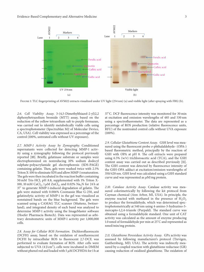
2.3. TLC Fingerprinting of AVS022 Formula Extracts. The fil-trate of sample solution was loaded to TLC silica gel 60 F 254(Merck, Germany) using sample applicator (Camag Linomat5, Switzerland). Solvent system of hexane: ethyl acetate: aceticacid (31 : 14 : 5 v/v/v) was used as mobile phase for phenolicseparation. The detection was examined under 254 nm UVlight and visible light after spraying with fast blue salt (FBS).The identification of phenolics in AVS022 was carried outby comparing its TLC chromatogram with those of phenolicreferencemarkers. Five phenolic referencemarkers includingcaffeic acid, ferulic acid, gallic acid, kaempferol, and quercetinwere used. The TLC chromatograms showed the presence ofcaffeic acid, ferulic acid, and GA in the AVS022 extracts, andcaffeic acid and GA were detected under both 254 nm UVlight and visible light (after spraying with FBS) as shown inFigure 1.
2.4. Cell Cultures and Treatment. HaCaT cells were culturedin DMEM/F12 medium supplemented with 10% fetal bovineserum (FBS) and 1% penicillin (100 units/mL)/streptomycin(100 𝜇g/mL) at 37∘C in a humidified air of 5% CO
2(𝑃CO
2
=40Torr) (a Forma Scientific CO
2Water-Jacketed Incubator).
Cells were treated with the AVS022 extract; each componentextract or GA used as the reference compound dissolvedin 80% ethanol, and the final concentration of ethanol inculture medium did not exceed 0.05% (v/v). To assess pho-toprotective and antioxidant effects, cells were treated withherbal extracts at concentrations up to 60 𝜇g/mL and GA upto 5 𝜇g/mL for 30min before UVA (330–400 nm) exposure.UVA intensity verification and selection of a UVA dose(4 J/cm2) and time point after irradiation were previouslydescribed [10]. GA was used as the reference phenolic in thisstudy because it was shown to possess stronger inhibitoryactivity than that of caffeic acid and ferulic acid againstoxidant formation in HaCaT cells exposed to UVA (4 J/cm2)(data not shown). Assays for cell viability, oxidant formation,GSH content, and antioxidant enzyme activities were carriedout at 1 h time point and forMMP-1 activity at 24 h time pointafter UVA exposure.
2.5. Cell Lysate Preparation. Cells were harvested by cen-trifugation and lysed with buffer containing 50mM Tris-HCl, 10mM ethylene diaminetetraacetic acid (EDTA), 1%(v/v) Triton X-100, phenylmethylsulfonyl fluoride (PMSF)(100mg/mL), and pepstatin A (1mg/mL) in DMSO andleupeptin (1mg/mL) in H
2O, pH 6.8. The cells were cen-
trifuged at 10,000 rpm for 10min and the supernatant wasthen collected. Protein concentration was determined usingthe Bio-Rad Protein Assay Kit (Bio-Rad, Germany).
Evidence-Based Complementary and Alternative Medicine 3
Kaempferol
Ferulic acid
QuercetinCaffeic acid
Gallic acid
Markers
1 2 3 4 Lane
UV 254nm
AVS0
22
(a)
Visible light
Ferulic acid
QuercetinCaffeic acid
KaempferolGallic acid
Markers
1 2 3 4 Lane
AVS0
22
(b)
Figure 1: TLC fingerprinting of AVS022 extracts visualized under UV light (254 nm) (a) and visible light (after spraying with FBS) (b).
2.6. Cell Viability Assay. 3-(4,5-Dimethylthiazol-2-yl)2,2diphenyltetrazolium bromide (MTT) assay, based on thereduction of the yellow tetrazolium salt to purple formazan,was carried out to identify metabolically viable cells usinga spectrophotometer (SpectraMax M2 of Molecular Device,CA, USA). Cell viability was expressed as a percentage of thecontrol (100%, untreated cells without UV exposure).
2.7. MMP-1 Activity Assay by Zymography. Conditionedsupernatants were collected for detecting MMP-1 activ-ity using a zymography following the protocol previouslyreported [10]. Briefly, gelatinase substrate or samples wereelectrophoresized on nonreducing 10% sodium dodecylsulphate-polyacrylamide gel electrophoresis (SDS-PAGE)containing gelatin. Then, gels were washed twice with 2.5%TritonX-100 to eliminate SDS and allowMMP-1 renaturation.The gels were then incubated in the reaction buffer containing50mM Tris-HCl, pH 8.8, supplemented with 1% Triton X-100, 10mM CaCl
2, 1 𝜇M ZnCl
2, and 0.02% Na
3N for 24 h at
37∘ to generate MMP-1-induced degradation of gelatin. Thegels were stained with 0.006% Coomassie Blue G-250, andgelatinolytic activity of MMP-1 in the gel was visualized asnonstained bands on the blue background. The gels werescanned using a CAMAG TLC scanner (Muttenz, Switzer-land), and integrated density of each band was analyzed todetermine MMP-1 activity using the ImageMaster software(Hoefer Pharmacia Biotech). Data was represented as arbi-trary densitometric units of MMP-1 activity per 1,000,000cells.
2.8. Assay for Cellular ROS Formation. Dichlorofluorescein(DCFH) assay, based on the oxidation of nonfluorescentDCFH by intracellular ROS to fluorescent 2,7-DCF, wasperformed to evaluate formation of ROS. After cells weresubjected to UVA (4 J/cm2), cells were incubated in DMEMwithout phenol red and loaded with 5 𝜇MDCFHDA for 1 h at
37∘C. DCF fluorescence intensity was monitored for 30minat excitation and emission wavelengths of 485 and 530 nmusing a spectrofluorometer. The data are represented as apercentage of ROS production (relative fluorescence units,RFU) of the nontreated control cells without UVA exposure(100%).
2.9. Cellular Glutathione Content Assay. GSH level was mea-sured using the fluorescent probe o-phthalaldehyde- (OPA-)based fluorometric method, principally by the reaction ofGSH with OPA at pH 8. The cell extracts were preparedusing 6.5% (w/v) trichloroacetic acid (TCA), and the GSHcontent assay was carried out as described previously [11].The GSH content was detected by fluorescence intensity ofthe GSH-OPA adduct at excitation/emission wavelengths of350/420 nm. GSH level was calculated using a GSH standardcurve and was represented as 𝜇M/mg protein.
2.10. Catalase Activity Assay. Catalase activity was mea-sured colorimetrically by following the kit protocol fromCayman chemical (Ann Arbor, MI, USA). In principle, theenzyme reacted with methanol in the presence of H
2O2
to produce the formaldehyde, which was determined spec-trophotometrically at 540 nm using 4-amino-3-hydrazino-5-mercapto-1,2,4-triazole (Purpald). The standard curve wasobtained using a formaldehyde standard. One unit of CATactivity was calculated as the amount of enzyme producing1.0 nmol of formaldehyde per min at 25∘C and represented asnmol/min/mg protein.
2.11. Glutathione Peroxidase Activity Assay. GPx activity wasassessed by following manufacturer’s protocol (Trevigen,Gaithersburg, MD, USA). The activity was indirectly mea-sured by a coupled reaction with glutathione reductase (GR)causing reduction of oxidized glutathione. The oxidation of
4 Evidence-Based Complementary and Alternative Medicine
AVS022
(𝜇g/mL)Control 0 7.5 15 30 60UVA (4 J/cm2)
Cel
l via
bilit
y (%
of c
ontro
l)120
100
80
60
40
20
0
∗∗∗
∗∗∗∗∗
### ###
(𝜇g/mL)Control 0 0.6 1.25 2.5 5
UVA (4 J/cm2)
120
100
80
60
40
20
0
GA
Cel
l via
bilit
y (%
of c
ontro
l)
∗∗∗
∗∗∗∗∗
∗∗∗∗∗∗∗ ∗ ∗
###
(𝜇g/mL)Control 0 7.5 15 30 60UVA (4 J/cm2)
120
100
80
60
40
20
0
H. perforateC. micracanthaC. indicum
F. racemosaT. triandra
Cel
l via
bilit
y (%
of c
ontro
l)
(a)
(b)
(c)
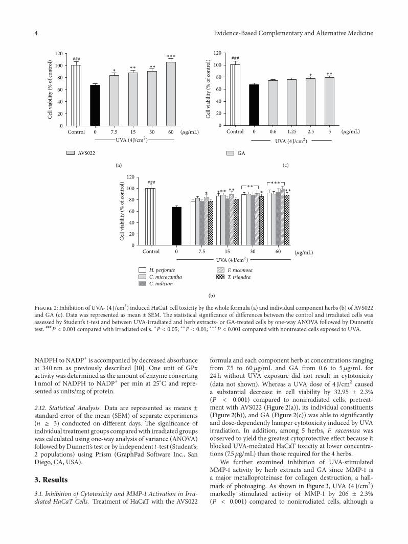
Figure 2: Inhibition of UVA- (4 J/cm2) induced HaCaT cell toxicity by the whole formula (a) and individual component herbs (b) of AVS022and GA (c). Data was represented as mean ± SEM. The statistical significance of differences between the control and irradiated cells wasassessed by Student’s 𝑡-test and between UVA-irradiated and herb extracts- or GA-treated cells by one-way ANOVA followed by Dunnett’stest. ###𝑃 < 0.001 compared with irradiated cells. ∗𝑃 < 0.05; ∗∗𝑃 < 0.01; ∗∗∗𝑃 < 0.001 compared with nontreated cells exposed to UVA.
NADPH to NADP+ is accompanied by decreased absorbanceat 340 nm as previously described [10]. One unit of GPxactivity was determined as the amount of enzyme converting1 nmol of NADPH to NADP+ per min at 25∘C and repre-sented as units/mg of protein.
2.12. Statistical Analysis. Data are represented as means ±standard error of the mean (SEM) of separate experiments(𝑛 ≥ 3) conducted on different days. The significance ofindividual treatment groups comparedwith irradiated groupswas calculated using one-way analysis of variance (ANOVA)followed byDunnett’s test or by independent 𝑡-test (Student’s;2 populations) using Prism (GraphPad Software Inc., SanDiego, CA, USA).
3. Results
3.1. Inhibition of Cytotoxicity and MMP-1 Activation in Irra-diated HaCaT Cells. Treatment of HaCaT with the AVS022
formula and each component herb at concentrations rangingfrom 7.5 to 60 𝜇g/mL and GA from 0.6 to 5 𝜇g/mL for24 h without UVA exposure did not result in cytotoxicity(data not shown). Whereas a UVA dose of 4 J/cm2 causeda substantial decrease in cell viability by 32.95 ± 2.3%(𝑃 < 0.001) compared to nonirradiated cells, pretreat-ment with AVS022 (Figure 2(a)), its individual constituents(Figure 2(b)), and GA (Figure 2(c)) was able to significantlyand dose-dependently hamper cytotoxicity induced by UVAirradiation. In addition, among 5 herbs, F. racemosa wasobserved to yield the greatest cytoprotective effect because itblocked UVA-mediated HaCaT toxicity at lower concentra-tions (7.5𝜇g/mL) than those required for the 4 herbs.
We further examined inhibition of UVA-stimulatedMMP-1 activity by herb extracts and GA since MMP-1 isa major metalloproteinase for collagen destruction, a hall-mark of photoaging. As shown in Figure 3, UVA (4 J/cm2)markedly stimulated activity of MMP-1 by 206 ± 2.3%(𝑃 < 0.001) compared to nonirradiated cells, although a

Evidence-Based Complementary and Alternative Medicine 5
(a)
(b)
(c)
MM
P-1
activ
ity (%
of c
ontro
l)
350
300
250
200
150
100
50
0
### ∗∗∗∗∗∗
∗∗∗
(𝜇g/mL)Control 0 1.25 2.5 5
UVA (4 J/cm2)
GA
0 15 30 60
###∗∗
∗∗∗
∗
(𝜇g/mL)Control
UVA (4 J/cm2)
MM
P-1
activ
ity (%
of c
ontro
l)
350
300
250
200
150
100
50
0
H. perforateC. micracanthaC. indicum
F. racemosaT. triandra
AVS022
(𝜇g/mL)Control 0 15 30 60UVA (4 J/cm2)
###
MM
P-1
activ
ity (%
of c
ontro
l) 350
300
250
200
150
100
50
0
∗∗
∗
Figure 3: Inhibition of UVA-stimulated MMP-1 activity in HaCaT cells by the whole formula (a) and individual component herbs (b) ofAVS022 and GA (c). Zymography analysis of secreted MMP-1 was performed as described in Section 2. Data was represented as mean ±SEM. The statistical significance of differences between the control and irradiated cells was evaluated by Student’s 𝑡-test and between UVA-irradiated and herb extracts- or GA-treated cells by one-way ANOVA followed by Dunnett’s test. ###𝑃 < 0.001 compared with irradiated cells.∗
𝑃 < 0.05; ∗∗𝑃 < 0.01; ∗∗∗𝑃 < 0.001 compared with nontreated cells exposed to UVA.
significant and dose-dependent reduction of MMP-1 activitywas observed in HaCaT cells pretreated with the wholeformulation of AVS022 (Figure 3(a)), its component herbsbut not T. triandra (Figure 3(b)), and GA (Figure 3(c))compared with unpretreated cells following UV irradiation.In agreement with the photoprotective effect on HaCaT cellcytotoxicity, among 5 herb components of AVS022 formula,F. racemosa presented the strongest protective activity againstUVA-induced enhanced MMP-1 activity since lower concen-trations (30 𝜇g/mL) of F. racemosa than those of other 4components were capable of suppressingMMP-1 stimulation.
3.2. Inhibition of ROS Formation and GSH Loss in IrradiatedHaCaT Cells. Level of cellular ROS and GSH is an importantmarker to indicate cellular redox status. We assessed whether
redox mechanisms were involved in the inhibitory effects ofherb extracts studied and GA on UVA-dependent cytotoxic-ity and MMP-1 upregulation. Figures 4 and 5 demonstratedthat, at 1 h postirradiation, UVA exposure (4 J/cm2) led to asubstantial increase in ROS by 38.54 ± 2.1% (𝑃 < 0.001) anda dramatic decline in GSH level by 49.3±1.3% (𝑃 < 0.001). Incontrast, pretreatment of HaCaT cells with the whole formula(Figures 4(a) and 5(a)) and the individual components ofAVS022 (Figures 4(b) and 5(b)) and GA (Figures 4(c) and5(c)) caused a significant and dose-dependent inhibition ofROS formation and GSH loss as compared to unpretreatedcells following UV irradiation. Furthermore, among all 5components of AVS022, F. racemosa was shown to have thehighest inhibitory effect on UVA-mediated reduced GSHcontent because the inhibitory concentrations (7.5 𝜇g/mL) ofF. racemosa were lower than those required for the 4 herbs.

6 Evidence-Based Complementary and Alternative Medicine
AVS022
(𝜇g/mL)Control 0 7.53.75 15 30 60
UVA (4 J/cm2)
Cel
lula
r oxi
dant
leve
l (%
of c
ontro
l) 150
125
100
75
50
25
0
∗∗∗∗∗∗
∗∗∗###
Oxi
dant
form
atio
n (%
of c
ontro
l) 150
125
100
75
50
25
0(𝜇g/mL)Control 0 7.5 15 30 60
UVA (4 J/cm2)
∗∗∗∗∗∗∗∗∗∗∗∗∗
∗∗
∗∗∗###
H. perforateC. micracanthaC. indicum
F. racemosaT. triandra
(𝜇g/mL)Control 0 0.6 1.25 2.5 5
UVA (4 J/cm2)
150
125
100
75
50
25
0Oxi
dant
form
atio
n (%
of c
ontro
l)
∗∗∗
∗∗
∗###
(a)
(b)
(c)
GA
Figure 4: Inhibition of UVA-induced cellular ROS formation in HaCaT cells by the whole formula (a) and individual component herbs (b)of AVS022 and GA (c). The fluorescent DCF as an indicator of ROS formation was measured at 485 nm excitation and 530 nm emission asdescribed in Section 2. Data were represented as a percentage of control (100%, nonirradiated and nontreated cells) using amicroplate reader.The statistical significance of differences between the control and irradiated cells was determined by Student’s 𝑡-test and between UVA-irradiated and herb extracts- or GA-treated cells by one-way ANOVA followed by Dunnett’s test. ###𝑃 < 0.001 compared with irradiated cells.∗
𝑃 < 0.05; ∗∗𝑃 < 0.01; ∗∗∗𝑃 < 0.001 compared with nontreated cells exposed to UVA.
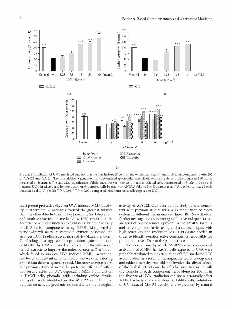
3.3. Inhibition of Catalase and Glutathione Peroxidase Inac-tivation in Irradiated HaCaT Cells. To further investigateredox mechanisms of herbal extracts studied and GA withrespect to modulation of endogenous antioxidants, as shownin Figures 6 and 7, enzymatic assays revealed that, comparedto nonirradiated control cells, UVA (4 J/cm2) irradiationdrastically reduced catalase activity by 43.53 ± 7.7% (𝑃 <0.001) and GPx activity by 66 ± 8.4% (𝑃 < 0.001). Never-theless, addition of AVS022, each component herb, and GAprior to UVA exposure was able to dose-dependently reverseinactivation of both catalase and GPx compared to irradiatedcells in the absence of herb extracts or GA. In agreement withour findings for cytotoxicity, MMP-1 activity, and GSH level,among all 5 components of AVS022, F. racemosa was shownto exert the most potent protection against UVA-dependentcatalase inactivation.
4. Discussion
Development of herbs employed in a traditional medicineas promising photoprotective agents has gained considerableattention in dermatology research because pharmacologicallyactive phytochemicals identified and isolated from severalmedicinal plants have been reported to yield antioxidantactions beneficial for the skin [12]. Since UVA irradiation-mediated oxidative stress of the skin is involved in ker-atinocyte toxicity and activation of MMP-1 accountable forphotoaged skin, we, therefore, explored redox mechanismsof the whole formula and individual component herbsof AVS022 and GA, a reference phenolic compound, inprotection against UVA-mediated cytotoxicity and MMP-1induction in keratinocyte HaCaT cells. Our study demon-strated that AVS022, its constituent herbs, and GA signif-icantly abrogated HaCaT cell toxicity mediated by UVA

Evidence-Based Complementary and Alternative Medicine 7
∗∗∗
∗∗∗
∗∗∗
∗∗∗
∗∗∗∗∗∗
∗∗∗###
Cel
lula
r GSH
cont
ent (
% o
f con
trol) 125
100
75
50
25
0
AVS022
(𝜇g/mL)Control 0 7.53.75 15 30 60
UVA (4 J/cm2)
∗∗∗∗∗∗∗∗∗
∗∗
###
Cel
lula
r GSH
cont
ent (
% o
f con
trol) 125
100
75
50
25
0(𝜇g/mL)Control 0 0.6 1.25 2.5 5
UVA (4 J/cm2)
∗
∗∗∗
∗∗∗
###
H. perforateC. micracanthaC. indicum
F. racemosaT. triandra
Cel
lula
r GSH
cont
ent (
% o
f con
trol)
175
150
125
100
75
50
25
0(𝜇g/mL)Control 0 7.5 15 30 60
UVA (4 J/cm2)
(a)
(b)
(c)
GA
Figure 5: Inhibition of UVA-dependent GSH loss in HaCaT cells by the whole formula (a) and individual component herbs (b) of AVS022and GA (c). GSH level was detected by fluorescence intensity of the GSH-OPA adduct at 350 nm excitation and 420 nm emission as describedin Section 2. The statistical significance of differences between the control and irradiated cells was evaluated by Student’s 𝑡-test and betweenUVA-irradiated and herb extracts- or GA-treated cells by one-way ANOVA followed by Dunnett’s test. ###𝑃 < 0.001 compared with irradiatedcells. ∗𝑃 < 0.05; ∗∗𝑃 < 0.01; ∗∗∗𝑃 < 0.001 compared with nontreated cells exposed to UVA.
(4 J/cm2). Stimulation of MMP-1 activity by UVA was alsosuppressed by the whole formula and its individual herbalcomponents except T. triandra component of AVS022 andGA. Previous studies reported that photooxidative stress ispossibly involved inMMP-1 regulation in skin cells includingkeratinocytes [13, 14], and improving antioxidant defensesystem may thus be mechanisms underlying the photo-protective effects of phytochemicals ubiquitously present inmedicinal plants. ROS accumulation in photoaged skinshas been suggested to associate with increased MMP-1expression, which could be reversed by promoting capacityof antioxidant defenses including catalase [15], GSH, andGPx [10, 16]. They are essential endogenous antioxidantdefenses controlling redox balance accountable for protectionagainst photooxidative stress in the keratinocytes and skincarcinogenesis [17, 18], and redox regulation ofMMP-1might,therefore, represent a strategy for photoaging prevention.We further investigated whether protective effects of the
whole formula and each component of AVS022 and GA onUVA-mediated increased ROS formation and GSH depletionas well as inactivation of catalase and GPx were involvedin the inhibition of MMP-1 activity. Our data indicatedthat pretreatment of irradiated HaCaT cells with the herbalextracts orGAabolishedUVA-dependentGSHdepletion andcatalase and GPx inactivation.
Since AVS022 is a polyherbal formulation composed of 5medicinal plants, combinations of multiple active ingredientsin different plants can make pharmacological action ofAVS022 complex. We, thus, examined the modulation ofMMP-1 and antioxidant defense capacity by AVS022 andindividual component in our study. Zymographic analysisof MMP-1 activity showed that combination of 5 herbsdid not yield synergistic protection against UVA-dependentenhanced MMP-1 activity and the F. racemosa componentwas primarily contributed to biological activities of theAVS022 formula because F. racemosa appeared to yield the
8 Evidence-Based Complementary and Alternative Medicine
Cata
lase
activ
ity (%
of c
ontro
l) 175
150
125
100
75
50
25
0
AVS022
(𝜇g/mL)Control 0 7.53.75 15 30 60UVA (4 J/cm2)
∗∗∗
∗∗∗∗∗∗
###
175
150
125
100
75
50
25
0(𝜇g/mL)Control 0 7.5 15 30 60
UVA (4 J/cm2)
∗∗∗
∗∗∗∗∗∗
∗∗∗ ∗∗∗
∗∗∗∗∗∗
∗∗∗∗∗∗
∗ ∗
###
Cata
lase
activ
ity (%
of c
ontro
l)
H. perforateC. micracanthaC. indicum
F. racemosaT. triandra
175
150
125
100
75
50
25
0(𝜇g/mL)Control 0 0.6 1.25 2.5 5
UVA (4 J/cm2)
∗∗∗∗∗∗
∗
###
Cata
lase
activ
ity (%
of c
ontro
l)
(a)
(b)
(c)
GA
Figure 6: Inhibition of UVA-mediated catalase inactivation in HaCaT cells by the whole formula (a) and individual component herbs (b)of AVS022 and GA (c). The formaldehyde generated was determined spectrophotometrically with Purpald as a chromogen at 540 nm asdescribed in Section 2. The statistical significance of differences between the control and irradiated cells was assessed by Student’s 𝑡-test andbetween UVA-irradiated and herb extracts- or GA-treated cells by one-way ANOVA followed by Dunnett’s test. ###𝑃 < 0.001 compared withirradiated cells. ∗𝑃 < 0.05; ∗∗𝑃 < 0.01; ∗∗∗𝑃 < 0.001 compared with nontreated cells exposed to UVA.
most potent protective effect on UVA-induced MMP-1 activ-ity. Furthermore, F. racemosa exerted the greatest abilitiesthan the other 4 herbs to inhibit cytotoxicity, GSH depletion,and catalase inactivation mediated by UVA irradiation. Inaccordance with our study on free radical scavenging activityof all 5 herbal components using DPPH (1,1-diphenyl-2-picrylhydrazyl) assay, F. racemosa extracts possessed thestrongest DPPH radical scavenging activity (data not shown).Our findings also suggested that protection against inductionof MMP-1 by UVA appeared to correlate to the abilities ofherbal extracts to improve the redox balance as T. triandra,which failed to suppress UVA-induced MMP-1 activation,had lower antioxidant activities than F. racemosa in restoringantioxidant defense system studied. Moreover, as reported inour previous study showing the protective effects of caffeicand ferulic acids on UVA-dependent MMP-1 stimulationin HaCaT cells, phenolic acids including caffeic, ferulic,and gallic acids identified in the AVS022 extracts couldbe possible active ingredients responsible for the biological
activity of AVS022. Our data in this study is also consis-tent with previous studies for GA in modulation of redoxsystem in different melanoma cell lines [19]. Nevertheless,further investigations concerning qualitative and quantitativeanalyses of phytochemicals present in the AVS022 formulaand its component herbs using analytical techniques withhigh sensitivity and resolution (e.g., HPLC) are needed inorder to identify possible active constituents responsible forphotoprotective effects of the plant extracts.
The mechanisms by which AVS022 extracts suppressedactivation of MMP-1 in HaCaT cells exposed to UVA wereprobably attributed to the attenuation of UVA-mediated ROSaccumulation as a result of the augmentation of endogenousantioxidant capacity and did not involve the direct effectsof the herbal extracts on the cells because treatment withthe formula or each component herbs alone for 30min inthe absence of UVA irradiation did not substantially affectMMP-1 activity (data not shown). Additionally, inhibitionof UV-induced MMP-1 activity and expression by natural

Evidence-Based Complementary and Alternative Medicine 9
GPx
activ
ity (%
of c
ontro
l)150
125
100
75
50
25
0
AVS022
(𝜇g/mL)Control 0 7.53.75 15 30 60UVA (4 J/cm2)
∗∗∗∗∗∗
∗∗∗∗∗∗
###
GPx
activ
ity (%
of c
ontro
l)
150
125
100
75
50
25
0(𝜇g/mL)Control 0 7.5 15 30 60
UVA (4 J/cm2)
∗∗∗
∗∗∗
∗∗∗
∗∗∗∗∗∗∗
∗∗∗###
H. perforateC. micracanthaC. indicum
F. racemosaT. triandra
GPx
activ
ity (%
of c
ontro
l)
150
125
100
75
50
25
0(𝜇g/mL)Control 0 0.6 1.25 2.5 5
UVA (4 J/cm2)
∗∗∗∗∗∗
∗∗∗
###
(a)
(b)
(c)
GA
Figure 7: Inhibition of UVA-dependent GPx inactivation in HaCaT cells by the whole formula (a) and individual component herbs (b) ofAVS022 and GA (c). GPx activity was evaluated as described in Section 2. The statistical significance of differences between the control andirradiated cells was determined by Student’s 𝑡-test and between UVA-irradiated and herb extracts- or GA-treated cells by one-way ANOVAfollowed by Dunnett’s test. ###𝑃 < 0.001 compared with irradiated cells. ∗𝑃 < 0.05; ∗∗𝑃 < 0.01; ∗∗∗𝑃 < 0.001 compared with nontreated cellsexposed to UVA.
products derived from medicinal plants could be regulatedby multiple signal pathways including AP-1 and NF-kappa Btranscription factors and MAP kinase [8, 14, 20]. Quercetin,a polyphenol commonly found in diet and medicinal plants,was demonstrated to block photocarcinogenesis in epidermalJB6 cells through downregulation of AP-1, NF-kappa B, andMAPK activities as well as activation of antioxidant tran-scription factor [21]. Further study is, thus, needed to explorean association between MMP-1 mediated by MAP kinaseand redox modulation at molecular levels in keratinocytesexposed to UV irradiation.
In conclusion, protective mechanisms by which AVS022,an oriental herbal formula, and its herbal componentsexerted inhibitory effects on UVA-induced MMP-1 activityinvolved regulation of endogenous antioxidants includingGSH, catalase, and GPx. Additionally, antioxidant potentialof the component herbs, particularly F. racemosa, and severalphenolic compounds (e.g., GA) may be contributed to thepharmacological actions ofAVS022 formula.This study could
provide pharmacological evidence for polyherbal formulaand its constituent herbs. Further identification of activecompounds to validate biological activities of the formulais needed in order to develop the herbal formula contain-ing antioxidant phytochemicals as effective photoprotectiveagents.
Conflict of Interests
The authors declare that there is no conflict of interests.
Acknowledgments
This work was supported byThailand Research Fund (Grantsnos. RSA5580012 and DBG5380040) and the “Chalerm-phrakiat” Grant, Faculty of Medicine Siriraj Hospital, Mahi-dol University. Appreciation is expressed to Assistant Profes-sor Chanitra Thuwajit, Department of Immunology, Faculty

10 Evidence-Based Complementary and Alternative Medicine
of Medicine Siriraj Hospital, Mahidol University, for valuablesuggestion and support concerning gelatin zymography.
References
[1] J. Koo and S. Arain, “Traditional chinese medicine for thetreatment of dermatologic disorders,” Archives of Dermatology,vol. 134, no. 11, pp. 1388–1393, 1998.
[2] N. Khan and S. Sultana, “Chemomodulatory effect of Ficus race-mosa extract against chemically induced renal carcinogenesisand oxidative damage response inWistar rats,” Life Sciences, vol.77, no. 11, pp. 1194–1210, 2005.
[3] S. C. Mandal, T. K. Maity, J. Das, B. P. Saba, and M. Pal, “Anti-inflammatory evaluation of Ficus racemosa Linn. leaf extract,”Journal of Ethnopharmacology, vol. 72, no. 1-2, pp. 87–92, 2000.
[4] V. P. Veerapur, K. R. Prabhakar, V. K. Parihar et al., “Ficusracemosa stem bark extract: a potent antioxidant and a probablenatural radioprotector,” Evidence-Based Complementary andAlternative Medicine, vol. 6, no. 3, pp. 317–324, 2009.
[5] R. W. Li, D. N. Leach, S. P. Myers, G. D. Lin, G. J. Leach, and P.C. Waterman, “A new anti-inflammatory glucoside from Ficusracemosa L,” Planta Medica, vol. 70, no. 5, pp. 421–426, 2004.
[6] S. Sureram, S. P. D. Senadeera, P. Hongmanee, C. Mahidol, S.Ruchirawat, and P. Kittakoop, “Antimycobacterial activity ofbisbenzylisoquinoline alkaloids from Tiliacora triandra againstmultidrug-resistant isolates of Mycobacterium tuberculosis,”Bioorganic and Medicinal Chemistry Letters, vol. 22, no. 8, pp.2902–2905, 2012.
[7] G. J. Fisher, T. Quan, T. Purohit et al., “Collagen fragmentationpromotes oxidative stress and elevates matrix metalloprotein-ase-1 in fibroblasts in aged human skin,” American Journal ofPathology, vol. 174, no. 1, pp. 101–114, 2009.
[8] Y. P. Hwang, K. N. Oh, H. J. Yun, and H. G. Jeong, “Theflavonoids apigenin and luteolin suppress ultraviolet A-inducedmatrix metalloproteinase-1 expression via MAPKs and AP-1-dependent signaling in HaCaT cells,” Journal of DermatologicalScience, vol. 61, no. 1, pp. 23–31, 2011.
[9] E. A. Offord, J. C. Gautier, O. Avanti et al., “Photoprotectivepotential of lycopene, 𝛽-carotene, vitamin E, vitamin C andcarnosic acid in UVA-irradiated human skin fibroblasts,” FreeRadical Biology and Medicine, vol. 32, no. 12, pp. 1293–1303,2002.
[10] T. Pluemsamran, T. Onkoksoong, and U. Panich, “Caffeic acidand ferulic acid inhibit UVA-induced matrix metalloprote-inase-1 through regulation of antioxidant defense system inkeratinocyte HaCaT cells,” Photochemistry and Photobiology,vol. 88, no. 4, pp. 961–968, 2012.
[11] U. Ketsawatsakul, “Modulation by bicarbonate the protectiveeffects of phenolic antioxidants on peroxynitrite-mediated cellcytotoxicity,” ScienceAsia, vol. 33, no. 3, pp. 273–282, 2007.
[12] V. M. Adhami, D. N. Syed, N. Khan, and F. Afaq, “Phyto-chemicals for prevention of solar ultraviolet radiation-induceddamages,” Photochemistry and Photobiology, vol. 84, no. 2, pp.489–500, 2008.
[13] J. H. Lee, J. H. Chung, and K. H. Cho, “The effects of epigallo-catechin-3-gallate on extracellular matrix metabolism,” Journalof Dermatological Science, vol. 40, no. 3, pp. 195–204, 2005.
[14] M. J. Piao, R. Zhang, N. H. Lee, and J. W. Hyun, “Phloro-glucinol attenuates ultraviolet B radiation-induced matrixmetalloproteinase-1 production in human keratinocytes viainhibitory actions against mitogen-activated protein kinases
and activator protein-1,” Photochemistry and Photobiology, vol.88, no. 2, pp. 381–388, 2012.
[15] M. H. Shin, G. E. Rhie, Y. K. Kim et al., “H2
O2
accumulationby catalase reduction changes MAP kinase signaling in agedhuman skin in vivo,” Journal of Investigative Dermatology, vol.125, no. 2, pp. 221–229, 2005.
[16] G. Park, D. S. Jang, and M. S. Oh, “Juglans mandshurica leafextract protects skin fibroblasts from damage by regulating theoxidative defense system,” Biochemical and Biophysical ResearchCommunications, vol. 421, no. 2, pp. 343–348, 2012.
[17] M. Isoir, V. Buard, P. Gasser, P. Voisin, E. Lati, and M.Benderitter, “Human keratinocyte radiosensitivity is linked toredox modulation,” Journal of Dermatological Science, vol. 41,no. 1, pp. 55–65, 2006.
[18] Y. Zhao, L. Chaiswing, T. D. Oberley et al., “Amechanism-basedantioxidant approach for the reduction of skin carcinogenesis,”Cancer Research, vol. 65, no. 4, pp. 1401–1405, 2005.
[19] U. Panich, T. Onkoksoong, S. Limsaengurai, P. Akarasereenont,and A. Wongkajornsilp, “UVA-induced melanogenesis andmodulation of glutathione redox system in different melanomacell lines: the protective effect of gallic acid,” Journal of Photo-chemistry and Photobiology B, vol. 108, pp. 16–22, 2012.
[20] J. Y. Bae, J. S. Choi, Y. J. Choi et al., “(-)Epigallocatechin gallatehampers collagen destruction and collagenase activation inultraviolet-B-irradiated humandermal fibroblasts: Involvementof mitogen-activated protein kinase,” Food and Chemical Toxi-cology, vol. 46, no. 4, pp. 1298–1307, 2008.
[21] M. Ding, J. Zhao, L. Bowman, Y. Lu, and X. Shi, “Inhibition ofAP-1 andMAPK signaling and activation of Nrf2/ARE pathwayby quercitrin,” International Journal of Oncology, vol. 36, no. 1,pp. 59–67, 2010.

Submit your manuscripts athttp://www.hindawi.com
Stem CellsInternational
Hindawi Publishing Corporationhttp://www.hindawi.com Volume 2014
Hindawi Publishing Corporationhttp://www.hindawi.com Volume 2014
MEDIATORSINFLAMMATION
of
Hindawi Publishing Corporationhttp://www.hindawi.com Volume 2014
Behavioural Neurology
EndocrinologyInternational Journal of
Hindawi Publishing Corporationhttp://www.hindawi.com Volume 2014
Hindawi Publishing Corporationhttp://www.hindawi.com Volume 2014
Disease Markers
Hindawi Publishing Corporationhttp://www.hindawi.com Volume 2014
BioMed Research International
OncologyJournal of
Hindawi Publishing Corporationhttp://www.hindawi.com Volume 2014
Hindawi Publishing Corporationhttp://www.hindawi.com Volume 2014
Oxidative Medicine and Cellular Longevity
Hindawi Publishing Corporationhttp://www.hindawi.com Volume 2014
PPAR Research
The Scientific World JournalHindawi Publishing Corporation http://www.hindawi.com Volume 2014
Immunology ResearchHindawi Publishing Corporationhttp://www.hindawi.com Volume 2014
Journal of
ObesityJournal of
Hindawi Publishing Corporationhttp://www.hindawi.com Volume 2014
Hindawi Publishing Corporationhttp://www.hindawi.com Volume 2014
Computational and Mathematical Methods in Medicine
OphthalmologyJournal of
Hindawi Publishing Corporationhttp://www.hindawi.com Volume 2014
Diabetes ResearchJournal of
Hindawi Publishing Corporationhttp://www.hindawi.com Volume 2014
Hindawi Publishing Corporationhttp://www.hindawi.com Volume 2014
Research and TreatmentAIDS
Hindawi Publishing Corporationhttp://www.hindawi.com Volume 2014
Gastroenterology Research and Practice
Hindawi Publishing Corporationhttp://www.hindawi.com Volume 2014
Parkinson’s Disease
Evidence-Based Complementary and Alternative Medicine
Volume 2014Hindawi Publishing Corporationhttp://www.hindawi.com