Embed Size (px)
Citation preview

Research ArticlePrevalence and Severity of Anaemia Stratified by Age andGender in Rural India
Gerardo Alvarez-Uria, Praveen K. Naik, Manoranjan Midde,Pradeep S. Yalla, and Raghavakalyan Pakam
Department of Infectious Diseases, Bathalapalli Rural Development Trust Hospital, Kadiri Road, Bathalapalli,Anantapur District, Andhra Pradesh 515661, India
Correspondence should be addressed to Gerardo Alvarez-Uria; [email protected]
Received 22 September 2014; Accepted 21 November 2014; Published 4 December 2014
Academic Editor: Aurelio Maggio
Copyright © 2014 Gerardo Alvarez-Uria et al. This is an open access article distributed under the Creative Commons AttributionLicense, which permits unrestricted use, distribution, and reproduction in any medium, provided the original work is properlycited.
Anaemia is a major public health problem in India. Although nearly three quarters of the Indian population live in rural areas, theepidemiology of anaemia in rural settings is notwell known.Weperformed a retrospective observational study using routine clinicaldata from patients attending the out-patient clinics of a rural hospital in India from June 2011 to August 2014. The study included73,795 determinations of haemoglobin. 49.5% of patients were female. The median haemoglobin concentration was 11.3 g/dL(interquartile range (IQR), 9.8–12.4) in females and 12.5 g/dL (IQR, 10.6–14.2) in males. Anaemia was present in the majority ofchildren <10 years, women after puberty, and older adults. Children <5 years had the highest prevalence of anaemia, especiallychildren aged 1-2 years. The high proportion of microcytic anaemia and the fact that gender differences were only seen after themenarche period in women suggest that iron deficiency was the main cause of anaemia. However, the prevalence of normocyticanaemia increased with age.The results of this study can be used by public health programmes to design target interventions aimedat reducing the huge burden of anaemia in India. Further studies are needed to clarify the aetiology of anaemia among older adults.
1. Introduction
According to the World Health Organization (WHO), thereare two billion people with anaemia in the world and halfof the anaemia is due to iron deficiency [1]. Anaemia is alate indicator of iron deficiency, so it is estimated that theprevalence of iron deficiency is 2.5 times that of anaemia[1, 2]. The estimated prevalence of anaemia in developingcountries is 39% in children <5 years, 48% in children 5–14 years, 42% in women 15–59 years, 30% in men 15–59years, and 45% in adults >60 years [1]. These staggeringfigures have important economic and health consequencesfor low- and middle-income countries. Anaemia and irondeficiency lead to substantial physical productivity losses inadults [2]. Iron deficiency during pregnancy is associatedwith maternal mortality, preterm labour, low birth-weight,and infant mortality [2]. In children, iron deficiency affectscognitive andmotor development and increases susceptibilityto infections [3].
Anaemia is a major health problem in India. In the 2005-2006 National Family Health Survey (NFHS-3), a householdsurvey aimed at having national and state representativedata on population health and nutrition; the prevalence ofanaemia was 70% in children aged 6–59 months, 55% infemales aged 15–49 years, and 24% in males aged 15–49 years[4]. Although the NFHS-3 showed that the prevalence ofanaemia was higher in rural areas, there is a paucity of dataabout the epidemiology of anaemia in rural settings [5]. Theaim of this study is to describe the prevalence of anaemiaamong patients who attended the outpatient clinics of a ruralhospital in Andhra Pradesh, India.
2. Methods
2.1. Setting. The study was performed in Anantapur, a districtsituated in the South border of Andhra Pradesh, India.Anantapur has a population of approximately four million
Hindawi Publishing CorporationAnemiaVolume 2014, Article ID 176182, 5 pageshttp://dx.doi.org/10.1155/2014/176182
2 Anemia
Table 1: Haemoglobin concentrations (g/dL) for the diagnosis ofanaemia and assessment of severity according to the World HealthOrganization.
Age Mild Moderate Severe6–59 months 10–10.9 7–9.9 <75–11 years 11–11.4 8–10.9 <812–14 years 11–11.9 8–10.9 <8Female >14 years 11–11.9 8–10.9 <8Male >14 years 11–12.9 8–10.9 <8
people. InAnantapur, 72%of the population live in rural areasand 36% are illiterate [5]. Rural Development Trust GeneralHospital is a nonprofit 220-bed hospital in Bathalapalli, arural village in Anantapur. The hospital belongs to a non-governmental organization called Rural Development Trust.
2.2. Study Design. We collected epidemiological data (ageand sex) and laboratory data from the hospital database ofpatients who attended outpatient clinics from June 1, 2011,to August 31, 2014. HIV infected patients were excluded.In patients who had more than one determination ofhaemoglobin during the study period, only one determina-tion per year of age was allowed in order to avoid repeatedmeasurements in the same patient. We used definitionsof anaemia according to recommendation from the WHO(Table 1) [6]. Microcytic anaemia was defined according tocut-offs proposed by the US Centers for Disease Control andPrevention (CDC) (1-2 years: <77 fL; 3–5 years: <79 fL; 6–11years: <80 fL; 12–15 years: <82 fL; >15 years: <85 fL) [7].
The study was approved by the Hospital Ethical Commit-tee. Statistical analysis was performed using Stata StatisticalSoftware (Stata Corporation. Release 12.1 College Station,Texas, USA).
3. Results
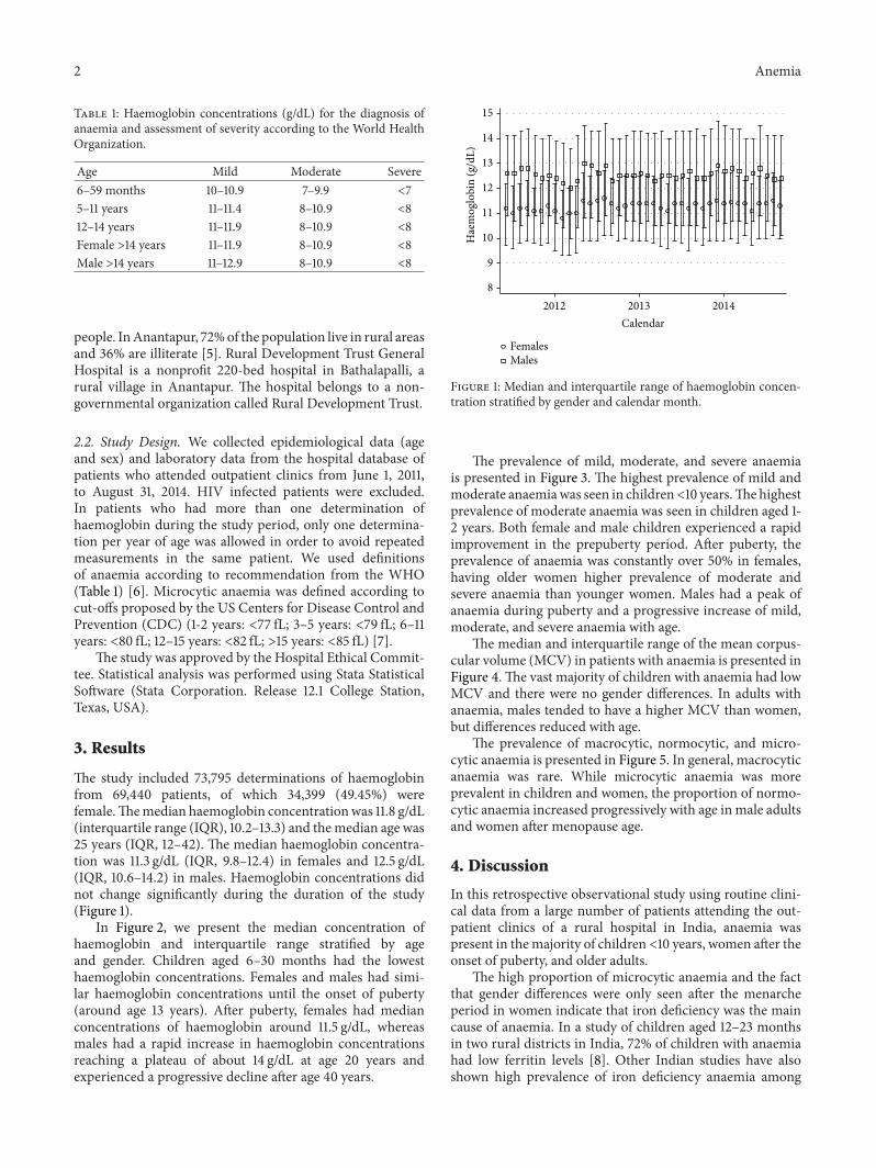
The study included 73,795 determinations of haemoglobinfrom 69,440 patients, of which 34,399 (49.45%) werefemale.Themedian haemoglobin concentrationwas 11.8 g/dL(interquartile range (IQR), 10.2–13.3) and themedian age was25 years (IQR, 12–42). The median haemoglobin concentra-tion was 11.3 g/dL (IQR, 9.8–12.4) in females and 12.5 g/dL(IQR, 10.6–14.2) in males. Haemoglobin concentrations didnot change significantly during the duration of the study(Figure 1).
In Figure 2, we present the median concentration ofhaemoglobin and interquartile range stratified by ageand gender. Children aged 6–30 months had the lowesthaemoglobin concentrations. Females and males had simi-lar haemoglobin concentrations until the onset of puberty(around age 13 years). After puberty, females had medianconcentrations of haemoglobin around 11.5 g/dL, whereasmales had a rapid increase in haemoglobin concentrationsreaching a plateau of about 14 g/dL at age 20 years andexperienced a progressive decline after age 40 years.
8
9
10
11
12
13
14
15
Hae
mog
lobi
n (g
/dL)
2012 2013 2014Calendar
FemalesMales
Figure 1: Median and interquartile range of haemoglobin concen-tration stratified by gender and calendar month.
The prevalence of mild, moderate, and severe anaemiais presented in Figure 3. The highest prevalence of mild andmoderate anaemiawas seen in children<10 years.Thehighestprevalence of moderate anaemia was seen in children aged 1-2 years. Both female and male children experienced a rapidimprovement in the prepuberty period. After puberty, theprevalence of anaemia was constantly over 50% in females,having older women higher prevalence of moderate andsevere anaemia than younger women. Males had a peak ofanaemia during puberty and a progressive increase of mild,moderate, and severe anaemia with age.
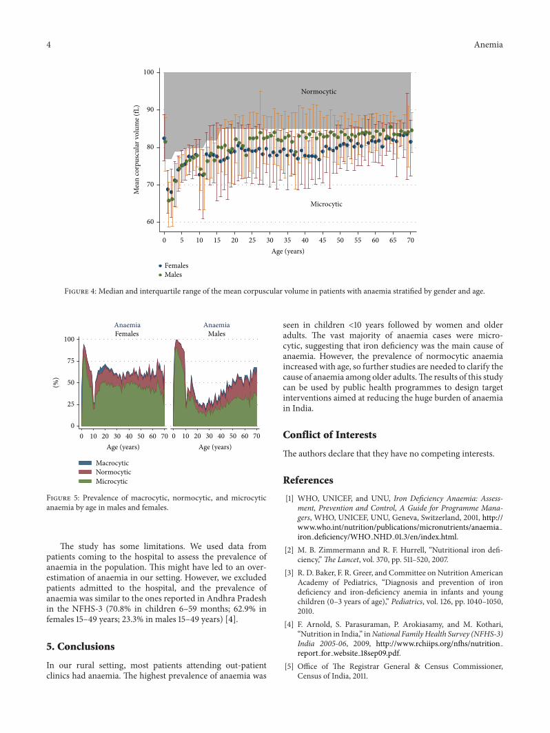
The median and interquartile range of the mean corpus-cular volume (MCV) in patients with anaemia is presented inFigure 4.The vast majority of children with anaemia had lowMCV and there were no gender differences. In adults withanaemia, males tended to have a higher MCV than women,but differences reduced with age.
The prevalence of macrocytic, normocytic, and micro-cytic anaemia is presented in Figure 5. In general, macrocyticanaemia was rare. While microcytic anaemia was moreprevalent in children and women, the proportion of normo-cytic anaemia increased progressively with age in male adultsand women after menopause age.
4. Discussion
In this retrospective observational study using routine clini-cal data from a large number of patients attending the out-patient clinics of a rural hospital in India, anaemia waspresent in themajority of children <10 years, women after theonset of puberty, and older adults.
The high proportion of microcytic anaemia and the factthat gender differences were only seen after the menarcheperiod in women indicate that iron deficiency was the maincause of anaemia. In a study of children aged 12–23 monthsin two rural districts in India, 72% of children with anaemiahad low ferritin levels [8]. Other Indian studies have alsoshown high prevalence of iron deficiency anaemia among

Anemia 3
Moderate anaemia
Mild
Mild (males)
8
9
10
11
12
13
14
15
Hae
mog
lobi
n (g
/dL)
0 5 10 15 20 25 30 35 40 45 50 55 60 65 70Age
FemalesMales
Figure 2: Median and interquartile range of haemoglobin concentration stratified by gender and age.
0
25
50
75
100
0 10 20 30 40 50 60 70 0 10 20 30 40 50 60 70
Females Males
MildModerateSevere
Age Age
Anaemia Anaemia
(%)
Figure 3: Prevalence of mild, moderate, and severe anaemia by agein males and females.
young women [9, 10]. The high prevalence of iron deficiencyanaemia among women in childbearing age has importantpublic health implications. It is estimated that anaemiaaccounts for 12.8% of maternal mortality in Asia [11]. Ironrequirements are greater in pregnancy, and iron deficiencyis associated with maternal death, preterm delivery, and lowbirth-weight [12, 13]. In India, only 28% of women consumemeat, fish, or eggs on a weekly basis [4], and the ironbioavailability of the vegetarian diet is poor [10, 14]. Effectivepublic health programmes aimed at reducing iron deficiencyamong young women could have a major impact in reducingmaternal and infant mortality [15].
The highest prevalence of anaemia was seen in chil-dren <10 years, especially in those <5 years. In India, over
95% of children are breastfed [4]. The WHO organizationrecommends introducing solid and semisolid food at theage of six months because breastfeeding does not sufficeto maintain optimal growth after this age. However, at age6–8 months only 45% of children receiving breastfeedingare given solid or semisolid food [4, 16]. Moreover, only10% of breastfeeding children and 20% of nonbreastfeedingchildren aged 6–35 months eat meat, fish, or eggs [4], whichare rich in haem iron with high bioavailability [17, 18].In the NFHS-3, only 14.6% of children aged 6–35 monthsconsumed food rich in iron in the previous 24 hours ofthe survey [4]. At this age, the effect of iron deficiency onthe neurological development can be not totally reversible[3, 19]. Consequently, the Indian Government recommendsiron and folic acid supplementations to younger children[20]. However, the programme implementation has beenpoor due to lack of logistic planning and accountability [20].In our study, we did not observe an increase in haemoglobinconcentrations during the study period suggesting that theprogramme has not achieved a reduction in the prevalenceof anaemia in our setting. Our results are in agreementwith other studies in India [21] and indicate that the ironsupplementation programme for children aged <24 monthsshould be better monitored.
In this study, we observed an increased prevalence ofanaemiawith age. Interestingly, the proportion of normocyticanaemia was highest in older adults, suggesting that othercauses than iron deficiency might have contributed to thehigh prevalence of anaemia in this group. Recent studieshave shown the poor bioavailability of vitamin B12 in thetypical Indian vegetarian diet [14] and substantial prevalenceof vitamin B12 deficiency in Indian patients with anaemia[9, 10, 22]. However, new studies investigating the aetiologyof anaemia among older adults are needed.
4 Anemia
Normocytic
Microcytic
60
70
80
90
100
Mea
n co
rpus
cula
r vol
ume (
fL)
0 5 10 15 20 25 30 35 40 45 50 55 60 65 70Age (years)
FemalesMales
Figure 4: Median and interquartile range of the mean corpuscular volume in patients with anaemia stratified by gender and age.
0 10 20 30 40 50 60 70 700 10 20 30 40 50 60
Females Males
MacrocyticNormocyticMicrocytic
Age (years)Age (years)
AnaemiaAnaemia
0
25
50
75
100
(%)
Figure 5: Prevalence of macrocytic, normocytic, and microcyticanaemia by age in males and females.
The study has some limitations. We used data frompatients coming to the hospital to assess the prevalence ofanaemia in the population. This might have led to an over-estimation of anaemia in our setting. However, we excludedpatients admitted to the hospital, and the prevalence ofanaemia was similar to the ones reported in Andhra Pradeshin the NFHS-3 (70.8% in children 6–59 months; 62.9% infemales 15–49 years; 23.3% in males 15–49 years) [4].
5. Conclusions
In our rural setting, most patients attending out-patientclinics had anaemia. The highest prevalence of anaemia was
seen in children <10 years followed by women and olderadults. The vast majority of anaemia cases were micro-cytic, suggesting that iron deficiency was the main cause ofanaemia. However, the prevalence of normocytic anaemiaincreased with age, so further studies are needed to clarify thecause of anaemia among older adults.The results of this studycan be used by public health programmes to design targetinterventions aimed at reducing the huge burden of anaemiain India.
Conflict of Interests
The authors declare that they have no competing interests.
References
[1] WHO, UNICEF, and UNU, Iron Deficiency Anaemia: Assess-ment, Prevention and Control, A Guide for Programme Mana-gers, WHO, UNICEF, UNU, Geneva, Switzerland, 2001, http://www.who.int/nutrition/publications/micronutrients/anaemiairon deficiency/WHO NHD 01.3/en/index.html.
[2] M. B. Zimmermann and R. F. Hurrell, “Nutritional iron defi-ciency,”The Lancet, vol. 370, pp. 511–520, 2007.
[3] R. D. Baker, F. R. Greer, and Committee on Nutrition AmericanAcademy of Pediatrics, “Diagnosis and prevention of irondeficiency and iron-deficiency anemia in infants and youngchildren (0–3 years of age),” Pediatrics, vol. 126, pp. 1040–1050,2010.
[4] F. Arnold, S. Parasuraman, P. Arokiasamy, and M. Kothari,“Nutrition in India,” inNational FamilyHealth Survey (NFHS-3)India 2005-06, 2009, http://www.rchiips.org/nfhs/nutritionreport for website 18sep09.pdf.
[5] Office of The Registrar General & Census Commissioner,Census of India, 2011.

Anemia 5
[6] WHO, Haemoglobin Concentrations for the Diagnosis of Ana-emia and Assessment of Severity, WHO, Geneva, Switzerland,2011, http://www.who.int/vmnis/indicators/haemoglobin/en/.
[7] “Recommendations to prevent and control iron deficiency inthe United States. Centers for Disease Control and Prevention,”MMWR Recommendations and Reports, vol. 47, pp. 1–29, 1998.
[8] S.-R. Pasricha, J. Black, S. Muthayya et al., “Determinants ofanemia among young children in rural India,” Pediatrics, vol.126, no. 1, pp. e140–e149, 2010.
[9] K. C. Menon, S. A. Skeaff, C. D. Thomson et al., “Concurrentmicronutrient deficiencies are prevalent in nonpregnant ruraland tribal women from central India,” Nutrition, vol. 27, no. 4,pp. 496–502, 2011.
[10] P. Thankachan, S. Muthayya, T. Walczyk, A. V. Kurpad, andR. F. Hurrell, “An analysis of the etiology of anemia and irondeficiency in young women of low socioeconomic status inBangalore, India,” Food and Nutrition Bulletin, vol. 28, no. 3, pp.328–336, 2007.
[11] K. S. Khan, D. Wojdyla, L. Say, A. M. Gulmezoglu, and P. F. vanLook, “WHO analysis of causes of maternal death: a systematicreview,”The Lancet, vol. 367, no. 9516, pp. 1066–1074, 2006.
[12] K. Kalaivani, “Prevalence & consequences of anaemia in preg-nancy,” Indian Journal of Medical Research, vol. 130, no. 5, pp.627–633, 2009.
[13] L. H. Allen, “Anemia and iron deficiency: effects on pregnancyoutcome,”TheAmerican Journal of Clinical Nutrition, vol. 71, no.5, pp. 1280s–1284s, 2000.
[14] K. Shridhar, P. K. Dhillon, L. Bowen et al., “Nutritional profileof Indian vegetarian diets—the Indian Migration Study (IMS),”Nutrition Journal, vol. 13, article 55, 2014.
[15] Z. A. Bhutta, J. K. Das, R. Bahl et al., “Can available interven-tions end preventable deaths in mothers, newborn babies, andstillbirths, and at what cost?”The Lancet, vol. 384, no. 9940, pp.347–370, 2014.
[16] C. J. Chantry, C. R. Howard, and P. Auinger, “Full breastfeedingduration and risk for iron deficiency in U.S. infants,” Breastfeed-ing Medicine, vol. 2, no. 2, pp. 63–73, 2007.
[17] M. B. Zimmermann, N. Chaouki, and R. F. Hurrell, “Iron defi-ciency due to consumption of a habitual diet low in bioavailableiron: a longitudinal cohort study in Moroccan children,” TheAmerican Journal of Clinical Nutrition, vol. 81, no. 1, pp. 115–121,2005.
[18] R. Hurrell, “How to ensure adequate iron absorption from iron-fortified food,”Nutrition Reviews, vol. 60, supplement 7, pp. S7–S15, 2002.
[19] S.More, V. B. Shivkumar, N. Gangane, and S. Shende, “Effects ofiron deficiency on cognitive function in school going adolescentfemales in rural area of central India,”Anemia, vol. 2013, ArticleID 819136, 5 pages, 2013.
[20] P. V. Kotecha, “Nutritional anemia in young children with focuson Asia and India,” Indian Journal of Community Medicine, vol.36, no. 1, pp. 8–16, 2011.
[21] R. K. Singh and S. Patra, “Extent of anaemia among preschoolchildren in EAG States, India: a challenge to policy makers,”Anemia, vol. 2014, Article ID 868752, 9 pages, 2014.
[22] A. Bhardwaj,D.Kumar, S. K. Raina, P. Bansal, S. Bhushan, andV.Chander, “Rapid assessment for coexistence of vitamin B12 andiron deficiency anemia among adolescent males and females innorthern himalayan state of India,” Anemia, vol. 2013, ArticleID 959605, 5 pages, 2013.

Submit your manuscripts athttp://www.hindawi.com
Stem CellsInternational
Hindawi Publishing Corporationhttp://www.hindawi.com Volume 2014
Hindawi Publishing Corporationhttp://www.hindawi.com Volume 2014
MEDIATORSINFLAMMATION
of
Hindawi Publishing Corporationhttp://www.hindawi.com Volume 2014
Behavioural Neurology
EndocrinologyInternational Journal of
Hindawi Publishing Corporationhttp://www.hindawi.com Volume 2014
Hindawi Publishing Corporationhttp://www.hindawi.com Volume 2014
Disease Markers
Hindawi Publishing Corporationhttp://www.hindawi.com Volume 2014
BioMed Research International
OncologyJournal of
Hindawi Publishing Corporationhttp://www.hindawi.com Volume 2014
Hindawi Publishing Corporationhttp://www.hindawi.com Volume 2014
Oxidative Medicine and Cellular Longevity
Hindawi Publishing Corporationhttp://www.hindawi.com Volume 2014
PPAR Research
The Scientific World JournalHindawi Publishing Corporation http://www.hindawi.com Volume 2014
Immunology ResearchHindawi Publishing Corporationhttp://www.hindawi.com Volume 2014
Journal of
ObesityJournal of
Hindawi Publishing Corporationhttp://www.hindawi.com Volume 2014
Hindawi Publishing Corporationhttp://www.hindawi.com Volume 2014
Computational and Mathematical Methods in Medicine
OphthalmologyJournal of
Hindawi Publishing Corporationhttp://www.hindawi.com Volume 2014
Diabetes ResearchJournal of
Hindawi Publishing Corporationhttp://www.hindawi.com Volume 2014
Hindawi Publishing Corporationhttp://www.hindawi.com Volume 2014
Research and TreatmentAIDS
Hindawi Publishing Corporationhttp://www.hindawi.com Volume 2014
Gastroenterology Research and Practice
Hindawi Publishing Corporationhttp://www.hindawi.com Volume 2014
Parkinson’s Disease
Evidence-Based Complementary and Alternative Medicine
Volume 2014Hindawi Publishing Corporationhttp://www.hindawi.com


![Research Article Prevalence of Anaemia among Pregnant Women …downloads.hindawi.com/journals/bmri/2014/849080.pdf · 2019-07-31 · of anaemia during pregnancy [ ]. e management](https://img.pdfslide.us/doc/110x75/5f7be4047395f42e6848e8a1/research-article-prevalence-of-anaemia-among-pregnant-women-2019-07-31-of-anaemia.jpg)

![Anaemia in Pregnancy - Welcome to JPAC...Anaemia during pregnancy is a global problem WHO region Prevalence of anaemia (%) in pregnant women [95% CI] Number of pregnant women affected](https://img.pdfslide.us/doc/110x75/5f048bb57e708231d40e80e4/anaemia-in-pregnancy-welcome-to-jpac-anaemia-during-pregnancy-is-a-global.jpg)