Embed Size (px)
Citation preview
1479Pharmacogenomics (2014) 15(11), 1479–1494 ISSN 1462-2416
part of
PharmacogenomicsResearch Article
10.2217/PGS.14.97 © 2014 Future Medicine Ltd
Pharmacogenomics
Research Article15
11
2014
Aim: Pharmacogenetics of methotrexate (MTX) contributes to interindividual differences in toxicity. We aimed to evaluate the impact of SNPs within the MTX pathway genes on MTX-induced toxicity and MTX plasma levels at 48 h following treatment in Asian adults with acute lymphoblastic leukemia or non-Hodgkin lymphoma. Patients & methods: Patients (n = 71) were genotyped for MTHFR C677T, MTHFR A1298C, SLC19A1 G80A, ABCG2 C421A and ABCB1 C3435T using the Sequenom MassARRAY® platform. Plasma MTX concentrations at 48 h were measured by fluorescence polarization immunoassay. Results: Forty-eight patients had hematopoietic toxicity, 51 had hepatic toxicity and 36 had mucositis. Patients homozygous for MTHFR 677TT were associated with increased risk of both hematopoietic (odds ratio [OR]: 9.03; 95% CI: 2.28–36.16; p = 0.002) and hepatic (OR: 3.92; 95% CI: 1.01–15.11; p = 0.036) toxicities. Hepatic toxicity was associated with SLC19A1 G80A (OR: 5.27, 95% CI: 1.21–22.72; p = 0.032) and ABCB1 C3435T (OR: 8.62; 95% CI: 1.96–37.57; p = 0.004). However, polymorphisms in MTHFR A1298C and ABCG2 C421A were not associated with any of the toxicities, and mucositis was not associated with any polymorphisms of the MTX pathway genes. Patients with MTHFR C677T and ABCB1 C3435T polymorphisms appear to have significantly higher MTX plasma concentrations (p < 0.05). Conclusion: Our results in Asian adults provides evidence for the contribution pharmacogenetics to the toxicity of high-dose MTX and plasma MTX concentrations at 48 h following treatment in patients with acute lymphoblastic leukemia or non-Hodgkin lymphoma. These results will contribute towards the effort of MTX therapy individualization.
Original submitted 24 April 2014; Revision submitted 6 June 2014
Keywords: high-dose methotrexate • methotrexate plasma concentration • methotrexate toxicity • NHL • non-Hodgkin lymphoma • single nucleotide polymorphism • SNP
BackgroundThe folate inhibitor drug, methotrex-ate (MTX) has been shown to be benefi-cial for the treatment of a variety of adult and childhood cancers, including breast cancer [1], osteo sarcoma [2], acute lympho-blastic leukemia (ALL) and lymphoma [3]. Intravenous infusion of high-dose MTX (HDMTX; ≥0.5 g/m2) in combination with other chemotherapeutic agents, as opposed to a conventional dose of <0.5 g/m2, has been shown to be effective in adult non-
Hodgkin lymphoma (NHL) and ALL [4]. However, HDMTX treatment can cause substantial toxicity; leading to nonadher-ence to treatment, and hence can lead to increased mortality and morbidity [3].
There is considerable interpatient vari-ability in occurrence of toxicity owing to HDMTX, causing unpredictable toxicity even when given in fixed doses to similar cohorts of patients [5]. Toxicity can affect the bone marrow causing myelosuppression, the liver leading to elevated liver enzymes and the
Effect of polymorphisms within methotrexate pathway genes on methotrexate toxicity and plasma levels in adults with hematological malignancies
Sujatha Suthandiram1, Gin-Gin Gan*,2, Shamsul Mohd Zain1, Ping-Chong Bee2, Lay-Hoong Lian3, Kian-Meng Chang4, Tee-Chuan Ong4 & Zahurin Mohamed1
1The Pharmacogenomics Laboratory,
Department of Pharmacology, University
of Malaya, Kuala Lumpur, Malaysia 2Department of Medicine, Faculty of
Medicine, University of Malaya, Kuala
Lumpur, Malaysia 3Department of Molecular Medicine,
Faculty of Medicine, University of
Malaya, Kuala Lumpur, Malaysia 4Department of Hematology, Ampang
Hospital, Selangor Darul Ehsan, Malaysia
*Author for correspondence:
Tel.: +603 79492585
Fax: +603 79494613
For reprint orders, please contact: [email protected]
1480 Pharmacogenomics (2014) 15(11) future science group
Research Article Suthandiram, Gan, Zain et al.
gastrointestinal tract causing oral mucositis [6]. MTX-induced toxicity is generally owing to the inhibition of normal cells and tissue growth adjacent to the target abnormal cells [7]. In normal clinical practice, plasma MTX monitoring is essential especially for patients who have been administered HDMTX in order to detect those who are at risk for HDMTX-related tox-icities and to determine the duration of leucovorin res-cue. Identifying genetic predictors of MTX toxicities is important for dosage adjustment and minimization of adverse effects.
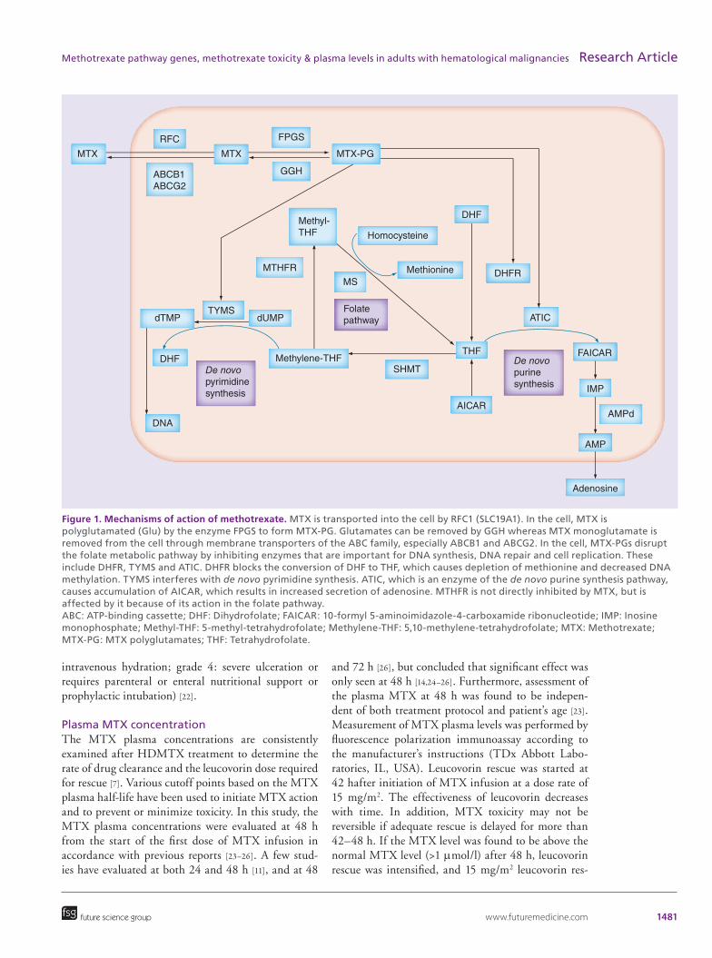
MTX action on the MTX pathway involves sev-eral metabolizing enzymes and transporters (Figure 1) whose functions have been suggested to be altered by genetic polymorphisms [3]. MTHFR is a key regulator enzyme that is essential for DNA synthesis and DNA methylation [8]. Solute carrier (SLC) transporters are involved in MTX uptake and may modify its toxicity and efficacy. SLC19A1 (also known as RFC1) has a general role in folate transport and mediates the trans-port of MTX into cells [9]. MTX is pumped out of the cell by a variety of ATP-binding cassette (ABC) efflux transporters [10]. ABCG2 (formerly known as BCRP) and ABCB1 (MDR1) are two ABC family genes. It had been shown that the expression patterns of the ABCB1 and ABCG2 genes affect the pharmacokinetics of MTX, which then significantly affects MTX activ-ity and toxicity [6,11]. Increased incidence of toxicity has been well documented in association with poly-morphisms in these MTX pathway genes [10–14]. Given these findings, the sheer scale of MTX toxicity might be profoundly affected by genetic polymorphisms in the MTX metabolism pathway.
The bulk of research on MTX to date presents con-flicting results, and there are marked differences in pharmacogenetics between various populations [6,15]. While pediatric evidence is abundant, there is limited data regarding adults [16]. Therefore, we investigated the association between polymorphisms in the MTX pathway genes with MTX-associated toxicity in a cohort of adult patients with ALL or NHL treated with HDMTX. In addition, our study provides data on the Asian population, in which there has only previously been one study carried out in adults [17].
Patients & methodsSubjectsThe patients included in this study were 71 Malaysian adults with ALL, peripheral T-cell lymphoma (PTCL) and Burkitt’s lymphoma. This study was a collabora-tion between the University Malaya Medical Centre (UMMC) and Ampang Hospital, both of which are located in the city of Kuala Lumpur, Malaysia. The study protocol was approved by the medical ethics com-
mittees of both centres. Patients were recruited from hematology clinics between September 2010 and Sep-tember 2013. The eligible criteria were age ≥18-years-old, not afflicted with other active malignancies and HIV free. A standardized extraction template was used to obtain demographic details, medical history, types of hematological malignancies and laboratory inves-tigations from the medical records. NHL types were classified according to the WHO 2008 classification system [18]. At the time of peripheral blood collection, written informed consent was given by all subjects.
Treatment protocolsThe multiagent chemotherapeutic protocols used were the modified German multicentre study group for treatment of adult ALL (GMALL) 07/2003, modified non-Hodgkin Lymphoma–Berlin–Frankfurt–Mün-ster (NHL-BFM) 90 protocol and hyperfractionated cyclophosphamide, vincristine, adriamycin and dexa-methasone (hyper-CVAD) protocol-course B [19,20]. All patients received high-dose MTX treatment, par-ticularly 1500 mg/m2 over 24 h or 1000 mg/m2 over 24 h for patients diagnosed with ALL, 1500 mg/m2 over 24 h for Burkitt’s lymphoma and 1000 mg/m2 over 24 h for PTCL patients, followed by leucovorin rescue. The infusion time between the different MTX treatments is the same for the three patient groups.
Toxicity evaluation of MTXToxicities were evaluated according to the National Cancer Institute common toxicity criteria (NCI-CTC) Version 2.0 [21] and included hematopoietic toxicity, as well as nonhematopoietic toxicity (hepatic toxicity and mucositis). MTX-induced toxicity was assessed for the period between the administration of MTX and the next course of chemotherapy, whereby the highest grade of toxicity observed in each patient was recorded. The subsequent course of chemotherapy were started during a period of 1–3 weeks after the MTX infu-sions following resolution of toxicities. Toxicity of any grade was considered as the clinical end point. How-ever, toxicity grade 3 or greater was not measured as an additional end point due to limitation in sample size, which would render the analysis underpowered. Hematologic toxicity was determined by the presence of polymorphnuclear leukocytes <0.5 × 10⁹/l, whereas hepatic toxicity was by the presence of an increase in ALP ≥2.5 × upper limit of normal (ULN), and/or bili-rubin ≥1.5 × ULN and/or ALT ≥2.5 × ULN and/or AST >2.5 × ULN. Stomatitis was graded according to its severity (grade 1: painless ulcer, erythema or mild soreness in the absence of lesions; grade 2: painful erythema, edema or ulcer, but able to eat or swallow; grade 3: painful erythema, edema or ulcer requiring
www.futuremedicine.com 1481
Figure 1. Mechanisms of action of methotrexate. MTX is transported into the cell by RFC1 (SLC19A1). In the cell, MTX is polyglutamated (Glu) by the enzyme FPGS to form MTX-PG. Glutamates can be removed by GGH whereas MTX monoglutamate is removed from the cell through membrane transporters of the ABC family, especially ABCB1 and ABCG2. In the cell, MTX-PGs disrupt the folate metabolic pathway by inhibiting enzymes that are important for DNA synthesis, DNA repair and cell replication. These include DHFR, TYMS and ATIC. DHFR blocks the conversion of DHF to THF, which causes depletion of methionine and decreased DNA methylation. TYMS interferes with de novo pyrimidine synthesis. ATIC, which is an enzyme of the de novo purine synthesis pathway, causes accumulation of AICAR, which results in increased secretion of adenosine. MTHFR is not directly inhibited by MTX, but is affected by it because of its action in the folate pathway. ABC: ATP-binding cassette; DHF: Dihydrofolate; FAICAR: 10-formyl 5-aminoimidazole-4-carboxamide ribonucleotide; IMP: Inosine monophosphate; Methyl-THF: 5-methyl-tetrahydrofolate; Methylene-THF: 5,10-methylene-tetrahydrofolate; MTX: Methotrexate; MTX-PG: MTX polyglutamates; THF: Tetrahydrofolate.
future science group
Methotrexate pathway genes, methotrexate toxicity & plasma levels in adults with hematological malignancies Research Article
intravenous hydration; grade 4: severe ulceration or requires parenteral or enteral nutritional support or prophylactic intubation) [22].
Plasma MTX concentrationThe MTX plasma concentrations are consistently examined after HDMTX treatment to determine the rate of drug clearance and the leucovorin dose required for rescue [7]. Various cutoff points based on the MTX plasma half-life have been used to initiate MTX action and to prevent or minimize toxicity. In this study, the MTX plasma concentrations were evaluated at 48 h from the start of the first dose of MTX infusion in accordance with previous reports [23–26]. A few stud-ies have evaluated at both 24 and 48 h [11], and at 48
and 72 h [26], but concluded that significant effect was only seen at 48 h [14,24–26]. Furthermore, assessment of the plasma MTX at 48 h was found to be indepen-dent of both treatment protocol and patient’s age [23]. Measurement of MTX plasma levels was performed by fluorescence polarization immunoassay according to the manufacturer’s instructions (TDx Abbott Labo-ratories, IL, USA). Leucovorin rescue was started at 42 hafter initiation of MTX infusion at a dose rate of 15 mg/m2. The effectiveness of leucovorin decreases with time. In addition, MTX toxicity may not be reversible if adequate rescue is delayed for more than 42–48 h. If the MTX level was found to be above the normal MTX level (>1 μmol/l) after 48 h, leucovorin rescue was intensified, and 15 mg/m2 leucovorin res-
RFC FPGS
GGH
MTX-PG
Methyl-THF
THF
Methionine
Homocysteine
DHF
DHFR
Adenosine
AMP
AMPd
IMP
FAICAR
ATIC
De novopurinesynthesis
MTHFR
MS
TYMSdTMP dUMP
SHMT
AICAR
Folatepathway
Methylene-THFDHF
DNA
De novopyrimidinesynthesis
MTXMTX
ABCB1ABCG2
1482 Pharmacogenomics (2014) 15(11) future science group
Research Article Suthandiram, Gan, Zain et al.
cue dose was given every 6 h until MTX plasma levels reached <0.05 μmol/l [27].
Genetic predictorsGenomic DNA was extracted from the collected blood samples using a QiAamp® DNA Blood Mini Kit (Qiagen, Hilden, Germany). The quality of DNA was checked consistently to confirm that 260/280 and 260/230 absorbance ratios exceed 1.8 to indi-cate high-quality DNA. The genomic DNA was then diluted to 10 and 20 ng/μl, for samples and duplicates, respectively and then placed in the well. A volume of 1 μl of DNA was used in every amplification reaction. The polymorphisms within MTX pathway genes that include MTHFR C677T, MTHFR A1298C, SLC19A1 G80A, ABCG2 C421A and ABCB1 C3435T were genotyped at the University of Hong Kong, Genome Research Centre using the Sequenom MassAR-RAY® technology platform with the iPLEX® GOLD chemistry (Sequenom, CA, USA) in the conditions recommended by the manufacturer. MassARRAY AssayDesign software package (v4.0) was used to design the specific assays with proximal SNPs filter-ing. The quality of the PCR fragment amplification and extension primer specificity was checked prior to running the reaction. Residual nucleotides were dephosphorylated prior to the iPLEX GOLD reaction. Based on a single-base extension, reaction products were desalted with SpectroClean resin (Sequenom), and 10 nl was spotted onto the SpectroCHIP® using the MassARRAY Nanodispenser. The MassARRAY Analyzer Compact MALDI-TOF mass spectrometer was used to determine the mass. The MassARRAY Typer 4.0 software was used for proper data acquisi-tion and analysis. Genotypes were called after cluster analysis using the default setting of the Gaussian mix-ture model. Inspection of the clusters was carried out to ensure a clear cluster separation with good signal to noise cutoff. A manual review was carried out to further clarify uncertain genotype calls. Assays with less than 80% call rate within the same SpectroCHIP was considered as having failed. A blank and five duplicates were introduced as quality controls. Spec-troCHIP with more than 25% call rate in the blank control or less than 99.5% concordance in duplicate checks, and more than 10% call rate in blank checks were considered to have failed and would be required to be repeated.
Statistical analysisAll values were presented as mean ± standard devia-tion for continuous data and as percentages for cat-egorical data. Deviation from the Hardy–Weinberg equilibrium was assessed using a goodness-of-fit χ2
where p < 0.05 is not consistent with Hardy–Wein-berg equilibrium. Logistic regression was employed to examine the association of the MTX pathway gene poly morphisms with MTX-induced toxicity and MTX plasma concentration at 48 h. Multivariate analysis and stepwise regression failed to indicate any contributing factor. Although our initial multivariate analysis did not reveal age and gender as factors, we still included these variables together with different types of diseases in our preanalysis for confirmation, as age and gender are known risk factors associated with adverse events. However, age and gender did not affect the results and there were no significant findings.
Correction for multiple testing was performed using Bonferroni method. All statistical tests were two-sided and results were considered significant if p < 0.05. All statistical analyses were performed using the SPSS software version 21.0 (SPSS, IBM Corp., IL, USA).
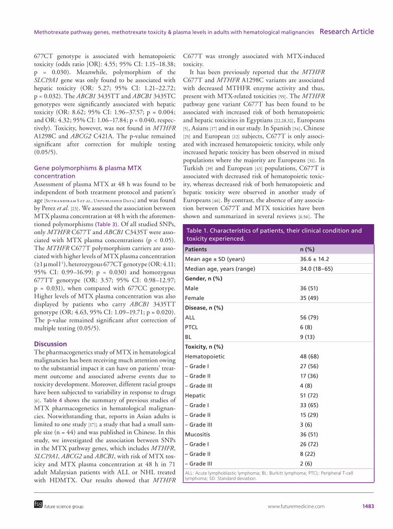
ResultsPatients & distribution of toxicityTable 1 provides a summary of the characteristics of the patients, their clinical condition and the toxicity experienced. There was no difference in the number of patients developing each kind of toxicity accord-ing to the treatment protocol used; thus all patients were analyzed together. The proportion of patients was considered equal between both gender with the median age of all subjects at 34 years. The majority of the patients had ALL (79%) and followed by Burkitt’s lymphoma (13%) and PTCL (8%). Hepatic toxicity was the most frequently observed toxicity appearing in 72% of the patients, followed closely by hemato-poietic toxicity (68%) and mucositis (51%). Of those with hepatic toxicity, 18 patients (35%) experienced grade 2 or greater. While for hematopoietic toxic-ity and mucositis, the number documented was 21 (44%) and 10 (28%). There were only four (8%), three (6%) and two (6%) subjects for toxicity grade 3 (hematopoietic, hepatic and mucositis, respec-tively). However, there are no subjects with toxicity of grade 4 and above.
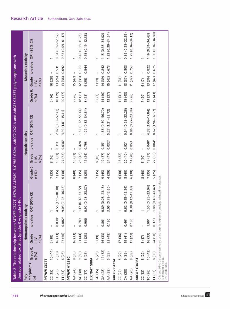
Pharmacogenetics of MTXThe relationship between SNPs within the MTX pathway genes and therapy-related toxicities among patients is shown in Table 2. All SNP markers studied were in Hardy–Weinberg equilibrium. The MTHFR 677TT genotype confers greater risk of hematopoietic toxicity (p = 0.002) and hepatic toxicity (p = 0.036) when compared with MTHFR 677CC. The risk for the 677TT homozygous patients as compared with 677CC is ninefold higher in the former and nearly fourfold higher in the latter. It is also notable that MTHFR
www.futuremedicine.com 1483future science group
Methotrexate pathway genes, methotrexate toxicity & plasma levels in adults with hematological malignancies Research Article
677CT genotype is associated with hematopoietic toxicity (odds ratio [OR]: 4.55; 95% CI: 1.15–18.38; p = 0.030). Meanwhile, polymorphism of the SLC19A1 gene was only found to be associated with hepatic toxicity (OR: 5.27; 95% CI: 1.21–22.72; p = 0.032). The ABCB1 3435TT and ABCB1 3435TC genotypes were significantly associated with hepatic toxicity (OR: 8.62; 95% CI: 1.96–37.57; p = 0.004; and OR: 4.32; 95% CI: 1.06–17.84; p = 0.040, respec-tively). Toxicity, however, was not found in MTHFR A1298C and ABCG2 C421A. The p-value remained significant after correction for multiple testing (0.05/5).
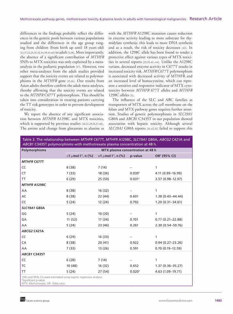
Gene polymorphisms & plasma MTX concentrationAssessment of plasma MTX at 48 h was found to be independent of both treatment protocol and patient’s age [Suthandiram S et al., Unpublished Data] and was found by Perez et al. [23]. We assessed the association between MTX plasma concentration at 48 h with the aforemen-tioned polymorphisms (Table 3). Of all studied SNPs, only MTHFR C677T and ABCB1 C3435T were asso-ciated with MTX plasma concentrations (p < 0.05). The MTHFR C677T polymorphism carriers are asso-ciated with higher levels of MTX plasma concentration (≥1 μmol l-1), heterozygous 677CT genotype (OR: 4.11; 95% CI: 0.99–16.99; p = 0.030) and homozygous 677TT genotype (OR: 3.57; 95% CI: 0.98–12.97; p = 0.031), when compared with 677CC genotype. Higher levels of MTX plasma concentration was also displayed by patients who carry ABCB1 3435TT geno type (OR: 4.63, 95% CI: 1.09–19.71; p = 0.020). The p-value remained significant after correction of multiple testing (0.05/5).
DiscussionThe pharmacogenetics study of MTX in hematological malignancies has been receiving much attention owing to the substantial impact it can have on patients’ treat-ment outcome and associated adverse events due to toxicity development. Moreover, different racial groups have been subjected to variability in response to drugs [6]. Table 4 shows the summary of previous studies of MTX pharmacogenetics in hematological malignan-cies. Notwithstanding that, reports in Asian adults is limited to one study [17]; a study that had a small sam-ple size (n = 44) and was published in Chinese. In this study, we investigated the association between SNPs in the MTX pathway genes, which includes MTHFR, SLC19A1, ABCG2 and ABCB1, with risk of MTX tox-icity and MTX plasma concentration at 48 h in 71 adult Malaysian patients with ALL or NHL treated with HDMTX. Our results showed that MTHFR
C677T was strongly associated with MTX-induced toxicity.
It has been previously reported that the MTHFR C677T and MTHFR A1298C variants are associated with decreased MTHFR enzyme activity and thus, present with MTX-related toxicities [55]. The MTHFR pathway gene variant C677T has been found to be associated with increased risk of both hematopoietic and hepatic toxicities in Egyptians [22,28,32], Europeans [5], Asians [17] and in our study. In Spanish [34], Chinese [25] and European [12] subjects, C677T is only associ-ated with increased hematopoietic toxicity, while only increased hepatic toxicity has been observed in mixed populations where the majority are Europeans [31]. In Turkish [39] and European [43] populations, C677T is associated with decreased risk of hematopoietic toxic-ity, whereas decreased risk of both hematopoietic and hepatic toxicity were observed in another study of Europeans [46]. By contrast, the absence of any associa-tion between C677T and MTX toxicities have been shown and summarized in several reviews [6,56]. The
Patients n (%)
Mean age ± SD (years) 36.6 ± 14.2
Median age, years (range) 34.0 (18–65)
Gender, n (%)
Male 36 (51)
Female 35 (49)
Disease, n (%)
ALL 56 (79)
PTCL 6 (8)
BL 9 (13)
Toxicity, n (%)
Hematopoietic 48 (68)
– Grade I 27 (56)
– Grade II 17 (36)
– Grade III 4 (8)
Hepatic 51 (72)
– Grade I 33 (65)
– Grade II 15 (29)
– Grade III 3 (6)
Mucositis 36 (51)
– Grade I 26 (72)
– Grade II 8 (22)
– Grade III 2 (6)
ALL: Acute lymphoblastic lymphoma; BL: Burkitt lymphoma; PTCL: Peripheral T-cell lymphoma; SD: Standard deviation.
Table 1. Characteristics of patients, their clinical condition and toxicity experienced.
1484 Pharmacogenomics (2014) 15(11) future science group
Research Article Suthandiram, Gan, Zain et al.
Poly
-m
orp
his
ms
(n)
Hem
ato
po
ieti
c to
xici
ty
H
epat
ic t
oxi
city
Mu
cosi
tis
toxi
city
Gra
de
0,
n (
%)
Gra
de
I–
IV,
n (
%)
p-v
alu
eO
R†
(95%
CI)
G
rad
e 0,
n
(%
)G
rad
e I–
IV,
n (
%)
p-v
alu
eO
R†
(95%
CI)
G
rad
e 0,
n
(%
)G
rad
e I–
IV,
n (
%)
p-v
alu
eO
R†
(95%
CI)
MTH
FR C
677
T
CC
(15
)10
(4
4)
5 (1
0)
–1
7
(35
)8
(16
)–
1
5 (1
4)
10 (
28)
–1
CT
(23
)7
(30
)16
(33
)0.
030‡
4.5
5 (1
.15
–18
.38
)
7 (3
5)
16 (
31)
0.31
12.
02 (
0.53
–57.
72)
10
(29
)13
(26
)0.
531
0.6
4 (0
.17–
12.5
2)
TT
(33
)6
(26
)27
(56
)0.
002
‡9.
03 (
2.28
–36.
16)
6
(30
)27
(53
)0.
036‡
3.92
(1.
01–1
5.11
)
20 (
57)
13 (
36)
0.09
20.
33 (
0.09
–01.
17)
MTH
FR A
129
8C
AA
(24
)8
(35
)16
(33
)–
1
8 (4
0)
16 (
31)
–1
9
(26
)15
(42
)–
1
AC
(30
)9
(39
)21
(4
4)
0.78
91.
17 (
0.37
–33.
72)
7
(35
)23
(45
)0.
424
1.62
(0.
52–5
5.4
4)
18
(51
)12
(33
)0.
100
0.42
(0.
13–1
1.23
)
CC
(17
)6
(26
)11
(23
)0.
90
00.
92 (
0.28
–23.
37)
5
(25
)12
(24
)0.
793
1.22
(0.
32–3
4.6
4)
8
(23
)9
(25
)0.
54
40.
65 (
0.19
–12.
38
)
SLC
19A
1 G
80A
GG
(15
)6
(26
)9
(19
)–
1
7 (3
5)
8 (1
6)
–1
8
(23
)7
(19
)–
1
GA
(28
)12
(52
)16
(33
)0.
861
0.89
(0.
28–2
3.18
)
9 (4
5)
19 (
37)
0.35
11.
86
(0.5
0–5
6.70
)
14 (
40
)14
(39
)0.
842
1.15
(0.
35–3
4.0
2)
AA
(28
)5
(22
)23
(4
8)
0.12
03.
05 (
0.78
–12.
60)
4
(20
)24
(47
)0.
032‡
5.27
(1.
21–2
2.72
)
13 (
37)
15 (
42)
0.67
31.
33 (
0.39
–34
.64
)
AB
CG
2 C
421
A
CC
(22
)5
(22
)17
(36
)–
1
6 (3
0)
16 (
32)
–1
11
(31
)11
(31
)–
1
CA
(28
)9
(39
)19
(41
)0.
461
0.62
(0.
18–1
2.24
)
8 (4
0)
20 (
40
)0.
921
0.9
4 (0
.28
–23.
26)
15
(43
)13
(37
)0.
803
0.8
6 (0
.25
–22.
65)
AA
(20
)9
(39
)11
(23
)0.
130
0.3
8 (0
.12–
11.3
3)
6
(30
)14
(28
)0.
853
0.8
6 (0
.27–
23.3
4)
9
(26
)11
(32
)0.
753
1.25
(0.
36–3
4.1
2)
AB
CB
1 C
3435
T
CC
(13
)5
(22
)8
(17)
–1
8
(40
)5
(10
)–
1
7 (2
0)
6 (1
7)–
1
TC (
26)
10 (
43)
16 (
33)
1.0
011.
00
(0.2
6–2
3.9
4)
7
(35
)19
(37
)0.
04
0‡4
.32
(1.0
6–1
7.8
4)
13
(37
)13
(36
)0.
822
1.16
(0.
31–3
4.4
3)
TT
(32
)8
(35
)24
(50
)0.
373
1.8
8 (0
.48
–47.
42)
5
(25
)27
(53
)0.
00
4‡
8.6
2 (1
.96
–37.
57)
15
(43
)17
(47
)0.
675
1.30
(0.
36–3
4.8
0)
†ORs and 95% CIs were estimated using logistic regression analysis adjusted for diseases.
‡Significant p-value.
OR: Odds ratio.
Tab
le 2
. Th
e re
lati
on
ship
bet
wee
n M
THFR
C67
7T,
MTH
FR A
129
8C, S
LC19
A1
G8
0A, A
BC
G2
C42
1A a
nd
AB
CB
1 C
3435
T p
oly
mo
rph
ism
s w
ith
th
erap
y-re
late
d t
oxi
citi
es (
gra
des
0 v
s g
rad
e I–
IV).
www.futuremedicine.com 1485future science group
Methotrexate pathway genes, methotrexate toxicity & plasma levels in adults with hematological malignancies Research Article
differences in the findings probably reflect the differ-ences in the genetic pools between various populations studied and the differences in the age group rang-ing from children (from birth up until 18 years old) [12,17,22,25,31,32,34,39,43,46] to adult [5,28]. More importantly, the absence of a significant contribution of MTHFR SNPs to MTX toxicities was only explained by a meta-analysis in the pediatric population [57]. However, two other meta-analyses from the adult studies provided support that the toxicity events are related to polymor-phisms in the MTHFR gene [15,16]. Our results from Asian adults therefore confirm the adult meta-analyses, thereby affirming that the toxicity events are related to the MTHFR C677T polymorphism. This should be taken into consideration in treating patients carrying the TT risk genotypes in order to prevent development of toxicity.
We report the absence of any significant associa-tion between MTHFR A1298C and MTX toxicities, which is supported by previous studies [10,22,29,35,37,46]. The amino acid change from glutamate to alanine as
with the MTHFR A1298C mutation causes reduction in enzyme activity leading to more substrate for thy-midylate synthesis; this leads to more DNA synthesis and as a result, the risk of toxicity decreases [42]. In addition, the 1298C allele has been found to render a protective effect against various types of MTX toxici-ties in several reports [25,33,42–44]. Unlike the A1298C variant, decreased enzyme activity in C677T results in increased toxicity risk. MTHFR C677T polymorphism is associated with decreased activity of MTHFR and an increased level of homocysteine, which can repre-sent a sensitive and responsive indicator of MTX cyto-toxicity between MTHFR 677T alleles and MTHFR 1298C alleles [5].
The influence of the SLC and ABC families as transporters of MTX across the cell membrane on the folate and MTX pathway genes requires further atten-tion. Studies of genetic polymorphisms in SLC19A1 G80A and ABCB1 C3435T in our population showed association with hepatic toxicity. Although several SLC19A1 G80A reports [41,42,52] failed to support this
Polymorphisms MTX plasma concentration at 48 h
≤1 μmol l-1, n (%) >1 μmol l-1, n (%) p-value OR† (95% CI)
MTHFR C677T
CC 8 (38) 7 (14) – 1
CT 7 (33) 18 (36) 0.030‡ 4.11 (0.99–16.99)
TT 6 (29) 25 (50) 0.031‡ 3.57 (0.98–12.97)
MTHFR A1298C
AA 8 (38) 16 (32) – 1
AC 8 (38) 22 (44) 0.601 1.38 (0.43–44.44)
CC 5 (24) 12 (24) 0.792 1.20 (0.31–34.61)
SLC19A1 G80A
GG 5 (24) 10 (20) – 1
GA 11 (52) 17 (34) 0.701 0.77 (0.21–22.88)
AA 5 (24) 23 (46) 0.261 2.30 (0.54–59.76)
ABCG2 C421A
CC 6 (29) 16 (33) – 1
CA 8 (38) 20 (41) 0.922 0.94 (0.27–23.26)
AA 7 (33) 13 (26) 0.591 0.70 (0.19–12.59)
ABCB1 C3435T
CC 6 (28) 7 (14) – 1
TC 10 (48) 16 (32) 0.652 1.37 (0.36–35.27)
TT 5 (24) 27 (54) 0.020‡ 4.63 (1.09–19.71)†ORs and 95% CIs were estimated using logistic regression analysis.‡Significant p-value.MTX: Methotrexate; OR: Odds ratio.
Table 3. The relationship between MTHFR C677T, MTHFR A1298C, SLC19A1 G80A, ABCG2 C421A and ABCB1 C3435T polymorphisms with methotrexate plasma concentration at 48 h.
1486 Pharmacogenomics (2014) 15(11) future science group
Research Article Suthandiram, Gan, Zain et al.
finding while only one study was consistent with the finding [14], caution should be exercised given that these reports were from pediatric studies. More studies, especially from adults, should be carried out in order to agree or refute this finding. On the other hand, there was one published report to date that found no asso-ciation between ABCB1 C3435T and MTX-related toxicity in patients with NHL [11]. Although ABCB1 C3435T has a major role in transcellular transport of anthracyclines (i.e., doxorubicin), these drugs were not administered on the same day as MTX and would therefore be unlikely to interfere with MTX toxicity. As for ABCG2 C421A, we found no relationship with MTX-related toxicity. Our results are consistent with the results seen in Japanese subjects with ALL or NHL treated with HDMTX [14].
The present study identified no significant influence of MTHFR A1298C, SLC19A1 G80A and ABCG2 C421A SNPs on MTX plasma levels at 48 h in patients with ALL and NHL. This observation supports find-ings from the previous report by Shimasaki and col-leagues [41]. Our results are also in agreement with a few reports that demonstrated no significant associa-tion with MTX plasma levels for SLC19A1 G80A in a Japanese [26] and a European population [42,50], and ABCG2 C421A SNP in a Spanish population [58], but contradict the higher MTX plasma levels found for SLC19A1 80AA in another European population [54]. However, we showed association between MTHFR 677CT and MTHFR 677TT with higher MTX plasma levels at 48 h. The MTX plasma level at 48 h has been found to be more prominent in those with the 677TT genotype in the Japanese population [14], which is simi-lar to our findings. Notably, further leucovorin rescue is usually administered when the MTX plasma con-centration is higher than 1 μmol l-1 [14] due to MTX elimination at the terminal stage that denotes MTX toxicity [59]. Our data suggest that MTHFR genotypes may allow us to adopt a precautionary treatment and predict the requirement for additional leucovorin res-cue in order to prevent toxicity events. In this study, we also showed ABCB1 3435TT genotype to be associated with higher MTX plasma levels at 48 h; however, this is in contrast to the previous study, which found no sig-nificant difference between ABCB1 3435TT genotype with MTX plasma levels [11].
Our results showed no significant association of chemotherapy-induced mucositis with any poly-morphisms in the MTX pathway genes. This lack of association found in our study is in agreement with results reported in published studies for MTHFR C677T in Asian [17,25], Egyptian [60] and European [36,45] populations. Our results are also in agree-ment with a report that demonstrated no significant
association between chemotherapy-induced mucosi-tis and the MTHFR A1298C, SLC19A1 G80A and ABCB1 C3435T SNPs in the European population [33]. Conversely, there was a published report regard-ing MTHFR C677T in the African [22] and European [33] populations and ABCB1 C3435T in the Turkish population [61] that failed to support this finding. Oral mucositis is a common, possibly severe, adverse effect caused by chemotherapy for hematological malignancies. Reactive oxygen species (ROS) pro-duction is the initial phase of chemo therapy-induced mucositis. Mucositis formation is linked with ele-vated ROS levels [62]. Although we did not measure ROS levels, lack of association between polymor-phisms of the MTX pathway genes and mucositis in our study has suggested that ROS levels might not be regulated. Studies have shown that polymorphisms of the MTX pathway genes caused an increase in ROS levels thereby inducing mucositis [61]. The limitation in the study sample size is readily acknowledged, which is usually the case with this type of study design and this limitation has been shared by most of the studies. Almost two-thirds of the studies have a sample size of less than 100 [6,15]. Most of the cur-rent studies have adopted the approach of reporting selected types of toxicity while trying not to lose the sample size. Conversely, the limited and heteroge-neous sample population in our study may contrib-ute to false-positive results. In addition, the relatively low incidence of the disease in our study population has made sampling more difficult. Although MTX is well acknowledged to result in hematopoietic, hepatic and mucositis toxicities, other chemothera-peutic agents may also contribute to similar adverse events. It appears that while other chemotherapeu-tic agents may contribute to the toxicity mentioned, they generally have a limited effect on mucositis as compared with MTX. MTX may be secreted in the saliva, leading to increased direct mucotoxicity [63]. As an example, anthracyclines largely cause cardio-toxicity [64] and vincristine can cause neuropathy [65]. Homocysteine and folate levels have been shown to affect the toxicity by regulating the MTHFR enzyme levels [7]. Although we did not address the homocys-teine and folate status in this study, it is possible that homozygosity of MTHFR C677T will decrease the enzyme activity thereby causing decreased folate and increased homocysteine concentrations, conditions favoring MTX toxicity [10]. Nevertheless, it must be emphasized that the influence of MTX pathway gene variants on MTX toxicity should be accompa-nied by the pharmaco kinetic findings, which was not assessed in our study as was the case with most of the reported studies. Moreover, the relationship between
www.futuremedicine.com 1487future science group
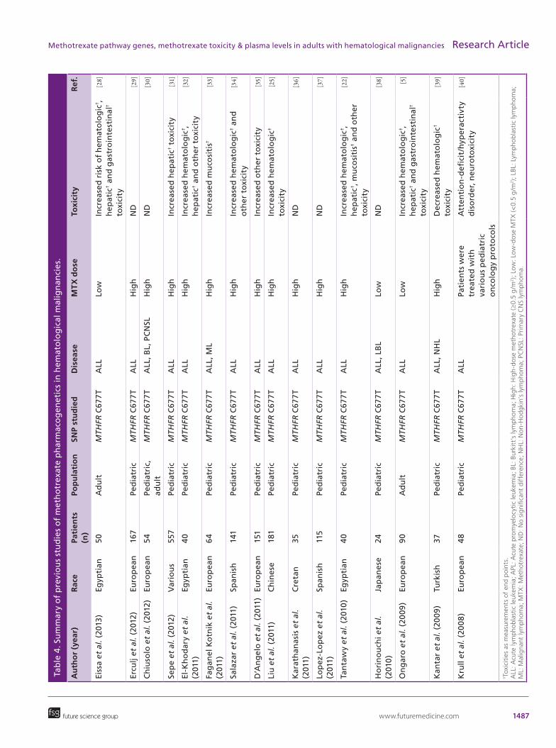
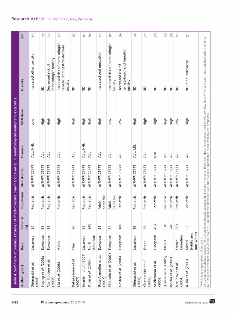
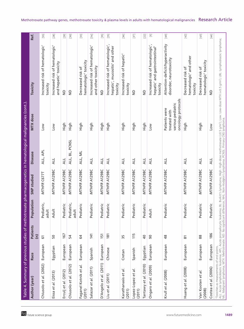
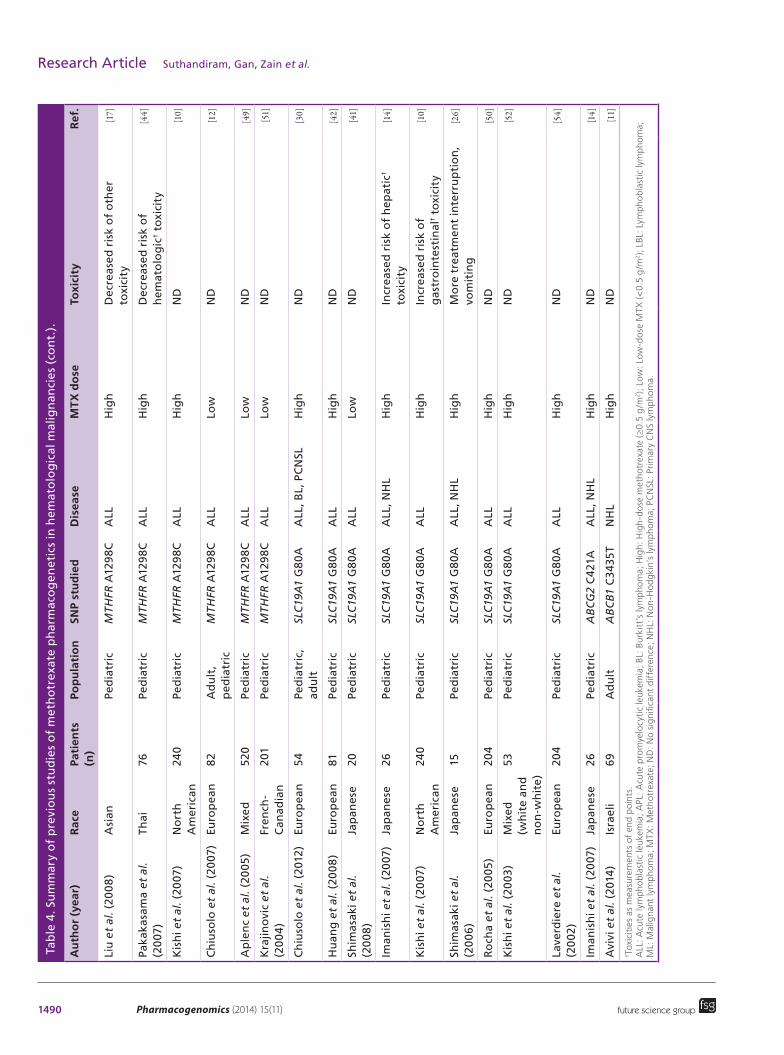
Methotrexate pathway genes, methotrexate toxicity & plasma levels in adults with hematological malignancies Research Article
Au
tho
r (y
ear)
Rac
ePa
tien
ts
(n)
Pop
ula
tio
nSN
P st
ud
ied
Dis
ease
MT
X d
ose
Toxi
city
Ref
.
Eiss
a et
al.
(201
3)
Egyp
tian
50A
du
ltM
THFR
C67
7T
ALL
Low
Incr
ease
d r
isk
of
hem
ato
log
ic† ,
h
epat
ic†
and
gas
tro
inte
stin
al†
toxi
city
[28]
Ercu
lj et
al.
(201
2)
Euro
pea
n16
7Pe
dia
tric
MTH
FR C
677
TA
LLH
igh
ND
[29]
Ch
iuso
lo e
t al
. (20
12)
Euro
pea
n5
4Pe
dia
tric
, ad
ult
MTH
FR C
677
TA
LL, B
L, P
CN
SLH
igh
ND
[30]
Sep
e et
al.
(201
2)
Var
iou
s55
7Pe
dia
tric
MTH
FR C
677
TA
LLH
igh
Incr
ease
d h
epat
ic†
toxi
city
[31]
El-K
ho
dar
y et
al.
(201
1)Eg
ypti
an4
0Pe
dia
tric
MTH
FR C
677
TA
LLH
igh
Incr
ease
d h
emat
olo
gic
† ,
hep
atic
† an
d o
ther
to
xici
ty[3
2]
Fag
anel
Ko
tnik
et
al.
(201
1)Eu
rop
ean
64
Ped
iatr
icM
THFR
C67
7T
ALL
, ML
Hig
hIn
crea
sed
mu
cosi
tis†
[33]
Sala
zar
et a
l. (2
011)
Span
ish
141
Ped
iatr
icM
THFR
C67
7T
ALL
Hig
hIn
crea
sed
hem
ato
log
ic†
and
o
ther
to
xici
ty[3
4]
D’A
ng
elo
et
al. (
2011
)Eu
rop
ean
151
Ped
iatr
icM
THFR
C67
7T
ALL
Hig
hIn
crea
sed
oth
er t
oxi
city
[35]
Liu
et
al. (
2011
)C
hin
ese
181
Ped
iatr
icM
THFR
C67
7T
ALL
Hig
hIn
crea
sed
hem
ato
log
ic†
toxi
city
[25]
Kar
ath
anas
is e
t al
. (2
011)
Cre
tan
35Pe
dia
tric
MTH
FR C
677
TA
LLH
igh
ND
[36]
Lop
ez-L
op
ez e
t al
. (2
011)
Span
ish
115
Ped
iatr
icM
THFR
C67
7T
ALL
Hig
hN
D[3
7]
Tan
taw
y et
al.
(201
0)
Egyp
tian
40
Ped
iatr
icM
THFR
C67
7T
ALL
Hig
hIn
crea
sed
hem
ato
log
ic† ,
h
epat
ic† ,
mu
cosi
tis†
and
oth
er
toxi
city
[22]
Ho
rin
ou
chi e
t al
. (2
010
)Ja
pan
ese
24Pe
dia
tric
MTH
FR C
677
TA
LL, L
BL
Low
ND
[38]
On
gar
o e
t al
. (20
09)
Euro
pea
n9
0A
du
ltM
THFR
C67
7T
ALL
Low
Incr
ease
d h
emat
olo
gic
† ,
hep
atic
† an
d g
astr
oin
test
inal
† to
xici
ty
[5]
Kan
tar
et a
l. (2
009
)Tu
rkis
h37
Ped
iatr
icM
THFR
C67
7T
ALL
, NH
LH
igh
Dec
reas
ed h
emat
olo
gic
† to
xici
ty[3
9]
Kru
ll et
al.
(20
08
)Eu
rop
ean
48
Ped
iatr
icM
THFR
C67
7T
ALL
Pati
ents
wer
e tr
eate
d w
ith
va
rio
us
ped
iatr
ic
on
colo
gy
pro
toco
ls
Att
enti
on
-defi
cit/
hyp
erac
tivt
y d
iso
rder
, neu
roto
xici
ty[4
0]
† Toxicities as measurements of end points.
ALL: Acute lymphoblastic leukemia; APL: Acute promyelocytic leukemia; BL: Burkitt’s lymphoma; High: High-dose methotrexate (≥0.5 g/m
2); Low: Low-dose MTX (<0.5 g/m
2); LBL: Lymphoblastic lymphoma;
ML: Malignant lymphoma; MTX: Methotrexate; ND: No significant difference; NHL: Non-Hodgkin’s lymphoma; PCNSL: Primary CNS lymphoma.
Tab
le 4
. Su
mm
ary
of
pre
vio
us
stu
die
s o
f m
eth
otr
exat
e p
har
mac
og
enet
ics
in h
emat
olo
gic
al m
alig
nan
cies
.
1488 Pharmacogenomics (2014) 15(11) future science group
Research Article Suthandiram, Gan, Zain et al.A
uth
or
(yea
r)R
ace
Pati
ents
(n
)Po
pu
lati
on
SNP
stu
die
dD
isea
seM
TX
do
seTo
xici
ty R
ef.
Shim
asak
i et
al.
(20
08
)Ja
pan
ese
20Pe
dia
tric
MTH
FR C
677
TA
LL, N
HL
Low
Incr
ease
d o
ther
to
xici
ty[4
1]
Hu
ang
et
al. (
200
8)
Euro
pea
n81
Ped
iatr
icM
THFR
C67
7T
ALL
Hig
hN
D[4
2]
Van
Ko
ote
n e
t al
. (2
00
8)
Euro
pea
n8
8Pe
dia
tric
MTH
FR C
677
TA
LLH
igh
Dec
reas
ed r
isk
of
hem
ato
log
ic†
toxi
city
[43]
Liu
et
al. (
200
8)
Asi
an
Ped
iatr
icM
THFR
C67
7T
ALL
Hig
hIn
crea
sed
ris
k o
f h
emat
olo
gic
† ,
hep
atic
† an
d g
astr
oin
test
inal
† to
xici
ty
[17]
Paka
kasa
ma
et a
l. (2
007
)Th
ai76
Ped
iatr
icM
THFR
C67
7T
ALL
Hig
hN
D[4
4]
Iman
ish
i et
al. (
2007
)Ja
pan
ese
26Pe
dia
tric
MTH
FR C
677
TA
LL, N
HL
Hig
hN
D[1
4]
Kis
hi e
t al
. (20
07)
No
rth
A
mer
ican
240
Ped
iatr
icM
THFR
C67
7T
ALL
Hig
hN
D[1
0]
Ru
iz-A
rgu
elle
s et
al.
(20
07)
Euro
pea
n28
Ad
ult
, p
edia
tric
MTH
FR C
677
TA
LLH
igh
Incr
ease
d o
ral m
uco
siti
s†[4
5]
Ch
iuso
lo e
t al
. (20
07)
Euro
pea
n82
Ad
ult
, p
edia
tric
MTH
FR C
677
TA
LLLo
wIn
crea
sed
ris
k o
f h
emat
olo
gic
† to
xici
ty[1
2]
Co
stea
et
al. (
200
6)
Euro
pea
n18
6Pe
dia
tric
MTH
FR C
677
TA
LLLo
wD
ecre
ased
ris
k o
f h
emat
olo
gic
† an
d h
epat
ic†
toxi
city
[46]
Shim
asak
i et
al.
(20
06
)Ja
pan
ese
15Pe
dia
tric
MTH
FR C
677
TA
LL, L
BL
Hig
hN
D[2
6]
Ch
atzi
dak
is e
t al
. (2
00
6)
Gre
ek4
6Pe
dia
tric
MTH
FR C
677
TA
LLH
igh
ND
[47]
Seid
eman
n e
t al
. (2
00
6)
Euro
pea
n4
84
Ped
iatr
icM
THFR
C67
7T
NH
LH
igh
ND
[48]
Ap
len
c et
al.
(20
05)
Mix
ed52
0Pe
dia
tric
MTH
FR C
677
TA
LLH
igh
ND
[49]
Ro
cha
et a
l. (2
005
)
246
Ped
iatr
icM
THFR
C67
7T
ALL
Hig
hN
D[5
0]
Kra
jino
vic
et a
l. (2
00
4)
Fren
ch-
Can
adia
n20
1Pe
dia
tric
MTH
FR C
677
TA
LLLo
wN
D[5
1]
Kis
hi e
t al
. (20
03)
Mix
ed
(wh
ite
and
n
on
-wh
ite
)
53Pe
dia
tric
MTH
FR C
677
TA
LLH
igh
ND
in n
euro
toxi
city
[52]
† Toxicities as measurements of end points.
ALL: Acute lymphoblastic leukemia; APL: Acute promyelocytic leukemia; BL: Burkitt’s lymphoma; High: High-dose methotrexate (≥0.5 g/m
2); Low: Low-dose MTX (<0.5 g/m
2); LBL: Lymphoblastic lymphoma;
ML: Malignant lymphoma; MTX: Methotrexate; ND: No significant difference; NHL: Non-Hodgkin’s lymphoma; PCNSL: Primary CNS lymphoma.
Tab
le 4
. Su
mm
ary
of
pre
vio
us
stu
die
s o
f m
eth
otr
exat
e p
har
mac
og
enet
ics
in h
emat
olo
gic
al m
alig
nan
cies
(co
nt.
).
www.futuremedicine.com 1489future science group
Methotrexate pathway genes, methotrexate toxicity & plasma levels in adults with hematological malignancies Research Article
Au
tho
r (y
ear)
Rac
ePa
tien
ts
(n)
Pop
ula
tio
nSN
P st
ud
ied
Dis
ease
MT
X d
ose
Toxi
city
Ref
.
Ch
iuso
lo e
t al
. (20
02)
Euro
pea
n61
Ped
iatr
ic,
adu
ltM
THFR
C67
7T
ALL
, APL
Low
Incr
ease
d r
isk
of
hem
ato
log
ic†
toxi
city
[53]
Eiss
a et
al.
(201
3)
Egyp
tian
50A
du
ltM
THFR
A12
98C
ALL
Low
Incr
ease
d r
isk
of
hem
ato
log
ic†
and
hep
atic
† to
xici
ty[2
8]
Ercu
lj et
al.
(201
2)
Euro
pea
n16
7Pe
dia
tric
MTH
FR A
129
8CA
LLH
igh
ND
[29]
Ch
iuso
lo e
t al
. (20
12)
Euro
pea
n5
4Pe
dia
tric
, ad
ult
MTH
FR A
129
8CA
LL, B
L, P
CN
SLH
igh
ND
[30]
Fag
anel
Ko
tnik
et
al.
(201
1)Eu
rop
ean
64
Ped
iatr
icM
THFR
A12
98C
ALL
, ML
Hig
hD
ecre
ased
ris
k o
f h
emat
olo
gic
† to
xici
ty[3
3]
Sala
zar
et a
l. (2
011)
Span
ish
141
Ped
iatr
icM
THFR
A12
98C
ALL
Hig
hIn
crea
sed
ris
k o
f h
emat
olo
gic
† an
d o
ther
to
xici
ty[3
4]
D’A
ng
elo
et
al. (
2011
)Eu
rop
ean
151
Ped
iatr
icM
THFR
A12
98C
ALL
Hig
hN
D[3
5]
Liu
et
al. (
2011
)C
hin
ese
181
Ped
iatr
icM
THFR
A12
98C
ALL
Hig
hIn
crea
sed
ris
k o
f h
emat
olo
gic
† ,
hep
atic
† , m
uco
siti
s† an
d o
ther
to
xici
ty
[25]
Kar
ath
anas
is e
t al
. (2
011)
Cre
tan
35Pe
dia
tric
MTH
FR A
129
8CA
LLH
igh
Incr
ease
d r
isk
of
hep
atic
† to
xici
ty[3
6]
Lop
ez-L
op
ez e
t al
. (2
011)
Span
ish
115
Ped
iatr
icM
THFR
A12
98C
ALL
Hig
hN
D[3
7]
Tan
taw
y et
al.
(201
0)
Egyp
tian
40
Ped
iatr
icM
THFR
A12
98C
ALL
Hig
hN
D[2
2]
On
gar
o e
t al
. (20
09)
Euro
pea
n9
0A
du
ltM
THFR
A12
98C
ALL
Low
Incr
ease
d r
isk
of
hem
ato
log
ic† ,
h
epat
ic†
and
gas
tro
inte
stin
al†
toxi
city
[5]
Kru
ll et
al.
(20
08
)Eu
rop
ean
48
Ped
iatr
icM
THFR
A12
98C
ALL
Pati
ents
wer
e tr
eate
d w
ith
va
rio
us
ped
iatr
ic
on
colo
gy
pro
toco
ls
Att
enti
on
-defi
cit/
hyp
erac
tivi
ty
dis
ord
er, n
euro
toxi
city
[40]
Hu
ang
et
al. (
200
8)
Euro
pea
n81
Ped
iatr
icM
THFR
A12
98C
ALL
Hig
hD
ecre
ased
ris
k o
f h
emat
olo
gic
† an
d o
ther
to
xici
ty
[42]
Van
Ko
ote
n e
t al
. (2
00
8)
Euro
pea
n8
8Pe
dia
tric
MTH
FR A
129
8CA
LLH
igh
Dec
reas
ed r
isk
of
hem
ato
log
ic†
toxi
city
[43]
Co
stea
et
al. (
200
6)
Euro
pea
n18
6Pe
dia
tric
MTH
FR A
129
8CA
LLLo
wN
D[4
6]
† Toxicities as measurements of end points.
ALL: Acute lymphoblastic leukemia; APL: Acute promyelocytic leukemia; BL: Burkitt’s lymphoma; High: High-dose methotrexate (≥0.5 g/m
2); Low: Low-dose MTX (<0.5 g/m
2); LBL: Lymphoblastic lymphoma;
ML: Malignant lymphoma; MTX: Methotrexate; ND: No significant difference; NHL: Non-Hodgkin’s lymphoma; PCNSL: Primary CNS lymphoma.
Tab
le 4
. Su
mm
ary
of
pre
vio
us
stu
die
s o
f m
eth
otr
exat
e p
har
mac
og
enet
ics
in h
emat
olo
gic
al m
alig
nan
cies
(co
nt.
).
1490 Pharmacogenomics (2014) 15(11) future science group
Research Article Suthandiram, Gan, Zain et al.A
uth
or
(yea
r)R
ace
Pati
ents
(n
)Po
pu
lati
on
SNP
stu
die
dD
isea
seM
TX
do
seTo
xici
ty R
ef.
Liu
et
al. (
200
8)
Asi
an
Ped
iatr
icM
THFR
A12
98C
ALL
Hig
hD
ecre
ased
ris
k o
f o
ther
to
xici
ty[1
7]
Paka
kasa
ma
et a
l. (2
007
)Th
ai76
Ped
iatr
icM
THFR
A12
98C
ALL
Hig
hD
ecre
ased
ris
k o
f h
emat
olo
gic
† to
xici
ty[4
4]
Kis
hi e
t al
. (20
07)
No
rth
A
mer
ican
240
Ped
iatr
icM
THFR
A12
98C
ALL
Hig
hN
D[1
0]
Ch
iuso
lo e
t al
. (20
07)
Euro
pea
n82
Ad
ult
, p
edia
tric
MTH
FR A
129
8CA
LLLo
wN
D[1
2]
Ap
len
c et
al.
(20
05)
Mix
ed52
0Pe
dia
tric
MTH
FR A
129
8CA
LLLo
wN
D[4
9]
Kra
jino
vic
et a
l. (2
00
4)
Fren
ch-
Can
adia
n20
1Pe
dia
tric
MTH
FR A
129
8CA
LLLo
wN
D[5
1]
Ch
iuso
lo e
t al
. (20
12)
Euro
pea
n5
4Pe
dia
tric
, ad
ult
SLC
19A
1 G
80A
ALL
, BL,
PC
NSL
Hig
hN
D[3
0]
Hu
ang
et
al. (
200
8)
Euro
pea
n81
Ped
iatr
icSL
C19
A1
G8
0AA
LLH
igh
ND
[42]
Shim
asak
i et
al.
(20
08
)Ja
pan
ese
20Pe
dia
tric
SLC
19A
1 G
80A
ALL
Low
ND
[41]
Iman
ish
i et
al. (
2007
)Ja
pan
ese
26Pe
dia
tric
SLC
19A
1 G
80A
ALL
, NH
LH
igh
Incr
ease
d r
isk
of
hep
atic
† to
xici
ty[1
4]
Kis
hi e
t al
. (20
07)
No
rth
A
mer
ican
240
Ped
iatr
icSL
C19
A1
G8
0AA
LLH
igh
Incr
ease
d r
isk
of
gas
tro
inte
stin
al†
toxi
city
[10]
Shim
asak
i et
al.
(20
06
)Ja
pan
ese
15Pe
dia
tric
SLC
19A
1 G
80A
ALL
, NH
LH
igh
Mo
re t
reat
men
t in
terr
up
tio
n,
vom
itin
g[2
6]
Ro
cha
et a
l. (2
005
)Eu
rop
ean
204
Ped
iatr
icSL
C19
A1
G8
0AA
LLH
igh
ND
[50]
Kis
hi e
t al
. (20
03)
Mix
ed
(wh
ite
and
n
on
-wh
ite
)
53Pe
dia
tric
SLC
19A
1 G
80A
ALL
Hig
hN
D[5
2]
Lave
rdie
re e
t al
. (2
002
)Eu
rop
ean
204
Ped
iatr
icSL
C19
A1
G8
0AA
LLH
igh
ND
[54]
Iman
ish
i et
al. (
2007
)Ja
pan
ese
26Pe
dia
tric
AB
CG
2 C
421A
ALL
, NH
LH
igh
ND
[14]
Avi
vi e
t al
. (20
14)
Isra
eli
69A
du
ltA
BC
B1
C3
435
TN
HL
Hig
hN
D[1
1]
† Toxicities as measurements of end points.
ALL: Acute lymphoblastic leukemia; APL: Acute promyelocytic leukemia; BL: Burkitt’s lymphoma; High: High-dose methotrexate (≥0.5 g/m
2); Low: Low-dose MTX (<0.5 g/m
2); LBL: Lymphoblastic lymphoma;
ML: Malignant lymphoma; MTX: Methotrexate; ND: No significant difference; NHL: Non-Hodgkin’s lymphoma; PCNSL: Primary CNS lymphoma.
Tab
le 4
. Su
mm
ary
of
pre
vio
us
stu
die
s o
f m
eth
otr
exat
e p
har
mac
og
enet
ics
in h
emat
olo
gic
al m
alig
nan
cies
(co
nt.
).
www.futuremedicine.com 1491future science group
Methotrexate pathway genes, methotrexate toxicity & plasma levels in adults with hematological malignancies Research Article
the MTHFR C677T polymorphism and MTX tox-icity can be described by disturbances in folate lev-els and by prolonged MTX exposure due to delayed MTX clearance. Most studies to date are limited to only gene effect and disregard the environmental effects of folate and homocysteine. Thus, the actual measure of effect size of the genetic polymorphisms is limited to the fact that these studies vary in terms of the underlying disease, age group, dosage of MTX, folate status, diet, concurrent medications and even toxicity threshold setting. A multicenter study with a larger sample size that combines and standardizes the study setting would be a better approach in the future in order to generate a consistent and definitive result.
In conclusion, this is the first study to identify a significant role of polymorphisms in the MTX path-way genes on MTX toxicity and MTX plasma con-centration at 48 h in Asian adults with hematologi-cal malignancies. Furthermore, the study confirmed the influence of MTHFR C677T on MTX-related toxicities and plasma levels. Our study suggests that pharmaco genetics modifies MTX-related toxicity and plasma levels in adult ALL or NHL patients. Such results could enable tailored therapy based on a pharmacogenetics approach in order to prevent toxic-ity events. In addition, polymorphisms of SLC19A1 G80A and ABCB1 C3435T need to be further inves-tigated as data has suggested that they may have significant roles in MTX toxicity and plasma MTX levels. Genotyping prior to treatment in adult ALL or NHL is likely to be valuable with the aim of adapt-ing MTX therapy and thus reducing MTX-induced toxicities.
ConclusionIn conclusion, this is the first study to identify a sig-nificant role of polymorphisms in the MTX pathway
genes on MTX toxicity and MTX plasma concen-tration at 48 h in Asian adults with hematological malignancies. Furthermore, the study confirmed the influence of MTHFR C677T on MTX-related tox-icities and plasma levels. Our study suggests that pharmacogenetics modify MTX-related toxicity and plasma levels in adult ALL or NHL. Such results could enable tailored therapy based on a pharmaco-genetics approach in order to prevent toxicity events. In addition, polymorphisms of the SLC19A1 G80A and ABCB1 C3435T polymorphisms need to be fur-ther investigated as data has suggested that they have significant roles in MTX toxicity and plasma MTX levels. Genotyping prior to treatment in adult ALL or NHL, is likely to be valuable with the aim of adapting MTX therapy and thus reducing the MTX-induced toxicities.
Future perspectiveOur findings may be significant for the further devel-opment of treatment strategy in adult ALL or NHL. Our data could help clinicians to determine individu-als at greater risk for MTX-associated toxicity. Clini-cians might therefore be able advise patients about the possibility of experiencing MTX-related toxicities.
AcknowledgementsThe authors would like to thank the Director of Health Malay-
sia for permission to publish this paper. The authors gratefully
acknowledge the study subjects for their participation in this
study and to the staff of UMMC and Ampang Hospital for
their assistance in subject recruitment.
Financial & competing interests disclosureThis study was supported by the HIR MOHE grant E000025-
20001, University Malaya Research Grant (UMRG)
(RG300/11HTM), University Malaya IPPP grant (PV072/2011B)
Executive summary
Background• High-dose methotrexate (HDMTX) has been shown to be useful in acute lymphoblastic leukemia (ALL) or
non-Hodgkin lymphoma (NHL); however, it causes toxicity in the bone marrow, liver and gastrointestinal tract.• Pharmacogenetics of methotrexate (MTX) and its relationship with plasma concentrations are conflicting, and
there is only one study in Asian adults.Patients & methods• In our study we investigated the association between polymorphisms in the MTX pathway genes with
MTX-associated toxicity in a cohort of adult patients with ALL or NHL treated with HDMTX.Results• Hepatic toxicity is significantly higher in patients with MTHFR C677T, SLC19A1 G80A and ABCB1 C3435T.• MTHFR C677T is also associated with risk of hematopoietic toxicity.• Higher levels of plasma MTX were found in those with MTHFR C677T and ABCB1 C3435T.Discussion• Our results in Asian adults provide evidence for the contribution of pharmacogenetics to the toxicity of
HDMTX and plasma MTX concentrations at 48 h following treatment in patients with ALL or NHL.
1492 Pharmacogenomics (2014) 15(11) future science group
Research Article Suthandiram, Gan, Zain et al.
and University Malaya IPPP grant (PS190/2010B). The authors
have no other relevant affiliations or financial involvement
with any organization or entity with a financial interest in or fi-
nancial conflict with the subject matter or materials discussed
in the manuscript apart from those disclosed.
No writing assistance was utilized in the production of this
manuscript.
Ethical conduct of researchThe authors state that they have obtained appropriate institu-
tional review board approval or have followed the principles
outlined in the Declaration of Helsinki for all human or animal
experimental investigations. In addition, for investigations in-
volving human subjects, informed consent has been obtained
from the participants involved.
ReferencesPapers of special note have been highlighted as: • of interest; •• of considerable interest
1 Bertino JR. Cancer research: from folate antagonism to molecular targets. Best Pract. Res. Clin. Haematol. 22(4), 577–582 (2009).
2 Patino-Garcia A, Zalacain M, Marrodan L, San-Julian M, Sierrasesumaga L. Methotrexate in pediatric osteosarcoma: response and toxicity in relation to genetic polymorphisms and dihydrofolate reductase and reduced folate carrier 1 expression. J. Pediatr. 154(5), 688–693 (2009).
3 Gervasini G, Vagace JM. Impact of genetic polymorphisms on chemotherapy toxicity in childhood acute lymphoblastic leukemia. Front. Genet. 3, 249 (2012).
4 Nathan PC, Whitcomb T, Wolters PL et al. Very high-dose methotrexate (33.6 g/m(2)) as central nervous system preventive therapy for childhood acute lymphoblastic leukemia: results of National Cancer Institute/Children’s Cancer Group trials CCG-191P, CCG-134P and CCG-144P. Leuk. Lymphoma 47(12), 2488–2504 (2006).
5 Ongaro A, De Mattei M, Della Porta MG et al. Gene polymorphisms in folate metabolizing enzymes in adult acute lymphoblastic leukemia: effects on methotrexate-related toxicity and survival. Haematologica 94(10), 1391–1398 (2009).
6 Schmiegelow K. Advances in individual prediction of methotrexate toxicity: a review. Br. J. Haematol. 146(5), 489–503 (2009).
7 Treon SP, Chabner BA. Concepts in use of high-dose methotrexate therapy. Clin. Chem. 42(8 Pt 2), 1322–1329 (1996).
8 Gemmati D, Ongaro A, Scapoli GL et al. Common gene polymorphisms in the metabolic folate and methylation pathway and the risk of acute lymphoblastic leukemia and non-Hodgkin’s lymphoma in adults. Cancer Epidemiol. Biomarkers Prev. 13(5), 787–794 (2004).
9 Ganapathy V, Smith SB, Prasad PD. SLC19: the folate/thiamine transporter family. Pflugers Arch. 447(5), 641–646 (2004).
10 Kishi S, Cheng C, French D et al. Ancestry and pharmacogenetics of antileukemic drug toxicity. Blood 109(10), 4151–4157 (2007).
11 Avivi I, Zuckerman T, Krivoy N, Efrati E. Genetic polymorphisms predicting methotrexate blood levels and toxicity in adult non-Hodgkin lymphoma. Leuk. Lymphoma 55(3), 565–570 (2014).
12 Chiusolo P, Reddiconto G, Farina G et al. MTHFR polymorphisms’ influence on outcome and toxicity in acute lymphoblastic leukemia patients. Leuk. Res. 31(12), 1669–1674 (2007).
13 Gemmati D, Ongaro A, Tognazzo S et al. Methylenetetrahydrofolate reductase C677T and A1298C gene variants in adult non-Hodgkin’s lymphoma patients: association with toxicity and survival. Haematologica 92(4), 478–485 (2007).
14 Imanishi H, Okamura N, Yagi M et al. Genetic polymorphisms associated with adverse events and elimination of methotrexate in childhood acute lymphoblastic leukemia and malignant lymphoma. J. Hum. Genet. 52(2), 166–171 (2007).
•• Correlatedgeneticpolymorphismswithhepatotoxicityorserumconcentrations.
15 Spyridopoulou KP, Dimou NL, Hamodrakas SJ, Bagos PG. Methylene tetrahydrofolate reductase gene polymorphisms and their association with methotrexate toxicity: a meta-analysis. Pharmacogenet. Genomics 22(2), 117–133 (2012).
16 Yang L, Hu X, Xu L. Impact of methylenetetrahydrofolate reductase (MTHFR) polymorphisms on methotrexate-induced toxicities in acute lymphoblastic leukemia: a meta-analysis. Tumour Biol. 33(5), 1445–1454 (2012).
17 Liu JX, Chen JP, Tan W, Lin DX. Association betweenmthfr gene polymorphisms and toxicity of HDMTX chemotherapy in acute lymphocytic leukemia. Zhongguo Shi Yan Xue Ye Xue Za Zhi 16(3), 488–492 (2008).
18 Jaffe ES. The 2008 WHO classification of lymphomas: implications for clinical practice and translational research. Hematology Am. Soc. Hematol. Educ. Program 523–531 (2009).
19 Thomas DA, Faderl S, Cortes J et al. Treatment of Philadelphia chromosome-positive acute lymphocytic leukemia with hyper-CVAD and imatinib mesylate. Blood 103(12), 4396–4407 (2004).
20 Garcia-Manero G, Kantarjian HM. The hyper-CVAD regimen in adult acute lymphocytic leukemia. Hematol. Oncol. Clin. North Am. 14(6), 1381–1396, x–xi (2000).
21 Trotti A, Byhardt R, Stetz J et al. Common toxicity criteria: version 2.0. an improved reference for grading the acute effects of cancer treatment: impact on radiotherapy. Int. J. Radiat. Oncol. Biol. Phys. 47(1), 13–47 (2000).
• Cleardescriptionoftheadverseeventsincancertreatment.
22 Tantawy AA, El-Bostany EA, Adly AA, Abou El Asrar M, El-Ghouroury EA, Abdulghaffar EE. Methylene tetrahydrofolate reductase gene polymorphism in Egyptian children with acute lymphoblastic leukemia. Blood Coagul. Fibrinolysis 21(1), 28–34 (2010).
23 Perez C, Wang YM, Sutow WW, Herson J. Significance of the 48-hour plasma level in high-dose methotrexate regimens. Cancer Clin. Trials 1(2), 107–111 (1978).
www.futuremedicine.com 1493future science group
Methotrexate pathway genes, methotrexate toxicity & plasma levels in adults with hematological malignancies Research Article
24 Liu Y, Yin Y, Sheng Q et al. Association of ABCC2 -24C>T polymorphism with high-dose methotrexate plasma concentrations and toxicities in childhood acute lymphoblastic leukemia. PLoS ONE 9(1), e82681 (2014).
25 Liu SG, Li ZG, Cui L, Gao C, Li WJ, Zhao XX. Effects of methylenetetrahydrofolate reductase gene polymorphisms on toxicities during consolidation therapy in pediatric acute lymphoblastic leukemia in a Chinese population. Leuk. Lymphoma 52(6), 1030–1040 (2011).
26 Shimasaki N, Mori T, Samejima H et al. Effects of methylenetetrahydrofolate reductase and reduced folate carrier 1 polymorphisms on high-dose methotrexate-induced toxicities in children with acute lymphoblastic leukemia or lymphoma. J. Pediatr. Hematol. Oncol. 28(2), 64–68 (2006).
27 Rahiem Ahmed Yaa YH. Prevention and management of high dose methotrexate toxicity. J. Cancer Sci. Ther. 5, 106–112 (2013).
28 Eissa DS, Ahmed TM. C677T and A1298C polymorphisms of the methylenetetrahydrofolate reductase gene: effect on methotrexate-related toxicity in adult acute lymphoblastic leukaemia. Blood Coagul. Fibrinolysis 24(2), 181–188 (2013).
29 Erculj N, Kotnik BF, Debeljak M, Jazbec J, Dolzan V. Influence of folate pathway polymorphisms on high-dose methotrexate-related toxicity and survival in childhood acute lymphoblastic leukemia. Leuk. Lymphoma 53(6), 1096–1104 (2012).
30 Chiusolo P, Giammarco S, Bellesi S et al. The role of MTHFR and RFC1 polymorphisms on toxicity and outcome of adult patients with hematological malignancies treated with high-dose methotrexate followed by leucovorin rescue. Cancer Chemother. Pharmacol. 69(3), 691–696 (2012).
31 Sepe DM, McWilliams T, Chen J et al. Germline genetic variation and treatment response on CCG-1891. Pediatr. Blood Cancer 58(5), 695–700 (2012).
32 El-Khodary NM, El-Haggar SM, Eid MA, Ebeid EN. Study of the pharmacokinetic and pharmacogenetic contribution to the toxicity of high-dose methotrexate in children with acute lymphoblastic leukemia. Med. Oncol. 29(3), 2053–2062 (2011).
33 Faganel Kotnik B, Grabnar I, Bohanec Grabar P, Dolzan V, Jazbec J. Association of genetic polymorphism in the folate metabolic pathway with methotrexate pharmacokinetics and toxicity in childhood acute lymphoblastic leukaemia and malignant lymphoma. Eur. J. Clin. Pharmacol. 67(10), 993–1006 (2011).
34 Salazar J, Altes A, Del Rio E et al. Methotrexate consolidation treatment according to pharmacogenetics of MTHFR ameliorates event-free survival in childhood acute lymphoblastic leukaemia. Pharmacogenomics J. 12(5), 379–385 (2011).
35 D’Angelo V, Ramaglia M, Iannotta A et al. Methotrexate toxicity and efficacy during the consolidation phase in paediatric acute lymphoblastic leukaemia and MTHFR polymorphisms as pharmacogenetic determinants. Cancer Chemother. Pharmacol. 68(5), 1339–1346 (2011).
36 Karathanasis NV, Stiakaki E, Goulielmos GN, Kalmanti M. The role of the methylenetetrahydrofolate reductase 677 and 1298 polymorphisms in Cretan children with
acute lymphoblastic leukemia. Genet. Test Mol. Biomarkers 15(1–2), 5–10 (2011).
37 Lopez-Lopez E, Martin-Guerrero I, Ballesteros J et al. Polymorphisms of the SLCO1B1 gene predict methotrexate-related toxicity in childhood acute lymphoblastic leukemia. Pediatr. Blood Cancer 57(4), 612–619 (2011).
38 Horinouchi M, Yagi M, Imanishi H et al. Association of genetic polymorphisms with hepatotoxicity in patients with childhood acute lymphoblastic leukemia or lymphoma. Pediatr. Hematol. Oncol. 27(5), 344–354 (2010).
39 Kantar M, Kosova B, Cetingul N et al. Methylenetetrahydrofolate reductase C677T and A1298C gene polymorphisms and therapy-related toxicity in children treated for acute lymphoblastic leukemia and non-Hodgkin lymphoma. Leuk. Lymphoma 50(6), 912–917 (2009).
40 Krull KR, Brouwers P, Jain N et al. Folate pathway genetic polymorphisms are related to attention disorders in childhood leukemia survivors. J. Pediatr. 152(1), 101–105 (2008).
41 Shimasaki N, Mori T, Torii C et al. Influence of MTHFR and RFC1 polymorphisms on toxicities during maintenance chemotherapy for childhood acute lymphoblastic leukemia or lymphoma. J. Pediatr. Hematol. Oncol. 30(5), 347–352 (2008).
42 Huang L, Tissing WJ, De Jonge R, Van Zelst BD, Pieters R. Polymorphisms in folate-related genes: association with side effects of high-dose methotrexate in childhood acute lymphoblastic leukemia. Leukemia 22(9), 1798–1800 (2008).
43 Van Kooten Niekerk PB, Schmiegelow K, Schroeder H. Influence of methylene tetrahydrofolate reductase polymorphisms and coadministration of antimetabolites on toxicity after high dose methotrexate. Eur. J. Hematol. 81(5), 391–398 (2008).
44 Pakakasama S, Kanchanakamhaeng K, Kajanachumpol S et al. Genetic polymorphisms of folate metabolic enzymes and toxicities of high dose methotrexate in children with acute lymphoblastic leukemia. Ann. Hematol. 86(8), 609–611 (2007).
45 Ruiz-Arguelles GJ, Coconi-Linares LN, Garces-Eisele J, Reyes-Nunez V. Methotrexate-induced mucositis in acute leukemia patients is not associated with the MTHFR 677T allele in Mexico. Hematology 12(5), 387–391 (2007).
46 Costea I, Moghrabi A, Laverdiere C, Graziani A, Krajinovic M. Folate cycle gene variants and chemotherapy toxicity in pediatric patients with acute lymphoblastic leukemia. Haematologica 91(8), 1113–1116 (2006).
47 Chatzidakis K, Goulas A, Athanassiadou-Piperopoulou F, Fidani L, Koliouskas D, Mirtsou V. Methylenetetrahydrofolate reductase C677T polymorphism: association with risk for childhood acute lymphoblastic leukemia and response during the initial phase of chemotherapy in greek patients. Pediatric Blood Cancer 47(2), 147–151 (2006).
48 Seidemann K, Book M, Zimmermann M et al. MTHFR 677 (C-->T) polymorphism is not relevant for prognosis or therapy-associated toxicity in pediatric NHL: results from 484 patients of multicenter trial NHL-BFM 95. Ann. Hematol. 85(5), 291–300 (2006).
1494 Pharmacogenomics (2014) 15(11)
49 Aplenc R, Thompson J, Han P et al. Methylenetetrahydrofolate reductase polymorphisms and therapy response in pediatric acute lymphoblastic leukemia. Cancer Res. 65(6), 2482–2487 (2005).
50 Rocha JC, Cheng C, Liu W et al. Pharmacogenetics of outcome in children with acute lymphoblastic leukemia. Blood 105(12), 4752–4758 (2005).
51 Krajinovic M, Lamothe S, Labuda D et al. Role of MTHFR genetic polymorphisms in the susceptibility to childhood acute lymphoblastic leukemia. Blood 103(1), 252–257 (2004).
52 Kishi S. Homocysteine, pharmacogenetics, and neurotoxicity in children with leukemia. J. Clin. Oncol. 21(16), 3084–3091 (2003).
53 Chiusolo P. Preponderance of methylenetetrahydrofolate reductase C677T homozygosity among leukemia patients intolerant to methotrexate. Ann. Oncol. 13(12), 1915–1918 (2002).
54 Laverdiere C, Chiasson S, Costea I, Moghrabi A, Krajinovic M. Polymorphism G80A in the reduced folate carrier gene and its relationship to methotrexate plasma levels and outcome of childhood acute lymphoblastic leukemia. Blood 100(10), 3832–3834 (2002).
55 Frosst P, Blom HJ, Milos R et al. A candidate genetic risk factor for vascular disease: a common mutation in methylenetetrahydrofolate reductase. Nat. Genet. 10(1), 111–113 (1995).
56 De Mattia E, Toffoli G. C677T and A1298C MTHFR polymorphisms, a challenge for antifolate and fluoropyrimidine-based therapy personalisation. Eur. J. Cancer 45(8), 1333–1351 (2009).
57 Lopez-Lopez E, Martin-Guerrero I, Ballesteros J, Garcia-Orad A. A systematic review and meta-analysis of MTHFR polymorphisms in methotrexate toxicity prediction in
pediatric acute lymphoblastic leukemia. Pharmacogenomics J. 13(6), 498–506 (2013).
58 Lopez-Lopez E, Ballesteros J, Pinan MA et al. Polymorphisms in the methotrexate transport pathway: a new tool for MTX plasma level prediction in pediatric acute lymphoblastic leukemia. Pharmacogenet. Genomics 23(2), 53–61 (2013).
59 Stoller RG, Hande KR, Jacobs SA, Rosenberg SA, Chabner BA. Use of plasma pharmacokinetics to predict and prevent methotrexate toxicity. N. Engl J. Med. 297(12), 630–634 (1977).
60 Ayad MW, El Naggar AA, El Naggar M. MTHFR C677T polymorphism: association with lymphoid neoplasm and effect on methotrexate therapy. Eur. J. Hematol. 93(1), 63–69 (2014).
61 Bektas-Kayhan K, Kucukhuseyin O, Karagoz G et al. Is the MDR1 C3435T polymorphism responsible for oral mucositis in children with acute lymphoblastic leukemia? Asian Pac. J. Cancer Prev. 13(10), 5251–5255 (2012).
62 Biswal BM. Current trends in the management of oral mucositis related to cancer treatment. Malays. J. Med. Sci. 15(3), 4–13 (2008).
63 Pico JL, Avila-Garavito A, Naccache P. Mucositis: its occurrence, consequences, and treatment in the oncology setting. Oncologist 3(6), 446–451 (1998).
64 Zinzani PL, Federico M, Oliva S et al. The more patients you treat, the more you cure: managing cardiotoxicity in the treatment of aggressive non-Hodgkin lymphoma. Leuk. Lymphoma doi:10.3109/10428194.2014.894187 (2014) (Epub ahead of print).
65 Dorchin M, Masoumi Dehshiri R, Soleiman S, Manashi M. Evaluation of neuropathy during intensive vincristine chemotherapy for non-Hodgkin’s lymphoma and acute lymphoblastic leukemia. Iran J. Pediatr. Hematol. Oncol. 3(4), 138–142 (2013).
future science group
Research Article Suthandiram, Gan, Zain et al.