Embed Size (px)
Citation preview
RESEARCH ARTICLE Open Access
Drug sensitivity patterns of HHV8 carrying bodycavity lymphoma cell linesRita Ötvös1,4*, Henriette Skribek1, Lorand L Kis1, Annunziata Gloghini2, Laszlo Markasz1, Emilie Flaberg1,Staffan Eksborg3, Jozsef Konya4, Lajos Gergely4, Antonino Carbone2 and Laszlo Szekely1
Abstract
Background: Primary effusion lymphoma (PEL) is a rare KSHV/HHV8-associated high-grade non-Hodgkin’slymphoma (NHL) of B-cell origin, characterized by serous effusions in body cavities. Most patients are HIV-infectedmen with severe immunosuppression and other HHV8-associated diseases such as Kaposi’s sarcoma (KS). Theprognosis for those infected is poor, with a median survival of less than 6 months in most cohorts. Sustainedcomplete remission is rare. High-dose chemotherapy regimens are used to improve remission rate and survival.The aim of the present study was to compare the drug sensitivity pattern of the available primary effusion (bodycavity based) lymphoma-derived cell lines in order to find additional, potentially effective drugs that are notincluded in current chemotherapy treatment protocols.
Methods: We have analyzed 11 cell lines against 27 frequently used cytostatic drugs in short term (3 days) survivalassays using automated high throughput confocal microscopy.
Results: All cell lines showed a distinct, individual drug sensitivity pattern. Considering the in vitro used andclinically achieved drug concentration, Vinorelbine, Paclitaxel, Epirubicin and Daunorubicin were the most effectivedrugs.
Conclusions: We suggest that inclusion of the above drugs into PEL chemotherapy protocols may be justified. Theheterogeneity in the drug response pattern however indicated that assay-guided individualized therapy might berequired to optimize therapeutic response.
BackgroundHuman herpesvirus 8 (HHV8) or Kaposi sarcoma her-pesvirus (KSHV) is the probable causative agent of twodistinct lymphoproliferative disorders: primary effusionlymphoma (PEL) and the plasma cell variant of multi-centric Castleman disease (MCD) in addition to Kaposisarcoma (KS) [1].Primary effusion lymphoma (PEL), or alternatively:
body cavity lymphoma is a non-Hodgkin’s lymphoma(NHL) of B-cell origin that develops predominantly inthe serous body cavities [2]. The lymphoma cells,although lacking many conventional B-cell markers,carry immunoglobulin gene rearrangement and expresssyndecans, suggesting pre-plasma cell origin. At the
clinico-pathological level, PEL is characterized by liquidgrowth in the serous body cavities associated withspreading along the serous membranes without infiltra-tive or destructive growth patterns [3,4]. Morphologi-cally, PEL bridges immunoblastic and anaplastic featuresand frequently displays a certain degree of plasmacelldifferentiation. In all known cases, the monoclonal B-cell population is infected with HHV-8. Half of the lym-phomas are dually infected with HHV-8 and Epstein-Barr virus (EBV) [5]. In the context of AIDS, most casesare associated with other KSHV/HHV8-related diseasessuch as Kaposi’s sarcoma (KS) or multicentric Castle-man’s disease (MCD). As PEL typically lacks a solidcomponent, its diagnosis rests on the cytological exami-nation of body fluid. Phenotypically, expression of theCD45 antigen (> 90% of cases) confirms the lymphoidderivation of PEL cells, which exhibit an indeterminateimmunophenotype, as they usually lack expression of B-and T-cell associated antigens (the majority of cases
* Correspondence: [email protected] of Microbiology, Tumor and Cell Biology (MTC) and Center forIntegrative Recognition in the Immune System (IRIS), Karolinska Institute, Box280 SE-17177 Stockholm, SwedenFull list of author information is available at the end of the article
Ötvös et al. BMC Cancer 2011, 11:441http://www.biomedcentral.com/1471-2407/11/441
© 2011 Ötvös et al; licensee BioMed Central Ltd. This is an Open Access article distributed under the terms of the Creative CommonsAttribution License (http://creativecommons.org/licenses/by/2.0), which permits unrestricted use, distribution, and reproduction inany medium, provided the original work is properly cited.
reported). There are, however, cases in the literaturethat had a B-cell or T-cell phenotype respectively [1].Conversely, PEL cells generally express various mar-
kers associated with activation, including CD30(approximately 75% of cases), CD38, CD71 and theepithelial membrane antigen. Moreover, PEL cellsexpress several plasma cell markers including CD138,VS38c and MUM-1/IRF4 [1].The prognosis of PEL is poor, as the median survival
in the previously published series does not exceed 3months [3,6-10].Given its rarity, however, there are very few longitudi-
nal observational series of patients with PEL and nolarge prospective trials have ever defined optimal treat-ment strategies [11].Prior to the introduction of antiretroviral therapy, the
therapeutic results were unsatisfactory in cohorts ofHIV+ patients, despite the use of aggressive polyche-motherapy regimens including anthracyclines. The sig-nificant improvement in the prognosis of AIDS-relatedlymphomas observed in the antiretroviral therapy eraalso applies to the PEL setting.In addition, the routine use of growth factors, such as
the granulocyte colony-stimulating factor (G-CSF), toavoid prolonged periods of neutropenia resulting fromchemotherapy is standard practice for all AIDS-relatedlymphoma (ARL) patients.Despite the improvement in therapeutical strategies
during the last few years, there is no evidence of a curefor PEL patients with conventional systemic chemother-apy addressed to aggressive NHL. The suggested benefitof high-dose Methotrexate in association with CHOP(Cyclophosphamide, Doxorubicin, Prednisolone andVincristine)-like regimens is negatively balanced by thehampered toxicity of Methotrexate in the presence ofserous effusions [1].Novel approaches for body cavity lymphoma therapy
outside traditional chemotherapy have been suggested aswell [11]. These include the addition of antiviral therapyas well as inhibition of specific cellular targets. Anti-tumor activity of the antiviral therapy directed againstKSHV/HHV8 infection has been reported. This experi-ence is based on single case reports. Patients with adiagnosis of PEL, related or not to HIV infection,experienced prolonged complete remission after theintracavitary administration of Cidofovir - an antiviralagent. Intracavitary Cidofovir, as well as interferon-a,may represent a reasonable choice in patients’ refractoryto conventional chemotherapy, or in elderly patients noteligible for more toxic systemic therapies [12].Another approach may be to target NF-�B through
the use of proteasome inhibition with drugs, such asBortezomib that induces apoptosis of PEL cell lines invitro [13].
In the present study we have investigated 11 differentprimary effusion (body cavity based) lymphoma-derivedcell lines to compare the drug sensitivity pattern, inorder to find new potentially successful chemotherapyagents, that are not used in current treatmentprotocols.
MethodsCell lines and culture conditionsThe following primary effusion (body cavity based) lym-phoma-derived cell lines were used in the present study.CRO-AP/2, CRO-AP/5, CRO-AP/6, BC-2, BC-3 wereestablished from pleural effusion, CRO-AP/3, HBL-6,BC-3, BCBL-1, JSC-1 were established from ascites fluidand BCP-1 from peripheral blood.Body-cavity cell lines were cultured in IMDM (Sigma),
supplemented with 20% heat-inactivated (at 56°C for 45min) fetal calf serum (FCS, Sigma), 100 IU/ml penicillin(Sigma), 100 μg/ml streptomycin (Sigma) and 2 mM L-glutamine (Sigma). Cell lines were grown at 37°C in thepresence of 5% CO2. Cultures were fed twice weeklywith the above-mentioned medium; maintained at ca.0.5 × 106 cells/ml. All cell lines were examined daily intheir culture vessels under an inverted microscope.Absence of mycoplasma contamination was routinelyassessed using staining with Hoechst 33258.
In vitro drug sensitivity assayIn vitro drug resistance of body-cavity cell lines wereassessed using a 3-day cell culture on microtiter plates.27 drugs (Table 1) were tested, each at 4 different con-centrations in triplicates on 384 well plates. Each wellwas loaded with 30 μl cell suspension containing 9000cells. After three days of incubation the living and deadcells were differentially stained using fluorescent Vital-Dye (Biomarker Hungary). The precise number of livingand dead cells was determined using a custom built-automated laser confocal fluorescent microscope (amodified Perkin-Elmer UltraView LCI) at the KarolinskaInstitute core Visualization Facility (KIVIF). The imageswere captured using the computer program QuantCap-ture 4.0 [14,15]. Image correction and counting of livingand dead cells was carried out by the program Quant-Count 5.0. All programs were created by the authors,using the symbol based graphical programming environ-ment OpenLab Automaton (Improvision). The 15 con-trol wells, that were used to determine the control cellsurvival (CCS), contained cells with only culture med-ium and 50 nl DMSO without drugs. 5 wells containedcells with culture medium alone. Comparing the twotypes of control wells no toxic effect of DMSO could beseen. Mean cell survival (MCS) was determined fromthe average of cell survival of all 11 body-cavity celllines (Table 2).
Ötvös et al. BMC Cancer 2011, 11:441http://www.biomedcentral.com/1471-2407/11/441
Page 2 of 11
DrugsFor the in vitro drug sensitivity test 27 drugs were used(summarized in Table 1). All the drugs were dissolvedin 50% dimethyl sulfoxide (DMSO) - 50% phosphatebuffered saline (PBS) and were printed on the 384 wellplates using high-density array replicator metal pinswith 50 nl replica volumes in a Biomek 2000 fluid dis-penser robot (Beckman). The same robot was used togenerate the drug masterplates containing the triplicatesof four different drug dilutions (1 ×, 5 ×, 25 ×, 125 ×)using a single tip automatic pipettor dispenser head.The starting concentration of the dilution series for theindividual drugs was initially determined based on thesolubility of the different agents.The drug plates that were used in this study were also
tested on a large number of in vitro tumor cell lines and
cells from primary tumor samples. In these assays wecould show that it was possible to find sensitive celllines for each individual drug, demonstrating that all thedrugs on the plate were active [16,17] (data not shown).To calculate the relationship between the in vitro drug
concentrations and the in vivo ones, we used area undercurve (AUC; area under the plasma, concentration curveversus time) values of the individual drugs. For this com-parison Quotient of Area Under Curve values (QAUC72
hr) were determined by the following formula:
in vitro used concentration × 72 hours(µg× hr/ml)/in vivo AUC72hr(µg× h/ml)
The in vivo AUC72 hr corresponds to the area undercurve value achieved in patients under a 72 hours per-iod. The in vivo AUC72 hr was established from the
Table 1 Chemotherapic agents used in the present study
Clinicaldose
Halftime
In vivoAUC72
hr
In vitro usedconcentrations
In vitroAUC72 hr
Ref QAUC
Antimetabolites Folic acid Methotrexate 12 g/m2 24 623,70 0,033 - 4,17 12,000-1499,976 [21] 0,019 - 2,405
Purine Cladribine 5 mg/m2 3 5,67 0,007 - 0,83 0,480-59,976 [22] 0,085-10,578
Fludarabine 25 mg/m2 2 27,72 0,167 - 20,83 12,000-1499,976 [23] 0,433-54,112
6-Mercaptopurin 85 mg/m2 4 138,60 0,556 - 69,44 39,997-4999,680 [24] 0,289-36,073
Pyrimidine Cytarabine 1 g/m2 2 221,76 0,133 - 16,66 9,596-1199,520 [25] 0,043-5,409
Fluorouracil 400 mg/m2 0.25 69,30 0,333 - 41,66 23,996-2999,520 [26] 0,346-43,283
Gemcitabine 1000 mg/m2 0.7 388,08 0,267 - 33,33 19,198-2399,760 [27] 0,049-6,184
Alkylating/alkylating-like Nitrogenmustards
Chlorambucil 0.2 mg/m2 2 33,60 0,667 - 83,33 47,998-5999,760 [28] 1,429-178,564
Platinum Carboplatin 360 mg/m2 3 665,28 0,007 - 0,83 0,480-59,976 [29] 0,001-0,090
Oxaliplatin 130 mg/m2 5.74 270,46 0,033 - 4,17 2,400-299,952 [30] 0,009-1,109
Spindle poison/mitoticinhibitor
Taxane Docetaxel 85 mg/m2 0.6 24,95 0,067 - 8,33 4,798-599,760 [31] 0,192-24,040
Paclitaxel 175 mg/m2 3 257,80 0,013 - 1,67 0,956-119,520 [27] 0,004-0,464
Vinca Vinblastin 1.7 mg/m2 0.83 1,71 0,00067 - 0,083 0,048-5,998 [32] 0,028-3,510
Vincristine 1.32 mg/m2 2 1,55 0,00067 - 0,083 0,480-59,976 [33] 0,309-38,636
Vinorelbine 80 mg/m2 40 498,96 0,007 - 0,83 4,798-599,760 [34] 0,010-1,202
Cytotoxic/antitumorantibiotics
Anthracyclin Daunorubicin 1.5 mg/kg 18 531,56 0,033 - 4,17 2,400-299,952 [35] 0,005-0,564
Doxorubicin 50 mg/m2 30 1091,48 0,013 - 1,66 0,956-119,520 [36] 0,001-0,110
Epirubicin 90 mg/m2 15 604,21 0,013 - 1,66 0,960-119,952 [27] 0,002-0,199
Streptomyces Dactinomycin 1.5 mg/m2 36 8,98 0,003 - 0,42 0,240-29,995 [37] 0,027-3,340
Bleomycin 8 IU/kg/day 6 33,26 0,008 - 1 0,576-72,000 [38] 0,017-2,165
Mitomycin 20 mg/m2 1 4,44 0,003 - 0,33 0,317-39,600 [39] 0,071-8,929
Hydroxyurea 15 mg/m2 2 630,00 0,333 - 41,66 23,996-2999,520 [40] 0,038-4,761
Topo-isomerase in-hibitors
Camptotheca Topotecan 1.2 mg/m2 3 2,49 0,007 - 0,83 0,054-6,718 [41] 0,022-2,693
Etoposide 100 mg/m2 4 221,76 0,133 - 16,66 9,596-1199,520 [25] 0,043-5,409
Other Asparaginase 30000 IU/m2 8 26,56 0,033 - 4,17 2,400-299,952 [42] 0,090-11,294
Bortezomib 1.45 mg/m2 40 21,62 0,007 - 0,83 0,480-59,976 [43] 0,022-2,774
Prednisolone 1 mg/kg/day 3 41.58 0,16666 - 20,83 11,998-1499,760 [44] 0,289-36,069
Ötvös et al. BMC Cancer 2011, 11:441http://www.biomedcentral.com/1471-2407/11/441
Page 3 of 11
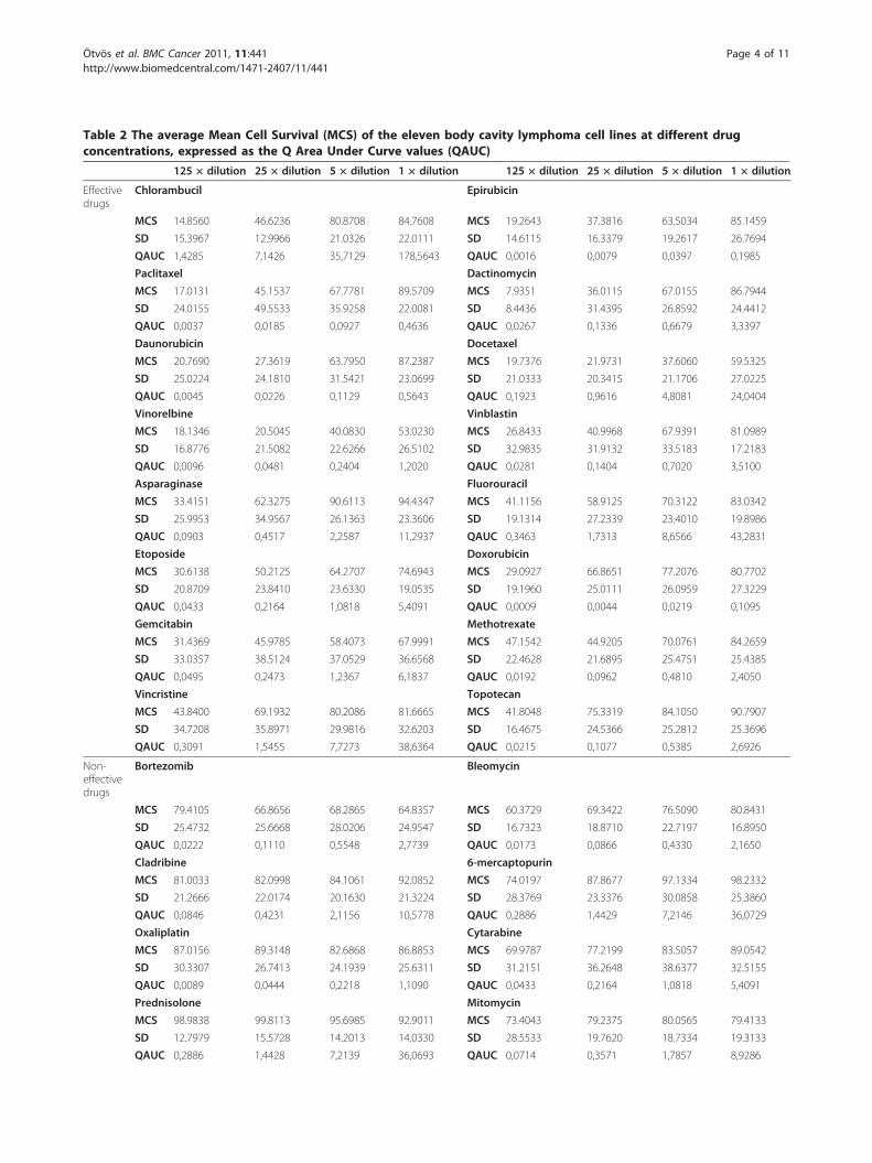
Table 2 The average Mean Cell Survival (MCS) of the eleven body cavity lymphoma cell lines at different drugconcentrations, expressed as the Q Area Under Curve values (QAUC)
125 × dilution 25 × dilution 5 × dilution 1 × dilution 125 × dilution 25 × dilution 5 × dilution 1 × dilution
Effectivedrugs
Chlorambucil Epirubicin
MCS 14.8560 46.6236 80.8708 84.7608 MCS 19.2643 37.3816 63.5034 85.1459
SD 15.3967 12.9966 21.0326 22.0111 SD 14.6115 16.3379 19.2617 26.7694
QAUC 1,4285 7,1426 35,7129 178,5643 QAUC 0,0016 0,0079 0,0397 0,1985
Paclitaxel Dactinomycin
MCS 17.0131 45.1537 67.7781 89.5709 MCS 7.9351 36.0115 67.0155 86.7944
SD 24.0155 49.5533 35.9258 22.0081 SD 8.4436 31.4395 26.8592 24.4412
QAUC 0,0037 0,0185 0,0927 0,4636 QAUC 0,0267 0,1336 0,6679 3,3397
Daunorubicin Docetaxel
MCS 20.7690 27.3619 63.7950 87.2387 MCS 19.7376 21.9731 37.6060 59.5325
SD 25.0224 24.1810 31.5421 23.0699 SD 21.0333 20.3415 21.1706 27.0225
QAUC 0,0045 0,0226 0,1129 0,5643 QAUC 0,1923 0,9616 4,8081 24,0404
Vinorelbine Vinblastin
MCS 18.1346 20.5045 40.0830 53.0230 MCS 26.8433 40.9968 67.9391 81.0989
SD 16.8776 21.5082 22.6266 26.5102 SD 32.9835 31.9132 33.5183 17.2183
QAUC 0,0096 0,0481 0,2404 1,2020 QAUC 0,0281 0,1404 0,7020 3,5100
Asparaginase Fluorouracil
MCS 33.4151 62.3275 90.6113 94.4347 MCS 41.1156 58.9125 70.3122 83.0342
SD 25.9953 34.9567 26.1363 23.3606 SD 19.1314 27.2339 23.4010 19.8986
QAUC 0,0903 0,4517 2,2587 11,2937 QAUC 0,3463 1,7313 8,6566 43,2831
Etoposide Doxorubicin
MCS 30.6138 50.2125 64.2707 74.6943 MCS 29.0927 66.8651 77.2076 80.7702
SD 20.8709 23.8410 23.6330 19.0535 SD 19.1960 25.0111 26.0959 27.3229
QAUC 0,0433 0,2164 1,0818 5,4091 QAUC 0,0009 0,0044 0,0219 0,1095
Gemcitabin Methotrexate
MCS 31.4369 45.9785 58.4073 67.9991 MCS 47.1542 44.9205 70.0761 84.2659
SD 33.0357 38.5124 37.0529 36.6568 SD 22.4628 21.6895 25.4751 25.4385
QAUC 0,0495 0,2473 1,2367 6,1837 QAUC 0,0192 0,0962 0,4810 2,4050
Vincristine Topotecan
MCS 43.8400 69.1932 80.2086 81.6665 MCS 41.8048 75.3319 84.1050 90.7907
SD 34.7208 35.8971 29.9816 32.6203 SD 16.4675 24.5366 25.2812 25.3696
QAUC 0,3091 1,5455 7,7273 38,6364 QAUC 0,0215 0,1077 0,5385 2,6926
Non-effectivedrugs
Bortezomib Bleomycin
MCS 79.4105 66.8656 68.2865 64.8357 MCS 60.3729 69.3422 76.5090 80.8431
SD 25.4732 25.6668 28.0206 24.9547 SD 16.7323 18.8710 22.7197 16.8950
QAUC 0,0222 0,1110 0,5548 2,7739 QAUC 0,0173 0,0866 0,4330 2,1650
Cladribine 6-mercaptopurin
MCS 81.0033 82.0998 84.1061 92.0852 MCS 74.0197 87.8677 97.1334 98.2332
SD 21.2666 22.0174 20.1630 21.3224 SD 28.3769 23.3376 30.0858 25.3860
QAUC 0,0846 0,4231 2,1156 10,5778 QAUC 0,2886 1,4429 7,2146 36,0729
Oxaliplatin Cytarabine
MCS 87.0156 89.3148 82.6868 86.8853 MCS 69.9787 77.2199 83.5057 89.0542
SD 30.3307 26.7413 24.1939 25.6311 SD 31.2151 36.2648 38.6377 32.5155
QAUC 0,0089 0,0444 0,2218 1,1090 QAUC 0,0433 0,2164 1,0818 5,4091
Prednisolone Mitomycin
MCS 98.9838 99.8113 95.6985 92.9011 MCS 73.4043 79.2375 80.0565 79.4133
SD 12.7979 15.5728 14.2013 14.0330 SD 28.5533 19.7620 18.7334 19.3133
QAUC 0,2886 1,4428 7,2139 36,0693 QAUC 0,0714 0,3571 1,7857 8,9286
Ötvös et al. BMC Cancer 2011, 11:441http://www.biomedcentral.com/1471-2407/11/441
Page 4 of 11
clinical dose and half-time using the standard trapezoi-dal rule calculation. The in vivo AUC72 hr data is sum-marized in the seventh column of Table 1. The detailedreferences to the clinical dose and to the in vivo half-time data are available at the Swedish pharmacologicalwebsite http://fass.se. A QAUC72 hr value higher than 1indicates that the in vitro drug concentration is higherthan the one used in the clinical practice. If this value is1, it means that the in vitro concentration correspondsto the clinically achieved in vivo concentration.
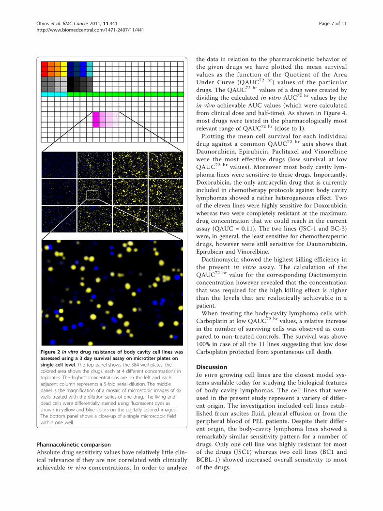
ResultsThe in vitro drug sensitivity assayWe have tested the drug sensitivity patterns of the bodycavity lymphoma lines in short term, in vitro survivalassays. The clinical origin and viral status of the indivi-dual lines is summarized in Figure 1. Each cell line wastested against 27 different drugs, in triplicates, at fourdifferent concentrations. The assay was carried out on384 well plates. After 3 days of incubation each indivi-dual well of the test plates was photographed using acustom developed, automated extended field confocalmicroscope. Living and dead cells were differentiallystained using viability dependent fluorescent dyes asshown in Figure 2. Each individual living or dead cellwas identified counted and their fluorescence intensitydistribution was recorded using automated image analy-tic and quantitation programs. For each well the percen-tage of surviving cells was calculated by comparing thenumber of living cells in the given well to the average ofliving cells in the untreated control wells.
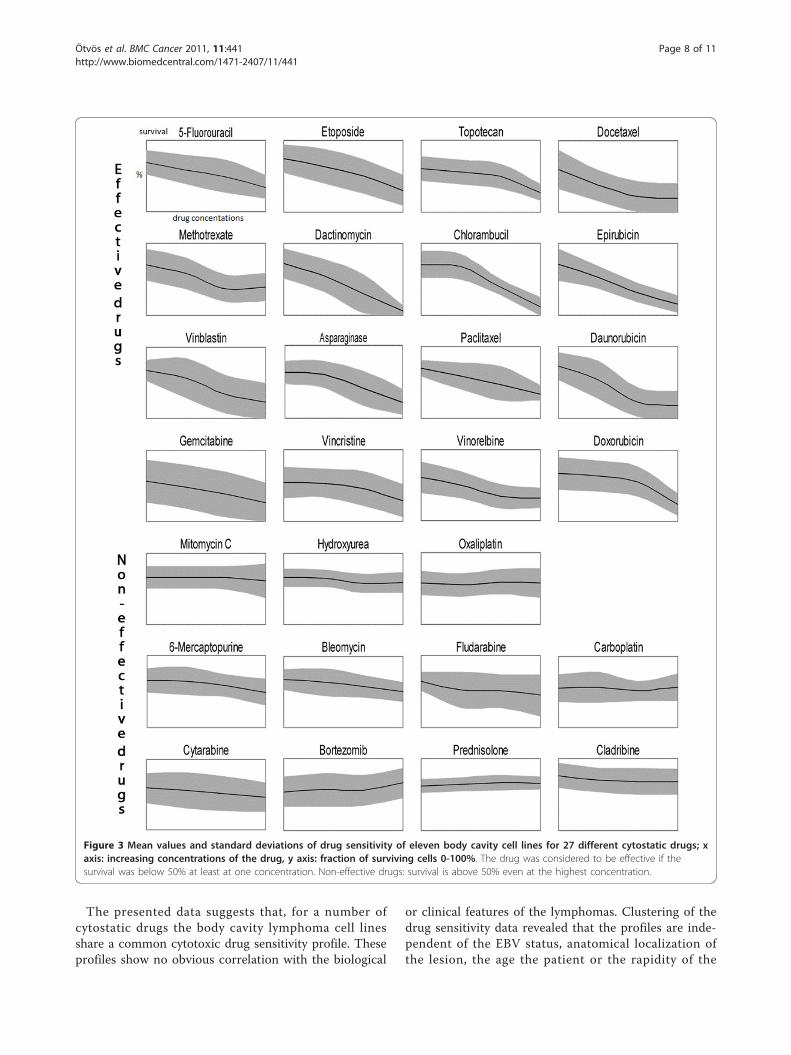
The summarized drug sensitivity pattern of the bodycavity lymphoma cell linesThe summarized cell survival data is shown in Figure 3.The middle line of the individual curves represents theMean Cell Survival (MCS) for all the cell lines alongwith the ± Standard Deviations of the means (SD - grayshaded area) for the four different dilutions of the 27drugs. Drugs were considered to be more universallyactive if they showed less standard deviation around themeans.
Most of the lines were sensitive for sixteen of the 27drugs where sensitivity was defined as less than 50%mean survival at any of the drug dilutions (effectivedrugs). If more than half of the cells were alive even atthe highest concentration than the drug was consideredto be ineffective.We found that the sixteen effective drugs against
body-cavity lymphoma were the following in the orderof effectiveness: Dactinomycin, Chlorambucil, Paclitaxel,Vinorelbine, Epirubicin, Docetaxel, Daunorubicin, Vin-blastin, Doxorubicin, Etoposide, Gemcitabine, Asparagi-nase, Fluorouracil, Topotecan, Vincristine andMethotrexate.Most body-cavity lymphoma lines were resistant to
Oxaliplatin, Bleomycin, 6-Mercaptopurine, Hydro-xyurea, Cladribine, Carboplatin, Bortezomib, Cytosine-arabinosid, Prednisolone, Mitomycin and Fludarabin.Although the Oxaliplatin, Cisplatin and Prednisolonedrugs were not effective against any of the body-cavitylymphoma lines these drugs show concentration-dependent growth-inhibitory effect on other cell linesor primary tumors in parallel experiments (data notshown) at the same concentration as used in thispaper [18].
Heat map of the cluster analysisIn order to identify possible co-segregation of the sensi-tivity patterns of the individual drugs as well as to sys-tematically compare all the lines with each other, wehave carried out unsupervised two-dimensional hier-archical clustering of the simplified drug sensitivity datausing the Cluster 3.0 program for MacOS X. The resultswere visualized using the program TreeView [19]. Thesensitivity to the drug was represented on a 5 step scalewhere every step represents less than 50% viability atthe four different drug dilutions. (Resistant - if morethan 50% survival at the highest concentration, maxi-mum sensitivity - if less than 50% survival at the lowestconcentration.) The graphical representation of the clus-tering results, along with the EBV, HIV status and thepresence of concomitant Kaposi sarcoma, are shown inFigure 1.
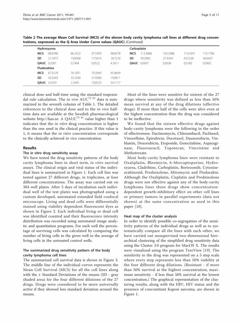
Table 2 The average Mean Cell Survival (MCS) of the eleven body cavity lymphoma cell lines at different drug concen-trations, expressed as the Q Area Under Curve values (QAUC) (Continued)
Hydroxyurea Carboplatin
MCS 88.8788 86.3322 97.5959 98.8578 MCS 113.3868 103.2988 113.4341 110.1786
SD 21.5473 19.8568 17.5610 18.7218 SD 39.2983 27.4344 43.5238 38.6427
QAUC 0,0381 0,1904 0,9522 4,7611 QAUC 0,0007 0,0036 0,0180 0,0902
Fludarabine
MCS 67.0129 76.1851 79.2040 95.8649
SD 43.0343 35.3340 37.6906 19.6811
QAUC 0,4329 2,1645 10,8223 54,1117
Ötvös et al. BMC Cancer 2011, 11:441http://www.biomedcentral.com/1471-2407/11/441
Page 5 of 11
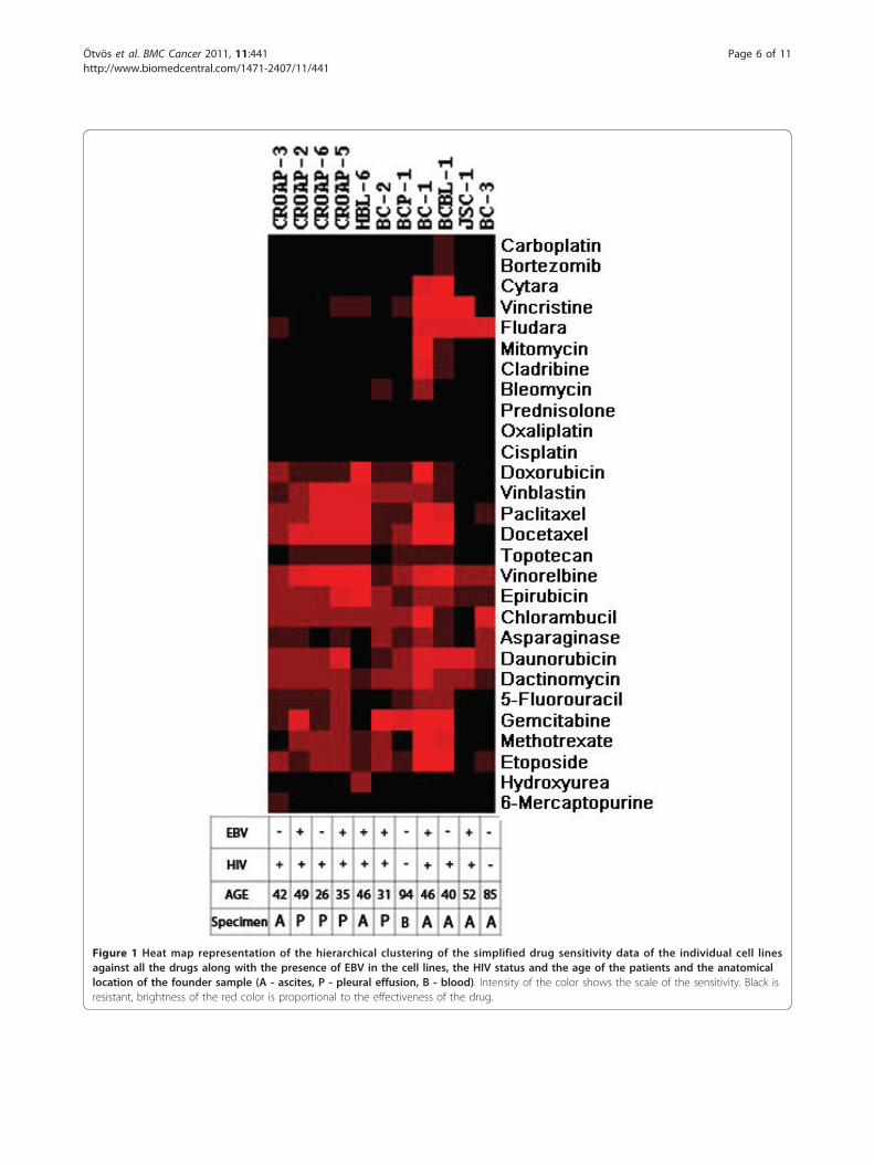
Figure 1 Heat map representation of the hierarchical clustering of the simplified drug sensitivity data of the individual cell linesagainst all the drugs along with the presence of EBV in the cell lines, the HIV status and the age of the patients and the anatomicallocation of the founder sample (A - ascites, P - pleural effusion, B - blood). Intensity of the color shows the scale of the sensitivity. Black isresistant, brightness of the red color is proportional to the effectiveness of the drug.
Ötvös et al. BMC Cancer 2011, 11:441http://www.biomedcentral.com/1471-2407/11/441
Page 6 of 11
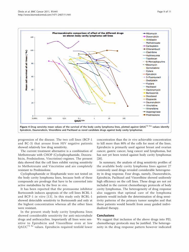
Pharmacokinetic comparisonAbsolute drug sensitivity values have relatively little clin-ical relevance if they are not correlated with clinicallyachievable in vivo concentrations. In order to analyze
the data in relation to the pharmacokinetic behavior ofthe given drugs we have plotted the mean survivalvalues as the function of the Quotient of the AreaUnder Curve (QAUC72 hr) values of the particulardrugs. The QAUC72 hr values of a drug were created bydividing the calculated in vitro AUC72 hr values by thein vivo achievable AUC values (which were calculatedfrom clinical dose and half-time). As shown in Figure 4.most drugs were tested in the pharmacologically mostrelevant range of QAUC72 hr (close to 1).Plotting the mean cell survival for each individual
drug against a common QAUC72 hr axis shows thatDaunorubicin, Epirubicin, Paclitaxel and Vinorelbinewere the most effective drugs (low survival at lowQAUC72 hr values). Moreover most body cavity lym-phoma lines were sensitive to these drugs. Importantly,Doxorubicin, the only antracyclin drug that is currentlyincluded in chemotherapy protocols against body cavitylymphomas showed a rather heterogeneous effect. Twoof the eleven lines were highly sensitive for Doxorubicinwhereas two were completely resistant at the maximumdrug concentration that we could reach in the currentassay (QAUC = 0.11). The two lines (JSC-1 and BC-3)were, in general, the least sensitive for chemotherapeuticdrugs, however were still sensitive for Daunorubicin,Epirubicin and Vinorelbine.Dactinomycin showed the highest killing efficiency in
the present in vitro assay. The calculation of theQAUC72 hr value for the corresponding Dactinomycinconcentration however revealed that the concentrationthat was required for the high killing effect is higherthan the levels that are realistically achievable in apatient.When treating the body-cavity lymphoma cells with
Carboplatin at low QAUC72 hr values, a relative increasein the number of surviving cells was observed as com-pared to non-treated controls. The survival was above100% in case of all the 11 lines suggesting that low doseCarboplatin protected from spontaneous cell death.
DiscussionIn vitro growing cell lines are the closest model sys-tems available today for studying the biological featuresof body cavity lymphomas. The cell lines that wereused in the present study represent a variety of differ-ent origin. The investigation included cell lines estab-lished from ascites fluid, pleural effusion or from theperipheral blood of PEL patients. Despite their differ-ent origin, the body-cavity lymphoma lines showed aremarkably similar sensitivity pattern for a number ofdrugs. Only one cell line was highly resistant for mostof the drugs (JSC1) whereas two cell lines (BC1 andBCBL-1) showed increased overall sensitivity to mostof the drugs.
Figure 2 In vitro drug resistance of body cavity cell lines wasassessed using a 3 day survival assay on microtiter plates onsingle cell level. The top panel shows the 384 well plates, thecolored area shows the drugs, each at 4 different concentrations intriplicates. The highest concentrations are on the left and eachadjacent column represents a 5-fold serial dilution. The middlepanel is the magnification of a mosaic of microscopic images of sixwells treated with the dilution series of one drug. The living anddead cells were differentially stained using fluorescent dyes asshown in yellow and blue colors on the digitally colored images.The bottom panel shows a close-up of a single microscopic fieldwithin one well.
Ötvös et al. BMC Cancer 2011, 11:441http://www.biomedcentral.com/1471-2407/11/441
Page 7 of 11
The presented data suggests that, for a number ofcytostatic drugs the body cavity lymphoma cell linesshare a common cytotoxic drug sensitivity profile. Theseprofiles show no obvious correlation with the biological
or clinical features of the lymphomas. Clustering of thedrug sensitivity data revealed that the profiles are inde-pendent of the EBV status, anatomical localization ofthe lesion, the age the patient or the rapidity of the
Figure 3 Mean values and standard deviations of drug sensitivity of eleven body cavity cell lines for 27 different cytostatic drugs; xaxis: increasing concentrations of the drug, y axis: fraction of surviving cells 0-100%. The drug was considered to be effective if thesurvival was below 50% at least at one concentration. Non-effective drugs: survival is above 50% even at the highest concentration.
Ötvös et al. BMC Cancer 2011, 11:441http://www.biomedcentral.com/1471-2407/11/441
Page 8 of 11
progression of the disease. The two cell lines (BCP-1and BC-3) that arouse from HIV negative patientsshowed relatively low drug sensitivity.The current treatment alternative is a combination of
Methotrexate with CHOP (Cyclophosphamide, Doxoru-bicin, Prednisolone, Vincristine)-regimes. The presentdata showed that the cell lines exhibit varying sensitivityto Methotrexate and Vincristine and are completelyresistant to Prednisolone.Cyclophosphamide or Ifosphamide were not tested on
the body cavity lymphoma lines, because both of thesecompounds are prodrugs that have to be converted intoactive metabolites by the liver in vivo.It has been reported that the proteasome inhibitor
Bortezomib induces apoptosis of the cell lines BCBL-1and BCP-1 in vitro [13]. In this study only BCBL-1showed detectable sensitivity to Bortezomib and only atthe highest concentration whereas all the other lineswere resistant.In the present study body cavity lymphoma lines
showed considerable sensitivity for anti-microtubuledrugs and anthracyclins. Importantly all lines were sen-sitive to Epirubicin and Vinorelbine even at lowQAUC72 hr values. Epirubicin required tenfold lower
concentration than the in vivo achievable concentrationto kill more than 80% of the cells for most of the lines.Epirubicin is primarily used against breast and ovariancancer, gastric cancer, lung cancer and lymphomas, buthas not yet been tested against body cavity lymphomas[20].In summary, the analysis of drug sensitivity profiles of
the available body cavity lymphoma lines against 27commonly used drugs revealed considerable heterogene-ity in drug response. Four drugs, namely, Daunorubicin,Epirubicin, Paclitaxel and Vinorelbine showed uniformlyhigh efficiency on the cell lines. These drugs are not yetincluded in the current chemotherapy protocols of bodycavity lymphomas. The heterogeneity of drug responsealso suggests that optimal care of the lymphomapatients would include the determination of drug sensi-tivity patterns of the primary tumor samples and thatthese patients would benefit from assay guided indivi-dualized therapy.
ConclusionsWe suggest that inclusion of the above drugs into PELchemotherapy protocols may be justified. The heteroge-neity in the drug response pattern however indicated
Figure 4 Drug sensivity mean values of the survival of the body cavity lymphoma lines, plotted against QAUC72 hrs values identifyEpirubicin, Daunorubicin, Vinorelbine and Paclitaxel as novel candidate drugs against body cavity lymphomas.
Ötvös et al. BMC Cancer 2011, 11:441http://www.biomedcentral.com/1471-2407/11/441
Page 9 of 11
that assay guided individualized therapy might berequired to optimize therapeutic response.
AcknowledgementsWe thank the Swedish Research Council, Cancer Research Institute/ConcernFoundation for Cancer Research and the IRIS center for economic support.The study sponsors had no role in the conduct of the study, in thecollection, management, analysis, or interpretation of data, or in thepreparation, review, or approval of the manuscript.
Author details1Department of Microbiology, Tumor and Cell Biology (MTC) and Center forIntegrative Recognition in the Immune System (IRIS), Karolinska Institute, Box280 SE-17177 Stockholm, Sweden. 2Dipartimento di Anatomia Patologica,Istituto Nazionale Tumori, via Venezian, Milano, Italy. 3Karolinska Pharmacy,and Department of Woman and Child Health, Childhood Cancer ResearchUnit, Karolinska Institutet, Karolinska University Hospital, Karolinksavagen,Stockholm, Sweden. 4Department of Medical Microbiology, Medical andHealth Science Center, University of Debrecen, Nagyerdei krt. 98, Debrecen,Hungary.
Authors’ contributionsThe project was conceived and designed by LS. The experiments weremainly carried out and/or coordinated by ÖR. LLK, AG and LM took part incell culturing, and preparation of the microtiter plates for in vitro drugsensitivity assays. EF was responsible for measuring the plates using theautomated laser confocal fluorescent microscope. LS and EF wrote thecomputer programs QuantCapture 4.0 and QuantCount 5.0. LM and HSanalysed and interpreted the data. SE made comparable the in vitro resultswith the in vivo data. JK, LG, AC together with the other authors have beeninvolved in the planning of the experimental details, and the drafting andcritical reading of the manuscript. All authors read and approved the finalmanuscript.
Competing interestsThe authors declare that they have no competing interests.
Received: 14 June 2011 Accepted: 12 October 2011Published: 12 October 2011
References1. Carbone A, Gloghini A: KSHV/HHV8-associated lymphomas. Br J Haematol
2008, 140(1):13-24.2. Boulanger E, Daniel MT, Agbalika F, Oksenhendler E: Combined
chemotherapy including high-dose methotrexate in KSHV/HHV8-associated primary effusion lymphoma. Am J Hematol 2003, 73(3):143-148.
3. Komanduri KV, Luce JA, McGrath MS, Herndier BG, Ng VL: The naturalhistory and molecular heterogeneity of HIV-associated primarymalignant lymphomatous effusions. J Acquir Immune Defic Syndr HumRetrovirol 1996, 13(3):215-226.
4. Morassut S, Vaccher E, Balestreri L, Gloghini A, Gaidano G, Volpe R, Tirelli U,Carbone A: HIV-associated human herpesvirus 8-positive primarylymphomatous effusions: radiologic findings in six patients. Radiology1997, 205(2):459-463.
5. Nador RG, Cesarman E, Chadburn A, Dawson DB, Ansari MQ, Sald J,Knowles DM: Primary effusion lymphoma: a distinct clinicopathologicentity associated with the Kaposi’s sarcoma-associated herpes virus.Blood 1996, 88(2):645-656.
6. Ansari MQ, Dawson DB, Nador R, Rutherford C, Schneider NR, Latimer MJ,Picker L, Knowles DM, McKenna RW: Primary body cavity-based AIDS-related lymphomas. Am J Clin Pathol 1996, 105(2):221-229.
7. Otsuki T, Kumar S, Ensoli B, Kingma DW, Yano T, Stetler-Stevenson M,Jaffe ES, Raffeld M: Detection of HHV-8/KSHV DNA sequences in AIDS-associated extranodal lymphoid malignancies. Leukemia 1996,10(8):1358-1362.
8. Karcher DS, Alkan S: Human herpesvirus-8-associated body cavity-basedlymphoma in human immunodeficiency virus-infected patients: aunique B-cell neoplasm. Hum Pathol 1997, 28(7):801-808.
9. Valencia ME, Martinez P, Moreno V, Laguna F, Lahoz JG: AIDS-related bodycavity-based lymphomas, herpesvirus-8 and HIV infection: a study ofseven cases. Aids 1999, 13(18):2603-2605.
10. Boulanger E, Agbalika F, Maarek O, Daniel MT, Grollet L, Molina JM,Sigaux F, Oksenhendler E: A clinical, molecular and cytogenetic study of12 cases of human herpesvirus 8 associated primary effusion lymphomain HIV-infected patients. Hematol J 2001, 2(3):172-179.
11. Chen YB, Rahemtullah A, Hochberg E: Primary effusion lymphoma.Oncologist 2007, 12(5):569-576.
12. Halfdanarson TR, Markovic SN, Kalokhe U, Luppi M: A non-chemotherapytreatment of a primary effusion lymphoma: durable remission afterintracavitary cidofovir in HIV negative PEL refractory to chemotherapy.Ann Oncol 2006, 17(12):1849-1850.
13. An J, Sun Y, Fisher M, Rettig MB: Antitumor effects of bortezomib (PS-341)on primary effusion lymphomas. Leukemia 2004, 18(10):1699-1704.
14. Flaberg E, Stuber G, Szekely L: Multi-dimensional laser confocalmicroscopy on live cells in submicroliter volumes using glass capillaries.Acta Histochem Cytochem 2006, 39(4):103-106.
15. Flaberg E, Sabelstrom P, Strandh C, Szekely L: Extended Field LaserConfocal Microscopy (EFLCM): combining automated Gigapixel imagecapture with in silico virtual microscopy. BMC Med Imaging 2008, 8:13.
16. Markasz L, Kis LL, Stuber G, Flaberg E, Otvos R, Eksborg S, Skribek H, Olah E,Szekely L: Hodgkin-lymphoma-derived cells show high sensitivity todactinomycin and paclitaxel. Leuk Lymphoma 2007, 48(9):1835-1845.
17. Markasz L, Stuber G, Flaberg E, Jernberg AG, Eksborg S, Olah E, Skribek H,Szekely L: Cytotoxic drug sensitivity of Epstein-Barr virus transformedlymphoblastoid B-cells. BMC Cancer 2006, 6:265.
18. Skribek H, Otvos R, Flaberg E, Nagy N, Markasz L, Eksborg S, Masszi T,Kozma A, Adam E, Miseta A, et al: Chronic lymphoid leukemia cells arehighly sensitive to the combination of prednisolone and daunorubicin,but much less to doxorubicin or epirubicin. Exp Hematol 2010,38(12):1219-1230.
19. de Hoon MJ, Imoto S, Nolan J, Miyano S: Open source clustering software.Bioinformatics 2004, 20(9):1453-1454.
20. Cersosimo RJ, Hong WK: Epirubicin: a review of the pharmacology,clinical activity, and adverse effects of an adriamycin analogue. J ClinOncol 1986, 4(3):425-439.
21. Crews KR, Liu T, Rodriguez-Galindo C, Tan M, Meyer WH, Panetta JC,Link MP, Daw NC: High-dose methotrexate pharmacokinetics andoutcome of children and young adults with osteosarcoma. Cancer 2004,100(8):1724-1733.
22. Albertioni F, Lindemalm S, Reichelova V, Pettersson B, Eriksson S,Juliusson G, Liliemark J: Pharmacokinetics of cladribine in plasma and its5’-monophosphate and 5’-triphosphate in leukemic cells of patients withchronic lymphocytic leukemia. Clin Cancer Res 1998, 4(3):653-658.
23. Hersh MR, Kuhn JG, Phillips JL, Clark G, Ludden TM, Von Hoff DD:Pharmacokinetic study of fludarabine phosphate (NSC 312887). CancerChemother Pharmacol 1986, 17(3):277-280.
24. Chan GL, Erdmann GR, Gruber SA, Stock P, Chen S, Ascher NL, Canafax DM:Pharmacokinetics of 6-thiouric acid and 6-mercaptopurine in renalallograft recipients after oral administration of azathioprine. Eur J ClinPharmacol 1989, 36(3):265-271.
25. Gruber A, Liliemark E, Tidefelt U, Paul C, Bjorkholm M, Peterson C,Liliemark J: Pharmacokinetics of mitoxantrone, etoposide and cytosinearabinoside in leukemic cells during treatment of acute myelogenousleukemia–relationship to treatment outcome and bone marrow toxicity.Leuk Res 1995, 19(10):757-761.
26. Casale F, Canaparo R, Serpe L, Muntoni E, Pepa CD, Costa M, Mairone L,Zara GP, Fornari G, Eandi M: Plasma concentrations of 5-fluorouracil andits metabolites in colon cancer patients. Pharmacol Res 2004,50(2):173-179.
27. Fogli S, Danesi R, Gennari A, Donati S, Conte PF, Del Tacca M: Gemcitabine,epirubicin and paclitaxel: pharmacokinetic and pharmacodynamicinteractions in advanced breast cancer. Ann Oncol 2002, 13(6):919-927.
28. GlaxoSmithKline Research Triangle Park N: Prescribing InformationLeukeran®.[http://www.drugs.com/monograph/leukeran.html].
29. Ghazal-Aswad S, Calvert AH, Newell DR: A single-sample assay for theestimation of the area under the free carboplatin plasma concentrationversus time curve. Cancer Chemother Pharmacol 1996, 37(5):429-434.
Ötvös et al. BMC Cancer 2011, 11:441http://www.biomedcentral.com/1471-2407/11/441
Page 10 of 11
30. Graham MA, Lockwood GF, Greenslade D, Brienza S, Bayssas M, Gamelin E:Clinical pharmacokinetics of oxaliplatin: a critical review. Clin Cancer Res2000, 6(4):1205-1218.
31. Rischin D, Ackland SP, Smith J, Garg MB, Clarke S, Millward MJ, Toner GC,Zalcberg J: Phase I and pharmacokinetic study of docetaxel incombination with epirubicin and cyclophosphamide in advancedcancer: dose escalation possible with granulocyte colony-stimulatingfactor, but not with prophylactic antibiotics. Ann Oncol 2002,13(11):1810-1818.
32. Bates SE, Bakke S, Kang M, Robey RW, Zhai S, Thambi P, Chen CC, Patil S,Smith T, Steinberg SM, et al: A phase I/II study of infusional vinblastinewith the P-glycoprotein antagonist valspodar (PSC 833) in renal cellcarcinoma. Clin Cancer Res 2004, 10(14):4724-4733.
33. Desai ZR, Van den Berg HW, Bridges JM, Shanks RG: Can severe vincristineneurotoxicity be prevented? Cancer Chemother Pharmacol 1982,8(2):211-214.
34. Freyer G, Delozier T, Lichinister M, Gedouin D, Bougnoux P, His P,Imadalou K, Trillet-Lenoir V: Phase II study of oral vinorelbine in first-lineadvanced breast cancer chemotherapy. J Clin Oncol 2003, 21(1):35-40.
35. Andersson B, Andersson I, Beran M, Ehrsson H, Eksborg S: Liquidchromatographic monitoring of daunorubicin and daunorubicinol inplasma from leukemic patients treated with daunorubicin or thedaunorubicin-DNA complex. Cancer Chemother Pharmacol 1979, 2(1):15-17.
36. Toffoli G, Corona G, Cattarossi G, Boiocchi M, Di Gennaro G, Tirelli U,Vaccher E: Effect of highly active antiretroviral therapy (HAART) onpharmacokinetics and pharmacodynamics of doxorubicin in patientswith HIV-associated non-Hodgkin’s lymphoma. Ann Oncol 2004,15(12):1805-1809.
37. Veal GJ, Cole M, Errington J, Parry A, Hale J, Pearson AD, Howe K,Chisholm JC, Beane C, Brennan B, et al: Pharmacokinetics of dactinomycinin a pediatric patient population: a United Kingdom Children’s CancerStudy Group Study. Clin Cancer Res 2005, 11(16):5893-5899.
38. Peng YM, Alberts DS, Chen HS, Mason N, Moon TE: Antitumour activityand plasma kinetics of bleomycin by continuous and intermittentadministration. Br J Cancer 1980, 41(4):644-647.
39. Kozuch P, Hoff PM, Hess K, Adams J, Newman RA, Lee F, Pazdur R: Phase Ibioequivalency study of MitoExtra and mitomycin C in patients withsolid tumors. Cancer 2001, 91(4):815-821.
40. Yan JH, Ataga K, Kaul S, Olson JS, Grasela DM, Gothelf S, Kutlar A,Orringer E: The influence of renal function on hydroxyureapharmacokinetics in adults with sickle cell disease. J Clin Pharmacol 2005,45(4):434-445.
41. Gerrits CJ, Schellens JH, Burris H, Eckardt JR, Planting AS, van der Burg ME,Rodriguez GI, Loos WJ, van Beurden V, Hudson I, et al: A comparison ofclinical pharmacodynamics of different administration schedules of oraltopotecan (Hycamtin). Clin Cancer Res 1999, 5(1):69-75.
42. Ylikangas P, Mononen I: Serious neutropenia in ALL patients treated withL-asparaginase may be avoided by therapeutic monitoring of theenzyme activity in the circulation. Ther Drug Monit 2002, 24(4):502-506.
43. Papandreou CN, Daliani DD, Nix D, Yang H, Madden T, Wang X, Pien CS,Millikan RE, Tu SM, Pagliaro L, et al: Phase I trial of the proteasomeinhibitor bortezomib in patients with advanced solid tumors withobservations in androgen-independent prostate cancer. J Clin Oncol2004, 22(11):2108-2121.
44. Penzak SR, Formentini E, Alfaro RM, Long M, Natarajan V, Kovacs J:Prednisolone pharmacokinetics in the presence and absence of ritonavirafter oral prednisone administration to healthy volunteers. J AcquirImmune Defic Syndr 2005, 40(5):573-580.
Pre-publication historyThe pre-publication history for this paper can be accessed here:http://www.biomedcentral.com/1471-2407/11/441/prepub
doi:10.1186/1471-2407-11-441Cite this article as: Ötvös et al.: Drug sensitivity patterns of HHV8carrying body cavity lymphoma cell lines. BMC Cancer 2011 11:441.
Submit your next manuscript to BioMed Centraland take full advantage of:
• Convenient online submission
• Thorough peer review
• No space constraints or color figure charges
• Immediate publication on acceptance
• Inclusion in PubMed, CAS, Scopus and Google Scholar
• Research which is freely available for redistribution
Submit your manuscript at www.biomedcentral.com/submit
Ötvös et al. BMC Cancer 2011, 11:441http://www.biomedcentral.com/1471-2407/11/441
Page 11 of 11
![Background Methods Results & ConclusionsMethods Design [quant → QUAL] Quant Data Collection. Phase 1: Quant. Phase 2: QUAL . Quant Data Analysis. QUAL Data Analysis . Integration](https://img.pdfslide.us/doc/110x75/6000faa49b2cd844807c19b1/background-methods-results-conclusions-methods-design-quant-a-qual-quant.jpg)