Embed Size (px)
Citation preview
Wojcinski et al. BMC Medical Imaging 2013, 13:36http://www.biomedcentral.com/1471-2342/13/36
RESEARCH ARTICLE Open Access
Diagnostic performance and inter-observerconcordance in lesion detection with theautomated breast volume scanner (ABVS)Sebastian Wojcinski1*, Samuel Gyapong2, André Farrokh2, Philipp Soergel1, Peter Hillemanns1
and Friedrich Degenhardt2
Abstract
Background: Automated whole breast ultrasound scanners of the latest generation have reached a level ofcomfortable application and high quality volume acquisition. Nevertheless, there is a lack of data concerning thistechnology. We investigated the diagnostic performance and inter-observer concordance of the Automated BreastVolume Scanner (ABVS) ACUSON S2000™ and questioned its implications in breast cancer diagnostics.
Methods: We collected 100 volume data sets and created a database containing 52 scans with no detectablelesions in conventional ultrasound (BI-RADS®-US 1), 30 scans with benign lesions (BI-RADS®-US 2) and 18 scans withbreast cancer (BI-RADS®-US 5).Two independent examiners evaluated the ABVS data on a separate workstation without any prior knowledge ofthe patients’ histories.
Results: The inter-rater reliability reached fair agreement (κ=0.36; 95% confidence interval (CI): 0.19-0.53). Withrespect to the true category, the conditional inter-rater validity coefficient was κ=0.18 (95% CI: 0.00-0.26) for thebenign cases and κ=0.80 (95% CI: 0.61-1.00) for the malignant cases.Combining the assessments of examiner 1 and examiner 2, the diagnostic accuracy (AC), sensitivity (SE) andspecificity (SP) for the experimental ABVS were AC = 79.0% (95% CI: 67.3-86.1), SE = 83.3% (95% CI: 57.7-95.6) andSP = 78.1% (% CI: 67.3-86.1), respectively.However, after the ABVS examination, there were a high number of requests for second-look ultrasounds in up to48.8% of the healthy women due to assumed suspicious findings in the volume data.In an exploratory analysis, we estimated that an ABVS examination in addition to mammography alone coulddetect a relevant number of previously occult breast cancers (about 1 cancer in 300 screened and otherwisehealthy women).
Conclusions: The ABVS is a reliable imaging method for the evaluation of the breast with high sensitivity and a fairinter-observer concordance. However, we have to overcome the problem of the high number of false-positiveresults. Therefore, further prospective studies in larger collectives are necessary to define standard procedures inimage acquisition and interpretation. Nevertheless, we consider the ABVS as being suitable for integration intobreast diagnostics as a beneficial and reliable imaging method.
Keywords: Breast cancer, Automated breast ultrasound, Automated breast volume scanner, ABVS, Inter-observerconcordance, Screening
* Correspondence: [email protected] of OB/GYN, Hannover Medical School, OE 6410,Carl-Neuberg-Straße 1, 30625 Hannover, GermanyFull list of author information is available at the end of the article
© 2013 Wojcinski et al.; licensee BioMed Central Ltd. This is an open access article distributed under the terms of the CreativeCommons Attribution License (http://creativecommons.org/licenses/by/2.0), which permits unrestricted use, distribution, andreproduction in any medium, provided the original work is properly cited.
Wojcinski et al. BMC Medical Imaging 2013, 13:36 Page 2 of 12http://www.biomedcentral.com/1471-2342/13/36
BackgroundToday, we can expect that more than one million womenwill be newly diagnosed with breast cancer each year [1].The most recent data were reported in 2000, when about400,000 women died from breast cancer, which repre-sented 1.6 percent of all female deaths [2]. Nations withthe highest cancer rates include the U.S.A, Italy, Australia,Germany, the Netherlands, Canada and France [3]. Sec-ondary prevention, i.e. the early detection of usually cur-able stages of breast cancer, has moved into the very focusof all healthcare systems. While the incidence of breastcancer remains considerably high, we notice a relevantdecline in cancer mortality in numerous countries. Thiseffort can partly be explained by new and innovative ther-apies, but there is sound evidence that advances in earlydetection probably play the most decisive role [4]. Hence,the early detection of breast cancer has moved into thecentral focus of primary healthcare. This success couldonly be achieved because of improvements in imagingtechnologies and a higher degree of health awareness.Breast cancer screening programs are based on radio-
logic examinations of the breast. Additional imagingmodalities are only indicated if suspicious or unclearfindings are present in the mammogram. Mammographyhas demonstrated excellent sensitivity, specificity andinter-observer concordance [5,6]. Nevertheless, the diag-nostic accuracy of breast ultrasound and magnetic res-onance imaging (MRI) is as good as mammography.However, these two modalities imply a number of majordisadvantages. Breast MRI is an expensive and complextechnology. Moreover, MRI has insufficient specificity and,consequently, produces a relevant number of false-positivefindings. Ultrasound, on the other hand, is observer-dependent, time-consuming and the examiner has to bepresent at the time of image acquisition. Furthermore, onlysubjectively chosen screenshots from the ultrasound exam-ination are printed and/or stored. Mammography has theadvantage that the examination is rapid, standardizableand cost efficient. The generation of the mammogram canbe performed by medical assistant personnel and thestored images allow second-readings and follow-ups. How-ever, merely focusing on detection rates and diagnosticaccuracy, breast ultrasound, breast MRI or even the com-bination of several imaging technologies may be superiorto mammography alone.So far, breast ultrasound is the most commonly ac-
cepted and reliable diagnostic method for women withclinically or radiologically suspicious breast lesions [7].Furthermore, bilateral whole breast ultrasound has evendemonstrated diagnostic advantages in screening asymp-tomatic women [8-12]. Nevertheless, after a comparisonof the advantages and disadvantages of the various im-aging techniques, mammography has become the mostcommon, reliable and efficient screening method. This
attitude in breast cancer screening may change someday,when technical advances allow further improvements ofultrasound and MRI. At this point, the concept ofautomated whole breast ultrasound emerges with thepotential capability to overcome the inherent deficits ofhand-held breast ultrasound. Automated breast scannerscan be easily operated by medical assistants. The storedvolume data allow comfortable and time-efficient evalu-ation at anytime by a medical professional. The perform-ance of second-readings by additional examiners andfollow-up evaluations are unproblematic. The concept ofautomated breast ultrasound dates back to the 1970s [13].In the current report, we present data concerning an
up-to-date technology in automated ultrasound, the Auto-mated Breast Volume Scanner (ACUSON S2000™ ABVS;Siemens Medical Solutions, Inc., CA, USA). The ABVS re-constructs 3D data sets of the entire breast volume fromautomatically acquired B-mode images. These data can bestored and analyzed on a separate workstation.We evaluated whether or not breast lesions, previously
detected by means of conventional ultrasound, couldalso be detected and correctly classified by independentexaminers who only used ABVS data. Furthermore, weanalyzed the inter-observer concordance and performeda model calculation to scrutinize the potential implica-tions of the ABVS in a screening setting.
Materials and methodsGeneral design and patient databaseOur study was carried out at the Franziskus HospitalBreast Cancer Center in Bielefeld, Germany, betweenMarch 2010 and July 2011. For the ultrasound examina-tions, we used the Siemens ACUSON S2000™ ultrasoundsystem with the integrated ABVS (Siemens Medical So-lutions, Inc., CA, USA). Patients are generally referredto our outpatient department on account of specificdiagnostic queries, such as palpable breast lesions, breastpain, suspicious mammograms and intensified screeningin low-risk and high-risk populations. Usually, patientsreceive a clinical examination, mammography and con-ventional breast ultrasound as the standard diagnosticmethods, as well as subsequent examinations whenevernecessary. Additionally, we offer an optional ABVS exam-ination to all our patients within the routine practice ofour breast cancer center. As a diagnostic standard, ultra-sound pictures are categorized according to the Breast Im-aging Reporting and Data System criteria of the AmericanCollege of Radiology (ACR BI-RADS®-US) [14]. Study par-ticipants were recruited from this population. Patientswith a final categorization of BI-RADS®-US 1, 2 or 5 in theconventional ultrasound examination were regarded assuitable for our study. BI-RADS®-US categories 0, 3 and 4involve breast lesions of questionable dignity. As the focusof our study was on the detection of evidently benign or
Wojcinski et al. BMC Medical Imaging 2013, 13:36 Page 3 of 12http://www.biomedcentral.com/1471-2342/13/36
malignant lesions, we excluded patients with BI-RADS®-US 0, 3 and 4. Patients with a bra cup size greater than D,a history of breast surgery, inflammatory conditions of thebreast, skin disorders or psychiatric disorders were alsoexcluded. Patients who met the inclusion criteria andagreed to receive an additional ABVS examination enteredour study. In this way, we performed 100 breast examina-tions with the ABVS and, consequently, we created a data-base containing 100 3D-scans with BI-RADS®-US 1, 2 or 5findings in the volume.All data were obtained using a standard of care clinical
protocol and approved equipment. Therefore, the respon-sible ethics committee did not demand additional approvalfor this non-interventional case study. Although the re-quirements of individual informed consent were waived,we decided that all study participants signed an additionalinformed consent form before the ABVS examination.Two independent examiners evaluated the cases from
the anonymized ABVS database. We compared the per-formance of these two examiners (ABVS, experimentalmethod) with each other and with the results from theconventional ultrasound (gold standard).
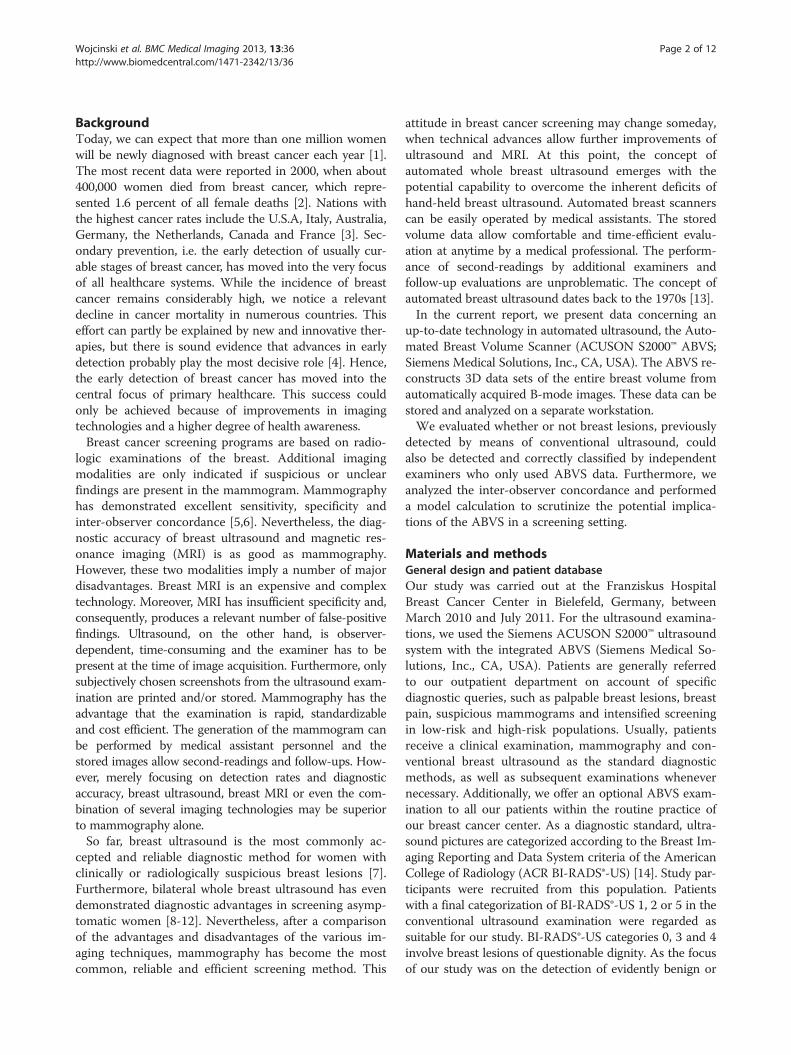
Technical background of the ABVS ultrasound systemFor our study we used the ACUSON S2000™ ABVS(Siemens Medical Solutions, Inc., CA, USA), an ultrasoundsystem that automatically acquires full-field volumes of the
Figure 1 ACUSON S2000™ ABVS. On the left-hand side is the ACUSON Svolume transducer attached to a mechanical arm.
breast (Figure 1). The system is equipped with an ultra-wide linear transducer (Siemens 14L5BV, 14 MHz, 15.4cm, and 768 piezoelectric elements). While automaticallysweeping over the breast, this ultrasound probe covers adistance of 16.8 cm in approximately one minute, acquir-ing about 300 high-resolution slices for post-processing(resolution: axial = 0.09 mm, lateral = 0.16 mm, sagittal =0.44 mm). These images are the source for the creation ofthe 3D data sets. A separate workstation provides com-prehensive tools for image analysis and manipulation ofthe volume data (Figure 2). The secondary images arecalculated from the acquisition volume in real-time. De-tails concerning the ABVS have been described else-where [15,16].
Conventional B-mode ultrasound examinations (goldstandard)Conventional B-mode ultrasound examinations wereperformed by the author SW, a DEGUM (DeutscheGesellschaft für Ultraschall in der Medizin, German so-ciety for ultrasound in medicine) level II certified seniorconsultant in gynecology with 8 years’ experience inbreast ultrasound [17]. This examiner also knew theresults of the other imaging modalities when available(mammography, magnetic resonance imaging) and, there-fore, defined the reference standard for the interpretationof the volume data sets. The conventional ultrasound
2000™ ultrasound machine, on the right-hand side is the 14L5BV
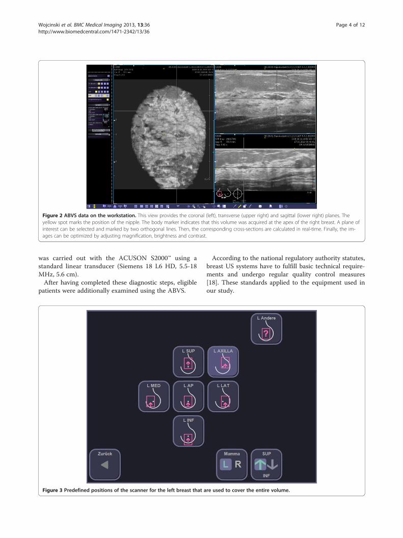
Figure 2 ABVS data on the workstation. This view provides the coronal (left), transverse (upper right) and sagittal (lower right) planes. Theyellow spot marks the position of the nipple. The body marker indicates that this volume was acquired at the apex of the right breast. A plane ofinterest can be selected and marked by two orthogonal lines. Then, the corresponding cross-sections are calculated in real-time. Finally, the im-ages can be optimized by adjusting magnification, brightness and contrast.
Wojcinski et al. BMC Medical Imaging 2013, 13:36 Page 4 of 12http://www.biomedcentral.com/1471-2342/13/36
was carried out with the ACUSON S2000™ using astandard linear transducer (Siemens 18 L6 HD, 5.5-18MHz, 5.6 cm).After having completed these diagnostic steps, eligible
patients were additionally examined using the ABVS.
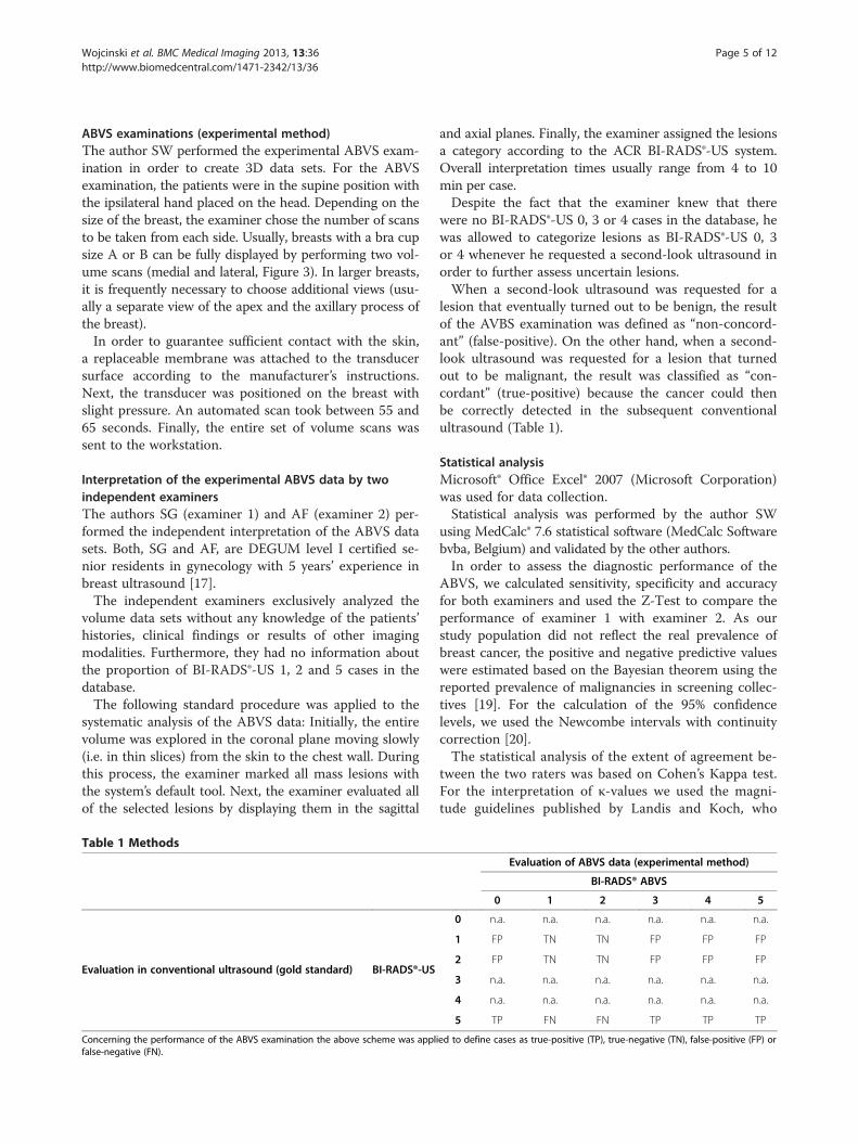
Figure 3 Predefined positions of the scanner for the left breast that a
According to the national regulatory authority statutes,breast US systems have to fulfill basic technical require-ments and undergo regular quality control measures[18]. These standards applied to the equipment used inour study.
re used to cover the entire volume.
Wojcinski et al. BMC Medical Imaging 2013, 13:36 Page 5 of 12http://www.biomedcentral.com/1471-2342/13/36
ABVS examinations (experimental method)The author SW performed the experimental ABVS exam-ination in order to create 3D data sets. For the ABVSexamination, the patients were in the supine position withthe ipsilateral hand placed on the head. Depending on thesize of the breast, the examiner chose the number of scansto be taken from each side. Usually, breasts with a bra cupsize A or B can be fully displayed by performing two vol-ume scans (medial and lateral, Figure 3). In larger breasts,it is frequently necessary to choose additional views (usu-ally a separate view of the apex and the axillary process ofthe breast).In order to guarantee sufficient contact with the skin,
a replaceable membrane was attached to the transducersurface according to the manufacturer’s instructions.Next, the transducer was positioned on the breast withslight pressure. An automated scan took between 55 and65 seconds. Finally, the entire set of volume scans wassent to the workstation.
Interpretation of the experimental ABVS data by twoindependent examinersThe authors SG (examiner 1) and AF (examiner 2) per-formed the independent interpretation of the ABVS datasets. Both, SG and AF, are DEGUM level I certified se-nior residents in gynecology with 5 years’ experience inbreast ultrasound [17].The independent examiners exclusively analyzed the
volume data sets without any knowledge of the patients’histories, clinical findings or results of other imagingmodalities. Furthermore, they had no information aboutthe proportion of BI-RADS®-US 1, 2 and 5 cases in thedatabase.The following standard procedure was applied to the
systematic analysis of the ABVS data: Initially, the entirevolume was explored in the coronal plane moving slowly(i.e. in thin slices) from the skin to the chest wall. Duringthis process, the examiner marked all mass lesions withthe system’s default tool. Next, the examiner evaluated allof the selected lesions by displaying them in the sagittal
Table 1 Methods
Evaluation in conventional ultrasound (gold standard) BI-RADS®-US
Concerning the performance of the ABVS examination the above scheme was applfalse-negative (FN).
and axial planes. Finally, the examiner assigned the lesionsa category according to the ACR BI-RADS®-US system.Overall interpretation times usually range from 4 to 10min per case.Despite the fact that the examiner knew that there
were no BI-RADS®-US 0, 3 or 4 cases in the database, hewas allowed to categorize lesions as BI-RADS®-US 0, 3or 4 whenever he requested a second-look ultrasound inorder to further assess uncertain lesions.When a second-look ultrasound was requested for a
lesion that eventually turned out to be benign, the resultof the AVBS examination was defined as “non-concord-ant” (false-positive). On the other hand, when a second-look ultrasound was requested for a lesion that turnedout to be malignant, the result was classified as “con-cordant” (true-positive) because the cancer could thenbe correctly detected in the subsequent conventionalultrasound (Table 1).
Statistical analysisMicrosoft® Office Excel® 2007 (Microsoft Corporation)was used for data collection.Statistical analysis was performed by the author SW
using MedCalc® 7.6 statistical software (MedCalc Softwarebvba, Belgium) and validated by the other authors.In order to assess the diagnostic performance of the
ABVS, we calculated sensitivity, specificity and accuracyfor both examiners and used the Z-Test to compare theperformance of examiner 1 with examiner 2. As ourstudy population did not reflect the real prevalence ofbreast cancer, the positive and negative predictive valueswere estimated based on the Bayesian theorem using thereported prevalence of malignancies in screening collec-tives [19]. For the calculation of the 95% confidencelevels, we used the Newcombe intervals with continuitycorrection [20].The statistical analysis of the extent of agreement be-
tween the two raters was based on Cohen’s Kappa test.For the interpretation of κ-values we used the magni-tude guidelines published by Landis and Koch, who
Evaluation of ABVS data (experimental method)
BI-RADS® ABVS
0 1 2 3 4 5
0 n.a. n.a. n.a. n.a. n.a. n.a.
1 FP TN TN FP FP FP
2 FP TN TN FP FP FP
3 n.a. n.a. n.a. n.a. n.a. n.a.
4 n.a. n.a. n.a. n.a. n.a. n.a.
5 TP FN FN TP TP TP
ied to define cases as true-positive (TP), true-negative (TN), false-positive (FP) or
Wojcinski et al. BMC Medical Imaging 2013, 13:36 Page 6 of 12http://www.biomedcentral.com/1471-2342/13/36
characterized the values of κ<0 as indicating no agree-ment, κ 0–0.20 slight, κ 0.21-0.40 fair, κ 0.41-0.60 mod-erate, κ 0.61-0.80 substantial, and κ 0.81-1 as almostperfect agreement [21].Furthermore, we assessed the correlation between the
expected and the observed rate of second-look ultra-sounds using the Chi-square test.Statistical significance was assumed as p < 0.05 for all
tests.
ResultsIn our study population, the age ranged from 19 to 86years (median 52 years). According to the BI-RADS®-UScategorization, 52% (n = 52) of our cases were assignedas BI-RADS®-US 1, 30% (n = 30) had BI-RADS®-US 2 le-sions and 18% (n = 18) of the cases had BI-RADS®-US 5lesions. All BI-RADS®-US 5 lesions were confirmed withhistological specimens. The mean tumor size for malig-nant and benign lesions was 22.0 mm (range 13 to 55)and 16.7 mm (range 8 to 36), respectively.
Inter-rater reliabilityThe concordance between examiner 1 and examiner 2concerning the correct clinical decision of whether thepatient should undergo a control ultrasound due to asuspicious finding or whether the patient should bedefined as healthy as there is no suspicious lesion isshown in (Table 2). The inter-rater reliability reached
Table 2 Concordance between examiner 1 and examiner2 concerning the correct clinical decision of whether thepatient should undergo a control ultrasound due to asuspicious finding in ABVS (due to BI-RADS® 0,3,4 or 5)or whether the patient should be defined as healthy asthere is no suspicious lesion in ABVS (i.e. BI-RADS® 1or 2)
Examiner 1 (SG)
BI-RADS® BI-RADS®
TotalABVS ABVS
1 or 2 0, 3, 4 or 5
(negative) (positive)
Examiner 2 (AF)
BI-RADS®
34a 8 42ABVS
1 or 2
(negative)
BI-RADS®
25 33a 58ABVS
0, 3, 4 or 5
(positive)
Total 59 41 100
Distribution of cases by rater and by category. The inter-rater reliability coeffi-cient calculates to κ = 0.36 (0.19-0.53).aconcordance between examiner 1 and examiner 2.
fair agreement and the Cohen’s Kappa value was κ=0.36(95% CI: 0.19-0.53).A more detailed breakdown of the concordance be-
tween examiner 1 and examiner 2 concerning the dis-tinct BI-RADS® category in the ABVS examination isgiven in (Table 3). In this analysis, the inter-rater reliabilityalso reached fair agreement and the Cohen’s Kappa valuewas κ = 0.27 (0.14-0.40).
Inter-rater validityWith respect to the true category (benign cases and malig-nant cases), the inter-rater validity coefficient calculated toκ=0.31 (95% CI: 0.21-0.41). Focusing on the benign cases(n = 82), the conditional inter-rater validity coefficient wasκ=0.18 (95% CI: 0.00-0.26), indicating slight agreement.Concerning the malignant cases, we found a Cohen’sKappa value of κ=0.80 (95% CI: 0.61-1.00), indicating sub-stantial to almost perfect agreement (Table 4).
Diagnostic performance of the ABVSThe sensitivity for examiner 1 and examiner 2 in detect-ing malignant lesions with the ABVS was 83.3% (95% CI:57.7-95.6) and 100% (95% CI: 78.1-100.0), respectively.The diagnostic accuracy of the method was 71.0% (95%CI: 60.9-79.4) and 60.0% (95% CI: 49.7-69.5), respect-ively. The differences between examiner 1 and examiner2 were statistically not significant. Nevertheless, specifi-city revealed to be quite low at 68.3% (95% CI: 57.0-77.9) and 51.2% (40.0-62.3), respectively, as there was arelevant number of requests for second-look ultrasoundsor further examinations in the group of healthy patientsafter the evaluation of the ABVS data. As previously de-scribed in the methods, these cases had to be classifiedas false-positives if there was no cancer (Table 1). More-over, specificity was significantly different between exam-iner 1 and examiner 2 (p = 0.001). The detailed results areshown in (Table 5).In order to investigate the effect of a second reading of
ABVS data, we performed a tentative analysis and com-bined the evaluations of examiner 1 and examiner 2. Withrespect to the low specificity, the following rules for thecombination of assessments were applied: If both exam-iners agreed that a scan was suspicious, the evaluation wasconsidered “positive”. If both examiners agreed that a scanwas unsuspicious or disagreed (and only one examinerregarded the scan as unsuspicious), the evaluation was con-sidered “negative”. In this scenario, the accuracy increasedto 79.0% (95% CI: 69.5-86.3), the specificity increased to78.1% (95% CI: 67.3-86.1) and the sensitivity remainedacceptably high at 83.3% (95% CI: 57.7-95.6) (Table 6).
Rate of second-look ultrasoundsWe expected 18 requests for second-look ultrasoundsafter the ABVS examination as there were 18 BI-RADS®-
Table 3 Concordance between examiner 1 and examiner 2 concerning the distinct BI-RADS® category in the ABVSexamination
BI-RADS® ABVS BI-RADS® ABVS BI-RADS® ABVS BI-RADS® ABVS
Total1 2 0, 3 or 4 5
(No finding) (Benign finding) (Unclear finding) (Malignant finding)
Examiner 2 (AF)
BI-RADS® ABVS
20a 2 4 2 281
(No Finding)
BI-RADS® ABVS
4 8a 2 0 142
(Benign finding)
BI-RADS® ABVS
14 7 13a 9 430, 3 or 4
(Unclear finding)
BI-RADS® ABVS
4 0 5 6a 155
(Malignant finding)
Total 42 17 24 17 100
Distribution of cases by rater and by detailed BI-RADS® ABVS category. Concerning this detailed evaluation, the inter-rater reliability coefficient calculates to κ =0.27 (0.14-0.40).aconcordance between examiner 1 and examiner 2.
Wojcinski et al. BMC Medical Imaging 2013, 13:36 Page 7 of 12http://www.biomedcentral.com/1471-2342/13/36
US 5 lesions in our database of 100 cases. We did notexpect requests for the other 82 cases (BI-RADS®-US 1or 2 lesions). However, the observed rate of second-lookultrasounds was significantly high, totaling 41 for exam-iner 1 and 58 for examiner 2, respectively (p < 0.001).Therefore, the rate of second-look ultrasounds in
healthy women (i.e. BI-RADS®-US 1 and 2) was 31.7%for examiner 1 and 48.8% for examiner 2.Regarding the BI-RADS®-US 1 and 2 cases separately,
there was a request for a second-look ultrasound in33.3% and 53.3% (examiner 1 and examiner 2) of thewomen with BI-RADS®-US 2 lesions and in 30.8% and46.2% of the women with no breast lesions at all (i.e. BI-RADS®-US 1).
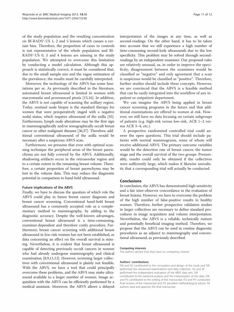
Model calculation and exploratory analysisBased on our findings, we performed a model calcula-tion to estimate how additional ABVS examinations
Table 4 Distribution of cases by rater, reported and true cate
Exa
ABVS
Examiner 2
ABVS negative
Benign cases (conventional ultrasound negative) 34a
Malignant cases (conventional ultrasound positive) 0a
Total 34
The inter-rater validity coefficient calculates to κ = 0.31 (95% CI: 0.21-0.41). The conbenign cases and κ=0.80 (95% CI: 0.61-1.00) for the malignant cases.aconcordance between examiner 1 and examiner 2.
could affect the detection rate of cancer in screeningcollectives.As described in the literature, the prevalence of occult
cancer that can be detected by additional ultrasound inwomen who already underwent mammography and clin-ical examination can be estimated to be between 0.3%and 0.4% [8-12]. Therefore, conventional ultrasound candetect one cancer in about 250 otherwise healthy women.Using these numbers, the positive predictive value in
a screening collective would calculate to 1.49% and thenegative predictive value to 99.91%, respectively. There-fore, the new false-negative rate in a diagnostic settingusing the ABVS would be about 0.09% instead of 0.4%(Table 7).If 10,000 women were additionally screened with the
ABVS, we could expect 2,219 positive scans resulting inthe same number of second-look-ultrasounds, finally lead-ing to 33 detected cancers. Therefore, one cancer would
gories
miner 1 Examiner 1
negative ABVS positive
Examiner 2 Examiner 2 Examiner 2Total
ABVS positive ABVS negative ABVS positive
22 8 18a 82
3 0 15a 18
25 8 33 100
ditional inter-rater validity coefficient is κ=0.18 (95% CI: 0.00-0.26) for the
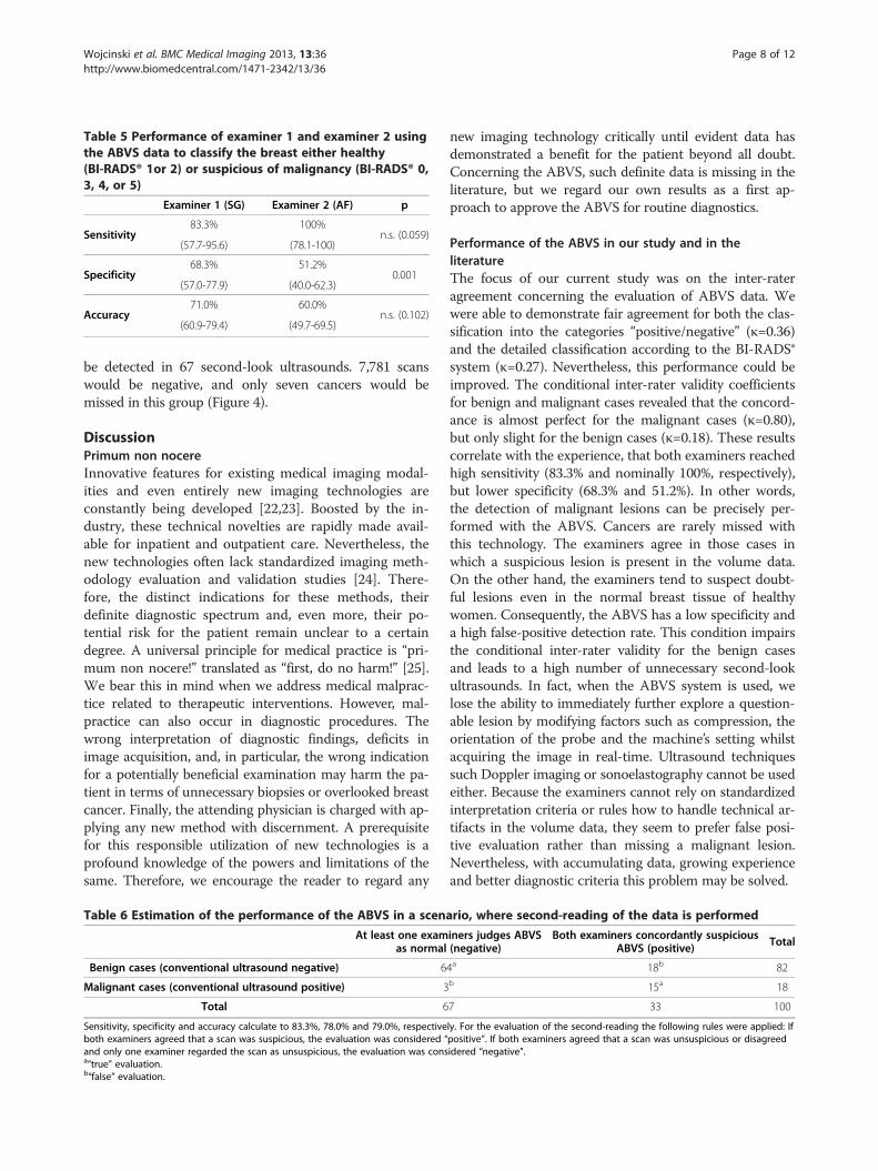
Table 5 Performance of examiner 1 and examiner 2 usingthe ABVS data to classify the breast either healthy(BI-RADS® 1or 2) or suspicious of malignancy (BI-RADS® 0,3, 4, or 5)
Examiner 1 (SG) Examiner 2 (AF) p
Sensitivity83.3% 100%
n.s. (0.059)(57.7-95.6) (78.1-100)
Specificity68.3% 51.2%
0.001(57.0-77.9) (40.0-62.3)
Accuracy71.0% 60.0%
n.s. (0.102)(60.9-79.4) (49.7-69.5)
Wojcinski et al. BMC Medical Imaging 2013, 13:36 Page 8 of 12http://www.biomedcentral.com/1471-2342/13/36
be detected in 67 second-look ultrasounds. 7,781 scanswould be negative, and only seven cancers would bemissed in this group (Figure 4).
DiscussionPrimum non nocereInnovative features for existing medical imaging modal-ities and even entirely new imaging technologies areconstantly being developed [22,23]. Boosted by the in-dustry, these technical novelties are rapidly made avail-able for inpatient and outpatient care. Nevertheless, thenew technologies often lack standardized imaging meth-odology evaluation and validation studies [24]. There-fore, the distinct indications for these methods, theirdefinite diagnostic spectrum and, even more, their po-tential risk for the patient remain unclear to a certaindegree. A universal principle for medical practice is “pri-mum non nocere!” translated as “first, do no harm!” [25].We bear this in mind when we address medical malprac-tice related to therapeutic interventions. However, mal-practice can also occur in diagnostic procedures. Thewrong interpretation of diagnostic findings, deficits inimage acquisition, and, in particular, the wrong indicationfor a potentially beneficial examination may harm the pa-tient in terms of unnecessary biopsies or overlooked breastcancer. Finally, the attending physician is charged with ap-plying any new method with discernment. A prerequisitefor this responsible utilization of new technologies is aprofound knowledge of the powers and limitations of thesame. Therefore, we encourage the reader to regard any
Table 6 Estimation of the performance of the ABVS in a scena
At least one examas normal
Benign cases (conventional ultrasound negative) 6
Malignant cases (conventional ultrasound positive) 3
Total 6
Sensitivity, specificity and accuracy calculate to 83.3%, 78.0% and 79.0%, respectiveboth examiners agreed that a scan was suspicious, the evaluation was considered “and only one examiner regarded the scan as unsuspicious, the evaluation was consa“true” evaluation.b“false” evaluation.
new imaging technology critically until evident data hasdemonstrated a benefit for the patient beyond all doubt.Concerning the ABVS, such definite data is missing in theliterature, but we regard our own results as a first ap-proach to approve the ABVS for routine diagnostics.
Performance of the ABVS in our study and in theliteratureThe focus of our current study was on the inter-rateragreement concerning the evaluation of ABVS data. Wewere able to demonstrate fair agreement for both the clas-sification into the categories “positive/negative” (κ=0.36)and the detailed classification according to the BI-RADS®system (κ=0.27). Nevertheless, this performance could beimproved. The conditional inter-rater validity coefficientsfor benign and malignant cases revealed that the concord-ance is almost perfect for the malignant cases (κ=0.80),but only slight for the benign cases (κ=0.18). These resultscorrelate with the experience, that both examiners reachedhigh sensitivity (83.3% and nominally 100%, respectively),but lower specificity (68.3% and 51.2%). In other words,the detection of malignant lesions can be precisely per-formed with the ABVS. Cancers are rarely missed withthis technology. The examiners agree in those cases inwhich a suspicious lesion is present in the volume data.On the other hand, the examiners tend to suspect doubt-ful lesions even in the normal breast tissue of healthywomen. Consequently, the ABVS has a low specificity anda high false-positive detection rate. This condition impairsthe conditional inter-rater validity for the benign casesand leads to a high number of unnecessary second-lookultrasounds. In fact, when the ABVS system is used, welose the ability to immediately further explore a question-able lesion by modifying factors such as compression, theorientation of the probe and the machine’s setting whilstacquiring the image in real-time. Ultrasound techniquessuch Doppler imaging or sonoelastography cannot be usedeither. Because the examiners cannot rely on standardizedinterpretation criteria or rules how to handle technical ar-tifacts in the volume data, they seem to prefer false posi-tive evaluation rather than missing a malignant lesion.Nevertheless, with accumulating data, growing experienceand better diagnostic criteria this problem may be solved.
rio, where second-reading of the data is performed
iners judges ABVS(negative)
Both examiners concordantly suspiciousABVS (positive) Total
4a 18b 82b 15a 18
7 33 100
ly. For the evaluation of the second-reading the following rules were applied: Ifpositive”. If both examiners agreed that a scan was unsuspicious or disagreedidered “negative”.
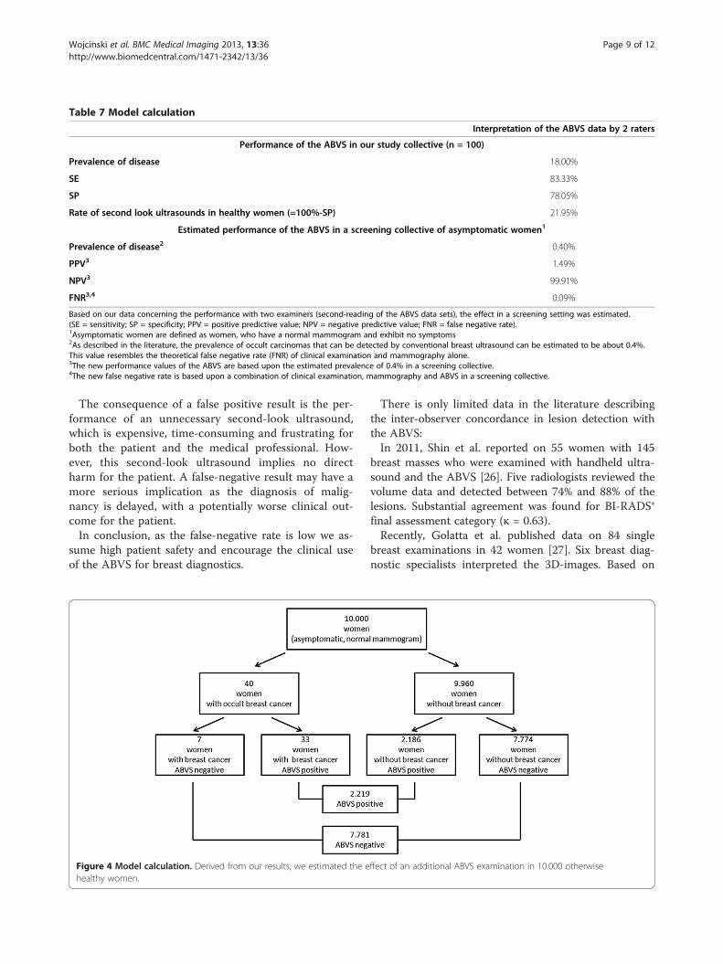
Table 7 Model calculation
Interpretation of the ABVS data by 2 raters
Performance of the ABVS in our study collective (n = 100)
Prevalence of disease 18.00%
SE 83.33%
SP 78.05%
Rate of second look ultrasounds in healthy women (=100%-SP) 21.95%
Estimated performance of the ABVS in a screening collective of asymptomatic women1
Prevalence of disease2 0.40%
PPV3 1.49%
NPV3 99.91%
FNR3,4 0.09%
Based on our data concerning the performance with two examiners (second-reading of the ABVS data sets), the effect in a screening setting was estimated.(SE = sensitivity; SP = specificity; PPV = positive predictive value; NPV = negative predictive value; FNR = false negative rate).1Asymptomatic women are defined as women, who have a normal mammogram and exhibit no symptoms2As described in the literature, the prevalence of occult carcinomas that can be detected by conventional breast ultrasound can be estimated to be about 0.4%.This value resembles the theoretical false negative rate (FNR) of clinical examination and mammography alone.3The new performance values of the ABVS are based upon the estimated prevalence of 0.4% in a screening collective.4The new false negative rate is based upon a combination of clinical examination, mammography and ABVS in a screening collective.
Wojcinski et al. BMC Medical Imaging 2013, 13:36 Page 9 of 12http://www.biomedcentral.com/1471-2342/13/36
The consequence of a false positive result is the per-formance of an unnecessary second-look ultrasound,which is expensive, time-consuming and frustrating forboth the patient and the medical professional. How-ever, this second-look ultrasound implies no directharm for the patient. A false-negative result may have amore serious implication as the diagnosis of malig-nancy is delayed, with a potentially worse clinical out-come for the patient.In conclusion, as the false-negative rate is low we as-
sume high patient safety and encourage the clinical useof the ABVS for breast diagnostics.
Figure 4 Model calculation. Derived from our results, we estimated the ehealthy women.
There is only limited data in the literature describingthe inter-observer concordance in lesion detection withthe ABVS:In 2011, Shin et al. reported on 55 women with 145
breast masses who were examined with handheld ultra-sound and the ABVS [26]. Five radiologists reviewed thevolume data and detected between 74% and 88% of thelesions. Substantial agreement was found for BI-RADS®final assessment category (κ = 0.63).Recently, Golatta et al. published data on 84 single
breast examinations in 42 women [27]. Six breast diag-nostic specialists interpreted the 3D-images. Based on
ffect of an additional ABVS examination in 10.000 otherwise
Table 8 Data in the literature concerning sensitivity (SE)and specificity (SP) of the ABVS
Author N SE SP
Wojcinski et al. 2011 [15] 50a 100% 52.8%
Lin et al. 2012 [28] 81b 100% 95.0%
Wang HY et al. 2012 [29] 239c 95.3% 80.5%
Wang ZL et al. 2012 [30] 165c 96.1% 91.9%
Current study, Examiner 1 100a 83.3% 68.3%
Current study, Examiner 2 100a 100% 51.2%avolume data sets (including scans without lesions, BI-RADS®-US 1).bpatients.clesions.
Wojcinski et al. BMC Medical Imaging 2013, 13:36 Page 10 of 12http://www.biomedcentral.com/1471-2342/13/36
the BI-RADS® classification the multiple kappa coeffi-cient was κ = 0.35.In our analysis, we found fair agreement between the
two examiners, which correlates with the latter results (κ =0.27). However, more data is needed to evaluate the per-formance of the ABVS in breast imaging convincingly.Moreover, there are several reports comparing the ABVS
with hand-held ultrasound [15,28-31]:The first detailed description of the technical back-
ground and performance of the ABVS was published byour study group in 2011 [15]. In 2011, 50 ABVS datasetswere evaluated by an independent examiner and accur-acy, sensitivity and specificity were calculated as 66.0%(95% CI: 52.9-79.1), 100% (95% CI: 73.2-100) and 52.8%(95% CI: 35.7-69.2), respectively. Concerning these vari-ables, our current study yielded comparable results(Table 5). Nevertheless, as both studies were conductedin the same institution, a comparison with the resultsfrom other study groups will be of greater interest.In 2012, Lin et al. published data on 81 patients and
compared ABVS to handheld ultrasound. The authorsdescribed a perfect sensitivity for both methods (ABVS:100%, hand-held ultrasound: 100%), high specificity(95.0% and 85.0%, respectively) and, consequently, a highdiagnostic accuracy (97.1% and 91.4%, respectively) [28].This performance appears extraordinarily high. We sug-gest that further standardized studies in larger collectivesshould investigate if these expectations can be fulfilled.In the same year, Wang HY et al. studied 239 lesions
in 213 women who were scheduled to undergo biopsy.In this study, ABVS was similar to hand-held ultrasoundin terms of sensitivity (ABVS: 95.3%, hand-held ultra-sound: 90.6%), specificity (80.5% and 82.5%, respectively),and accuracy (85.8% and 85.3%, respectively) [29].Recently, Wang ZL et al. published data on 153 pa-
tients with 165 breast lesions. The patients underwentmammography, ABVS and hand-held ultrasound. Theauthors reported no significant differences betweenABVS and conventional ultrasound concerning sensitiv-ity (ABVS: 96.1%, hand-held ultrasound: 93.2%), specifi-city (91.9% and 88.7%, respectively), and accuracy (94.5%and 91.5%, respectively) [30,31].Compared to our data, there is a discrepancy mainly
concerning the specificity, which was lower in our investi-gation (Table 8). Actually, we had a relevant number ofhealthy women without breast lesions (i.e. BI-RADS®-US 1)in our collective. This condition automatically increasesthe false-positive rate and actually reduces specificity.Hence, the main difference of our study design in
comparison to the above-mentioned studies is that wedid not focus on the evaluation of known lesions alone,but also on the detection. Therefore, our examiners didnot know whether there was a lesion in the particularvolume or not. In the studies from the literature, the
examiners were well aware, that there is definitely a le-sion in the volume that requires biopsy (i.e. BI-RADS® 3and above). This knowledge certainly has an influenceon the evaluation of the lesion and the overall perform-ance of the ABVS.Our interpretation: the almost perfect performance of
the ABVS as described in the literature is only valid forcollectives of women with already pre-diagnosed breastlesions. In realistic collectives that also involve womenwithout breast lesions (i.e. BI-RADS®-US 1) or with clearlybenign breast lesions (i.e. BI-RADS®-US 2), our data maybe more convincing.Concerning the inter-observer agreement, Zhang et al.
published data on 234 breast lesions from 208 patientswho were examined with the ABVS [32]. Zhang et al. in-vestigated the inter-observer agreement concerning thedescription of known breast masses according to the BI-RADS®-US lexicon. They found substantial agreementfor lesion shape, orientation, margin, echo pattern, poster-ior acoustic features, calcification and final assessment,and fair agreement for retraction phenomenon and lesionboundary, respectively. Our study design did not focus thedetailed description of lesions, but we showed fair agree-ment for the detection of breast masses. This aspect wasnot analyzed by Zhang et al.Furthermore, there are reports in the literature about
the ABVS that concentrate on the optimal scanningtechnique [16], the accuracy of measuring the cancer ex-tent [33] and the detection of lesions located behind thenipple [34].
Limitations of our study and limitations of the ABVSAll previously mentioned studies have the essential limi-tation of unicentric design and small patient collectives.This limitation also applies to our current study. Futurestudy concepts should include multicenter design andlarger, well-defined patient populations as described else-where [15]. Furthermore, we compared only two exam-iners. Although the results demonstrated fair agreement,the concordance should be confirmed with more ob-servers. Another limitation of our study is the selection
Wojcinski et al. BMC Medical Imaging 2013, 13:36 Page 11 of 12http://www.biomedcentral.com/1471-2342/13/36
of the study population and the resulting concentrationon BI-RADS®-US 1, 2 and 5 lesions which causes a cer-tain bias. Therefore, the proportion of cases to controlsis not representative of the whole population and BI-RADS®-US 0, 3 and 4 lesions are missing in the studypopulation. We attempted to overcome this limitationby conducting a model calculation. Although this ap-proach is statistically correct, it must be considered that,due to the small sample size and the vague estimation ofthe prevalence, the results must be carefully interpreted.Moreover, the technology of the ABVS has some limi-
tations per se. As previously described in the literature,automated breast ultrasound is limited in women withmacromastia and pronounced ptosis [15,16]. In addition,the ABVS is not capable of scanning the axillary region.Today, sentinel node biopsy is the standard therapy forwomen that were preoperatively staged with a negativenodal status, which requires ultrasound of the axilla [35].Furthermore, lymph node alterations may be the first signin mammographically and/or sonographically occult breastcancer or other malignant diseases [36,37]. Therefore, add-itional conventional ultrasound of the axilla would benecessary after a suspicious ABVS scan.Furthermore, we presume that even with optimal scan-
ning technique the peripheral areas of the breast paren-chyma are not fully covered by the ABVS. Additionally,shadowing artifacts occur in the retroareolar region andto a certain extent in the remaining breast volume. There-fore, a certain proportion of breast parenchyma may belost in the volume data. This may reduce the diagnosticpotential in comparison to hand-held ultrasound.
Future implications of the ABVSFinally, we have to discuss the question of which role theABVS could play in future breast cancer diagnosis andbreast cancer screening. Conventional hand-held breastultrasound has a commonly accepted role as a comple-mentary method to mammography, by adding to thediagnostic accuracy. Despite the well-known advantages,conventional breast ultrasound is a time-consuming,examiner-dependent and therefore costly procedure. Fur-thermore, breast cancer screening with additional breastultrasound in low-risk women has not been established, asdata concerning an effect on the overall survival is miss-ing. Nevertheless, it is evident that breast ultrasound iscapable of detecting previously occult cancers in womenwho had already undergone mammography and clinicalexamination [8,9,11,12]. However, screening larger collec-tives with conventional ultrasound is plainly not feasible.With the ABVS, we have a tool that could principallyovercome these problems, and the ABVS may make ultra-sound available to a larger number of women. Image ac-quisition with the ABVS can be efficiently performed by amedical assistant. Moreover, the ABVS allows a delayed
interpretation of the images at any time, as well assecond-readings. On the other hand, it has to be takeninto account that we still experience a high number oftime-consuming second-look ultrasounds due to the lowspecificity. This problem may be solved through second-readings by an independent examiner. Our proposed rulesare relatively unusual, as, in order to improve the speci-ficity, disagreement between the examiners would beclassified as “negative” and only agreement that a scanis suspicious would be classified as “positive”. Therefore,further studies should include these concepts. However,we are convinced that the ABVS is a feasible methodthat can be easily integrated into the workflow of any in-patient or outpatient department.We can imagine the ABVS being applied in breast
cancer screening programs in the future and that add-itional examinations are offered to women at risk. How-ever, we still have no data focusing on certain subgroupsof patients (e.g. high-risk versus low-risk, ACR 1–2 ver-sus ACR 3–4, etc.).A prospective randomized controlled trial could an-
swer the open questions. This trial should include pa-tients with normal mammograms, some of whom willreceive additional ABVS. The primary outcome variableswould be the detection rate of breast cancer, the tumorstage and the overall survival of the two groups. Presum-ably, results could only be obtained if the collectiveswere sufficiently large, which makes it likewise unrealis-tic that a corresponding trial will actually be conducted.
ConclusionsIn conclusion, the ABVS has demonstrated high sensitivityand a fair inter-observer concordance in the evaluation ofbreast lesions. However, we have to overcome the problemof the high number of false-positive results in healthywomen. Therefore, further prospective validation studiesin larger collectives are necessary to define standard pro-cedures in image acquisition and volume interpretation.Nevertheless, the ABVS is a reliable, technically matureand potentially beneficial imaging method. Therefore, wepropose that the ABVS can be used in routine diagnosticprocedures as an adjunct to mammography and conven-tional ultrasound, as previously described.
Competing interestsThe authors declare that they have no competing interest.
Authors’ contributionsSW and SG contributed to the conception and design of the study and SWperformed the ultrasound examinations and data collection. SG and AFperformed the independent evaluation of the ABVS data sets. SWcontributed to the statistical analysis and the interpretation of the data. SWand PS contributed to the writing of the manuscript. FD and PH conductedfinal reviews of the manuscript and FD provided methodological advice. Allauthors read and approve the final manuscript.
Wojcinski et al. BMC Medical Imaging 2013, 13:36 Page 12 of 12http://www.biomedcentral.com/1471-2342/13/36
AcknowledgementsPublication costs were covered by a grant of the DFG (German ResearchFoundation) within the project “Open Access Publications” at MHH(Hannover Medical School, Germany).
Author details1Department of OB/GYN, Hannover Medical School, OE 6410,Carl-Neuberg-Straße 1, 30625 Hannover, Germany. 2Department of OB/GYN,Franziskus Hospital, Bielefeld, Germany.
Received: 2 September 2012 Accepted: 8 November 2013Published: 12 November 2013
References1. Parkin DM, Bray F, Ferlay J, Pisani P: Global cancer statistics, 2002.
CA Cancer J Clin 2005, 55(2):74–108.2. IARC: World cancer report 2008. Lyon: International Agency for Research on
Cancer, WHO Press; 2008.3. IARC: Globocan database. Lyon: International Agency for Research on
Cancer; 2008.4. Parkin DM, Fernandez LM: Use of statistics to assess the global burden of
breast cancer. Breast J 2006, 12(1):S70–80.5. Duijm LE, Louwman MW, Groenewoud JH, van de Poll-Franse LV, Frache-
boud J, Coebergh JW: Inter-observer variability in mammography screen-ing and effect of type and number of readers on screening outcome.Br J Cancer 2009, 100(6):901–907.
6. Independent UK Panel on Breast Cancer Screening: The benefits andharms of breast cancer screening: an independent review. Lancet 2012,380(9855):1778–1786.
7. Zonderland HM, Coerkamp EG, Hermans J, van de Vijver MJ, vanVoorthuisen AE: Diagnosis of breast cancer: contribution of US as anadjunct to mammography. Radiology 1999, 213(2):413–422.
8. Buchberger W, Niehoff A, Obrist P, DeKoekkoek-Doll P, Dunser M: Clinicallyand mammographically occult breast lesions: detection and classificationwith high-resolution sonography. Semin Ultrasound CT MR 2000,21(4):325–336.
9. Corsetti V, Ferrari A, Ghirardi M, Bergonzini R, Bellarosa S, Angelini O, Bani C,Ciatto S: Role of ultrasonography in detecting mammographically occultbreast carcinoma in women with dense breasts. Radiol Med 2006,111(3):440–448.
10. Gordon PB, Goldenberg SL: Malignant breast masses detected only byultrasound: a retrospective review. Cancer 1995, 76(4):626–630.
11. Kolb TM, Lichy J, Newhouse JH: Occult cancer in women with densebreasts: detection with screening US–diagnostic yield and tumorcharacteristics. Radiology 1998, 207(1):191–199.
12. Nothacker M, Duda V, Hahn M, Warm M, Degenhardt F, Madjar H,Weinbrenner S, Albert US: Early detection of breast cancer: benefits andrisks of supplemental breast ultrasound in asymptomatic women withmammographically dense breast tissue: a systematic review. BMC Cancer2009, 9:335.
13. Maturo VG, Zusmer NR, Gilson AJ, Smoak WM, Janowitz WR, Bear BE,Goddard J, Dick DE: Ultrasound of the whole breast utilizing a dedicatedautomated breast scanner. Radiology 1980, 137(2):457–463.
14. Mendelson EB, Baum JK, Berg WA, Merritt CR, Rubin E: BI-RADS: ultrasound.In Breast imaging reporting and data system: ACR BI-RADS - breast imagingatlas. Edited by D’Orsi CJ, Mendelson EB, Ikeda DM. Reston, VA: AmericanCollege of Radiology; 2002.
15. Wojcinski S, Farrokh A, Hille U, Wiskirchen J, Gyapong S, Soliman AA,Degenhardt F, Hillemanns P: The automated breast volume scanner(ABVS): initial experiences in lesion detection compared withconventional handheld B-mode ultrasound: a pilot study of 50 cases.International journal of women’s health 2011, 3:337–346.
16. Tozaki M, Isobe S, Yamaguchi M, Ogawa Y, Kohara M, Joo C, Fukuma E:Optimal scanning technique to cover the whole breast using anautomated breast volume scanner. Jpn J Radiol 2010, 28(4):325–328.
17. DEGUM (deutsche gesellschaft für ultraschall in der medizin, germanassociation for ultrasound in medicine) - mehrstufenkonzept mammasonogra-phie. http://www.degum.de/Mehrstufenkonzept_Mammasonogra.634.0.html.
18. KBV (kassenaerztliche bundesvereinigung, German federal association of funddoctors) -ultraschallvereinbarung, ultrasound regulations. http://www.kbv.de/2488.html.
19. Zhou XH, Obuchowski NA, McClish DK: Statistical methods in diagnosticmedicine: 2nd ed. New York: Wiley; 2011.
20. Newcombe RG: Interval estimation for the difference betweenindependent proportions: comparison of eleven methods. Stat Med 1998,17(8):873–890.
21. Landis JR, Koch GG: The measurement of observer agreement forcategorical data. Biometrics 1977, 33(1):159–174.
22. Hahn M, Roessner L, Krainick-Strobel U, Gruber IV, Kramer B, Gall C,Siegmann KC, Wallwiener D, Kagan KO: Sonographic criteria for the differ-entiation of benign and malignant breast lesions using real-time spatialcompound imaging in combination with XRES adaptive image process-ing. Ultraschall Med 2012, 33(3):270–274.
23. Farrokh A, Wojcinski S, Degenhardt F: Diagnostic value of strain ratiomeasurement in the differentiation of malignant and benign breastlesions. Ultraschall Med 2011, 32(4):400–405.
24. Wojcinski S, Farrokh A, Peisker U, Thomas A, Degenhardt F, Hahn M: Neuediagnostische verfahren - sono-elastografie der mamma. Senologie -Zeitschrift für Mammadiagnostik und -therapie 2011, 7:15–18.
25. Smith CM: Origin and uses of primum non nocere–above all, do noharm! J Clin Pharmacol 2005, 45(4):371–377.
26. Shin HJ, Kim HH, Cha JH, Park JH, Lee KE, Kim JH: Automated ultrasound ofthe breast for diagnosis: interobserver agreement on lesion detectionand characterization. AJR Am J Roentgenol 2011, 197(3):747–754.
27. Golatta M, Franz D, Harcos A, Junkermann H, Rauch G, Scharf A, Schuetz F,Sohn C, Heil J: Interobserver reliability of automated breast volumescanner (ABVS) interpretation and agreement of ABVS findings withhand held breast ultrasound (HHUS), mammography and pathologyresults. Eur J Radiol 2013, 82(8):332–336.
28. Lin X, Wang J, Han F, Fu J, Li A: Analysis of eighty-one cases with breastlesions using automated breast volume scanner and comparison withhandheld ultrasound. Eur J Radiol 2012, 81(5):873–878.
29. Wang HY, Jiang YX, Zhu QL, Zhang J, Dai Q, Liu H, Lai XJ, Sun Q:Differentiation of benign and malignant breast lesions: a comparisonbetween automatically generated breast volume scans and handheldultrasound examinations. Eur J Radiol 2012, 81(11):3190–3200.
30. Wang ZL, Xu JH, Li JL, Huang Y, Tang J: Erratum to: comparison ofautomated breast volume scanning to hand-held ultrasound and mam-mography. Radiol Med 2012, 117(8):1443.
31. Wang ZL, Xw JH, Li JL, Huang Y, Tang J: Comparison of automated breastvolume scanning to hand-held ultrasound and mammography.Radiol Med 2012, 117(8):1287–1293.
32. Zhang J, Lai XJ, Zhu QL, Wang HY, Jiang YX, Liu H, Dai Q, You SS, Xiao MS,Sun Q: Interobserver agreement for sonograms of breast lesionsobtained by an automated breast volume scanner. Eur J Radiol 2012,81(9):2179–2183.
33. Tozaki M, Fukuma E: Accuracy of determining preoperative cancer extentmeasured by automated breast ultrasonography. Jpn J Radiol 2010,28(10):771–773.
34. Isobe S, Tozaki M, Yamaguchi M, Ogawa Y, Homma K, Satomi R, Saito M,Joo C, Fukuma E: Detectability of breast lesions under the nipple usingan automated breast volume scanner: comparison with handheldultrasonography. Jpn J Radiol 2011, 29(5):361–365.
35. Kuehn T, Bembenek A, Decker T, Munz DL, Sautter-Bihl ML, Untch M, Wallwi-ener D: Consensus committee of the german society of, senology: a con-cept for the clinical implementation of sentinel lymph node biopsy inpatients with breast carcinoma with special regard to quality assurance.Cancer 2005, 103(3):451–461.
36. de Bresser J, de Vos B, van der Ent F, Hulsewe K: Breast MRI in clinicallyand mammographically occult breast cancer presenting with an axillarymetastasis: a systematic review. Eur J Surg Oncol 2010, 36(2):114–119.
37. Schwab FD, Burger H, Isenschmid M, Kuhn A, Mueller MD, Gunthert AR:Suspicious axillary lymph nodes in patients with unremarkable imagingof the breast. Eur J Obstet Gynecol Reprod Biol 2010, 150(1):88–91.
doi:10.1186/1471-2342-13-36Cite this article as: Wojcinski et al.: Diagnostic performance and inter-observer concordance in lesion detection with theautomated breast volume scanner (ABVS). BMC Medical Imaging2013 13:36.