Embed Size (px)
Citation preview
January 2015, Vol.13, No.1 34 Journal of Integrative Medicine
Journal homepage: www.jcimjournal.com/jimwww.elsevier.com/locate/issn/20954964
Available also online at www.sciencedirect.com. Copyright © 2015, Journal of Integrative Medicine Editorial Office. E-edition published by Elsevier (Singapore) Pte Ltd. All rights reserved.
● Research ArticleEvaluation of chemopreventive potentials of ethanolic extract of Ruta graveolens against A375 skin melanoma cells in vitro and induced skin cancer in mice in vivoSamrat Ghosh, Sourav Sikdar, Avinaba Mukherjee, Anisur Rahman Khuda-Bukhsh
Cytogenetics and Molecular Biology Laboratory, Department of Zoology, University of Kalyani, Kalyani-741235, West Bengal, India
ABSTRACT
OBJECTIVE: Chemopreventive approach with natural products, particularly plants and plant-derived ones, is receiving increasing attention for their effective role against cancer without any palpable side effects.Inthisstudy,efficacyofethanolicextractofRuta graveolens (RG) on skin melanoma cells (A375) in vitro and on 7,12-dimethylbenz(a)anthracene (DMBA)-induced skin cancer in vivo has been tested in Swiss albino mice.
METHODS: Studies on cell viability, apoptosis and autophagy induction were conducted in vitro. To checkapoptosis,assayslikealterationinmitochondrialmembranepotential,annexinV-fluoresceinisothiocyanate/propidium iodide assay and immunoblot were performed. Fluorescence microscopic and immunoblotassayswereperformedtoconfirmautophagyinduction.TheeffectsofRGweredeterminedby evaluating body weight, tumor incidence, tumor volume and tumor burden in mice. Enzymatic and non-enzymatic antioxidant status was assessed. The role of some relevant signaling proteins was also analyzed.
RESULTS: RG caused death of A375 cells through induction of caspase 3-mediated apoptosis and Beclin-1-associated autophagy. Moreover, RG administration (75 mg/kg body weight) which showed no acuteorchronictoxicity,showedsignificantreductionintheskintumorburdenofDMBA-paintedmice.RGalso demonstrated potent anti-lipid peroxidative and antioxidant functions during the course of skin cancer induction by DMBA.
CONCLUSION: Chemopreventive potential of RG was demonstrated from overall results of this study, indicating its possible use in therapeutic formulation of an effective drug to treat skin cancer.
Keywords: Ruta graveolens; plant extracts; chemoprevention; skin neoplasms; apoptosis; autophagy
Citation: Ghosh S, Sikdar S, Mukherjee A, Khuda-Bukhsh AR. Evaluation of chemopreventive potentials of ethanolic extract of Ruta graveolens against A375 skin melanoma cells in vitro and induced skin cancer in mice in vivo. J Integr Med. 2015; 13(1): 34–44.
http://dx.doi.org/10.1016/S2095-4964(15)60156-XReceived July 26, 2014; accepted November 21, 2014.Correspondence: Anisur Rahman Khuda-Bukhsh, PhD, Professor; Tel: +91-33-2582-8750 Extn.315; E-mail: [email protected]; [email protected].

January 2015, Vol.13, No.135Journal of Integrative Medicine
www.jcimjournal.com/jim
1 Introduction
Unwanted side effects of many currently used drugs make them unsuitable or of limited use in the treatment of cancer. The search for drugs relatively free of side effects leads to the arena of alternative mode of treatment for cancer, which mainly uses certain medicinal plants and plant-derived products for this purpose. Ruta graveolens is a medicinal and culinary plant growing in the Mediterranean region of southern Europe and northern Africa. It is used for the treatment of inflammatory conditions, eczema, ulcers, arthritis and fibromyalgia and as an antidote for venoms, insect repellent, and also as an abortifacient[1–3]. Extract of this plant is considered as an effective ingredient in medicinal preparations of rue oil and infusions that are used as antispasmodics and emmenagogues[4].
A major therapeutic goal of recent cancer treatment is to trigger tumor-selective cell death. This target can be achieved by two major ways: by apoptosis (type I pro-grammed cell death; PCD I) and/or by autophagy (type II programmed cell death; PCD II). Drugs having the ability to induce apoptosis are preferred in medical oncology. In order to survive, cancer cells many a time develop intricate mechanisms that give them the ability to escape apoptosis and to develop resistance against anticancer drugs[5]. For this reason, in many cases, anticancer drugs fail to curb cancer spread after initial success. Therefore, drugs that can destroy cancer cells by inducing both apoptosis and autophagy are considered to have added advantage, in view of the fact that cancer cells can be killed by autophagy even if they escape apoptosis[6]. Thus, study to determine the efficacy of some medicinal plants and plant-derived products to stop proliferation of cancer cells or to annihilate them by apoptotic, necrotic, or autophagic means has achieved considerable importance.
Plants and plant-derived products with chemopreventive activities are gaining importance in the field of anticancer drug research. Certain chemopreventive agents are known to act through their anti-lipid-peroxidative and antioxidant properties[7]. For experimental induction of skin cancer in Swiss albino mice, 7,12-dimethylbenz(a)anthracene (DMBA) is generally used as a site- and organ-specific carcinogen, either as an initiator or as a promoter[8,9].
In this study, the hypotheses tested were: if ethanolic extract of Ruta graveolens (RG) could annihilate skin melanoma cells A375 in vitro, if RG could combat DMBA-induced skin cancer in Swiss albino mice in vivo, and if it could delineate the possible mechanism of action.
2 Materials and methods
2.1 Drug usedRG was obtained from Boiron Laboratory, Lyon,
France. For the preparation of dried extract, the ethanol content was first evaporated under reduced pressure in a rotary evaporator; a concentrated ingredient that looked dark brownish in color and semisolid in nature, was obtained. Then a solution of the extract was made by adding distilled water and gently stirring. The solution was kept in 4 ℃ for further use in experiments as the ‘stock solution’ of the drug in case of in vitro experiments; the same was also used for feeding the experimental mice in vivo.2.2 In vitro experiments2.2.1 Cell line and cell culture
Skin melanoma (A375) and skin keratinocyte (HaCaT) cell lines were procured from National Centre for Cell Science (Pune, India); peripheral blood mononuclear cells (PBMCs) were isolated from human blood sample by the standard Ficoll Paque density gradient method[10]. Cell-culture method (routine adherent cell culture method) followed has been described elsewhere[11].2.2.2 Cell viability assay
Cell viability assay was made following 3-(4,5-dimeth-ylthiazol-2-yl)-2,5-diphenyltetrazolium bromide (MTT) assay as per the technique described previously[11]. The dose of RG that reduced the viability of cells at 48 h to about 50% was determined. The viability of A375 cells exposed to 3-methyladenine (3-MA; autophagy inhibitor, 10 mmol/L, Santa Cruz Biotechnology Inc., CA, USA) for 2 h before RG administration was also assessed.2.2.3 Alteration in mitochondrial membrane potential
Flow cytrometric analysis was done to estimate quan-titatively alteration in mitochondrial membrane potential (MMP) in the RG-exposed A375 cells. At first, cells were exposed to 44.80 µg/mL of RG for different periods of time, starting from 0 h through 12, 24, 36 and 48 h. Then fixation of the treated cells was done in 70% ethanol. After that fixed cells were incubated with 10 μmol/L rhodamine 123 for 30 min at 4 ℃ in the dark and the fluorescence intensity was measured by flow cytometer using FL-1H filter. Cyflogic software (CyFlo Ltd, Turku, Finland) was used for analyzing the data.2.2.4 Annexin V-fluorescein isothiocyanate/propidium iodide dual-stain assay
A375 cells were exposed to RG of three specified doses, one being the IC50 dose, one below and one above the IC50 dose (D1 = 35 µg/mL, D2 = 44.80 µg/mL, and D3 = 50 µg/mL) for 48 h of time interval. The cells were then processed for the dual stain assay[12]. Briefly, at 4 ℃ in the dark, 15-minute incubation was done after adding 10 μmol/L annexin V-fluorescein isothiocyanate (FITC, Santa Cruz Biotechnology Inc., CA, USA) and 5 μmol/L of propidium iodide (PI) to the cell suspensions. A flow cytometer (BD FACS Calibur, USA) with FL-1 (530 nm) and FL-2 (585 nm) filters was used to determine the flu-orescence intensities and the distribution frequencies of

www.jcimjournal.com/jim
January 2015, Vol.13, No.1 36 Journal of Integrative Medicine
live and dead cells were analyzed with Cyflogic software (CyFlo Ltd, Turku, Finland).2.2.5 Expression study of signaling proteins by immunoblot analysis
Anti-Bcl-2 (mouse IgG2a), anti-BAX (mouse IgG1), anti-cleaved poly ADP ribose polymerase (PARP) (rabbit IgG), anti-cytochrome c (mouse IgG1), anti-Akt (mouse IgG1), anti-caspase 3 (mouse IgG2a), anti-pAkt (rabbit IgG), anti-glyceraldehyde-3-phosphate dehydrogenase (GAPDH) (mouse IgG1), anti-voltage-dependent anion-selective channel protein 1 (VDAC1) (anti-goat IgG), anti-mechanistic target of rapamycin (mTOR) (rabbit IgG) (Santa Cruz Biotechnology Inc., CA, USA), anti-LC3 (rabbit IgG), anti-Atg5 (rabbit IgG), anti-Beclin-1 (rabbit IgG), and anti-Atg7 (rabbit IgG) (Cell Signaling Technology, Inc.) antibodies were used for immunoblot study[13]. For cytosolic and mitochondrial protein expression analysis, GAPDH and VDAC1 served the purpose of loading control, respectively.2.2.6 Detection of acidic vesicular organelles
The vital staining method with acridine orange (AO) was used to detect the acidic vesicular organelles (AVOs) in RG-treated cells (both in presence or absence of 3-MA). The cytoplasm and the acidic compartments fluoresce as bright green and bright red objects, respectively presented in AO-stained cells[14]. Chilled ethanol (70%) was used to fix the RG-exposed cells. Then 5 mmol/L AO was used for the staining of fixed cells. After that fluorescence of the stained cells was measured with a fluorescence microscope (Axisocope plus 2, Zeiss). 2.3 In vivo experiments 2.3.1 Animals
Six/eight weeks old healthy inbred strains of Swiss albino mice (Mus musculus) weighing about 25 g were kept for at least 14 d in an environmentally controlled room (temperature (24±28) ℃; humidity 55%±5%, 12-hour light/dark cycle), and were allowed to take in food and water ad libitum. All experiments were conducted as per the guidelines cleared by the Animal Ethics Committee of the University of Kalyani, India (Registration number-892/OC/05/CPCSEA) and under the supervision of the Animal Welfare Committee (Department of Zoology, University of Kalyani, India). Randomized mice of both sexes were used in experiments more or less in equal numbers.2.3.2 Acute toxicity study of RG
Normal mice were force fed with different doses of RG (75, 100, 125, and 150 mg/kg body weight) through gavage to check acute toxicity of RG, if any, produced by any of these doses. Any change in behavior pattern or mortality in mice over the next 24 h was considered as indicative sign of acute toxicity. However, none of experimental doses of RG showed acute toxicity; of these, the lowest dose (75 mg/kg body weight) that did not show
slightest of toxicity has been chosen for treatment for the longer periods. 2.3.3 Experimental design
Thirty animals used in the present study were divided into 5 groups, each consisting of 6 mice. Development of skin papilloma was induced by following the standard practice of chemical carcinogenesis[15]. Hair of skin was first removed by applying depilatory cream over the surface of back of all experimental and control mice. Acetone (0.1 mL/mouse) was rubbed on the depilated back of the group II mice twice a week for 8 weeks (vehicle-treated control). DMBA (25 μg in 0.1 mL acetone per mouse) was applied on depilated back of the groups III and IV mice, twice a week for 8 weeks. While no treatment was given to the group III mice, the group IV mice were force fed RG at a dose of 75 mg/kg body weight for 1 week before they were subjected to the DMBA treatment continuously for the next 25 weeks, 3 times a week on alternate days. To maintain another group of control, the group V mice were orally administered with RG only at the same dose of the experimental mice (75 mg/kg body weight) throughout the time course of the experimental study. To maintain a negative control, back of mice of group I was painted with neither acetone nor DMBA and this group of mice was not fed RG. Through cervical dislocation all mice were sacrificed at the end of experiment.2.3.4 Body weight, tumor incidence, tumor volume and measurement of tumor burden
The body weight of all the experimental mice was measured before and after the time period of RG administration.
The tumor volume was determined by using the following formula: (4π/3)×(D1/2)×(D2/2)×(D3/2), where D1, D2 and D3 represented the three diameters (in mm) of the tumors.
Tumor burden was evaluated through multiplication of the value of tumor volume with the number of tumors per animal. 2.3.5 Skin and liver tissue homogenates preparation
From experimental mice, the skin and liver tissues were collected and homogenized in lysis buffer and then centrifuged at 12 000×g for 30 min at 4 ℃. The supernatants were collected and used further. The total protein was estimated by the method of Bradford[16]. Skin and liver tissue homogenates were used for various enzymatic assays. Spectrophotometric analysis of catalase (CAT)[17], superoxide dismutase (SOD)[18], reduced glutathione (GSH)[19], lipid peroxidase (LPO)[20], glutathione S-transferase (GST)[21] and glutathione peroxidase (GPX)[22] was undertaken with skin tissue homogenates whereas spectrophotometric analysis of GSH and GST was performed with liver tissue homogenates according to the standard protocols.2.3.6 Immunoblot assay
Immunoblot assay was done as described in the “In vitro experiments” section.

January 2015, Vol.13, No.137Journal of Integrative Medicine
www.jcimjournal.com/jim
2.4 Statistical analysisData are represented as mean ± standard error of mean.
Significance of the differences between the mean values was determined by one-way analysis of variance with Dunnett’s t post-hoc test, using SPSS 14.0 software. P < 0.05 was considered as statistically significant.
3 Results
3.1 In vitro experiments3.1.1 RG reduced A375 cell viability
RG was non-cytotoxic to normal PBMCs and HaCaT cells whereas it reduced A375 cell viability significantly. Upon exposure to (44.80±0.81) µg/mL of RG for 48 h, the cell viability was reduced to about 50% (Figure 1).3.1.2 RG altered MMP
The histogram of flow cytometric data revealed shifting of fluorescence peak towards left (Y-axis) upon RG adminis-tration, indicating marked decreases in fluorescence intensity. Histogram implied the MMP alteration between 12 and 24 h of RG administration (Figure 2).
3.1.3 RG induced externalization of phosphatidyl serine Annexin V and PI stain assay showed that cell population
shifted from lower left co-ordinates to the right side co-ordinates (annexin V+) upon RG exposure (Figure 3), revealing the fact of externalization of phosphatidyl serine. Annexin V does not bind with the normal cells because it can only bind with phosphatidyl serine which is available in the inner membrane of the cell. When the cells become apoptotic, the phosphatidyl serine comes out to the external membrane surface (externalization of phosphatidyl serine) and readily binds with the annexin V, resulting in the shift of cell population towards the right side quadrants. Thus this result (Table 1) indicates the ability of RG to induce apoptosis in the cells. 3.1.4 RG modulated expression of certain proteins related to apoptosis
Significant up-regulation of BAX, cytochrome c (cytosolic fraction), cleaved caspase 3 and cleaved PARP expressions was observed from immunoblot study (Figure 4). Down-regulated expressions of Bcl-2 and cytochrome c (mitochondrial fraction) were observed upon RG administration (Table 2).3.1.5 RG induced autophagy
Autolysosomes (fusion of autophagosomes with lysosomes) are forms of AVOs. During autophagosome formation, conversion of LC3 (microtubule-associated protein light chain 3) and activation of Beclin-1 take place.
The presence of AVOs in RG-treated cells was observed from AO stain distribution (orange-red fluorescence) (Figure 5), and there was very much reduction in fluorescence intensity upon administration of 3-MA (autophagy inhibitor) with RG (Figure 5).
Conversion of LC3-I (18 kD) to LC3-II (16 kD) upon RG administration (Figure 6) was confirmed from result of immunoblot assay. During the time period 18–24 h, expression of LC3-I was reduced and that of LC3-II increased, indicating the autophagic activation upon RG administration.
Upon RG administration up-regulated expression of Atg7, Beclin-1, and Atg5 and down-regulated expression
Figure 2 Mitochondrial membrane potential alteration study by flow cytometryA375 cells were exposed to Ruta graveolens extracts (44.80 µg/mL) for various time intervals (0, 12, 24, 36 and 48 h). Histogram showed that fluorescence peak was shifted towards the Y-axis upon Ruta graveolens extracts exposure to cells. The cell counts, within an arbitrary area along the X-axis, were calculated in percentage value (percentage of visibility) and were mentioned in the figure.
Figure 1 Results of cell viability assay (MTT assay)Data are expressed as mean ± standard error of mean, n=6; *P<0.05, vs control.MTT: 3-(4,5-dimethylthiazol-2-yl)-2,5-diphenyltetrazolium bromide; PBMCs: peripheral blood mononuclear cells.

www.jcimjournal.com/jim
January 2015, Vol.13, No.1 38 Journal of Integrative Medicine
Table 1 Cell population percentages in specific quadrants obtained from annexin V-FITC/PI stain assay
Treatment Live (LL) Early apoptotic (LR) Late apoptotic (UR) Dead (UL)
Control 98.75 1.08 0.01 0.16D1 80.50 19.35 0.14 0.01D2 53.91 41.88 4.05 0.16D3 40.56 44.89 9.93 4.62
Data represent percentages of cell population in specific quadrants. LL: lower left; LR: lower right; UR: upper right; UL: upper left; D1: 35 μg/mL dose of Ruta graveolens; D2: 44.80 μg/mL dose of Ruta graveolens; D3: 50 μg/mL dose of Ruta graveolens.
Figure 4 Results of immunoblottingA375 cells were treated with D1 (35 µg/mL), D2 (44.80 µg/mL) and D3 (50 µg/mL) doses of Ruta graveolens for 48 h. For cytosolic and mitochondrial protein expression analysis, GAPDH and VDAC1 respectively served the purpose of equal loading protein.VDAC1: voltage-dependent anion-selective channel protein 1; PARP: poly ADP ribose polymerase; mTOR: mechanistic target of rapamycin; GAPDH: glyceraldehyde-3-phosphate dehydrogenase.
Figure 3 Annexin V-FITC/PI stain assayExternalization of phosphatidyl serine was assessed by conducting FACS analysis (FL1 and FL2 filters). Cells were treated for 48 h with D1 (35 µg/mL), D2 (44.80 µg/mL) and D3 (50 µg/mL) doses of Ruta graveolens. LL-lower left (Annexin V-/PI-), UL-upper left (Annexin V-/PI+), LR-lower right (Annexin V+/PI-), and UR-upper right (annexin V+/PI+) represent the specific quadrants. Annexin V and PI stain assay showed that cell population shifted from lower left coordinates to the right side coordinates (annexin V+) upon Ruta graveolens extract exposure, revealing the fact of externalization of phosphatidyl serine. FITC: fluorescein isothiocyanate; PI: propidium iodide.
of mTOR and Bcl-2 occurred, as revealed from immuno-blots (Figure 4 and Table 2). 3.1.6 RG-induced cell death involved autophagy
To find out whether RG-induced autophagy was involved in A375 cell death, cell viability was assessed by exposing the cells to both RG and 3-MA (autophagy inhibitor). The cell viability increased to 70.58% upon administration of IC50 dose (44.80 µg/mL) of RG in presence of 3-MA,
specifying thereby autophagy induction led the cells to death (Figure 7).3.2 In vivo experimental results3.2.1 Effect on body weight
The body weight of mice of the group III (DMBA-treated) was significantly decreased as compared to the control ones (group I) (Table 3), whereas RG administration significantly prevented loss of body weight in the DMBA-treated mice

January 2015, Vol.13, No.139Journal of Integrative Medicine
www.jcimjournal.com/jim
Figure 6 Immunoblotting of LC3A375 cells were treated with RG (44.80 µg/mL) for various time intervals (0–48 h). GAPDH served as the loading control. The densitometric plot represents the expression of LC3-I and LC3-II. Band intensity was measured by ImageJ software. The values are represented as mean ± standard error of mean, n=3; *P<0.05, vs GAPDH.RG: Ruta graveolens; GAPDH: glyceraldehyde-3-phosphate dehydrogenase.
Figure 5 Fluorescence microscopic studyCells were treated for 48 h with or without 3-MA (10 mmol/L) in addition of 44.80 µg/mL of RG. Acridine orange (5 mmol/L) was used for cell staining. Photographs were captured with fluorescence microscope (400×). Orange-red fluorescence indicates presence of acidic vesicular organelles.3-MA: 3-methyladenine; RG: Ruta graveolens.
Table 2 Relative band intensities of corresponding immunoblots of Figure 4
Name of the proteinsBand intensity (%)
Control D1 D2 D3VDAC1 100 98.329±0.483 99.488±0.360 102.603±0.275Cytochrome c (mitochondrial fraction) 100 37.967±0.097* 34.408±0.122* 27.729±0.057*
Cytochrome c (cytosolic fraction) 100 110.499±0.575* 115.589±3.459* 143.190±0.895*
Cleaved PARP 100 129.752±0.602* 172.766±0.970* 177.100±0.309*
Cleaved caspase 3 100 136.599±0.960* 143.998±0.653* 188.298±0.157*
BAX 100 158.640±0.265* 178.426±0.787* 214.164±0.251*
Bcl-2 100 59.932±0.221* 41.879±0.295* 38.956±0.345*
mTOR 100 78.067±0.668* 73.451±0.036* 52.875±0.310*
Beclin-1 100 128.316±0.876* 135.266±0.577* 150.618±0.086*
Atg5 100 135.362±0.796* 156.723±0.671* 170.345±0.443*
Atg7 100 196.600±0.219* 246.660±0.041* 290.086±0.115*
GAPDH 100 101.706±0.576 98.878±0.260 99.376±0.264
Band intensity was measured by ImageJ software. Taking the value of untreated control as 100%, percentages of other groups were calculated. Data are expressed as mean ± standard error of mean, n=6; *P<0.05, vs control group.VDAC1: voltage-dependent anion-selective channel protein 1; PARP: poly ADP ribose polymerase; mTOR: mechanistic target of rapamycin; GAPDH: glyceraldehyde-3-phosphate dehydrogenase.
www.jcimjournal.com/jim
January 2015, Vol.13, No.1 40 Journal of Integrative Medicine
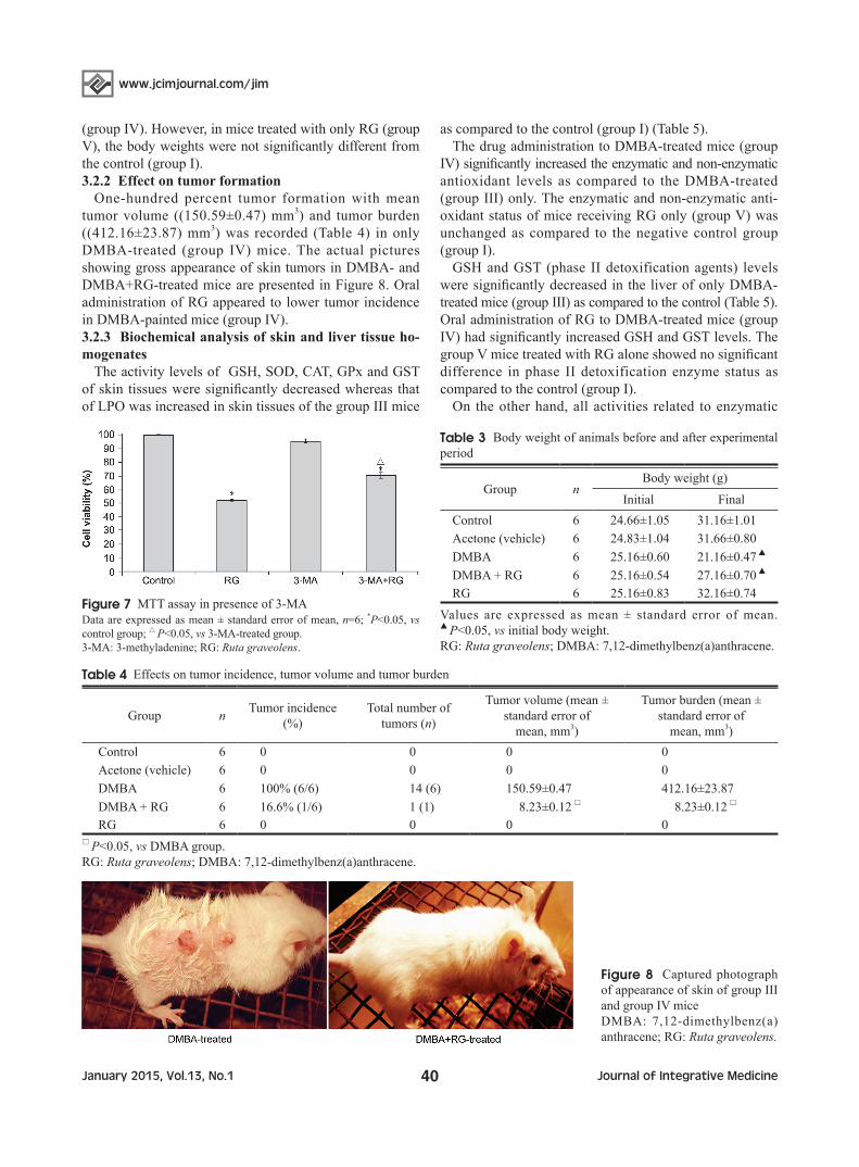
(group IV). However, in mice treated with only RG (group V), the body weights were not significantly different from the control (group I).3.2.2 Effect on tumor formation
One-hundred percent tumor formation with mean tumor volume ((150.59±0.47) mm3) and tumor burden ((412.16±23.87) mm3) was recorded (Table 4) in only DMBA-treated (group IV) mice. The actual pictures showing gross appearance of skin tumors in DMBA- and DMBA+RG-treated mice are presented in Figure 8. Oral administration of RG appeared to lower tumor incidence in DMBA-painted mice (group IV).3.2.3 Biochemical analysis of skin and liver tissue ho-mogenates
The activity levels of GSH, SOD, CAT, GPx and GST of skin tissues were significantly decreased whereas that of LPO was increased in skin tissues of the group III mice
as compared to the control (group I) (Table 5).The drug administration to DMBA-treated mice (group
IV) significantly increased the enzymatic and non-enzymatic antioxidant levels as compared to the DMBA-treated (group III) only. The enzymatic and non-enzymatic anti-oxidant status of mice receiving RG only (group V) was unchanged as compared to the negative control group (group I).
GSH and GST (phase II detoxification agents) levels were significantly decreased in the liver of only DMBA-treated mice (group III) as compared to the control (Table 5). Oral administration of RG to DMBA-treated mice (group IV) had significantly increased GSH and GST levels. The group V mice treated with RG alone showed no significant difference in phase II detoxification enzyme status as compared to the control (group I).
On the other hand, all activities related to enzymatic
Table 3 Body weight of animals before and after experimental period
Group nBody weight (g)
Initial FinalControl 6 24.66±1.05 31.16±1.01Acetone (vehicle) 6 24.83±1.04 31.66±0.80DMBA 6 25.16±0.60 21.16±0.47 ▲
DMBA + RG 6 25.16±0.54 27.16±0.70 ▲
RG 6 25.16±0.83 32.16±0.74
Values are expressed as mean ± standard error of mean. ▲ P<0.05, vs initial body weight.RG: Ruta graveolens; DMBA: 7,12-dimethylbenz(a)anthracene.
Table 4 Effects on tumor incidence, tumor volume and tumor burden
Group n Tumor incidence (%)
Total number of tumors (n)
Tumor volume (mean ± standard error of
mean, mm3)
Tumor burden (mean ± standard error of
mean, mm3)Control 6 0 0 0 0Acetone (vehicle) 6 0 0 0 0DMBA 6 100% (6/6) 14 (6) 150.59±0.47 412.16±23.87
DMBA + RG 6 16.6% (1/6) 1 (1) 8.23±0.12 □ 8.23±0.12 □
RG 6 0 0 0 0□ P<0.05, vs DMBA group.RG: Ruta graveolens; DMBA: 7,12-dimethylbenz(a)anthracene.
Figure 7 MTT assay in presence of 3-MAData are expressed as mean ± standard error of mean, n=6; *P<0.05, vs control group; △ P<0.05, vs 3-MA-treated group.3-MA: 3-methyladenine; RG: Ruta graveolens.
Figure 8 Captured photograph of appearance of skin of group III and group IV miceDMBA: 7,12-dimethylbenz(a)anthracene; RG: Ruta graveolens.

January 2015, Vol.13, No.141Journal of Integrative Medicine
www.jcimjournal.com/jim
and non-enzymatic activities remained unchanged in the acetone-painted group II (vehicle control). The same was true for body weights in this group as compared to the control.3.2.4 Effect of RG on expression of constitutive and activated Akt
Significant down-regulation of expression of both constitutive and active forms of Akt was noted after RG administration to DMBA-painted mice (group IV) as compared to the DMBA-treated group III (Figure 9).
Relative band intensities of the immunoblots are shown in Table 6.
4 Discussion
For effective control and management of the deadly disease cancer, chemopreventive drugs, particularly derived from the plant kingdom, are being harnessed throughout the world, and some amount of success has also been achieved with systematic research. Results of the present
Table 6 Relative band intensities of corresponding immunoblots of Figure 9
Group n Akt pAkt GAPDH
Control 6 100 100 100Acetone 6 100.927±0.337 98.960±0.764 99.443±0.515DMBA 6 252.145±0.638* 151.990±0.241* 98.831±0.596DMBA+RG 6 122.305±0.019*▲ 110.331±0.766*▲ 100.008±0.898RG 6 100.870±0.145 98.759±0.807 99.419±0.485
Taking the value of untreated control as 100%, percentages of other groups were calculated. Values are expressed as mean ± standard error of mean, n=6; *P<0.05, vs control group; ▲ P<0.05, vs DMBA group. GAPDH: glyceraldehyde-3-phosphate dehydrogenase; RG: Ruta graveolens; DMBA: 7,12-dimethylbenz(a)anthracene.
Table 5 Status of enzymatic and non-enzymatic antioxidants in skin and liver tissues
Group n GSH (mg/mL) SOD (μmol/(L·min·mg)) CAT (μmol/(L·min·mg)) LPO (μmol/(L·min·mg))
Control 6 100 100 100 100
Acetone 6 99.293±0.490 100.730±0.249 100.447±0.573 92.920±0.135
DMBA 6 52.966±0.740* 60.330±0.460* 53.395±0.984* 161.371±0.470*
DMBA+RG 6 81.355±0.556 ▲ 77.092±0.708 ▲ 73.706±0.697 ▲ 125.093±0.144 ▲
RG 6 100.423±0.441 98.030±0.351 91.493±0.388 105.790±0.068
Group n GPx (μmol/(L·min·mg)) GST (μmol/(L·min·mg)) GSH (liver) GST (liver)
Control 6 100 100 100 100Acetone 6 104.236±0.383 98.788±0.057 99.508±0.980 98.788±0.619
DMBA 6 60.085±1.580* 56.220±0.079* 36.836±0.509* 37.480±0.079*
DMBA+RG 6 75.725±0.569 ▲ 87.166±0.061 ▲ 68.369±0.179 ▲ 78.864±0.341 ▲
RG 6 96.032±0.700 99.901±0.055 100.294±0.049 100.370±0.277
Values are expressed as mean ± standard error of mean. *P<0.05, vs control group; ▲ P<0.05, vs DMBA group.RG: Ruta graveolens; DMBA: 7,12-dimethylbenz(a)anthracene; GSH: reduced glutathione; SOD: superoxide dismutase; CAT: cata-lase; LPO: lipid peroxidase; GPX: glutathione peroxidase; GST: glutathione S-transferase.
Figure 9 Results of Western blotBand intensity was measured by ImageJ software. GAPDH served as loading control. Significant down-regulation of expression of Akt and pAkt was noted after RG administration to DMBA-painted mice (group IV) as compared to the DMBA-treated only.GAPDH: glyceraldehyde-3-phosphate dehydrogenase; RG: Ruta graveolens; DMBA: 7,12-dimethylbenz(a)anthracene.
www.jcimjournal.com/jim
January 2015, Vol.13, No.1 42 Journal of Integrative Medicine
study reveals that RG was able to cause cell death of skin melanoma (A375) cells in vitro through induction of apoptosis and autophagy to a significant extent and also plays a protective role through modulation of enzymatic and non-enzymatic antioxidants against skin papilloma formation in mice in vivo.
Present research directions are mostly aimed at innovative targets at the molecular level, so that more specific drugs capable of hitting these targets can be developed. In view of the characteristic immortality attained by cancer cells, one major target of drug discovery involves the ability of the drug to annihilate cancer cells through induction of programmed cell death like apoptosis or autophagy. The findings of the present study are encouraging as RG demonstrated its profound ability of killing cancer cells by inducing apoptosis or/and by triggering molecular mechanism of autophagy. The ability of a drug to generate cytotoxicity in cancer cells, but not much in the normal cells is an important criterion in control and management of cancer. Most orthodox drugs lack in this property. Most of these drugs can effectively kill cancer cells by producing cytotoxicity, but the normal cells are not spared in this process, precluding their use to a large extent. Therefore, the results showing considerable ability of RG to produce preferential cytotoxicity in cancer cells over normal cells are significant findings.
Most anticancer drugs act through induction of apoptosis/necrosis in cancer cells as a major means. However, many a recent report suggest autophagy to have role in cell killing as well[23,24], although autophagy is generally known as a cell-survival mechanism induced by stresses. Therefore, drugs having both apoptotic and autophagic effects are of added advantage, because even after apoptosis is blocked by cancer cells, autophagy can play the cell death process. Therefore, in recent years molecular mechanisms of induction of autophagy through autolysosomes (forms of AVOs) pathway have received considerable attention[23–25]. In this investigation, existence of AVOs was confirmed from the result of fluorescence microscopic study with AO stain. Presence of cellular autophagosome punctae containing LC3-II is another confirmation of activation of autophagy[26]. In this study, conversion of LC3-I to LC3-II in RG-exposed cells was confirmed from the immunoblot study. Result of the present study showed upon exposure to RG, expression of mTOR, which prevents induction of autophagy[27], was down-regulated. Up-regulated expression of Atg5 and Atg7, two crucial molecules of the autophagic vacuole[27], was revealed from immunoblot assay. Findings in this study revealed down-regulated expression of Bcl-2 that inhibits both apoptosis and autophagy[28] in RG-treated cells. Beclin-1 plays a crucial role in the process of autophagy induction[29]. Up-regulation of Beclin-1 expression was found in RG-exposed cells. All these outcomes collectively
confirmed that there was induction of autophagy in RG-exposed A375 cells.
To find out whether this RG-mediated autophagy induction was to conduct cell death, cell viability was assessed after treating cells with RG and 3-MA, an autophagy inhibitor. A noticeable increase in viability was evident from the result which confirmed that RG-induced autophagy led the cells to the cell death pathway.
The cancer cells develop the property to evade apoptotic signals or signals that can trigger autophagy. On the other hand, they tend to show hyperactivity in response to all signals that promote cell divisional activities, causing them to divide and re-divide relentlessly propagating thereby a rapid growth. Thus agents that can target molecular mechanism leading to blockage of divisional activity, or antagonize all kinds of growth-promoting signals, or promote apoptotic signals, have considerable role to play in formulation of anticancer drugs. Apoptosis is mainly characterized by depolarized MMP, externalization of phosphatidyl serine (PS) from mitochondrial inner membrane to outer membrane, release of cytochrome c from mitochondria and massive caspase activation[30]. BAX, a Bcl-2 family protein, once activated by upstream pro-apoptotic signals[31], relocates from the cytosol to the surface of mitochondria and forms pores in the mitochondrial outer membrane[32]. This causes terminal release of pro-apoptotic factors, particularly cytochrome c[33], from the mitochondrial inner-membrane space. Released cytochrome c in cytosol now activates the caspase cascade and the activated caspase 3 in turn cleaves PARP, as a result of which fragmentation of DNA and ultimately induction of apoptosis ensues. In the present study, flow cytometry assay with rhodamine 123 staining indicated depolarization of MMP, and results of annexin V/PI dual stain assay confirmed the externalization of PS in RG-exposed cells. Immunoblot assay suggested BAX activation, cytochrome c release and activation of caspase 3. Increased expression of cleaved PARP (85 kD) was also found in RG-treated A375 cells. All these findings collectively suggest that RG induces mitochondria-dependent caspase 3-mediated apoptosis in A375 cells.
To test efficacy of RG in a mammalian model in vivo, skin cancer was induced in mice (Mus musculus). RG administration alone (75 mg/kg body weight) did not show any acute or chronic toxicity in normal mice, but showed significant reduction in the skin tumor burden in DMBA-painted mice. While 100% tumor incidence was noted in RG-untreated DMBA-painted mice, much less cancer burden was observed when they were also administered with RG. Our results thus suggest that RG had the ability to actively interfere with skin cell proliferation induced by DMBA.
Akt, an important regulator of proliferation and differ-

January 2015, Vol.13, No.143Journal of Integrative Medicine
www.jcimjournal.com/jim
entiation[34], is thought to be involved in DMBA-induced skin cancer. Results of the present study showed Akt expression was modulated upon RG administration. RG treatment down-regulated both constitutive and activated (pAkt) forms of Akt as compared to the DMBA treatment alone, indicative of its potential for use in anticancer drug formulation.
Liver is an organ where detoxification process takes place, but many orthodox chemotherapeutic drugs are known to deposit their byproducts in liver, resulting in further toxicity. Thus a critical study of toxicity biomarkers in liver was considered important for determining their status during the process of carcinogenesis. In this study therefore the multiple biochemical mechanisms including phase-II detoxification enzyme induction and antioxidant activities[35,36] were considered. Studies reported earlier that chemopreventive agents convert DNA damaging entities through the induction of detoxification agents such as GST[37]. In this study, enzyme activities of GSH and GST were significantly decreased in the liver of tumor-bearing animals (group III), but improved considerably upon RG administration in DMBA-painted animals (group IV) as compared to the control.
Over production of reactive oxygen species in the cells is one of the major reasons for DNA damage that is also implicated in carcinogenesis. Decreased activities of enzymatic antioxidants and drop in non-enzymatic anti-oxidant levels were well documented in skin cancer[38]. In this study it was revealed that RG administration at a dose of 75 mg/kg body weight to DMBA-painted mice significantly improved the status of lipid peroxidation and antioxidants, further confirming its anticancer role.
5 Conclusion
Overall results would suggest that RG is a strong candidate for being used in formulation of anticancer drugs, particularly against skin cancer.
6 Acknowledgements
The authors are sincerely thankful to Boiron Laboratories, Lyon, France for financially (partial) supporting the work.
7 Competing interests
The authors declare that they have no competing interests.
REFERENCES
1 Eickhorst K, DeLeo V, Csaposs J. Rue the herb: Ruta graveolens ― associated phytophototoxicity. Dermatitis. 2007; 18(1): 52–57.
2 Oliva A, Meepagala KM, Wedge DE, Harries D, Hale AL, Aliotta G, Duke SO. Natural fungicides from Ruta graveolens
L. leaves, including a new quinolone alkaloid. J Agric Food Chem. 2003; 51(4): 890–896.
3 Wessner D, Hofmann H, Ring J. Phytophotodermatitis due to Ruta graveolens applied as protection against evil spells. Contact Dermatitis. 1999; 41(4): 232.
4 International Program on Chemical Safety. Chemical safety information from intergovernmental organizations. 1989.
5 Lefranc F, Facchini V, Kiss R. Proautophagic drugs: a novel means to combat apoptosis-resistant cancers, with a special emphasis on glioblastomas. Oncologist. 2007; 12(12): 1395–1403.
6 Ghosh S, Bishayee K, Khuda-Bukhsh AR. Graveoline isolated from ethanolic extract of Ruta graveolens triggers apoptosis and autophagy in skin melanoma cells: A novel apoptosis-independent autophagic signalling pathway. Phytother Res. 2014; 28(8): 1153–1162.
7 Naithani R, Huma L, Moriarty RM, McCormick DL, Mehta RG. Comprehensive review of cancer chemopreventive agents evaluated in experimental chemoprevention models and clinical trials. Curr Med Chem. 2008; 15(11): 1044–1071.
8 Renju GL, Manoharan S, Balakrishnan S, Senthil N. Chemo-preventive and antilipidperoxidative potential of Clerodendron inerme (L) Gaertn in 7,12-dimethylbenz(a)anthracene induced skin carcinogenesis in Swiss albino mice. Pak J Biol Sci. 2007; 10(9): 1465–1470.
9 Alias LM, Manoharan S, Vellaichamy L, Balakrishnan S, Ramachandran CR. Protective effect of ferulic acid on 7,12-dimethylbenz(a)anthracene-induced skin carcinogenesis in Swiss albino mice. Exp Toxicol Pathol. 2009; 61(3): 205–214.
10 Ruitenberg JJ, Mulder CB, Maino VC, Landay AL, Ghanekar SA. VACUTAINER CPT and Ficoll density gradient separation perform equivalently in maintaining the quality and function of PBMC from HIV seropositive blood samples. BMC Immunol. 2006; 7: 11.
11 Ghosh S, Bishayee K, Paul A, Mukherjee A, Sikdar S, Chakraborty D, Boujedaini N, Khuda-Bukhsh AR. Homeopathic mother tincture of Phytolacca decandra induces apoptosis in skin melanoma cells by activating caspase-mediated signaling via reactive oxygen species elevation. J Integr Med. 2013; 11(2): 116–124.
12 Ghosh S, Bishayee K, Khuda-Bukhsh AR. Oleanolic acid isolated from ethanolic extract of Phytolacca decandra induces apoptosis in A375 skin melanoma cells: drug-DNA interaction and signaling cascade. J Integr Med. 2014; 12(2): 102–114.
13 Kanzawa T, Kondo Y, Ito H, Kondo S, Germano I. Induction of autophagic cell death in malignant glioma cells by arsenic trioxide. Cancer Res. 2003; 63(9): 2103–2108.
14 Chakraborty D, Bishayee K, Ghosh S, Biswas R, Mandal SK, Khuda-Bukhsh AR. [6]-Gingerol induces caspase 3 dependent apoptosis and autophagy in cancer cells: drug-DNA interaction and expression of certain signal genes in HeLa cells. Eur J Pharmacol. 2012; 694(1–3): 20–29.
15 Azuine MA, Bhide SV. Chemopreventive effect of turmeric against stomach and skin tumors induced by chemical carcinogens in Swiss mice. Nutr Cancer. 1992; 17(1): 77–83.
16 Bradford MM. A rapid and sensitive method for the quantitation

www.jcimjournal.com/jim
January 2015, Vol.13, No.1 44 Journal of Integrative Medicine
of microgram quantities of protein utilizing the principle of protein-dye binding. Anal Biochem.1976; 72: 248–254.
17 Chance B, Maehly AC. Assay of catalase and peroxidase. Methods Enzymol. 1955; 2: 764–775.
18 Kakkar P, Das B, Viswanathan PN. A modified spectropho-tometric assay of superoxide dismutase. Indian J Biochem Biophys. 1984; 21(2): 130–132.
19 Ellman GL. Tissue sulfhydryl groups. Arch Biochem Biophys. 1959; 82(1): 70–77.
20 Buege JA, Aust SD. Microsomal lipid peroxidation. Methods Enzymol. 1984; 105: 302–310.
21 Habig WH, Pabst MJ, Jakoby WB. Glutathione-S-transferase, the first enzymatic step in mercapturic acid formation. J Biol Chem. 1974; 249(22): 7130–7139.
22 Paglia DE, Valentine WN. Studies on the quantitative and qualitative characterisation of erythrocyte glutathione peroxidase. J Lab Clin Med. 1967; 70(1): 158–169.
23 Gozuacik D, Kimchi A. Autophagy as a cell death and tumor suppressor mechanism. Oncogene. 2004; 23(16): 2891–2906.
24 Tsujimoto Y, Shimizu S. Another way to die: autophagic programmed cell death. Cell Death Differ. 2005; 12(Suppl 2): 1528–1534.
25 Mizushima N. Methods for monitoring autophagy. Int J Biochem Cell Biol. 2004; 36(12): 2491–2502.
26 Kabeya Y, Mizushima N, Ueno T, Yamamoto A, Kirisako T, Noda T, Kominami E, Ohsumi Y, Yoshimori T. LC3, a mammalian homologue of yeast Apg8p, is localized in autophagosome membranes after processing. EMBO J. 2000; 19(21): 5720–5728.
27 Kung CP, Budina A, Balaburski G, Bergenstock MK, Murphy M. Autophagy in tumor suppression and cancer therapy. Crit Rev Eukaryot Gene Expr. 2011; 21(1): 71–100.
28 Pattingre S, Tassa A, Qu X, Garuti R, Liang XH, Mizushima N, Packer M, Schneider MD, Levine B. Bcl-2 antiapoptotic proteins inhibit Beclin 1-dependent autophagy. Cell. 2005; 122(6): 927–939.
29 Levine B, Sinha S, Kroemer G. Bcl-2 family members: dual
regulators of apoptosis and autophagy. Autophagy. 2008; 4(5): 600–606.
30 Green DR, Kroemer G. The central executioner of apoptosis: mitochondria or caspases? Trends Cell Biol. 1998; 8(7): 267–271.
31 Oltvai ZN, Milliman CL, Korsmeyer SJ. Bcl-2 heterodimerizes in vivo with a conserved homolog, Bax, that accelerates programmed cell death. Cell. 1993; 74(4): 609–619.
32 Antonsson B, Conti F, Ciavatta A, Montessuit S, Lewis S, Martinou I, Bernasconi L, Bernard A, Mermod JJ, Mazzei G, Maundrell K, Gambale F, Sadoul R, Martinou JC. Inhibition of Bax channel-forming activity by Bcl-2. Science. 1997; 277(5324): 370–372.
33 Dejean LM, Martinez-Caballero S, Guo L, Hughes C, Teijido O, Ducret T, Ichas F, Korsmeyer SJ, Antonsson B, Jonas EA, Kinnally KW. Oligomeric Bax is a component of the putative cytochrome c release channel MAC, mitochondrial apoptosis-induced channel. Mol Biol Cell. 2005; 16(5): 2424–2432.
34 Xu N, Lao Y, Zhang Y, Gillespie DA. Akt: a double-edged sword in cell proliferation and genome stability. J Oncol. 2012; 2012: 951724.
35 Keeney S, McKenna H, Fleming P, McIlfatrick S. Attitudes, knowledge and behaviours with regard to skin cancer: a literature review. Eur J Oncol Nurs. 2009; 13(1): 29–35.
36 Meijerman I, Beijnen JH, Schellens JH. Combined action and regulation of phase II enzymes and multidrug resistance proteins in multidrug resistance in cancer. Cancer Treat Rev. 2008; 34(6): 505–520.
37 Choi SC, Yun KJ, Kim TH, Kim HJ, Park SG, Oh GJ, Chae SC, Oh GJ, Nah YH, Kim JJ, Chung HT. Prognostic potential of glutathione S-transferase M1 and T1 null genotypes for gastric cancer progression. Cancer Lett. 2003; 195(2): 169–175.
38 Das I, Saha T. Effect of garlic on lipid peroxidation and antioxidation enzymes in DMBA-induced skin carcinoma. Nutrition. 2009; 25(4): 459–471.
Submission Guide
Journal of Integrative Medicine (JIM) is an international, peer-reviewed, PubMed-indexed journal, publishing papers on all aspects of integrative medicine, such as acupuncture and traditional Chinese medicine, Ayurvedic medicine, herbal medicine, homeopathy, nutrition, chiropractic, mind-body medicine, Taichi, Qigong, meditation, and any other modalities of complementary and alternative medicine (CAM). Article
types include reviews, systematic reviews and meta-analyses, randomized controlled and pragmatic trials, translational and patient-centered effectiveness outcome studies, case series and reports, clinical trial protocols, preclinical and basic science studies, papers on methodology and CAM history or education, editorials, global views, commentaries, short communications, book reviews, conference proceedings, and letters to the editor.
● No submission and page charges ● Quick decision and online first publication
For information on manuscript preparation and submission, please visit JIM website. Send your postal address by e-mail to [email protected], we will send you a complimentary print issue upon receipt.