Embed Size (px)
Citation preview
Research ArticleInterim Clinical Outcomes in Nanocomposite Bone MaterialRepairing Large Proximal Femoral Defect of Fibrous Dysplasia
Yun Lang,1 Ze-ping Yu,1 Yan Xiong,1 Chong-qi Tu,1 Cheng Ren,1 Bin Zhang,1
Hong-sheng Yang,1 Fang Yuan,2 Hong Li,3 Yong-gang Yan,3 and Hong Duan1
1Deparment of Orthopedics, West China Hospital, Sichuan University, No. 37 Guo Xue Lane, Chengdu, Sichuan 610064, China2Department of Radiology, West China Hospital, Sichuan University, Chengdu, China3School of Physical Science and Technology, Sichuan University, Chengdu, China
Correspondence should be addressed to Chong-qi Tu; [email protected] and Hong Duan; [email protected]
Received 14 April 2015; Revised 23 June 2015; Accepted 29 June 2015
Academic Editor: Faik Oktar
Copyright © 2015 Yun Lang et al.This is an open access article distributed under theCreative CommonsAttribution License, whichpermits unrestricted use, distribution, and reproduction in any medium, provided the original work is properly cited.
Background and Objectives. To evaluate the clinical effectiveness and safety of using nanocomposite bone material in the repair oflarge proximal femoral defects that are due to fibrous dysplasia.Method.Thirty-one patientswere analyzed retrospectively, including13 males and 18 females, and the mean age was 30.9 years (13–59). The median follow-up period was 50 months (30–78) and themasses of artificial bone transplants were in the range of 15∼40 g (average of 23.4 g). Functional and radiographic outcomes wereevaluated. Results. All wounds healed to grade A.There were no infections, nonspecific inflammatory reactions, rejection reactions,or fractures. One case had fat liquefaction and healed after dressing. All patients had no recurrence until the last follow-up. At thelast follow-up, the mean Musculoskeletal Tumor Society’s (MSTS) 93 score was 28.42 ± 1.31, the mean Harris hip score was 84.23± 8.97, and mean radiopaque density ratio was 0.78 ± 0.09. Radiologic analysis indicated that nanocomposite bone material hadbeen completely incorporated with the host bone within a year. Conclusions. This study indicated that the nanocomposite bonematerial had biological safety and good biocompatibility. In conclusion, the nanocomposite bone material is an ideal artificial bonesubstitute and worthy of promotion in the field of orthopedics.
1. Introduction
Bones are an important organ in our bodies and provide usthe freedom to do the things that wewant to do. Bones help usto stand up straight, walk, and jump. Many health problems,such as trauma, infection, and tumors, can cause brokenbones [1]. Bone tissue can repair itself for small defects, andsometimes cannot heal large bone defects. The treatment oflarge bone defects represents a considerable challenge in clin-ical practice [2, 3]. This situation necessitates the use of bonegrafts, including autografts, allografts, and bone substitutesfor healing [4]. Autografts have long been acknowledged asthe gold standard for bone grafts due to their outstandingosteoinductivity, osteoconductivity, and osteogenicity. How-ever, the following limitations restrict their extensive clinicalapplication: limited supply, donor site injury, and potentialrisks of infection. Allografts, providing scaffolds for graftsites, are relatively abundant in supply, are osteoconductive,
and have been used successfully in bone grafting procedures.However, allografts have their limitations as well, including aslowly creeping substitution process compared to autografts,high risks of disunion or delayed union, and potential riskof disease transmission and antigenicity that induces hostrejections. Bone graft substitutes include organic materials,inorganic materials, and composites. Surgeons encounter aserious challenge in searching for the ideal artificial bonesubstitute for the repair of large bone defects. Biomaterials,due to their comparability with human bone structure, havebeen demonstrated to be popular in orthopedic surgery [4].
Fibrous dysplasia (FD) of bone is a rare spontaneousbenign skeletal disorder characterized by a focal prolifer-ation of fibrous tissue in the bone marrow that manifestsin childhood or early adult life and can affect one bone(monostotic), or multiple bones (polyostotic) [5]. Accordingto Ippolito and colleagues [6], in monostotic FD, the mostcommonly appearing site is on the femur. Clinical symptoms
Hindawi Publishing CorporationJournal of NanomaterialsVolume 2015, Article ID 385612, 9 pageshttp://dx.doi.org/10.1155/2015/385612
2 Journal of Nanomaterials
Table 1: Surgical approach of 31 patients.
Lesion type Proximal femur Femoral neck and proximal femur TotalBone material grafting 7 4 11Bone material grafting + internal fixation 8 9 17Bone material + allograft cortical bone plate grafting + internal fixation 2 1 3Total 17 14 31
(a) (b)
Figure 1: The morphology of nanocomposite bone material. Notes: (a) nanocomposite bone material with a size of 4mm × 2mm × 2mm;(b) SEM image (×3,000 magnification) of nanocomposite bone material with a porosity of 75%–85%.SEM: scanning electron microscope.
of FD include bone pain, bone deformities, and pathologicalfractures [7]. It is well-known that the surgical manage-ment of fibrous dysplasia includes intralesional curettage,correction of the deformity, bone grafting, and rigid internalfixation. Various types of surgical treatment are reported,ranging from lesion curettage after bone grafting to massivecortical bone grafting, particularly in lesions of the femoralneck and for intramedullary fixation in extended lesions withdeformations [8–10].Themultiple treatmentmodalitiesmakeFD difficult to manage, especially in the proximal femur.
Nanocomposite bone material resembles human bone interms of structure and mechanical strength, which is usefulas a bone repairing material because it has biocompatibility,bioactivity, and good biomechanical properties as confirmedin various research [11–17].
We reviewed 31 patients and reported on our experiencein applying nanocomposite bone material (the porous n-HA/PA66 composite) to repair large proximal femoral defectsof FD.
2. Materials and Methods
2.1. Material Introduction. Nanocomposite bone material,which was provided by the Sichuan National Nanotechnol-ogy Co., Ltd. (Chengdu, People’s Republic of China) andmet biological safety standards according to the ChineseGB/T16886 and GB/T16175, was the biomaterial compositeand consisted of nanohydroxyapatite (n-HA) and polyamide66 (PA66) (Figure 1). The diameters of n-HA nanoparticlesrange from 80 nm to 100 nm and the molecular weight is1,000, while PA66 has a molecular weight range from 18,000to 20,000.Themass ratios of n-HAandPA66 in the compositewere 35 to 40. The composite pore size, porosity ratio, and
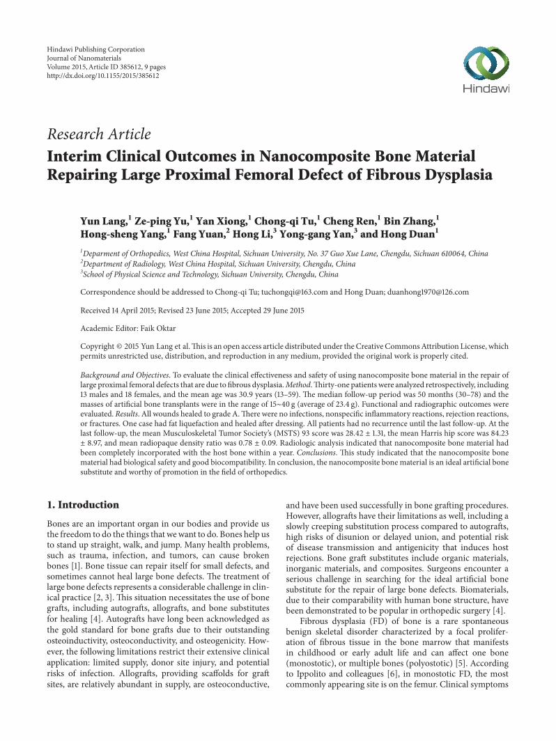
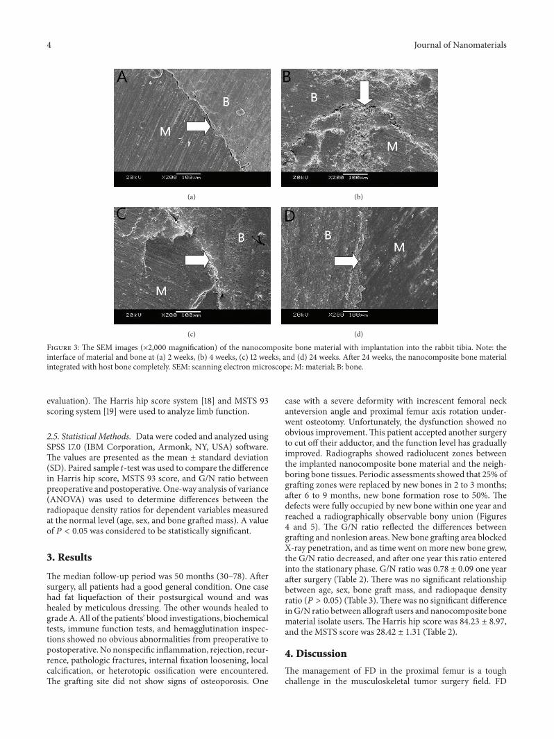
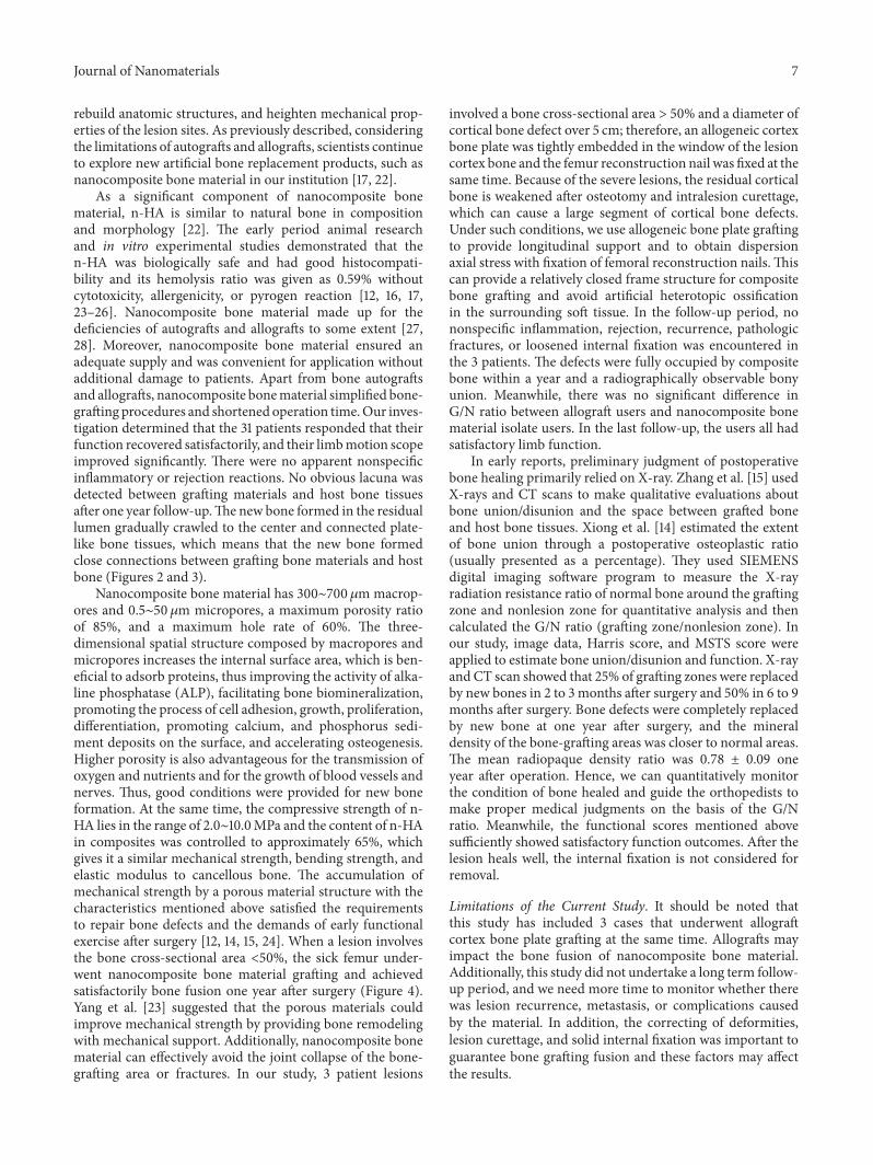
compressive strength were 300 to 700 𝜇m, 75% to 85%, and2.0 to 10.0mPa, respectively. For the nanocomposite bonematerial, it was demonstrated that there was no cytotoxicityor pyrogen. Additionally, the material’s hemolysis ratio was0.59% [17]. Early animal implantation studies showed thata rapid developing process of bone repair was observed inthe nanocomposite, the primer mineralization started at theimplant near to the neighboring bone tissue, and the newbone started increasing at 4 weeks. After 12 weeks, new boneformed in the pores, progressively linked with each other,and directly connected with materials. After 24 weeks, thenew bone gradually reached toward the center of the implantand exhibited good osteoinductivity and osteocompatibility(Figures 2 and 3). The treatments were undertaken with theunderstanding and written consent of each subject accordingto the World Medical Association Declaration of Helsinki(version 2008). The study has been independently reviewedand approved by the Ethics committee atWest ChinaHospitalof Sichuan University.
2.2. Clinical Data. From December 2007 to October 2011,there were 31 consecutive patients who fulfilled the followinginclusion criteria: (1) the diagnosis of FD should be confirmedby clinical feature, imaging data and pathology; (2) lesionsshould undergo intralesional curettage, biopsy, nanocompos-ite bone material transplantation, or rigid internal fixationand allograft used according to the type of lesion; (3)there was complete follow-up data. Exclusion criteria wereas follows: (1) the patient showed recurrence; (2) patientscontraindicated to operation. There were 13 males and 18females and the mean age was 30.9 years (13–59). The lesionlocations are listed in Table 1. The size of the lesions rangedfrom 6.0 cm × 2.5 cm × 2.5 cm to 10.0 cm × 3.5 cm × 3.5 cm.
Journal of Nanomaterials 3
(a) (b)
(c) (d)
Figure 2: Figure showing how the amount of new bone and calcium crystals increased over time. Note: bone and calcium crystals at (a) 2weeks, (b) 4 weeks, (c) 12 weeks, and (d) 24 weeks. Masson staining was used, ×200 magnification. After 24 weeks, interconnected porositywas filled completely with new bone tissue. M: material; B: bone.
The average of the implant amount of nanocomposite bonematerial was 23.4 g (15–40). We used a software program(Syngo version V35; Siemens Medical Systems, Erlangen,Germany) for quantitative analysis to measure the X-rayradiopaque density value. The value was measured in thegrafting section and nonlesion bone area near the operationsection, and the grafting-to-nonlesion count ratio (G/N ratio)was then calculated. The normal value of the G/N ratio was1.00 [14]. Both preoperatively and postoperatively, all patientsunderwent blood investigations, biochemical tests, immunefunction tests, and hemagglutination inspections.
2.3. Surgical Methods. This procedure included four follow-ing steps: orderly intralesional curettage, biopsy, inactivated,and filled with nanocomposite bone material. If the corticalbone was seriously affected, the combination application ofallogeneic cortex bone plate and nanocomposite bone mate-rial grafting was recommended. The femur reconstructionnail or proximal femoral nail antirotation (PFNA) wouldbe planted for those with a deformity, pathologic fracture,or obviously weakened bone strength. Additionally, thedeformity cases underwent valgus osteotomy prior to thesesteps. The surgery procedure option is shown in Table 1,which all patients underwent. 22 patients with hip and/orthe femoral varus deformity underwent a single level wedge-shaped valgus osteotomy at subtrochanteric region to ensure
a neck-shaft angle of 120∘; if the deformity combined thefemur neck and the subtrochanteric region, double-levelvalgus osteotomy was performed with the first level at thesubtrochanteric region and the second around the dome ofthe varus femur deformity. Hence, to ensure the healing ofthe osteotomy site, the second osteotomy site was determinedby the host bone quality, with our goal of double-levelosteotomy being to restore the neck-shaft angle more than90∘ and rebuild the femur alignment. Three cases withlesions involving a bone cross-section over 50% and corticalbone defects over 5 cm underwent double-level osteotomy.Meanwhile, allogeneic cortex bone plate grafting and femurreconstruction nail fixation were performed.
2.4. Postoperative Treatment and Follow-Up. Postoperatively,all patients accepted intravenous infusion cefathiamidine toprevent infection, early isotope exercise of quadriceps, andpassive exercise of hips and knees. The clinical evaluationincluded wound healing, blood investigations, biochemicaltests, immune function tests, and hemagglutination inspec-tions one, 2, 3, 6, 9, and 12 months and then every six monthsafter surgery. At the same time, patients were followed up bypanoramicX-ray and computed tomography (CT) plain scan,and spiral CT 3D reconstruction was performed to observethe healing of grafted bone and calculate theG/N ratio (imagemeasurement data and statistical data using a double-blind
4 Journal of Nanomaterials
(a) (b)
(c) (d)
Figure 3: The SEM images (×2,000 magnification) of the nanocomposite bone material with implantation into the rabbit tibia. Note: theinterface of material and bone at (a) 2 weeks, (b) 4 weeks, (c) 12 weeks, and (d) 24 weeks. After 24 weeks, the nanocomposite bone materialintegrated with host bone completely. SEM: scanning electron microscope; M: material; B: bone.
evaluation). The Harris hip score system [18] and MSTS 93scoring system [19] were used to analyze limb function.
2.5. Statistical Methods. Data were coded and analyzed usingSPSS 17.0 (IBM Corporation, Armonk, NY, USA) software.The values are presented as the mean ± standard deviation(SD). Paired sample 𝑡-test was used to compare the differencein Harris hip score, MSTS 93 score, and G/N ratio betweenpreoperative and postoperative. One-way analysis of variance(ANOVA) was used to determine differences between theradiopaque density ratios for dependent variables measuredat the normal level (age, sex, and bone grafted mass). A valueof 𝑃 < 0.05 was considered to be statistically significant.
3. Results
The median follow-up period was 50 months (30–78). Aftersurgery, all patients had a good general condition. One casehad fat liquefaction of their postsurgical wound and washealed by meticulous dressing. The other wounds healed tograde A. All of the patients’ blood investigations, biochemicaltests, immune function tests, and hemagglutination inspec-tions showed no obvious abnormalities from preoperative topostoperative. No nonspecific inflammation, rejection, recur-rence, pathologic fractures, internal fixation loosening, localcalcification, or heterotopic ossification were encountered.The grafting site did not show signs of osteoporosis. One
case with a severe deformity with increscent femoral neckanteversion angle and proximal femur axis rotation under-went osteotomy. Unfortunately, the dysfunction showed noobvious improvement. This patient accepted another surgeryto cut off their adductor, and the function level has graduallyimproved. Radiographs showed radiolucent zones betweenthe implanted nanocomposite bone material and the neigh-boring bone tissues. Periodic assessments showed that 25% ofgrafting zones were replaced by new bones in 2 to 3 months;after 6 to 9 months, new bone formation rose to 50%. Thedefects were fully occupied by new bone within one year andreached a radiographically observable bony union (Figures4 and 5). The G/N ratio reflected the differences betweengrafting and nonlesion areas. New bone grafting area blockedX-ray penetration, and as time went on more new bone grew,the G/N ratio decreased, and after one year this ratio enteredinto the stationary phase. G/N ratio was 0.78 ± 0.09 one yearafter surgery (Table 2). There was no significant relationshipbetween age, sex, bone graft mass, and radiopaque densityratio (𝑃 > 0.05) (Table 3). There was no significant differenceinG/N ratio between allograft users and nanocomposite bonematerial isolate users. The Harris hip score was 84.23 ± 8.97,and the MSTS score was 28.42 ± 1.31 (Table 2).
4. Discussion
The management of FD in the proximal femur is a toughchallenge in the musculoskeletal tumor surgery field. FD
Journal of Nanomaterials 5
(a) (b) (c)
(d) (e)
(f) (g)
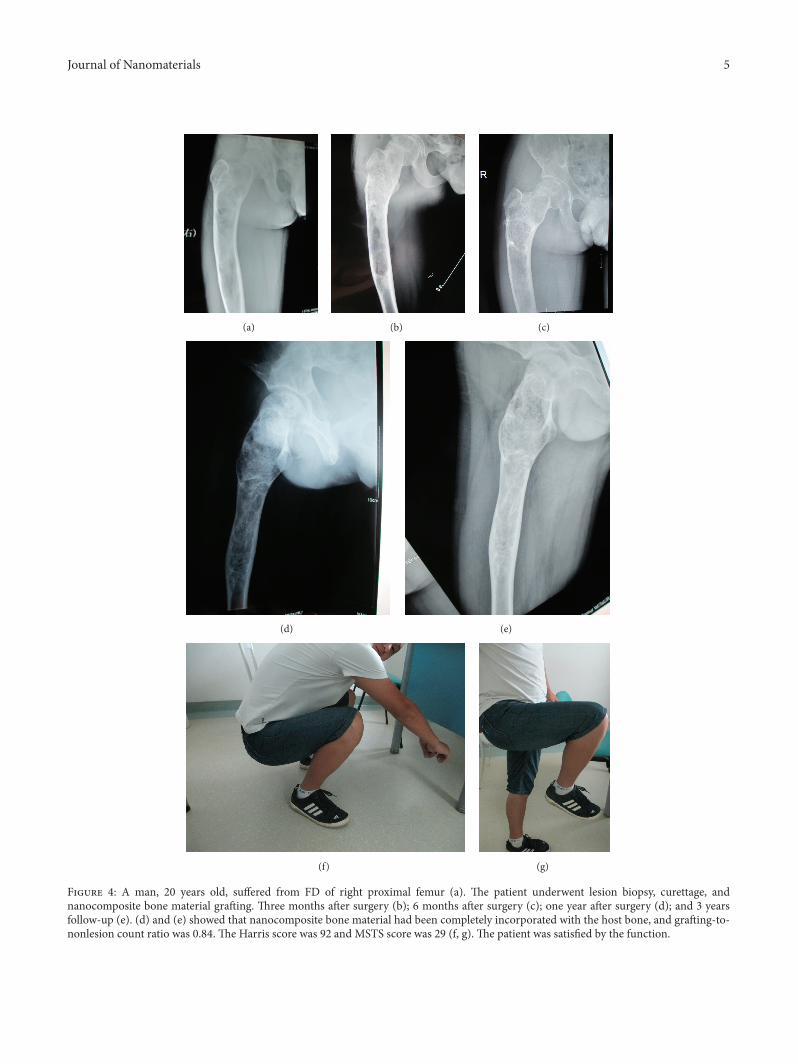
Figure 4: A man, 20 years old, suffered from FD of right proximal femur (a). The patient underwent lesion biopsy, curettage, andnanocomposite bone material grafting. Three months after surgery (b); 6 months after surgery (c); one year after surgery (d); and 3 yearsfollow-up (e). (d) and (e) showed that nanocomposite bone material had been completely incorporated with the host bone, and grafting-to-nonlesion count ratio was 0.84. The Harris score was 92 and MSTS score was 29 (f, g). The patient was satisfied by the function.
6 Journal of Nanomaterials
(a1) (a2)
(a)
(b1) (b2)
(b)
(c1) (c2)
(c)
(d1) (d2)
(d)
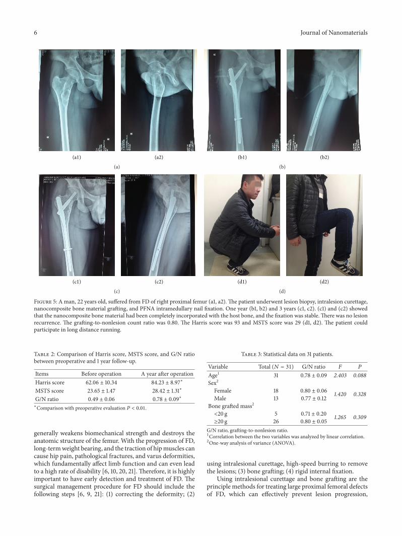
Figure 5: A man, 22 years old, suffered from FD of right proximal femur (a1, a2). The patient underwent lesion biopsy, intralesion curettage,nanocomposite bone material grafting, and PFNA intramedullary nail fixation. One year (b1, b2) and 3 years (c1, c2). (c1) and (c2) showedthat the nanocomposite bone material had been completely incorporated with the host bone, and the fixation was stable. There was no lesionrecurrence. The grafting-to-nonlesion count ratio was 0.80. The Harris score was 93 and MSTS score was 29 (d1, d2). The patient couldparticipate in long distance running.
Table 2: Comparison of Harris score, MSTS score, and G/N ratiobetween preoperative and 1 year follow-up.
Items Before operation A year after operationHarris score 62.06 ± 10.34 84.23 ± 8.97∗
MSTS score 23.65 ± 1.47 28.42 ± 1.31∗
G/N ratio 0.49 ± 0.06 0.78 ± 0.09∗∗Comparison with preoperative evaluation 𝑃 < 0.01.
generally weakens biomechanical strength and destroys theanatomic structure of the femur. With the progression of FD,long-termweight bearing, and the traction of hipmuscles cancause hip pain, pathological fractures, and varus deformities,which fundamentally affect limb function and can even leadto a high rate of disability [6, 10, 20, 21].Therefore, it is highlyimportant to have early detection and treatment of FD. Thesurgical management procedure for FD should include thefollowing steps [6, 9, 21]: (1) correcting the deformity; (2)
Table 3: Statistical data on 31 patients.
Variable Total (𝑁 = 31) G/N ratio 𝐹 𝑃
Age1 31 0.78 ± 0.09 2.403 0.088Sex2
Female 18 0.80 ± 0.06 1.420 0.328Male 13 0.77 ± 0.12
Bone grafted mass2
<20 g 5 0.71 ± 0.20 1.265 0.309≥20 g 26 0.80 ± 0.05
G/N ratio, grafting-to-nonlesion ratio.1Correlation between the two variables was analyzed by linear correlation.2One-way analysis of variance (ANOVA).
using intralesional curettage, high-speed burring to removethe lesions; (3) bone grafting; (4) rigid internal fixation.
Using intralesional curettage and bone grafting are theprinciple methods for treating large proximal femoral defectsof FD, which can effectively prevent lesion progression,
Journal of Nanomaterials 7
rebuild anatomic structures, and heighten mechanical prop-erties of the lesion sites. As previously described, consideringthe limitations of autografts and allografts, scientists continueto explore new artificial bone replacement products, such asnanocomposite bone material in our institution [17, 22].
As a significant component of nanocomposite bonematerial, n-HA is similar to natural bone in compositionand morphology [22]. The early period animal researchand in vitro experimental studies demonstrated that then-HA was biologically safe and had good histocompati-bility and its hemolysis ratio was given as 0.59% withoutcytotoxicity, allergenicity, or pyrogen reaction [12, 16, 17,23–26]. Nanocomposite bone material made up for thedeficiencies of autografts and allografts to some extent [27,28]. Moreover, nanocomposite bone material ensured anadequate supply and was convenient for application withoutadditional damage to patients. Apart from bone autograftsand allografts, nanocomposite bonematerial simplified bone-grafting procedures and shortened operation time.Our inves-tigation determined that the 31 patients responded that theirfunction recovered satisfactorily, and their limbmotion scopeimproved significantly. There were no apparent nonspecificinflammatory or rejection reactions. No obvious lacuna wasdetected between grafting materials and host bone tissuesafter one year follow-up.The new bone formed in the residuallumen gradually crawled to the center and connected plate-like bone tissues, which means that the new bone formedclose connections between grafting bone materials and hostbone (Figures 2 and 3).
Nanocomposite bone material has 300∼700𝜇mmacrop-ores and 0.5∼50𝜇m micropores, a maximum porosity ratioof 85%, and a maximum hole rate of 60%. The three-dimensional spatial structure composed by macropores andmicropores increases the internal surface area, which is ben-eficial to adsorb proteins, thus improving the activity of alka-line phosphatase (ALP), facilitating bone biomineralization,promoting the process of cell adhesion, growth, proliferation,differentiation, promoting calcium, and phosphorus sedi-ment deposits on the surface, and accelerating osteogenesis.Higher porosity is also advantageous for the transmission ofoxygen and nutrients and for the growth of blood vessels andnerves. Thus, good conditions were provided for new boneformation. At the same time, the compressive strength of n-HA lies in the range of 2.0∼10.0MPa and the content of n-HAin composites was controlled to approximately 65%, whichgives it a similar mechanical strength, bending strength, andelastic modulus to cancellous bone. The accumulation ofmechanical strength by a porous material structure with thecharacteristics mentioned above satisfied the requirementsto repair bone defects and the demands of early functionalexercise after surgery [12, 14, 15, 24]. When a lesion involvesthe bone cross-sectional area <50%, the sick femur under-went nanocomposite bone material grafting and achievedsatisfactorily bone fusion one year after surgery (Figure 4).Yang et al. [23] suggested that the porous materials couldimprove mechanical strength by providing bone remodelingwith mechanical support. Additionally, nanocomposite bonematerial can effectively avoid the joint collapse of the bone-grafting area or fractures. In our study, 3 patient lesions
involved a bone cross-sectional area > 50% and a diameter ofcortical bone defect over 5 cm; therefore, an allogeneic cortexbone plate was tightly embedded in the window of the lesioncortex bone and the femur reconstruction nail was fixed at thesame time. Because of the severe lesions, the residual corticalbone is weakened after osteotomy and intralesion curettage,which can cause a large segment of cortical bone defects.Under such conditions, we use allogeneic bone plate graftingto provide longitudinal support and to obtain dispersionaxial stress with fixation of femoral reconstruction nails.Thiscan provide a relatively closed frame structure for compositebone grafting and avoid artificial heterotopic ossificationin the surrounding soft tissue. In the follow-up period, nononspecific inflammation, rejection, recurrence, pathologicfractures, or loosened internal fixation was encountered inthe 3 patients. The defects were fully occupied by compositebone within a year and a radiographically observable bonyunion. Meanwhile, there was no significant difference inG/N ratio between allograft users and nanocomposite bonematerial isolate users. In the last follow-up, the users all hadsatisfactory limb function.
In early reports, preliminary judgment of postoperativebone healing primarily relied on X-ray. Zhang et al. [15] usedX-rays and CT scans to make qualitative evaluations aboutbone union/disunion and the space between grafted boneand host bone tissues. Xiong et al. [14] estimated the extentof bone union through a postoperative osteoplastic ratio(usually presented as a percentage). They used SIEMENSdigital imaging software program to measure the X-rayradiation resistance ratio of normal bone around the graftingzone and nonlesion zone for quantitative analysis and thencalculated the G/N ratio (grafting zone/nonlesion zone). Inour study, image data, Harris score, and MSTS score wereapplied to estimate bone union/disunion and function. X-rayand CT scan showed that 25% of grafting zones were replacedby new bones in 2 to 3 months after surgery and 50% in 6 to 9months after surgery. Bone defects were completely replacedby new bone at one year after surgery, and the mineraldensity of the bone-grafting areas was closer to normal areas.The mean radiopaque density ratio was 0.78 ± 0.09 oneyear after operation. Hence, we can quantitatively monitorthe condition of bone healed and guide the orthopedists tomake proper medical judgments on the basis of the G/Nratio. Meanwhile, the functional scores mentioned abovesufficiently showed satisfactory function outcomes. After thelesion heals well, the internal fixation is not considered forremoval.
Limitations of the Current Study. It should be noted thatthis study has included 3 cases that underwent allograftcortex bone plate grafting at the same time. Allografts mayimpact the bone fusion of nanocomposite bone material.Additionally, this study did not undertake a long term follow-up period, and we need more time to monitor whether therewas lesion recurrence, metastasis, or complications causedby the material. In addition, the correcting of deformities,lesion curettage, and solid internal fixation was important toguarantee bone grafting fusion and these factors may affectthe results.
8 Journal of Nanomaterials
5. Conclusions
Nanocomposite bone material has biological safety and goodbiocompatibility. The material can achieve self-repairingthrough bone conduction and osteogenic induction and isan efficient bone-grafting material that presents satisfactoryeffects in the repair of large proximal femoral defects thatare due to fibrous dysplasia and should be generalized in theorthopedic field.
Conflict of Interests
The authors declare that they have no conflict of interests.
Authors’ Contribution
Yun Lang and Ze-ping Yu participated in the collection ofclinical data, performed patient follow-ups, and drafted thepaper. Yan Xiong made substantial contributions to concep-tion and design of this research and has reviewed the paperfor important intellectual content and given final approvalof the version to be published. Hong Li and Yong-gang Yanprovided the data of the composites’ in vitro and animalexperiments. Fang Yuan analysed the image data. HongDuanand Chong-qi Tu were responsible for these operations andparticipated in project coordination and assisted with thepaper. Each author has participated sufficiently in this workto take public responsibility for the appropriate portions ofthe paper. All authors read and approved the final paper. YunLang and Ze-ping Yu contributed equally to this work andshould be considered cofirst authors.
Acknowledgment
The result described in this paper was supported by theNational Science and Technology Support Program of thePeople’s Republic of China (2007BAE131304).
References
[1] A. C. Looker, B. Dawson-Hughes, A. N. A. Tosteson, H.Johansson, J. A. Kanis, and L. J. Melton III, “Hip fracturerisk in older US adults by treatment eligibility status based onnew National Osteoporosis Foundation guidance,”OsteoporosisInternational, vol. 22, no. 2, pp. 541–549, 2011.
[2] W. R. Moore, S. E. Graves, and G. I. Bain, “Synthetic bone graftsubstitutes,” ANZ Journal of Surgery, vol. 71, no. 6, pp. 354–361,2001.
[3] A. Schorr, W. Campbell, and M. Schenk, CommunicationResearch and Media Science in Europe: Perspectives for Researchand Academic Training in Europe’s Changing Media Reality,Mouton de Gruyter, Berlin, Germany, 2003.
[4] A. S. Greenwald, S. D. Boden, V. M. Goldberg, Y. Khan, C.T. Laurencin, and R. N. Rosier, “Bone-graft substitutes: facts,fictions, and applications,” Journal of Bone and Joint Surgery A,vol. 83, supplement 2, pp. 98–103, 2001.
[5] L. Lichtenstein, “Polyostotic fibrous dysplasia,” Archives ofSurgery, vol. 36, no. 5, pp. 874–898, 1938.
[6] E. Ippolito, E. W. Bray, A. Corsi et al., “Natural historyand treatment of fibrous dysplasia of bone: a multicenter
clinicopathologic study promoted by the European PediatricOrthopaedic Society,” Journal of Pediatric Orthopaedics, Part B,vol. 12, no. 3, pp. 155–177, 2003.
[7] M. Riminucci, I. Saggio, P. G. Robey, and P. Bianco, “Fibrousdysplasia as a stem cell disease,” Journal of Bone and MineralResearch, vol. 21, supplement 2, pp. 125–131, 2006.
[8] W. F. Enneking and P. F. Gearen, “Fibrous dysplasia of thefemoral neck. Treatment by cortical bone-grafting,”The Journalof Bone & Joint Surgery—American Volume, vol. 68, no. 9, pp.1415–1422, 1986.
[9] L. Yang, Y. Jing, D. Hong, and T. Chong-Qi, “Valgus osteotomycombined with intramedullary nail for Shepherd’s crook defor-mity in fibrous dysplasia: 14 femurs with a minimum of 4 yearsfollow-up,” Archives of Orthopaedic and Trauma Surgery, vol.130, no. 4, pp. 497–502, 2010.
[10] J. T. Guille, S. J. Kumar, and G. D. Macewen, “Fibrous dysplasiaof the proximal part of the femur: long-term results of curettageand bone-grafting and mechanical realignment,”The Journal ofBone& Joint Surgery—AmericanVolume, vol. 80, no. 5, pp. 648–658, 1998.
[11] X. Yang, Q. Chen, L.-M. Liu et al., “Comparison of ante-rior cervical fusion by titanium mesh cage versus nano-hydroxyapatite/polyamide cage following single-level corpec-tomy,” International Orthopaedics, vol. 37, no. 12, pp. 2421–2427,2013.
[12] H. Li, Y. B. Li, Y. G. Yan, G. Zhou, M. Wang, and L. Cheng,“Preparation and biological safety evaluation of porous n-HA/PA66 composite,” Journal of Biomedical Engineering, vol. 25, no.5, pp. 1126–1129, 2008.
[13] Y. Zhang, Z.-X. Quan, Z.-H. Zhao et al., “Evaluation of anteriorcervical reconstruction with titanium mesh cages versus nano-hydroxyapatite/polyamide66 cages after 1- or 2-level corpec-tomy for multilevel cervical spondylotic myelopathy: a retro-spective study of 117 patients,” PLoS ONE, vol. 9, no. 5, ArticleID e96265, 2014.
[14] Y. Xiong, C. Ren, B. Zhang et al., “Analyzing the behaviorof a porous nano-hydroxyapatite/polyamide 66 (n-HA/PA66)composite for healing of bone defects,” International Journal ofNanomedicine, vol. 9, no. 1, pp. 485–494, 2014.
[15] S.-L. Zhang, Y. Zhou, H. Duan et al., “Repairing bone defectwith nano-hydroxyapatite and polyamide 66 composite aftergiant cell tumor operation,” Journal of Sichuan University—Medical Science Edition, vol. 43, no. 3, pp. 373–377, 2012.
[16] C.-Y. Meng, H. An, D.-M. Jiang et al., “Repair of bone defectwith porous composite of nano-hydroxyapatite and polymide,”Chinese Journal of Orthopaedic Trauma, vol. 21, no. 3, pp. 187–191, 2005.
[17] H. Wang, Y. Li, Y. Zuo, J. Li, S. Ma, and L. Cheng,“Biocompatibility and osteogenesis of biomimetic nano-hydroxyapatite/polyamide composite scaffolds for bone tissueengineering,” Biomaterials, vol. 28, no. 22, pp. 3338–3348, 2007.
[18] N. N. Mahomed, D. C. Arndt, B. J. McGrory, and W. H. Harris,“The Harris hip score: comparison of patient self-report withsurgeon assessment,” The Journal of Arthroplasty, vol. 16, no. 5,pp. 575–580, 2001.
[19] W. F. Enneking, W. Dunham, M. C. Gebhardt, M. Malawar,and D. J. Pritchard, “A system for the functional evaluation ofreconstructive procedures after surgical treatment of tumors ofthe musculoskeletal system,” Clinical Orthopaedics and RelatedResearch, vol. 1, no. 286, pp. 241–246, 1993.
Journal of Nanomaterials 9
[20] T. Lejman and J. Sulko, “Orthopedic management in childrenwith fibrous dysplasia of bone,” Chirurgia Narzadow Ruchu iOrtopedia Polska, vol. 64, no. 3, pp. 303–310, 1999.
[21] R. P. Stanton, E. Ippolito, D. Springfield, L. Lindaman, S.Wientroub, and A. Leet, “The surgical management of fibrousdysplasia of bone,” Orphanet Journal of Rare Diseases, vol. 7,supplement 1, article S1, 2012.
[22] L. Wu, Y.-B. Li, W.-H. Yang, L. Zhang, and J.-M. Han, “Qual-itative and quantitative comparison of human cortical bone,nano-HA and nano-HA/PA66,” Chinese Journal of FunctionalMaterial, vol. 36, no. 6, pp. 892–895, 2005.
[23] K. Yang, J.Wei, C.-Y.Wang, andY. Li, “A study on in vitro and invivo bioactivity of nanohydroxyapatite/polymer biocomposite,”Chinese Science Bulletin, vol. 52, no. 2, pp. 267–271, 2007.
[24] Q. Xu, H. Lu, J. Zhang, G. Lu, Z. Deng, and A. Mo, “Tis-sue engineering scaffold material of porous nanohydroxyap-atite/polyamide 66,” International Journal of Nanomedicine, vol.5, pp. 331–335, 2010.
[25] B. Qiao, J. Li, Q. Zhu et al., “Bone plate composed of aternary nano-hydroxyapatite/polyamide 66/glass fiber compos-ite: biomechanical properties and biocompatibility,” Interna-tional Journal of Nanomedicine, vol. 9, no. 1, pp. 1423–1432, 2014.
[26] Y. Qu, P. Wang, Y. Man, Y. Li, Y. Zuo, and J. Li, “Preliminarybiocompatible evaluation of nano-hydroxyapatite/polyamide66 composite porous membrane,” International Journal ofNanomedicine, vol. 5, no. 1, pp. 429–435, 2010.
[27] T. J. Cypher and J. P. Grossman, “Biological principles of bonegraft healing,” Journal of Foot and Ankle Surgery, vol. 35, no. 5,pp. 413–417, 1996.
[28] H. J. Mankin, F. J. Hornicek, and K. A. Raskin, “Infectionin massive bone allografts,” Clinical Orthopaedics and RelatedResearch, vol. 3, no. 432, pp. 210–216, 2005.

Submit your manuscripts athttp://www.hindawi.com
ScientificaHindawi Publishing Corporationhttp://www.hindawi.com Volume 2014
CorrosionInternational Journal of
Hindawi Publishing Corporationhttp://www.hindawi.com Volume 2014
Polymer ScienceInternational Journal of
Hindawi Publishing Corporationhttp://www.hindawi.com Volume 2014
Hindawi Publishing Corporationhttp://www.hindawi.com Volume 2014
CeramicsJournal of
Hindawi Publishing Corporationhttp://www.hindawi.com Volume 2014
CompositesJournal of
NanoparticlesJournal of
Hindawi Publishing Corporationhttp://www.hindawi.com Volume 2014
Hindawi Publishing Corporationhttp://www.hindawi.com Volume 2014
International Journal of
Biomaterials
Hindawi Publishing Corporationhttp://www.hindawi.com Volume 2014
NanoscienceJournal of
TextilesHindawi Publishing Corporation http://www.hindawi.com Volume 2014
Journal of
NanotechnologyHindawi Publishing Corporationhttp://www.hindawi.com Volume 2014
Journal of
CrystallographyJournal of
Hindawi Publishing Corporationhttp://www.hindawi.com Volume 2014
The Scientific World JournalHindawi Publishing Corporation http://www.hindawi.com Volume 2014
Hindawi Publishing Corporationhttp://www.hindawi.com Volume 2014
CoatingsJournal of
Advances in
Materials Science and EngineeringHindawi Publishing Corporationhttp://www.hindawi.com Volume 2014
Smart Materials Research
Hindawi Publishing Corporationhttp://www.hindawi.com Volume 2014
Hindawi Publishing Corporationhttp://www.hindawi.com Volume 2014
MetallurgyJournal of
Hindawi Publishing Corporationhttp://www.hindawi.com Volume 2014
BioMed Research International
MaterialsJournal of
Hindawi Publishing Corporationhttp://www.hindawi.com Volume 2014
Nano
materials
Hindawi Publishing Corporationhttp://www.hindawi.com Volume 2014
Journal ofNanomaterials