Embed Size (px)
Citation preview
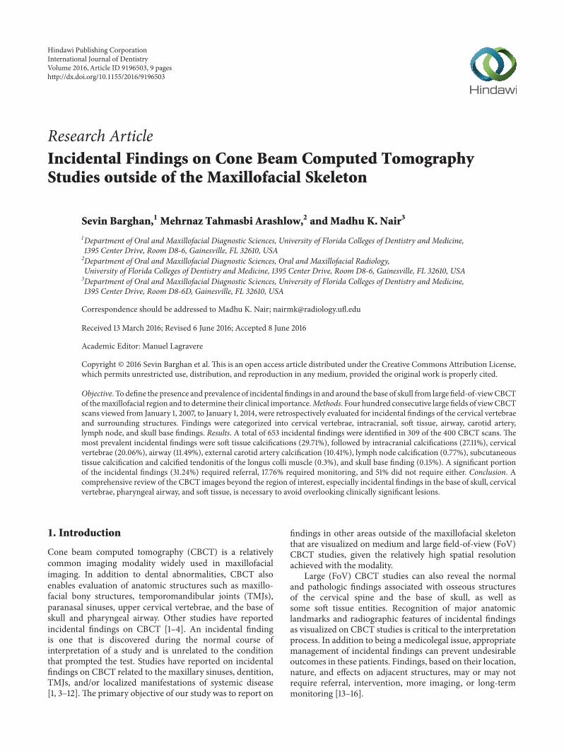
Research ArticleIncidental Findings on Cone Beam Computed TomographyStudies outside of the Maxillofacial Skeleton
Sevin Barghan,1 Mehrnaz Tahmasbi Arashlow,2 and Madhu K. Nair3
1Department of Oral and Maxillofacial Diagnostic Sciences, University of Florida Colleges of Dentistry and Medicine,1395 Center Drive, Room D8-6, Gainesville, FL 32610, USA2Department of Oral and Maxillofacial Diagnostic Sciences, Oral and Maxillofacial Radiology,University of Florida Colleges of Dentistry and Medicine, 1395 Center Drive, Room D8-6, Gainesville, FL 32610, USA3Department of Oral and Maxillofacial Diagnostic Sciences, University of Florida Colleges of Dentistry and Medicine,1395 Center Drive, Room D8-6D, Gainesville, FL 32610, USA
Correspondence should be addressed to Madhu K. Nair; [email protected]
Received 13 March 2016; Revised 6 June 2016; Accepted 8 June 2016
Academic Editor: Manuel Lagravere
Copyright © 2016 Sevin Barghan et al. This is an open access article distributed under the Creative Commons Attribution License,which permits unrestricted use, distribution, and reproduction in any medium, provided the original work is properly cited.
Objective. To define the presence and prevalence of incidental findings in and around the base of skull from large field-of-viewCBCTof themaxillofacial region and to determine their clinical importance.Methods. Four hundred consecutive large fields of viewCBCTscans viewed from January 1, 2007, to January 1, 2014, were retrospectively evaluated for incidental findings of the cervical vertebraeand surrounding structures. Findings were categorized into cervical vertebrae, intracranial, soft tissue, airway, carotid artery,lymph node, and skull base findings. Results. A total of 653 incidental findings were identified in 309 of the 400 CBCT scans. Themost prevalent incidental findings were soft tissue calcifications (29.71%), followed by intracranial calcifications (27.11%), cervicalvertebrae (20.06%), airway (11.49%), external carotid artery calcification (10.41%), lymph node calcification (0.77%), subcutaneoustissue calcification and calcified tendonitis of the longus colli muscle (0.3%), and skull base finding (0.15%). A significant portionof the incidental findings (31.24%) required referral, 17.76% required monitoring, and 51% did not require either. Conclusion. Acomprehensive review of the CBCT images beyond the region of interest, especially incidental findings in the base of skull, cervicalvertebrae, pharyngeal airway, and soft tissue, is necessary to avoid overlooking clinically significant lesions.
1. Introduction
Cone beam computed tomography (CBCT) is a relativelycommon imaging modality widely used in maxillofacialimaging. In addition to dental abnormalities, CBCT alsoenables evaluation of anatomic structures such as maxillo-facial bony structures, temporomandibular joints (TMJs),paranasal sinuses, upper cervical vertebrae, and the base ofskull and pharyngeal airway. Other studies have reportedincidental findings on CBCT [1–4]. An incidental findingis one that is discovered during the normal course ofinterpretation of a study and is unrelated to the conditionthat prompted the test. Studies have reported on incidentalfindings on CBCT related to the maxillary sinuses, dentition,TMJs, and/or localized manifestations of systemic disease[1, 3–12]. The primary objective of our study was to report on
findings in other areas outside of the maxillofacial skeletonthat are visualized on medium and large field-of-view (FoV)CBCT studies, given the relatively high spatial resolutionachieved with the modality.
Large (FoV) CBCT studies can also reveal the normaland pathologic findings associated with osseous structuresof the cervical spine and the base of skull, as well assome soft tissue entities. Recognition of major anatomiclandmarks and radiographic features of incidental findingsas visualized on CBCT studies is critical to the interpretationprocess. In addition to being a medicolegal issue, appropriatemanagement of incidental findings can prevent undesirableoutcomes in these patients. Findings, based on their location,nature, and effects on adjacent structures, may or may notrequire referral, intervention, more imaging, or long-termmonitoring [13–16].
Hindawi Publishing CorporationInternational Journal of DentistryVolume 2016, Article ID 9196503, 9 pageshttp://dx.doi.org/10.1155/2016/9196503
2 International Journal of Dentistry
This study evaluated the prevalence and nature of inci-dental findings in and around the base of skull includingbut not limited to the cervical spine, lateral neck region,pharyngeal airway, and intracranial structures in CBCTstudies which are usually reviewed by dentists and dentalspecialists.
2. Material and Methods
The study protocol was reviewed and approved by Universityof Florida Institutional Review Board (IRB). The images of400 consecutive large FoVCBCT studieswere reviewed in theOral and Maxillofacial Radiology Clinic at the University ofFlorida, College of Dentistry from January 1, 2007, to January1, 2014, for incidental findings related to the cervical spine,paravertebral regions, lateral neck region, pharyngeal airway,and intracranial structures. The chosen sample was deemedappropriate in size by comparison with similar studies inthe literature. The images were viewed with InVivo Dentalsoftware (Anatomage, Inc., San Jose, CA, USA). The CBCTstudies included in this study were the iCAT� (Imaging Sci-ences International, Hatfield, PA,USA), CS 9300 (CarestreamHealth, Atlanta, GA), NewTom QR-DVT-9000 (QR-NIMs.r.l., Verona, Italy), and the CS9500 (Carestream Health,Atlanta, GA). Exposure parameters include 70–125 kVp and12–40mAs, with differences in the fields of view and acqui-sition voxel sizes. Patients’ demographic data, indications forimaging, and the types of CBCT units used were recorded.Findings related to the dentition, periodontium, paranasalsinuses, and temporomandibular joint diseases were notrecorded. Findings directly related to the primary indicationsfor CBCT scans were excluded. Images with poor imagequality were also excluded. Diagnoses were based entirely onradiographic appearance using well-established radiographicinterpretation processes. In radiology, incidental findingsare defined as an occult entity discovered unexpectedlyon an imaging examination performed for an unrelatedreason [5]. The cervical vertebrae were evaluated for thepresence of degenerative changes, malalignment, fusion, lossof intervertebral space, and any other pathoses. The airwaywas evaluated for the presence of narrowing, asymmetry,masses, and lymphoid hyperplasia. The neck region wasinvestigated for the presence of any masses, asymmetry,lateralization or narrowing of the pharyngeal airway, ordystrophic calcification. In addition, the clivus, base of skull,and intracranial anatomywere evaluated for any abnormality.All studies had reports dictated earlier by board-certified oraland maxillofacial radiologists. However, this study involvedevaluation of the datasets by two oral and maxillofacial radi-ology residents in their second and third years of training viaindependent sessions and under optimal viewing conditions.Findings were tabulated and compared with those reportedby the radiologists. All cases with intracranial findings hadbeen reviewed by board-certifiedneuroradiologistswith headand neck fellowship training as well. Any additional findingsor confounders observed by the residents were broughtto the attention of the oral and maxillofacial radiologistand neuroradiologists as part of their training process. Noadditional findings were reported. Residents were included
0.00
5.00
10.00
15.00
20.00
25.00
Num
ber o
f pat
ient
s (%
)
Age category
<10
10,19
20
–29
30
–39
40
–49
50
–59
60
–69
70
–79
80
–89
≥90
Figure 1: Age distribution of patients.
Table 1: Indications for CBCT examination.
Diagnostic tasks Number of patients PercentageImplants 152 38Pathology 121 30Orthodontics 96 24TMJ 31 8Total 400 100
in the process to assess the extent to which nonradiologistswould potentially miss incidental findings. After reviewingthe types and prevalence of incidental findings in the regionsof interest, findings were divided into three categories: thosethat required appropriate referral to a physician or specialist,those that required follow-up in the form of continuedmonitoring, and those that required no referral, follow-up,or monitoring but required documentation.
Following data collection, data analysis was performedwith the help of Statistical Package for Social Sciences (SPSS)version 22. Qualitative analysis was done.
3. Results
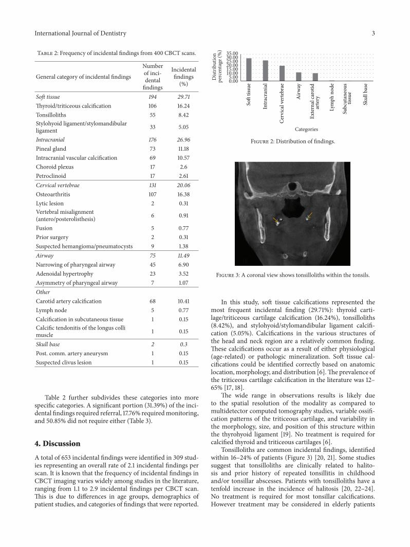
Out of 400 patients, 146 (36.5%) were males and 254 (63.5%)were females. Mean age of patients referred for CBCT was47.08 years. Most frequently referred patient age groupswere the 60–69 age group (19.75%) (Figure 1). Patients werereferred for orthodontic intervention (𝑛 = 96), implanttreatment planning (𝑛 = 152), TMJ evaluation (𝑛 = 31), andevaluation of suspected pathoses (𝑛 = 121). The indicationsfor CBCT examination are shown in Table 1. A total of 653incidental findings were identified in 309 of the 400 CBCTstudies (77%), representing an overall rate of 1.63 incidentalfindings per study. Ninety-one (22.75%) scans showed noincidental findings of relevance. The most prevalent inci-dental findings included soft tissue calcifications (29.71%),followed by intracranial calcifications (26.96%), cervical ver-tebral pathoses (20.06%), airway findings (11.49%), externalcarotid artery calcifications (10.41%), lymph node calcifica-tions (0.77%), calcifications in subcutaneous tissue, calcifictendonitis of the longus colli muscle (0.3%), and skull baselesions (0.15%) (Figure 2).
International Journal of Dentistry 3
Table 2: Frequency of incidental findings from 400 CBCT scans.
General category of incidental findings
Numberof inci-dentalfindings
Incidentalfindings(%)
Soft tissue 194 29.71Thyroid/triticeous calcification 106 16.24Tonsilloliths 55 8.42Stylohyoid ligament/stylomandibularligament 33 5.05
Intracranial 176 26.96Pineal gland 73 11.18Intracranial vascular calcification 69 10.57Choroid plexus 17 2.6Petroclinoid 17 2.61Cervical vertebrae 131 20.06Osteoarthritis 107 16.38Lytic lesion 2 0.31Vertebral misalignment(antero/posterolisthesis) 6 0.91
Fusion 5 0.77Prior surgery 2 0.31Suspected hemangioma/pneumatocysts 9 1.38Airway 75 11.49Narrowing of pharyngeal airway 45 6.90Adenoidal hypertrophy 23 3.52Asymmetry of pharyngeal airway 7 1.07OtherCarotid artery calcification 68 10.41Lymph node 5 0.77Calcification in subcutaneous tissue 1 0.15Calcific tendonitis of the longus collimuscle 1 0.15
Skull base 2 0.3Post. comm. artery aneurysm 1 0.15Suspected clivus lesion 1 0.15
Table 2 further subdivides these categories into morespecific categories. A significant portion (31.39%) of the inci-dental findings required referral, 17.76% requiredmonitoring,and 50.85% did not require either (Table 3).
4. Discussion
A total of 653 incidental findings were identified in 309 stud-ies representing an overall rate of 2.1 incidental findings perscan. It is known that the frequency of incidental findings inCBCT imaging varies widely among studies in the literature,ranging from 1.1 to 2.9 incidental findings per CBCT scan.This is due to differences in age groups, demographics ofpatient studies, and categories of findings that were reported.
0.005.00
10.0015.0020.0025.0030.0035.00
Dist
ribut
ion
perc
enta
ge (%
)
Categories
Soft
tissu
e
Intr
acra
nial
Cer
vica
l ver
tebr
ae
Airw
ay
arte
ryEx
tern
al ca
rotid
Lym
ph n
ode
tissu
eSu
bcut
aneo
us
Skul
l bas
e
Figure 2: Distribution of findings.
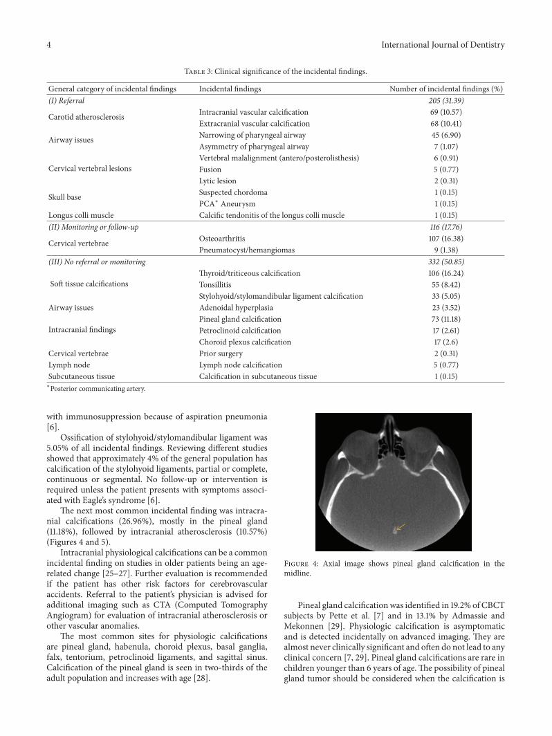
Figure 3: A coronal view shows tonsilloliths within the tonsils.
In this study, soft tissue calcifications represented themost frequent incidental finding (29.71%): thyroid carti-lage/triticeous cartilage calcification (16.24%), tonsilloliths(8.42%), and stylohyoid/stylomandibular ligament calcifi-cation (5.05%). Calcifications in the various structures ofthe head and neck region are a relatively common finding.These calcifications occur as a result of either physiological(age-related) or pathologic mineralization. Soft tissue cal-cifications could be identified correctly based on anatomiclocation, morphology, and distribution [6].The prevalence ofthe triticeous cartilage calcification in the literature was 12–65% [17, 18].
The wide range in observations results is likely dueto the spatial resolution of the modality as compared tomultidetector computed tomography studies, variable ossifi-cation patterns of the triticeous cartilage, and variability inthe morphology, size, and position of this structure withinthe thyrohyoid ligament [19]. No treatment is required forcalcified thyroid and triticeous cartilages [6].
Tonsilloliths are common incidental findings, identifiedwithin 16–24% of patients (Figure 3) [20, 21]. Some studiessuggest that tonsilloliths are clinically related to halito-sis and prior history of repeated tonsillitis in childhoodand/or tonsillar abscesses. Patients with tonsilloliths have atenfold increase in the incidence of halitosis [20, 22–24].No treatment is required for most tonsillar calcifications.However treatment may be considered in elderly patients
4 International Journal of Dentistry
Table 3: Clinical significance of the incidental findings.
General category of incidental findings Incidental findings Number of incidental findings (%)(I) Referral 205 (31.39)
Carotid atherosclerosis Intracranial vascular calcification 69 (10.57)Extracranial vascular calcification 68 (10.41)
Airway issues Narrowing of pharyngeal airway 45 (6.90)Asymmetry of pharyngeal airway 7 (1.07)
Cervical vertebral lesionsVertebral malalignment (antero/posterolisthesis) 6 (0.91)Fusion 5 (0.77)Lytic lesion 2 (0.31)
Skull base Suspected chordoma 1 (0.15)PCA∗ Aneurysm 1 (0.15)
Longus colli muscle Calcific tendonitis of the longus colli muscle 1 (0.15)(II) Monitoring or follow-up 116 (17.76)
Cervical vertebrae Osteoarthritis 107 (16.38)Pneumatocyst/hemangiomas 9 (1.38)
(III) No referral or monitoring 332 (50.85)
Soft tissue calcificationsThyroid/triticeous calcification 106 (16.24)Tonsillitis 55 (8.42)Stylohyoid/stylomandibular ligament calcification 33 (5.05)
Airway issues Adenoidal hyperplasia 23 (3.52)
Intracranial findingsPineal gland calcification 73 (11.18)Petroclinoid calcification 17 (2.61)Choroid plexus calcification 17 (2.6)
Cervical vertebrae Prior surgery 2 (0.31)Lymph node Lymph node calcification 5 (0.77)Subcutaneous tissue Calcification in subcutaneous tissue 1 (0.15)∗Posterior communicating artery.
with immunosuppression because of aspiration pneumonia[6].
Ossification of stylohyoid/stylomandibular ligament was5.05% of all incidental findings. Reviewing different studiesshowed that approximately 4% of the general population hascalcification of the stylohyoid ligaments, partial or complete,continuous or segmental. No follow-up or intervention isrequired unless the patient presents with symptoms associ-ated with Eagle’s syndrome [6].
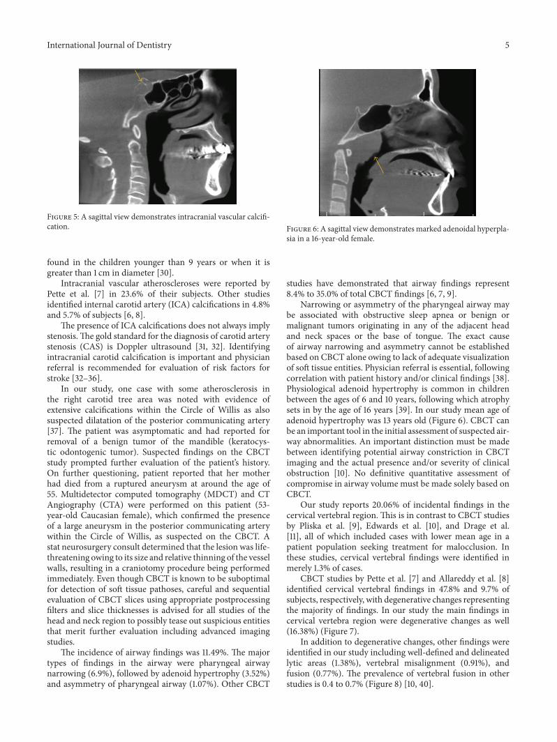
The next most common incidental finding was intracra-nial calcifications (26.96%), mostly in the pineal gland(11.18%), followed by intracranial atherosclerosis (10.57%)(Figures 4 and 5).
Intracranial physiological calcifications can be a commonincidental finding on studies in older patients being an age-related change [25–27]. Further evaluation is recommendedif the patient has other risk factors for cerebrovascularaccidents. Referral to the patient’s physician is advised foradditional imaging such as CTA (Computed TomographyAngiogram) for evaluation of intracranial atherosclerosis orother vascular anomalies.
The most common sites for physiologic calcificationsare pineal gland, habenula, choroid plexus, basal ganglia,falx, tentorium, petroclinoid ligaments, and sagittal sinus.Calcification of the pineal gland is seen in two-thirds of theadult population and increases with age [28].
Figure 4: Axial image shows pineal gland calcification in themidline.
Pineal gland calcificationwas identified in 19.2% of CBCTsubjects by Pette et al. [7] and in 13.1% by Admassie andMekonnen [29]. Physiologic calcification is asymptomaticand is detected incidentally on advanced imaging. They arealmost never clinically significant and often do not lead to anyclinical concern [7, 29]. Pineal gland calcifications are rare inchildren younger than 6 years of age.The possibility of pinealgland tumor should be considered when the calcification is
International Journal of Dentistry 5
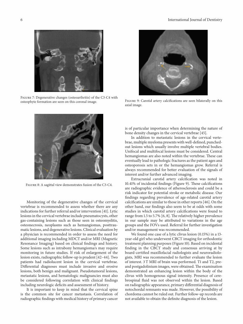
Figure 5: A sagittal view demonstrates intracranial vascular calcifi-cation.
found in the children younger than 9 years or when it isgreater than 1 cm in diameter [30].
Intracranial vascular atheroscleroses were reported byPette et al. [7] in 23.6% of their subjects. Other studiesidentified internal carotid artery (ICA) calcifications in 4.8%and 5.7% of subjects [6, 8].
The presence of ICA calcifications does not always implystenosis.The gold standard for the diagnosis of carotid arterystenosis (CAS) is Doppler ultrasound [31, 32]. Identifyingintracranial carotid calcification is important and physicianreferral is recommended for evaluation of risk factors forstroke [32–36].
In our study, one case with some atherosclerosis inthe right carotid tree area was noted with evidence ofextensive calcifications within the Circle of Willis as alsosuspected dilatation of the posterior communicating artery[37]. The patient was asymptomatic and had reported forremoval of a benign tumor of the mandible (keratocys-tic odontogenic tumor). Suspected findings on the CBCTstudy prompted further evaluation of the patient’s history.On further questioning, patient reported that her motherhad died from a ruptured aneurysm at around the age of55. Multidetector computed tomography (MDCT) and CTAngiography (CTA) were performed on this patient (53-year-old Caucasian female), which confirmed the presenceof a large aneurysm in the posterior communicating arterywithin the Circle of Willis, as suspected on the CBCT. Astat neurosurgery consult determined that the lesion was life-threatening owing to its size and relative thinning of the vesselwalls, resulting in a craniotomy procedure being performedimmediately. Even though CBCT is known to be suboptimalfor detection of soft tissue pathoses, careful and sequentialevaluation of CBCT slices using appropriate postprocessingfilters and slice thicknesses is advised for all studies of thehead and neck region to possibly tease out suspicious entitiesthat merit further evaluation including advanced imagingstudies.
The incidence of airway findings was 11.49%. The majortypes of findings in the airway were pharyngeal airwaynarrowing (6.9%), followed by adenoid hypertrophy (3.52%)and asymmetry of pharyngeal airway (1.07%). Other CBCT
Figure 6: A sagittal view demonstrates marked adenoidal hyperpla-sia in a 16-year-old female.
studies have demonstrated that airway findings represent8.4% to 35.0% of total CBCT findings [6, 7, 9].
Narrowing or asymmetry of the pharyngeal airway maybe associated with obstructive sleep apnea or benign ormalignant tumors originating in any of the adjacent headand neck spaces or the base of tongue. The exact causeof airway narrowing and asymmetry cannot be establishedbased on CBCT alone owing to lack of adequate visualizationof soft tissue entities. Physician referral is essential, followingcorrelation with patient history and/or clinical findings [38].Physiological adenoid hypertrophy is common in childrenbetween the ages of 6 and 10 years, following which atrophysets in by the age of 16 years [39]. In our study mean age ofadenoid hypertrophy was 13 years old (Figure 6). CBCT canbe an important tool in the initial assessment of suspected air-way abnormalities. An important distinction must be madebetween identifying potential airway constriction in CBCTimaging and the actual presence and/or severity of clinicalobstruction [10]. No definitive quantitative assessment ofcompromise in airway volume must be made solely based onCBCT.
Our study reports 20.06% of incidental findings in thecervical vertebral region. This is in contrast to CBCT studiesby Pliska et al. [9], Edwards et al. [10], and Drage et al.[11], all of which included cases with lower mean age in apatient population seeking treatment for malocclusion. Inthese studies, cervical vertebral findings were identified inmerely 1.3% of cases.
CBCT studies by Pette et al. [7] and Allareddy et al. [8]identified cervical vertebral findings in 47.8% and 9.7% ofsubjects, respectively, with degenerative changes representingthe majority of findings. In our study the main findings incervical vertebra region were degenerative changes as well(16.38%) (Figure 7).
In addition to degenerative changes, other findings wereidentified in our study including well-defined and delineatedlytic areas (1.38%), vertebral misalignment (0.91%), andfusion (0.77%). The prevalence of vertebral fusion in otherstudies is 0.4 to 0.7% (Figure 8) [10, 40].
6 International Journal of Dentistry
Figure 7: Degenerative changes (osteoarthritis) of the C3-C4 withosteophyte formation are seen on this coronal image.
Figure 8: A sagittal view demonstrates fusion of the C3-C4.
Monitoring of the degenerative changes of the cervicalvertebrae is recommended to assess whether there are anyindications for further referral and/or intervention [41]. Lyticlesions in the cervical vertebrae include pneumatocysts, othergas-containing lesions such as those seen in osteomyelitis,osteonecrosis, neoplasms such as hemangiomas, posttrau-matic lesions, and degenerative lesions. Clinical evaluation bya physician is recommended in order to assess the need foradditional imaging including MDCT and/or MRI (MagneticResonance Imaging) based on clinical findings and history.Some lesions such as intrabony hemangioma/s may requiremonitoring in future studies. If risk of enlargement of thelesion exists, radiographic follow-up is prudent [42–44]. Twopatients had radiolucent lesion in the cervical vertebrae.Differential diagnoses must include invasive and erosivelesions, both benign and malignant. Pseudotumoral lesions,metastatic lesions, and hematologic malignancies must alsobe considered following correlation with clinical findingsincluding neurologic deficits and assessment of history.
It is important to keep in mind that the cervical spineis the common site for cancer metastasis. Correlation ofradiographic findings withmedical history of primary cancer
Figure 9: Carotid artery calcifications are seen bilaterally on thisaxial image.
is of particular importance when determining the nature ofbone density changes in the cervical vertebrae [45].
In addition to metastatic lesions in the cervical verte-brae, multiplemyeloma presents with well-defined, punched-out lesions which usually involve multiple vertebral bodies.Unifocal and multifocal lesions must be considered. Centralhemangiomas are also noted within the vertebrae. These caneventually lead to pathologic fractures as the patient ages andosteoporosis sets in or the hemangiomas grow. Referral isalways recommended for better evaluation of the signals ofinterest and/or further advanced imaging.
Extracranial carotid artery calcification was noted in10.41% of incidental findings (Figure 9). These calcificationsare radiographic evidence of atherosclerosis and could be arisk indicator for potential stroke or metabolic disease. Ourfindings regarding prevalence of age-related carotid arterycalcifications are similar to those in other reports [46]. On theother hand, our findings also seem to be at odds with somestudies in which carotid artery calcifications were found torange from 1.5 to 5.7% [6, 8]. The relatively higher prevalencein our sample may be attributed to variations in the agegroups and the FOVs used. Referral for further investigationand/or management was recommended.
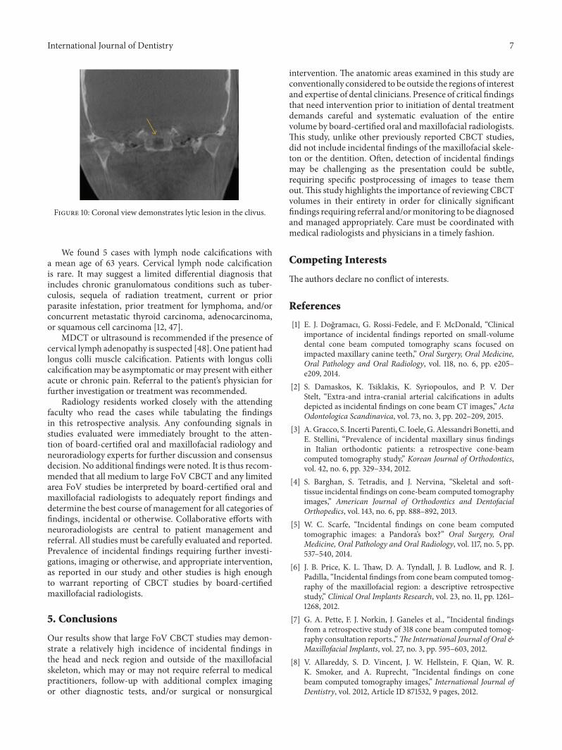
We found one case of a lytic clivus lesion (0.15%) in a 13-year-old girl who underwent CBCT imaging for orthodontictreatment planning purposes (Figure 10). Based on incidentalfinding in the CBCT study and consensus arriving at byboard-certified maxillofacial radiologists and neuroradiolo-gists, MRI was recommended to further evaluate the lesionof interest. 3 T MRI of brain was performed. T1 and T2, pre-and postgadolinium images, were obtained.The examinationdemonstrated an enhancing lesion within the body of theclivus with homogenous signal intensity. Presence of cere-brospinal fluid was not observed within the lesion. Basedon radiographic appearance, primary differential diagnosis ofnotochordal remnants was made. However, the possibility ofchordoma cannot be ruled out. Further follow-up records arenot available to obtain the definite diagnosis of the lesion.
International Journal of Dentistry 7
Figure 10: Coronal view demonstrates lytic lesion in the clivus.
We found 5 cases with lymph node calcifications witha mean age of 63 years. Cervical lymph node calcificationis rare. It may suggest a limited differential diagnosis thatincludes chronic granulomatous conditions such as tuber-culosis, sequela of radiation treatment, current or priorparasite infestation, prior treatment for lymphoma, and/orconcurrent metastatic thyroid carcinoma, adenocarcinoma,or squamous cell carcinoma [12, 47].
MDCT or ultrasound is recommended if the presence ofcervical lymph adenopathy is suspected [48]. One patient hadlongus colli muscle calcification. Patients with longus collicalcificationmay be asymptomatic ormay present with eitheracute or chronic pain. Referral to the patient’s physician forfurther investigation or treatment was recommended.
Radiology residents worked closely with the attendingfaculty who read the cases while tabulating the findingsin this retrospective analysis. Any confounding signals instudies evaluated were immediately brought to the atten-tion of board-certified oral and maxillofacial radiology andneuroradiology experts for further discussion and consensusdecision. No additional findings were noted. It is thus recom-mended that all medium to large FoV CBCT and any limitedarea FoV studies be interpreted by board-certified oral andmaxillofacial radiologists to adequately report findings anddetermine the best course of management for all categories offindings, incidental or otherwise. Collaborative efforts withneuroradiologists are central to patient management andreferral. All studies must be carefully evaluated and reported.Prevalence of incidental findings requiring further investi-gations, imaging or otherwise, and appropriate intervention,as reported in our study and other studies is high enoughto warrant reporting of CBCT studies by board-certifiedmaxillofacial radiologists.
5. Conclusions
Our results show that large FoV CBCT studies may demon-strate a relatively high incidence of incidental findings inthe head and neck region and outside of the maxillofacialskeleton, which may or may not require referral to medicalpractitioners, follow-up with additional complex imagingor other diagnostic tests, and/or surgical or nonsurgical
intervention. The anatomic areas examined in this study areconventionally considered to be outside the regions of interestand expertise of dental clinicians. Presence of critical findingsthat need intervention prior to initiation of dental treatmentdemands careful and systematic evaluation of the entirevolume by board-certified oral andmaxillofacial radiologists.This study, unlike other previously reported CBCT studies,did not include incidental findings of the maxillofacial skele-ton or the dentition. Often, detection of incidental findingsmay be challenging as the presentation could be subtle,requiring specific postprocessing of images to tease themout.This study highlights the importance of reviewing CBCTvolumes in their entirety in order for clinically significantfindings requiring referral and/ormonitoring to be diagnosedand managed appropriately. Care must be coordinated withmedical radiologists and physicians in a timely fashion.
Competing Interests
The authors declare no conflict of interests.
References
[1] E. J. Dogramacı, G. Rossi-Fedele, and F. McDonald, “Clinicalimportance of incidental findings reported on small-volumedental cone beam computed tomography scans focused onimpacted maxillary canine teeth,” Oral Surgery, Oral Medicine,Oral Pathology and Oral Radiology, vol. 118, no. 6, pp. e205–e209, 2014.
[2] S. Damaskos, K. Tsiklakis, K. Syriopoulos, and P. V. DerStelt, “Extra-and intra-cranial arterial calcifications in adultsdepicted as incidental findings on cone beam CT images,” ActaOdontologica Scandinavica, vol. 73, no. 3, pp. 202–209, 2015.
[3] A.Gracco, S. Incerti Parenti, C. Ioele, G. Alessandri Bonetti, andE. Stellini, “Prevalence of incidental maxillary sinus findingsin Italian orthodontic patients: a retrospective cone-beamcomputed tomography study,” Korean Journal of Orthodontics,vol. 42, no. 6, pp. 329–334, 2012.
[4] S. Barghan, S. Tetradis, and J. Nervina, “Skeletal and soft-tissue incidental findings on cone-beam computed tomographyimages,” American Journal of Orthodontics and DentofacialOrthopedics, vol. 143, no. 6, pp. 888–892, 2013.
[5] W. C. Scarfe, “Incidental findings on cone beam computedtomographic images: a Pandora’s box?” Oral Surgery, OralMedicine, Oral Pathology and Oral Radiology, vol. 117, no. 5, pp.537–540, 2014.
[6] J. B. Price, K. L. Thaw, D. A. Tyndall, J. B. Ludlow, and R. J.Padilla, “Incidental findings from cone beam computed tomog-raphy of the maxillofacial region: a descriptive retrospectivestudy,” Clinical Oral Implants Research, vol. 23, no. 11, pp. 1261–1268, 2012.
[7] G. A. Pette, F. J. Norkin, J. Ganeles et al., “Incidental findingsfrom a retrospective study of 318 cone beam computed tomog-raphy consultation reports.,”The International Journal of Oral &Maxillofacial Implants, vol. 27, no. 3, pp. 595–603, 2012.
[8] V. Allareddy, S. D. Vincent, J. W. Hellstein, F. Qian, W. R.K. Smoker, and A. Ruprecht, “Incidental findings on conebeam computed tomography images,” International Journal ofDentistry, vol. 2012, Article ID 871532, 9 pages, 2012.
8 International Journal of Dentistry
[9] B. Pliska, M. DeRocher, and B. E. Larson, “Incidence ofsignificant findings on CBCT scans of an orthodontic patientpopulation,” Northwest Dentistry, vol. 90, no. 2, pp. 12–16, 2011.
[10] R. Edwards, N. Alsufyani, G. Heo, and C. Flores-Mir, “Thefrequency and nature of incidental findings in large-field conebeam computed tomography scans of an orthodontic sample,”Progress in orthodontics, vol. 15, article 37, 2014.
[11] N. Drage, S. Rogers, C. Greenall, and R. Playle, “Incidentalfindings on cone beam computed tomography in orthodonticpatients,” Journal of Orthodontics, vol. 40, no. 1, pp. 29–37, 2013.
[12] Z. A. Newaz, S. Barghan, R. A. Katkar, J. A. Bennett, andM. K. Nair, “Incidental findings of skull-base abnormalities incone-beam computed tomography scans with consultation bymaxillofacial radiologists,” American Journal of Orthodonticsand Dentofacial Orthopedics, vol. 147, no. 1, pp. 127–131, 2015.
[13] R. Barboza, J. H. Fox, L. E. T. Shaffer, J. M. Opalek, and S.Farooki, “Incidental findings in the cervical spine at CT fortrauma evaluation,”American Journal of Roentgenology, vol. 192,no. 3, pp. 725–729, 2009.
[14] T. Ergun and H. Lakadamyali, “The prevalence and clinicalimportance of incidental soft-tissue findings in cervical CTscans of trauma population,” Dentomaxillofacial Radiology, vol.42, no. 10, Article ID 20130216, 2013.
[15] H. Popat, N. Drage, and P. Durning, “Mid-line clefts of thecervical vertebrae—an incidental finding arising from conebeam computed tomography of the dental patient,” BritishDental Journal, vol. 204, no. 6, pp. 303–306, 2008.
[16] B. Cakur, M. A. Sumbullu, S. Dagistan, and D. Durna, “Theimportance of cone beam CT in the radiological detection ofosteomalacia,” Dentomaxillofacial Radiology, vol. 41, no. 1, pp.84–88, 2012.
[17] V. Soerdjbalie-Maikoe and R. R. van Rijn, “Embryology, normalanatomy, and imaging techniques of the hyoid and larynx withrespect to forensic purposes: a review article,” Forensic Science,Medicine, and Pathology, vol. 4, no. 2, pp. 132–139, 2008.
[18] M. Ahmad, R. Madden, and L. Perez, “Triticeous cartilage:prevalence on panoramic radiographs and diagnostic criteria,”Oral Surgery, OralMedicine, Oral Pathology, Oral Radiology andEndodontology, vol. 99, no. 2, pp. 225–230, 2005.
[19] E. Alqahtani, D. E. Marrero, W. L. Champion, A. Alawaji,P. D. Kousoubris, and J. E. Small, “Triticeous cartilage CTimaging characteristics, prevalence, extent, and distribution ofossification,” Otolaryngology—Head and Neck Surgery, vol. 154,no. 1, pp. 131–137, 2016.
[20] F. Aspestrand and A. Kolbenstvedt, “Calcifications of the pala-tine tonsillary region: CT demonstration,” Radiology, vol. 165,no. 2, pp. 479–480, 1987.
[21] M.-A. Fauroux, C. Mas, P. Tramini, and J.-H. Torres, “Preva-lence of palatine tonsilloliths: a retrospective study on 150consecutive CT examinations,” Dentomaxillofacial Radiology,vol. 42, no. 7, Article ID 20120429, 2013.
[22] A. C. C. Dal Rio, E. M. D. Nicola, and A. R. F. Teixeira,“Halitosis—an assessment protocol proposal,” Brazilian Journalof Otorhinolaryngology, vol. 73, no. 6, pp. 835–842, 2007.
[23] M. P. Caldas, E. G. Neves, F. R. Manzi, S. M. de Almeida, F. N.Boscolo, and F.Haiter-Neto, “Tonsillolith—report of an unusualcase,” British Dental Journal, vol. 202, no. 5, pp. 265–267, 2007.
[24] T. Ansai and T. Takehara, “Tonsillolith as a halitosis-inducingfactor,” British Dental Journal, vol. 198, no. 5, pp. 263–264, 2005.
[25] M. H. Daghighi, V. Rezaei, S. Zarrintan, and H. Pourfathi,“Intracranial physiological calcifications in adults on computed
tomography in Tabriz, Iran,” Folia Morphologica, vol. 66, no. 2,pp. 115–119, 2007.
[26] A. T. Turgut, H. M. Karakas, Y. Ozsunar et al., “Age-relatedchanges in the incidence of pineal gland calcification in Turkey:a prospectivemulticenterCT study,”Pathophysiology, vol. 15, no.1, pp. 41–48, 2008.
[27] P. P. Sedghizadeh, M. Nguyen, and R. Enciso, “Intracranialphysiological calcifications evaluated with cone beam CT,”Dentomaxillofacial Radiology, vol. 41, no. 8, pp. 675–678, 2012.
[28] Y. Kıroglu, C. Callı, N. Karabulut, and C. Oncel, “Intracranialcalcifications on CT,” Diagnostic and Interventional Radiology,vol. 16, no. 4, pp. 263–269, 2010.
[29] D. Admassie and A. Mekonnen, “Incidence of normal pinealand chroids plexus calcification on brain CT (computerizedtomography) at Tikur Anbessa Teaching Hospital Addis Ababa,Ethiopia,” Ethiopian Medical Journal, vol. 47, no. 1, pp. 55–60,2009.
[30] S. Deepak, B. Jayakumar, and Shanavas, “Extensive intracranialcalcification,” Journal of Association of Physicians of India, vol.53, article 948, 2005.
[31] D. M. Almog, T. Horev, K. A. Illig, R. M. Green, and L.C. Carter, “Correlating carotid artery stenosis detected bypanoramic radiography with clinically relevant carotid arterystenosis determined by duplex ultrasound,” Oral Surgery, OralMedicine, Oral Pathology, Oral Radiology, and Endodontics, vol.94, no. 6, pp. 768–773, 2002.
[32] D. Bos, M. J. M. van der Rijk, T. E. A. Geeraedts et al.,“Intracranial carotid artery atherosclerosis: prevalence and riskfactors in the general population,” Stroke, vol. 43, no. 7, pp. 1878–1884, 2012.
[33] A. H. Friedlander, D. S. Liebeskind, H. Q. Tran, and S. M.Mallya, “What are the potential implications of identifyingintracranial internal carotid artery atherosclerotic lesions oncone-beam computed tomography? A systematic review andillustrative case studies,” Journal of Oral and MaxillofacialSurgery, vol. 72, no. 11, pp. 2167–2177, 2014.
[34] D. Bos, M. L. P. Portegies, A. van der Lugt et al., “Intracranialcarotid artery atherosclerosis and the risk of stroke in whites:the Rotterdam Study,” JAMA Neurology, vol. 71, no. 4, pp. 405–411, 2014.
[35] J. F. Arenillas, “Intracranial atherosclerosis: current concepts,”Stroke, vol. 42, supplement 1, pp. S20–S23, 2011.
[36] J.-M. Bugnicourt, C. Leclercq, J.-M. Chillon et al., “Presence ofintracranial artery calcification is associated with mortality andvascular events in patients with ischemic stroke after hospitaldischarge: a cohort study,” Stroke, vol. 42, no. 12, pp. 3447–3453,2011.
[37] M. K. Nair, J. C. Pettigrew Jr., and A. A. Mancuso, “Intracranialaneurysm as an incidental finding,” Dentomaxillofacial Radiol-ogy, vol. 36, no. 2, pp. 107–112, 2007.
[38] S. P. Patil, H. Schneider, A. R. Schwartz, and P. L. Smith, “Adultobstructive sleep apnea: pathophysiology and diagnosis,” Chest,vol. 132, no. 1, pp. 325–337, 2007.
[39] N. Yildirim,M. Sahan, and Y. Karslioglu, “Adenoid hypertrophyin adults: clinical and morphological characteristics,” Journal ofInternational Medical Research, vol. 36, no. 1, pp. 157–162, 2008.
[40] D. Bebnowski, M. P. Hanggi, G. Markic, M. Roos, and T.Peltomaki, “Cervical vertebrae anomalies in subjects with classII malocclusion assessed by lateral cephalogram and cone beamcomputed tomography,” European Journal of Orthodontics, vol.34, no. 2, pp. 226–231, 2012.
International Journal of Dentistry 9
[41] P. Soni, V. Sharma, and J. Sengupta, “Cervical vertebraeanomalies—incidental findings on lateral cephalograms,” AngleOrthodontist, vol. 78, no. 1, pp. 176–180, 2008.
[42] T. Nakayama, S. Ehara, and H. Hama, “Spontaneous progres-sion of vertebral intraosseous pneumatocysts to fluid-filledcysts,” Skeletal Radiology, vol. 30, no. 9, pp. 523–526, 2001.
[43] L. Laufer, H. Schulman, and Y. Hertzanu, “Vertebral pneumato-cyst. A case report,” Spine, vol. 21, no. 3, pp. 389–391, 1996.
[44] M. A. Husain, S. Tetradis, and S. M. Mallya, “Intraosseouspneumatocysts of the cervical spine: a report of four cases andreviewof literature,”Oral Surgery, OralMedicine, Oral Pathologyand Oral Radiology, vol. 119, no. 1, pp. e49–e54, 2015.
[45] L. G. Jenis, E. J. Dunn, and H. S. An, “Metastatic disease ofthe cervical spine. A review,” Clinical Orthopaedics and RelatedResearch, no. 359, pp. 89–103, 1999.
[46] A. B. Wells, Incidence of soft tissue calcifications of the head andneck region on maxillofacial cone beam computed tomography[M.S. thesis], University of Louisville, 2011.
[47] B. L. Eisenkraft and P. M. Som, “The spectrum of benign andmalignant etiologies of cervical node calcification,” AmericanJournal of Roentgenology, vol. 172, no. 5, pp. 1433–1437, 1999.
[48] S. Jank, P. Robatscher, R. Emshoff, H. Strobl, G. Gojer, andB. Norer, “The diagnostic value of ultrasonography to detectoccult lymph node involvement at different levels in patientswith squamous cell carcinoma in the maxillofacial region,”International Journal of Oral and Maxillofacial Surgery, vol. 32,no. 1, pp. 39–42, 2003.

Submit your manuscripts athttp://www.hindawi.com
Hindawi Publishing Corporationhttp://www.hindawi.com Volume 2014
Oral OncologyJournal of
DentistryInternational Journal of
Hindawi Publishing Corporationhttp://www.hindawi.com Volume 2014
Hindawi Publishing Corporationhttp://www.hindawi.com Volume 2014
International Journal of
Biomaterials
Hindawi Publishing Corporationhttp://www.hindawi.com Volume 2014
BioMed Research International
Hindawi Publishing Corporationhttp://www.hindawi.com Volume 2014
Case Reports in Dentistry
Hindawi Publishing Corporationhttp://www.hindawi.com Volume 2014
Oral ImplantsJournal of
Hindawi Publishing Corporationhttp://www.hindawi.com Volume 2014
Anesthesiology Research and Practice
Hindawi Publishing Corporationhttp://www.hindawi.com Volume 2014
Radiology Research and Practice
Environmental and Public Health
Journal of
Hindawi Publishing Corporationhttp://www.hindawi.com Volume 2014
The Scientific World JournalHindawi Publishing Corporation http://www.hindawi.com Volume 2014
Hindawi Publishing Corporationhttp://www.hindawi.com Volume 2014
Dental SurgeryJournal of
Drug DeliveryJournal of
Hindawi Publishing Corporationhttp://www.hindawi.com Volume 2014
Hindawi Publishing Corporationhttp://www.hindawi.com Volume 2014
Oral DiseasesJournal of
Hindawi Publishing Corporationhttp://www.hindawi.com Volume 2014
Computational and Mathematical Methods in Medicine
ScientificaHindawi Publishing Corporationhttp://www.hindawi.com Volume 2014
PainResearch and TreatmentHindawi Publishing Corporationhttp://www.hindawi.com Volume 2014
Preventive MedicineAdvances in
Hindawi Publishing Corporationhttp://www.hindawi.com Volume 2014
EndocrinologyInternational Journal of
Hindawi Publishing Corporationhttp://www.hindawi.com Volume 2014
Hindawi Publishing Corporationhttp://www.hindawi.com Volume 2014
OrthopedicsAdvances in


![Fundamentals of cone beam computed tomography for a ...Cone beam computed tomography (CBCT, also referred to as C-arm computed tomography [CT], cone beam volume CT, or flat panel CT)](https://img.pdfslide.us/doc/110x75/611ad245d6c77f53c63c9117/fundamentals-of-cone-beam-computed-tomography-for-a-cone-beam-computed-tomography.jpg)