Embed Size (px)
Citation preview
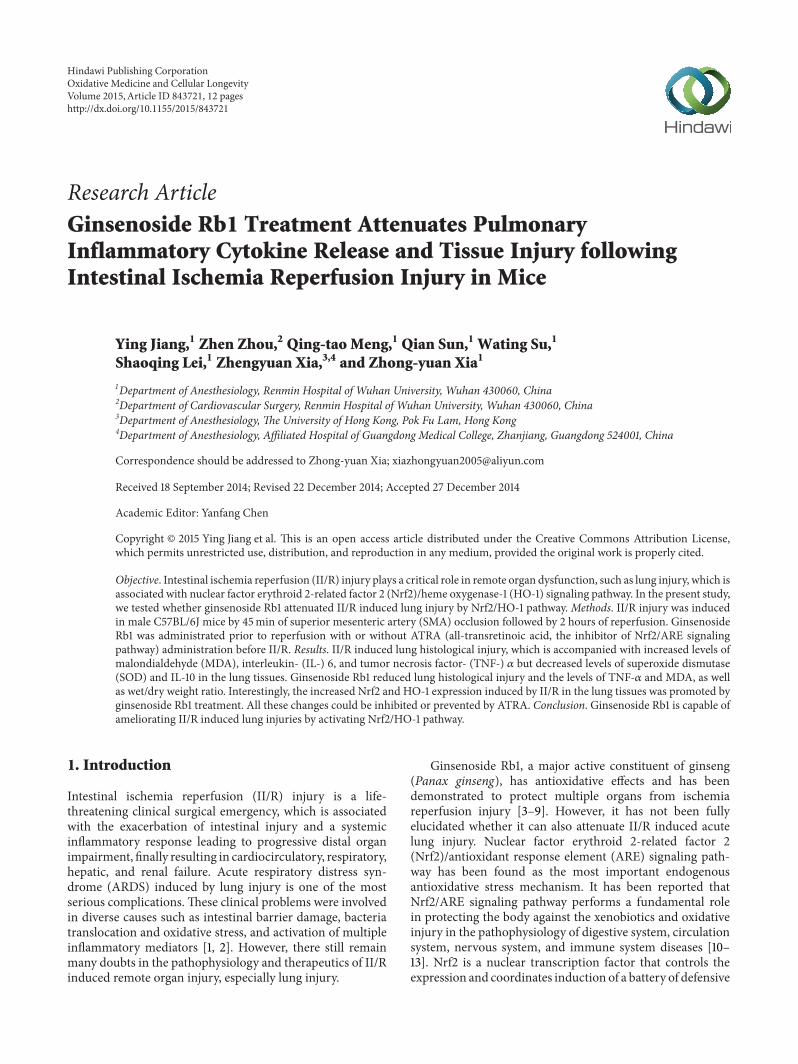
Research ArticleGinsenoside Rb1 Treatment Attenuates PulmonaryInflammatory Cytokine Release and Tissue Injury followingIntestinal Ischemia Reperfusion Injury in Mice
Ying Jiang,1 Zhen Zhou,2 Qing-tao Meng,1 Qian Sun,1 Wating Su,1
Shaoqing Lei,1 Zhengyuan Xia,3,4 and Zhong-yuan Xia1
1Department of Anesthesiology, Renmin Hospital of Wuhan University, Wuhan 430060, China2Department of Cardiovascular Surgery, Renmin Hospital of Wuhan University, Wuhan 430060, China3Department of Anesthesiology, The University of Hong Kong, Pok Fu Lam, Hong Kong4Department of Anesthesiology, Affiliated Hospital of Guangdong Medical College, Zhanjiang, Guangdong 524001, China
Correspondence should be addressed to Zhong-yuan Xia; [email protected]
Received 18 September 2014; Revised 22 December 2014; Accepted 27 December 2014
Academic Editor: Yanfang Chen
Copyright © 2015 Ying Jiang et al. This is an open access article distributed under the Creative Commons Attribution License,which permits unrestricted use, distribution, and reproduction in any medium, provided the original work is properly cited.
Objective. Intestinal ischemia reperfusion (II/R) injury plays a critical role in remote organ dysfunction, such as lung injury, which isassociated with nuclear factor erythroid 2-related factor 2 (Nrf2)/heme oxygenase-1 (HO-1) signaling pathway. In the present study,we tested whether ginsenoside Rb1 attenuated II/R induced lung injury by Nrf2/HO-1 pathway.Methods. II/R injury was inducedin male C57BL/6J mice by 45min of superior mesenteric artery (SMA) occlusion followed by 2 hours of reperfusion. GinsenosideRb1 was administrated prior to reperfusion with or without ATRA (all-transretinoic acid, the inhibitor of Nrf2/ARE signalingpathway) administration before II/R. Results. II/R induced lung histological injury, which is accompanied with increased levels ofmalondialdehyde (MDA), interleukin- (IL-) 6, and tumor necrosis factor- (TNF-) 𝛼 but decreased levels of superoxide dismutase(SOD) and IL-10 in the lung tissues. Ginsenoside Rb1 reduced lung histological injury and the levels of TNF-𝛼 and MDA, as wellas wet/dry weight ratio. Interestingly, the increased Nrf2 and HO-1 expression induced by II/R in the lung tissues was promoted byginsenoside Rb1 treatment. All these changes could be inhibited or prevented by ATRA. Conclusion. Ginsenoside Rb1 is capable ofameliorating II/R induced lung injuries by activating Nrf2/HO-1 pathway.
1. Introduction
Intestinal ischemia reperfusion (II/R) injury is a life-threatening clinical surgical emergency, which is associatedwith the exacerbation of intestinal injury and a systemicinflammatory response leading to progressive distal organimpairment, finally resulting in cardiocirculatory, respiratory,hepatic, and renal failure. Acute respiratory distress syn-drome (ARDS) induced by lung injury is one of the mostserious complications.These clinical problems were involvedin diverse causes such as intestinal barrier damage, bacteriatranslocation and oxidative stress, and activation of multipleinflammatory mediators [1, 2]. However, there still remainmany doubts in the pathophysiology and therapeutics of II/Rinduced remote organ injury, especially lung injury.
Ginsenoside Rb1, a major active constituent of ginseng(Panax ginseng), has antioxidative effects and has beendemonstrated to protect multiple organs from ischemiareperfusion injury [3–9]. However, it has not been fullyelucidated whether it can also attenuate II/R induced acutelung injury. Nuclear factor erythroid 2-related factor 2(Nrf2)/antioxidant response element (ARE) signaling path-way has been found as the most important endogenousantioxidative stress mechanism. It has been reported thatNrf2/ARE signaling pathway performs a fundamental rolein protecting the body against the xenobiotics and oxidativeinjury in the pathophysiology of digestive system, circulationsystem, nervous system, and immune system diseases [10–13]. Nrf2 is a nuclear transcription factor that controls theexpression and coordinates induction of a battery of defensive
Hindawi Publishing CorporationOxidative Medicine and Cellular LongevityVolume 2015, Article ID 843721, 12 pageshttp://dx.doi.org/10.1155/2015/843721
2 Oxidative Medicine and Cellular Longevity
genes encoding detoxifying enzymes and antioxidant pro-teins [14]. In response to stimulation of oxidative stress, Nrf2translocates from the cytoplasm into the nucleus and thenbinds to a cis-acting enhancer sequence designated as AREand regulates AREmediated antioxidant enzyme gene such asheme oxygenase-1 (HO-1) expression and induction [15, 16].HO-1 belongs to a member of the heat shock protein familyand plays a significant protective role against inflammatoryprocesses and oxidative tissue injury [17].
In this study, we established a superior mesenteric artery(SMA) occlusion/reperfusion mice model to induce lunginjury. We used ATRA (all-transretinoic acid) as inhibitorof Nrf2/ARE signaling pathway, which interfered in therecruitment ofNrf2 to theARE, thus disrupting the activationof ARE-driven genes [18]. With the treatment of ginsenosideRb1, we aim to investigatewhether ginsenoside Rb1 attenuatesacute lung injury (ALI) induced by II/R inmice viaNrf2/AREpathway.
2. Material and Methods
2.1. Mice. The current study was approved by the Ani-mal Care Committee of Wuhan University, China, andwas performed in accordance with National Institutes ofHealth guidelines for the use of experimental animals. MaleC57BL/6mice (9–12 weeks old; 17–22 g) were purchased fromHUNAN SLAC JD Laboratory Animal Co. Ltd., China. Theywere housed under standard laboratory conditions at 22–24∘C, relative humidity of 50 ± 15%, and kept on a 12 hday/night rhythm with free access to water and food. Allexperimental protocols conducted in the mice were carriedout in accordance with the Guide for the Care and Use ofLaboratoryAnimals by theNational Institutes ofHealth (NIHPublication number 80-23).
2.2. Surgical Preparation. Animals were anesthetized intra-peritoneally with pentobarbital sodium (50mg/kg bodyweight). Amidline laparotomywas performed; then the supe-rior mesenteric artery (SMA) was isolated. The II/R injurywas established by occluding SMA with a microvascularclip for 45 minutes followed by 2 hours of reperfusion aspreviously described [19]. Ischemia was recognized by theexistence of pulseless or pale color of the small intestine. Thereturn of pulses and the reestablishment of the pink colorwere assumed to indicate valid reperfusion of the intestine.The Sham group underwent the same surgical process, apartfrom occlusion of SMA. After 2 h reperfusion, the mice werekilled. A median sternotomy was performed; the lung andintestine samples were obtained for further analysis.
2.3. Experimental Protocol. The mice were randomly allo-cated into eight groups (𝑛 = 8 in each group) (Figure 1):(1) Sham surgical preparation including isolation of the SMAwithout occlusion was performed (Sham); (2) mice weresubjected to II/R without treatment (II/R); (3) mice weresubjected to II/R with treatment of normal saline 10 minutesbefore reperfusion (II/R + NS); (4), (5) mice were treated
IschemiaReperfusion
Normal saline
Rb1 (30mg/kg)
Rb1 (60mg/kg)
Rb1 (60mg/kg)
ATRA i.p. 2 weeks
ATRA i.p. 2 weeks
ATRA i.p. 2 weeks
(1) Sham
(2) II/R
(3) II/R + NS
(4) II/R + Rb1-30
(5) II/R + Rb1-60
(6) ATRA + sham
(7) ATRA + II/R
(8) ATRA + II/R + Rb-60
45 min min120
45 min min120
45 min min120
45 min min120
45 min min120
45 min min120
45 min min120
45 min min120
Figure 1: Experimental protocols. Mice were subjected to 45minof SMA occlusion followed by 2 h of reperfusion. II/R: intestinalischemia/reperfusion, NS: normal saline, Rb1: ginsenoside Rb1, andATRA: all-transretinoic acid.
with 30mg/kg (II/R + Rb1-30) or 60mg/kg (II/R + Rb1-60) ginsenoside Rb1, in which surgery was performed asin the II/R group with administration of the ginsenosideRb1 intraperitoneally 10 minutes before reperfusion; (6) micewere subjected to Sham surgery and treated with ATRA(ATRA+Sham), which is the inhibitor ofNrf2/ARE signalingpathway; (7) mice were subjected to II/R and treated withATRA (ATRA + II/R); (8) mice were subjected to II/R andtreated with ATRA and 60mg/kg ginsenoside Rb1 as group5 (ATRA + II/R + Rb1-60). During the last two weeks beforethe operation, themice in the group 6, 7, 8 received ATRA i.p.daily at 10mg/kg and fed on a vitamin A-deficient diet, andthe mice in the other groups received the equivalent volumeof corn oil and fed on a control normal diet [18].
2.4. Lung Histology. The left lung was removed and fixed in10% formalin. Following embedding in paraffin, the sectionsof 4 𝜇m were stained with hematoxylin and eosin for lightmicroscopy. Semiquantitative analysis of lung histopathologywas performed by scoring the tissues based on lung edema,infiltration of inflammatory cells, alveolar hemorrhage, hya-line membrane, and atelectasis: no lesion, 0; injured area ⩽25%, 1; injured area 26–50%, 2; injured area 51–70%, 3; injuredarea 71–90%, 4; injured area > 90%, 5. A total of three fieldswere randomly selected for each slide and the average wasused as the histopathology score [20].
2.5. Histopathological Assessment of Intestines. After reperfu-sion, 1 cm of small intestine without adipose tissue was takenfrom the same place at the distal end of ileum and fixed in4% formaldehyde. After embedding in paraffin, 4𝜇msectionswere stained with hematoxylin and eosin before assessmentby light microscopy (original magnification ×200, OlympusBX50; Olympus Optical, Tokyo, Japan).
Oxidative Medicine and Cellular Longevity 3
(a) (b) (c) (d)
(e) (f) (g) (h)
Sham II/R
II/R
+N
S
II/R
+Rb
1-30
II/R
+Rb
1-60
ATRA
+sh
am
ATRA
+II
/R
ATRA
+II
/R+
Rb-60
6
4
2
0
Chiu
’s sc
ore
∗ ∗
∗# ∗#
∗$ ∗$
(i)
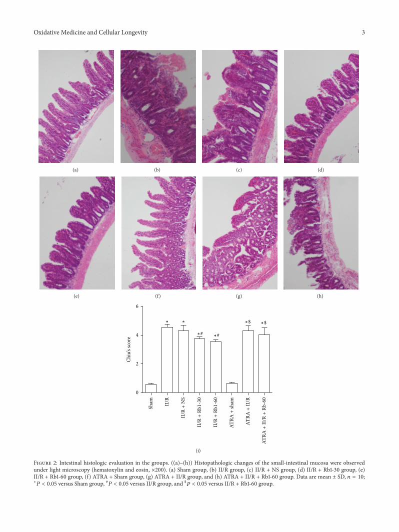
Figure 2: Intestinal histologic evaluation in the groups. ((a)–(h)) Histopathologic changes of the small-intestinal mucosa were observedunder light microscopy (hematoxylin and eosin, ×200). (a) Sham group, (b) II/R group, (c) II/R + NS group, (d) II/R + Rb1-30 group, (e)II/R + Rb1-60 group, (f) ATRA + Sham group, (g) ATRA + II/R group, and (h) ATRA + II/R + Rb1-60 group. Data are mean ± SD, 𝑛 = 10;∗
𝑃 < 0.05 versus Sham group, #𝑃 < 0.05 versus II/R group, and $𝑃 < 0.05 versus II/R + Rb1-60 group.

4 Oxidative Medicine and Cellular Longevity
(a) (b) (c)
(d) (e) (f)
(g) (h)
Sham II/R
II/R
+N
S
II/R
+Rb
1-30
II/R
+Rb
1-60
ATRA
+sh
am
ATRA
+II
/R
ATRA
+II
/R+
Rb-60
∗∗
∗#∗#
∗$ ∗$
Lung
inju
ry sc
ore
4
3
2
1
0
(i)
Figure 3: Histopathologic changes in mice lung under light microscopy (hematoxylin and eosin, ×200). (a) Sham group, (b) II/R group, (c)II/R + NS group, (d) II/R + Rb1-30 group, (e) II/R + Rb1-60 group, (f) ATRA + Sham group, (g) ATRA + II/R group, and (h) ATRA + II/R +Rb1-60 group. Data are mean ± SD, 𝑛 = 10; ∗𝑃 < 0.05 versus Sham group, #𝑃 < 0.05 versus II/R group, and $
𝑃 < 0.05 versus II/R + Rb1-60group.

Oxidative Medicine and Cellular Longevity 5
Sham II/R
II/R
+N
S
II/R
+Rb
1-30
II/R
+Rb
1-60
ATRA
+sh
am
ATRA
+II
/R
ATRA
+II
/R+
Rb-60
∗ ∗
∗#∗#
∗$ ∗$
Lung
wet
/dry
ratio
8
6
4
2
0
Figure 4:The effects of ginsenoside Rb1 on the lung wet/dry weightratio. Data are mean ± SD, 𝑛 = 5; ∗𝑃 < 0.05 versus Sham group,#𝑃 < 0.05 versus II/R group, and $
𝑃 < 0.05 versus II/R + Rb1-60group.
Using the improved Chiu score method [21] to evaluateintestinal mucosal damage, higher scores are interpreted toindicatemore severe damage. Criteria of Chiu grading systemconsist of 5 subdivisions according to the changes of villusand gland of intestinal mucosa: grade 0, normal mucosa;grade 1, development of subepithelial Gruenhagen’s space atthe tip of villus; grade 2, extension of the space withmoderateepithelial lifting; grade 3, massive epithelial lifting with a fewdenuded villi; grade 4, denuded villi with exposed capillaries;and grade 5, disintegration of the lamina propria, ulceration,and hemorrhage.
2.6. Assessment of Pulmonary Edema. The left lung washarvested. After the lung wet weight was measured, thelungs were placed in a calorstat at 60∘C for 48 h, and thenthe specimen was reweighed. The pulmonary edema wasestimated by lung wet/dry weight ratio [22].
2.7. Immunohistochemical Assessment. Paraffin-embeddedlung sections were stained using the streptavidin-biotincomplex immunohistochemistry technique for HO-1 andNrf2 detection. Brown staining in the cytoplasm and/ornucleus was considered an indicator of positive expression.With the Image-Pro Plus version 6.0, results were evaluatedsemiquantitatively according to optical density values ofpositive expression.
2.8. Western Blot Analysis. The right lungs were removedand nuclear fractions were prepared using a Nuclear andCytoplasmic Protein Extraction Kit (Beyotime Institute ofBiotechnology, China) according to the manufacturer’s pro-tocol. Western blot analysis was carried out as described [23].Primary antibodies (working concentration) usedwere rabbitpolyclonal antibodies against mice Nrf2 (1 : 2000, H-300,
Santa Cruz Biotechnology, CA), HO-1 (1 : 1000, H-105, SantaCruzBiotechnology, CA), andLaminB1 (1 : 2000,H-90, SantaCruz Biotechnology, CA). The HRP-conjugated secondaryantibody was goat anti-rabbit IgG (Beyotime Institute ofBiotechnology, China) used at 1 : 2000. Lamin B was usedas an internal control. The ECL Western blotting detectionreagents (Beyotime Institute of Biotechnology, China) wereused for visualization of the protein bands. The intensities ofthe bandswere analyzedwith quantity one software (Bio-Rad,Hercules, CA).
2.9. Determination of Tissue Tumor Necrosis Factor Alpha(TNF-𝛼), Interlukin-6 (IL-10), and Interlukin-6 (IL-6). Tissuelevels of TNF-𝛼, IL-10, and IL-6 were determined usingcommercially available ELISA kits (R&D, Minneapolis, MN)according to manufacturer’s instructions. The results wereexpressed as pg/mL.
2.10. Determination of Tissue MDA Level and SOD Activity.The right lung tissues were homogenized on ice in normalsaline. The homogenates were centrifuged at 4000 g⋅min−1at 4∘C for 10min. The MDA level in the supernatantswas determined by the measurement of thiobarbituric acid-reactive substances levels (assay kits were supplied byNanjingJiancheng Corp., China) as previously described [24]. Theresults were calculated as nmol⋅100mg−1 protein. The SODactivity in the supernatants was evaluated by inhibition ofnitroblue tetrazolium (NBT) reduction by O2− generatedby the xanthine/xanthine oxidase system (assay kits weresupplied by Nanjing Jiancheng Corp., China) in accordancewith the previous method [24].The results were expressed byU⋅100mg−1 protein.
2.11. Statistical Analysis. Data are presented as mean ± SD.Statistical comparison amongmultiple groupswas performedby one-way analysis of variance (ANOVA) followed byNewman-Keulsmultiple comparison test using theGraphPadPrism 5.0 software (GraphPad Software Inc., San Diego, CA,USA). A value of 𝑃 < 0.05 was considered statisticallysignificant.
3. Results
3.1. Histopathological Assessment of Intestines. The II/R groupshowed edema in the villi, inflammatory cells infiltration, anddamaged areas interspersed with hemorrhage. In addition,the gap between epithelial cells significantly increased andcapillaries and lymph vessels were markedly dilated. Normalvilli were seen in the intestine of the Sham group andATRA+Sham group under the light microscope. Ginsenoside Rb1 atthe dose of 30mg/kg and 60mg/kg both significantly atten-uated the histological intestine injury (Figure 2). However,there is little amelioration of the intestine injury induced byII/R in the ATRA + II/R + Rb1-60 group.
3.2. Pathologic Alternations of Lung Tissue. The lungs fromII/R group showed damaged areas interspersed with hemor-rhage, inflammatory cell infiltration, and pulmonary edema,

6 Oxidative Medicine and Cellular Longevity
Sham II/R
II/R
+N
S
II/R
+Rb
1-30
II/R
+Rb
1-60
ATRA
+sh
am
ATRA
+II
/R
ATRA
+II
/R+
Rb-60
∗ ∗
∗#∗#
∗$ ∗$
SOD
(U/m
g pr
ot)
150
100
50
0
(a)
MD
A (n
mol
/mg
prot
)
Sham II/R
II/R
+N
S
II/R
+Rb
1-30
II/R
+Rb
1-60
ATRA
+sh
am
ATRA
+II
/R
ATRA
+II
/R+
Rb-60
∗∗
∗#∗#
∗$ ∗$
2.5
2.0
1.5
1.0
0.5
0.0
(b)
Figure 5: The changes of lung tissue SOD activity and MDA levels. Data are mean ± SD, 𝑛 = 10; ∗𝑃 < 0.05 versus Sham group, #𝑃 < 0.05versus II/R group, and $
𝑃 < 0.05 versus II/R + Rb1-60 group.
while little damage was seen in the lungs of the shamgroup and ATRA + Sham group under the light micro-scope. Ginsenoside Rb1 at the both doses of 30mg/kg and60mg/kg significantly attenuated the histological lung injury(Figure 3). However, there is little amelioration of the lunginjury induced by II/R in the ATRA + II/R + Rb1-60 group.This indicates that ATRA attenuated the protective actionof ginsenoside Rb1 against II/R induced lung damage in themice.
3.3. Changes of LungWet/DryWeight Ratio. Wenext assessedthe lung wet/dry weight ratio as indicators of lung permeabil-ity damage. As shown in Figure 4, the lung wet/dry weightratio was significantly higher in the II/R group than the Shamgroup (𝑃 < 0.05). Compared with the II/R group, the lungwet/dry ratio was decreased significantly after the treatmentof ginsenoside Rb1 (𝑃 < 0.05, II/R + Rb1-30 or II/R + Rb1-60versus II/R). This decrease was reversed by administration ofATRA (𝑃 > 0.05, ATRA + II/R + Rb1-60 versus II/R). Therewas no significant difference between the Sham and ATRA +Sham group or II/R and II/R + NS group (𝑃 > 0.05).
3.4. Changes of the Level of MDA and the Activity of SOD.Oxidative stress has been proposed as an important mech-anism of the development of II/R induced organ damage.Reperfusion or reoxygenation will activate recovery of aero-bic metabolism and results in an overload of reactive oxygenspecies (ROS). Robust ROS generation generates excessivehydroxyl radicals, which are very unstable and have a highpotential to damage cellular structures, enzymes, or channelproteins on the cellular membrane. We examined the effectsof ginsenosideRb1 on lung tissue lipidic peroxidation product(MDA) levels and the antioxidative SOD activity. As shown in
Figure 5, treatment with 30mg/kg and 60mg/kg ginsenosideRb1 significantly reduced MDA levels and increased SODactivity, and this effect was inhibited by administration ofATRA.
3.5. Changes of Tissue TNF-𝛼, IL-10, and IL-6 Levels. Ina number of clinical studies, microinflammation has beenfound to be associated with processes that may be related toII/R caused injury. As shown in Figure 6, the level of tissuesTNF-𝛼 and IL-6 in the II/R group was significantly higherthan that in the Sham group.However, the level of tissue IL-10was significantly reduced in the II/R group compared to thatin the Sham group. Treatment with 30mg/kg and 60mg/kgginsenoside Rb1 significantly reduced TNF-𝛼 and IL-6 levelsand increased IL-10 levels. After treatment with ATRA, thiseffect was inhibited.
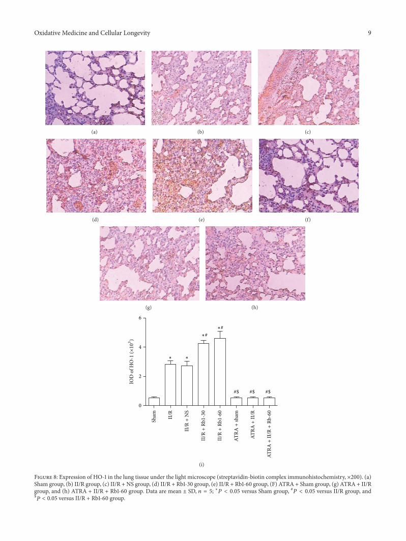
3.6. Effects of Ginsenoside Rb1 on HO-1 and Nrf2 Expressionin Lung Tissue by Immunohistochemical Detection. The lungtissue was obtained to measure the expression of Nrf2 andHO-1 by immunohistochemical assay. In the II/R group,both cytoplasm and nuclei of the lung tissue showed Nrf2expression, but the expression of HO-1 was showed in thecytoplasm. Compared with the Sham group, the expressionof Nrf2 and HO-1 in the II/R group increased significantly.After treatment with ginsenoside Rb1 at dose 30 or 60mg/kg,the expression ofNrf2 andHO-1wasmuchhigher than that inthe II/R group. In theATRA+ II/R andATRA+ II/R+Rb1-60groups, Nrf2 was also expressed obviously in the cytoplasmand nuclei, though mild expression of HO-1 could be seen inthe cytoplasm of lung tissue in these groups (Figures 7 and 8).

Oxidative Medicine and Cellular Longevity 7
Sham II/R
II/R
+N
S
II/R
+Rb
1-30
II/R
+Rb
1-60
ATRA
+sh
am
ATRA
+II
/R
ATRA
+II
/R+
Rb-60
∗∗
∗#∗#
∗$ ∗$
4
3
2
1
0
TNF-𝛼
(pg/
mg)
(a)
Sham II/R
II/R
+N
S
II/R
+Rb
1-30
II/R
+Rb
1-60
ATRA
+sh
am
ATRA
+II
/R
ATRA
+II
/R+
Rb-60
∗ ∗
∗#
∗#
∗$ ∗$
4
5
3
2
1
0
IL-6
(pg/
mg)
(b)
Sham II/R
II/R
+N
S
II/R
+Rb
1-30
II/R
+Rb
1-60
ATRA
+sh
am
ATRA
+II
/R
ATRA
+II
/R+
Rb-60
∗ ∗
∗#∗#
∗$ ∗$
IL-1
0 (p
g/m
g)
2.0
1.5
1.0
0.5
0.0
(c)
Figure 6: Cytokine levels in lung from mice. Cytokine levels were determined in lung homogenate using multiplex analysis: (a) TNF-𝛼, (b)IL-6, and (c) IL-10. Data are mean ± SD, 𝑛 = 10; ∗𝑃 < 0.05 versus Sham group, #𝑃 < 0.05 versus II/R group, and $
𝑃 < 0.05 versus II/R +Rb1-60 group.
3.7. Effects of Ginsenoside Rb1 on Cytoplasmic HO-1 andNuclear Nrf2 Expression in Lung Tissue by Western Blot-ting Analysis. To further confirm the protective effect ofginsenoside Rb1 on the lung tissue against II/R injury,protein expression of nuclear Nrf2 and cytoplasmic HO-1was examined by Western blot. As shown in Figure 9, Nrf2and HO-1 expression were both increased markedly in theII/R group as compared with the Sham group. II/R with Rb1intervention further increased the expression of Nrf2 andHO-1 significantly. ATRA administration has no effects onthe cytoplasmic HO-1 expression as compared with the Shamgroup. This indicated that Rb1 induced cytoplasmic HO-1expression was inhibited by ATRA. There was no significant
difference inNrf2 expression between theATRA+ II/R groupand the II/R group or between the ATRA + II/R + Rb1-60group and the II/R group.
4. Discussion
In this study, we have demonstrated in a mice model that45min occlusion of SMA followed by 2 h of reperfusioncaused significant lung injury as evidenced by pathologicmorphologic changes seen in the lung tissue, as well as theincreased lung wet/dry ratio, which is in accordance with theprevious reports [25]. We found that postischemia treatment

8 Oxidative Medicine and Cellular Longevity
(a) (b) (c)
(d) (e) (f)
(g) (h)
Sham II/R
II/R
+N
S
II/R
+Rb
1-30
II/R
+Rb
1-60
ATRA
+sh
am
ATRA
+II
/R
ATRA
+II
/R+
Rb-60
∗ ∗
∗#∗#
∗$ ∗$
15
10
5
0
IOD
of N
rf2
(×10
3)
#$
(i)
Figure 7: Expression of Nrf2 in the lung tissue under the light microscope (streptavidin-biotin complex immunohistochemistry, ×200). (a)Sham group, (b) II/R group, (c) II/R + NS group, (d) II/R + Rb1-30 group, (e) II/R + Rb1-60 group, (f) ATRA + Sham group, (g) ATRA + II/Rgroup, and (h) ATRA + II/R + Rb1-60 group. Data are mean ± SD, 𝑛 = 5; ∗𝑃 < 0.05 versus Sham group, #𝑃 < 0.05 versus II/R group, and$𝑃 < 0.05 versus II/R + Rb1-60 group.
Oxidative Medicine and Cellular Longevity 9
(a) (b) (c)
(d) (e) (f)
(g) (h)
IOD
of H
O-1
(×10
3)
Sham II/R
II/R
+N
S
II/R
+Rb
1-30
II/R
+Rb
1-60
ATRA
+sh
am
ATRA
+II
/R
ATRA
+II
/R+
Rb-60
∗ ∗
∗#∗#
#$ #$ #$
6
4
2
0
(i)
Figure 8: Expression of HO-1 in the lung tissue under the light microscope (streptavidin-biotin complex immunohistochemistry, ×200). (a)Sham group, (b) II/R group, (c) II/R + NS group, (d) II/R + Rb1-30 group, (e) II/R + Rb1-60 group, (F) ATRA + Sham group, (g) ATRA + II/Rgroup, and (h) ATRA + II/R + Rb1-60 group. Data are mean ± SD, 𝑛 = 5; ∗𝑃 < 0.05 versus Sham group, #𝑃 < 0.05 versus II/R group, and$𝑃 < 0.05 versus II/R + Rb1-60 group.

10 Oxidative Medicine and Cellular Longevity
Nrf2
Lamin B1
(a)
HO-1
𝛽-Actin
(b)
Sham II/R
II/R
+N
S
II/R
+Rb
1-30
II/R
+Rb
1-60
ATRA
+sh
am
ATRA
+II
/R
ATRA
+II
/R+
Rb-60
∗ ∗
∗#∗#
#$
1.5
1.0
0.5
0.0
∗$ ∗$
Nuc
lear
Nrf2
/lam
in B
1
(c)
Sham II/R
II/R
+N
S
II/R
+Rb
1-30
II/R
+Rb
1-60
ATRA
+sh
am
ATRA
+II
/R
ATRA
+II
/R+
Rb-60
∗ ∗
∗#
∗#
#$ #$ #$
1.5
2.0
1.0
0.5
0.0
Cyto
plas
mic
HO
-1/𝛽
-act
in
(d)
Figure 9:Western blotting analysis of the presence of Nrf2 in nuclear proteins andHO-1 in cytoplasmic proteins in themice lung tissue. Dataobtained from quantitative densitometry were presented as mean ± SD. 𝑛 = 10, ∗𝑃 < 0.05 versus Sham group, #𝑃 < 0.05 versus II/R group,and $𝑃 < 0.05 versus II/R + Rb1-60 group.
with ginsenoside Rb1 enhanced Nrf2 translocation to thenucleus in the lung tissues of mice and Rb1 treatment couldreduce pulmonary morphologic damage, alleviate injuriesinduced by oxidative stress, and modulate inflammatoryreactions. Further, Nrf2 function inhibition with ATRAreverted the pulmonary protective effects of ginsenosideRb1, indicating that ginsenoside Rb1 confers its respiratoryprotection via Nrf2/ARE signaling in the II/R induced acutelung injury.
The mechanisms of acute lung injury induced by II/R arecomplex. It is thought that the damage of intestinal mucosalbarrier following II/R causes the dislocation of bacteria orendogenous endotoxin, thus leading to increased oxidativestress and systemic inflammatory reaction, which is one ofthe main reasons for acute lung injury.
Ginseng is one of the most widely used herbal medicines.Ginsenosides, the major active ingredient of ginseng, havebeen noticed for their multiple pharmacological effects onantioxidation, signal transduction pathways, and interactionwith receptors [26]. Oxidative stress refers to themismatchedredox equilibrium between the production of free radicalsand the ability of cells to defend against them. One feasibleway to prevent free radical mediated cellular injuries is toaugment the oxidative defense capacity through intake ofantioxidants. Moreover, the induction of endogenous phaseII detoxifying enzymes or antioxidative proteins seems tobe a reasonable strategy for delaying disease progression.
Activation of Nrf2/ARE plays an important role in protectingcells from oxidative stress [27, 28]. The ability of Nrf2 toupregulate the expression of antioxidant genes via AREsuggests that increasing Nrf2 activity may provide a usefulsystem for combating oxidative insults. Several recent reportshave demonstrated that coordinate upregulation of ARE-driven genes protects organs from ischemia reperfusioninjury [29–31]. Accumulating evidence also suggests thatupregulation of HO-1 expression and the subsequent increasein HO activity may confer an adaptive survival responseagainst oxidative insults. Our previous studies showed thatRb1 reduces renal apoptosis and alleviates renal dysfunctionafter II/R in part through the Nrf2/ARE pathway [32]. Wanget al. demonstrated that Rb1 attenuates lung injury inducedby II/R via inhibiting NF-𝜅B activation [33]. To determinethe mechanism by which postischemia treatment with Rb1reduces II/R-induced ROS generation, we examined theeffect of ginsenoside Rb1 on Nrf2 and HO-1 expression inmice lung tissues. Our present study demonstrated that Rb1increased nuclearNrf2 protein and cytoplasmicHO-1 proteinexpressions in lung tissues of mice after II/R. The increase ofHO-1 expression byRb1 conferred cytoprotection against II/Rinduced oxidative stress. In addition, previous studies haveshown that ATRA does not affect the half-life of Nrf2 or itsnuclear translocation. ATRA inhibits Nrf2 function by stim-ulating the formation of Nrf2:RAR𝛼-containing complexesthat do not bind to the ARE [18]. We showed that ATRA, as a

Oxidative Medicine and Cellular Longevity 11
potent inhibitor for combination of Nrf2 with ARE, partiallyreversed the protective effects of Rb1, thus providing furtherevidence for Nrf2/ARE as a possible cytoprotective pathwayfor Rb1.
Ginseng extract was reported to have immunomodu-latory effects [34]. Smolinski and Pestka [35] reported thatimmunologic effects include modulation of lipopolysac-charide-induced proinflammatory cytokine production invitro and in vivo by ginsenoside Rb1.This was also confirmedwith our study in which Rb1 significantly reduced the tissuelevel of TNF-𝛼, IL-6, and IL-10. These results show that Rb1may have multiple mechanisms of action that affect cytopro-tection by both reducing ROS generation and increasing theanti-inflammatory effect.
In summary, our present study indicates that ginsenosideRb1 alleviates acute lung injury following II/R via activatingNrf2/ARE pathway.The experiment data may help us furtherunderstand the pharmacological effects of Rb1 and alsosuggest a new therapeutic target to protect the body fromII/R injury. However, further studies need to be performed intransfection of lung endothelial cells and intestinal epithelialcells with the siRNA and expressing plasmid for Nrf2 toconfirm the findings of the current study.
Disclosure
All authors have no financial, personal, or other relationshipswith other people or organizations that could inappropriatelyinfluence the work.
Conflict of Interests
The authors declare that they have no conflict of interests.
Authors’ Contribution
Ying Jiang and Zhen Zhou contributed equally to this work.
Acknowledgment
This studywasmainly supported by a grant from theNationalNatural Science Foundation of China (NSFC) 81170768 andpartly by NSFC 81471844 and 81300674.
References
[1] H. T. Hassoun, B. C. Kone, D. W. Mercer, F. G. Moody, N. W.Weisbrodt, and F. A.Moore, “Post-injurymultiple organ failure:the role of the gut,” Shock, vol. 15, no. 1, pp. 1–10, 2001.
[2] A. Pierro and S. Eaton, “Intestinal ischemia reperfusion injuryand multisystem organ failure,” Seminars in Pediatric Surgery,vol. 13, no. 1, pp. 11–17, 2004.
[3] T.-H. Lan, Z.-W. Xu, Z. Wang, Y.-L. Wu, W.-K. Wu, andH.-M. Tan, “Ginsenoside Rb1 prevents homocysteine-inducedendothelial dysfunction via PI3K/Akt activation and PKCinhibition,” Biochemical Pharmacology, vol. 82, no. 2, pp. 148–155, 2011.
[4] Y. P. Hwang and H. G. Jeong, “Ginsenoside Rb1 pro-tects against 6-hydroxydopamine-induced oxidative stress by
increasing heme oxygenase-1 expression through an estrogenreceptor-related PI3K/Akt/Nrf2-dependent pathway in humandopaminergic cells,” Toxicology and Applied Pharmacology, vol.242, no. 1, pp. 18–28, 2010.
[5] Y. Wu, Z.-Y. Xia, J. Dou et al., “Protective effect of gin-senoside Rb1 against myocardial ischemia/reperfusion injuryin streptozotocin-induced diabetic rats,” Molecular BiologyReports, vol. 38, no. 7, pp. 4327–4335, 2011.
[6] Z. Jiang, Y. Wang, X. Zhang et al., “Preventive and therapeuticeffects of ginsenoside Rb1 for neural injury during cerebralinfarction in rats,” The American Journal of Chinese Medicine,vol. 41, no. 2, pp. 341–352, 2013.
[7] L. Shen, Y. Xiong, D. Q.-H. Wang et al., “Ginsenoside Rb1reduces fatty liver by activating AMP-activated protein kinasein obese rats,” Journal of Lipid Research, vol. 54, no. 5, pp. 1430–1438, 2013.
[8] R. Hashimoto, J. Yu, H. Koizumi, Y. Ouchi, and T. Okabe,“Ginsenoside Rb1 prevents MPP+-induced apoptosis in PC12cells by stimulating estrogen receptors with consequent activa-tion of ERK1/2, Akt and inhibition of SAPK/JNK, p38 MAPK,”Evidence-Based Complementary and Alternative Medicine, vol.2012, Article ID 693717, 8 pages, 2012.
[9] D. Liu, H. Zhang, W. Gu, Y. Liu, and M. Zhang, “Neuro-protective effects of ginsenoside rb1 on high glucose-inducedneurotoxicity in primary cultured rat,” PLoS ONE, vol. 8, no. 11,Article ID e79399, 2013.
[10] P. Yao, A. Nussler, L. Liu et al., “Quercetin protects humanhepatocytes from ethanol-derived oxidative stress by inducingheme oxygenase-1 via the MAPK/Nrf2 pathways,” Journal ofHepatology, vol. 47, no. 2, pp. 253–261, 2007.
[11] A. S. Pachori, L. G. Melo, L. Zhang, S. D. Solomon, andV. J. Dzau, “Chronic recurrent myocardial ischemic injury issignificantly attenuated by pre-emptive adeno-associated virusheme oxygenase-1 gene delivery,” Journal of the AmericanCollege of Cardiology, vol. 47, no. 3, pp. 635–643, 2006.
[12] S. Lee and K. Suk, “Heme oxygenase-1 mediates cytoprotectiveeffects of immunostimulation in microglia,” Biochemical Phar-macology, vol. 74, no. 5, pp. 723–729, 2007.
[13] H. J. Kang, Y. B. Hong, H. J. Kim, and I. Bae, “CR6-interactingfactor 1 (CRIF1) regulates NF-E2-related factor 2 (NRF2)protein stability by proteasome-mediated degradation,” Journalof Biological Chemistry, vol. 285, no. 28, pp. 21258–21268, 2010.
[14] J.W. Kaspar, S. K. Niture, and A. K. Jaiswal, “Nrf2:INrf2 (Keap1)signaling in oxidative stress,” Free Radical Biology andMedicine,vol. 47, no. 9, pp. 1304–1309, 2009.
[15] S. Dhakshinamoorthy, D. J. Long II, and A. K. Jaiswal, “Antioxi-dant regulation of genes encoding enzymes that detoxify xeno-biotics and carcinogens,” Current Topics in Cellular Regulation,vol. 36, pp. 201–216, 2001.
[16] D. Bloom, S. Dhakshinamoorthy, W. Wang, C. M. Celli, and A.K. Jaiswal, “Role of NFE2 related factors in oxidative stress,” inCell and Molecular Responses to Stress. Volume 2: Protein Adap-tation and Signal Transduction, K. B. Storey and J. M. Storey,Eds., pp. 229–238, Elsevier, Amsterdam,TheNetherlands, 2001.
[17] Y.-F. Liao,W. Zhu, D.-P. Li, and X. Zhu, “Heme oxygenase-1 andgut ischemia/reperfusion injury: a short review,”World Journalof Gastroenterology, vol. 19, no. 23, pp. 3555–3561, 2013.
[18] X. J. Wang, J. D. Hayes, C. J. Henderson, and C. R. Wolf,“Identification of retinoic acid as an inhibitor of transcriptionfactor Nrf2 through activation of retinoic acid receptor alpha,”Proceedings of the National Academy of Sciences of the UnitedStates of America, vol. 104, no. 49, pp. 19589–19594, 2007.

12 Oxidative Medicine and Cellular Longevity
[19] Q. Sun, Q. T. Meng, Y. Jiang, and Z.-Y. Xia, “Ginsenoside Rb1attenuates intestinal ischemia reperfusion induced renal injuryby activating Nrf2/ARE pathway,” Molecules, vol. 17, no. 6, pp.7195–7205, 2012.
[20] J. Shen, G. Fu, L. Jiang, J. Xu, L. Li, and G. Fu, “Effectof dexmedetomidine pretreatment on lung injury followingintestinal ischemia-reperfusion,” Experimental andTherapeuticMedicine, vol. 6, no. 6, pp. 1359–1364, 2013.
[21] C. J. Chiu, A. H. McArdle, R. Brown, H. J. Scott, and F. N.Gurd, “Intestinal mucosal lesion in low-flow states. I. A mor-phological, hemodynamic, and metabolic reappraisal,” Archivesof Surgery, vol. 101, no. 4, pp. 478–483, 1970.
[22] M. L. Pearce, J. Yamashita, and J. Beazell, “Measurement ofpulmonary edema,” Circulation Research, vol. 16, pp. 482–488,1965.
[23] A. J. L. Chia, C. E. Goldring, N. R. Kitteringham, S. Q. Wong, P.Morgan, and B. K. Park, “Differential effect of covalent proteinmodification and glutathione depletion on the transcriptionalresponse of Nrf2 and NF-𝜅B,” Biochemical Pharmacology, vol.80, no. 3, pp. 410–421, 2010.
[24] K. X. Liu, Y. S. Li, W. Q. Huang, C. Li, and J.-X. Liu, “Immediatebut not delayed postconditioning during reperfusion attenuatesacute lung injury induced by intestinal ischemia/reperfusionin rats: comparison with ischemic preconditioning,” Journal ofSurgical Research, vol. 157, no. 1, pp. e55–e62, 2009.
[25] B. C. Guido, M. Zanatelli, W. Tavares-de-Lima, S. M. Oliani,and A. S. Damazo, “Annexin-A1 peptide down-regulates theleukocyte recruitment and up-regulates interleukin-10 releaseinto lung after intestinal ischemia-reperfusion in mice,” Journalof Inflammation, vol. 10, no. 1, article 10, 2013.
[26] J. M. Lu, Q. Yao, and C. Chen, “Ginseng compounds: an updateon their molecular mechanisms and medical applications,”Current Vascular Pharmacology, vol. 7, no. 3, pp. 293–302, 2009.
[27] W. F. Yao, G. J. Luo, G. S. Zhu et al., “Propofol activation ofthe Nrf2 pathway is associated with amelioration of acute lunginjury in a rat liver transplantation model,” Oxidative Medicineand Cellular Longevity, vol. 2014, Article ID 258567, 9 pages,2014.
[28] H. R. Potteti, N. M. Reddy, T. K. Hei, D. V. Kalvakolanu, and S.P. Reddy, “The NRF2 activation and antioxidative response arenot impaired overall during hyperoxia-induced lung epithelialcell death,”OxidativeMedicine and Cellular Longevity, vol. 2013,Article ID 798401, 11 pages, 2013.
[29] B. Ke, X.-D. Shen, Y. Zhang et al., “KEAP1-NRF2 complexin ischemia-induced hepatocellular damage of mouse livertransplants,” Journal of Hepatology, vol. 59, no. 6, pp. 1200–1207,2013.
[30] W. Wang, J. Kang, H. Li et al., “Regulation of endoplasmicreticulum stress in rat cortex by p62/ZIP through the Keap1-Nrf2-ARE signalling pathway after transient focal cerebralischaemia,” Brain Injury, vol. 27, no. 7-8, pp. 924–933, 2013.
[31] B. F. Peake, C. K. Nicholson, J. P. Lambert et al., “Hydrogensulfide preconditions the db/db diabetic mouse heart againstischemia-reperfusion injury by activating Nrf2 signaling in anErk-dependent manner,”The American Journal of Physiology—Heart and Circulatory Physiology, vol. 304, no. 9, pp. H1215–H1224, 2013.
[32] Q. Sun, Q. T. Meng, Y. Jiang et al., “Protective effect of ginseno-side Rb1 against intestinal ischemia-reperfusion induced acuterenal injury inmice,”PLoSONE, vol. 8, no. 12, Article ID e80859,2013.
[33] J. Wang, L. Qiao, S. Li, and G. Yang, “Protective effect of gin-senoside Rb1 against lung injury induced by intestinal ischemia-reperfusion in rats,”Molecules, vol. 18, no. 1, pp. 1214–1226, 2013.
[34] A. Rhule, B. Rase, J. R. Smith, and D. M. Shepherd, “Toll-likereceptor ligand-induced activation of murine DC2.4 cells isattenuated by Panax notoginseng,” Journal of Ethnopharmacol-ogy, vol. 116, no. 1, pp. 179–186, 2008.
[35] A. T. Smolinski and J. J. Pestka, “Modulation of lipopolysac-charide-induced proinflammatory cytokine production in vitroand in vivo by the herbal constituents apigenin (chamomile),ginsenoside Rb
1
(ginseng) and parthenolide (feverfew),” Foodand Chemical Toxicology, vol. 41, no. 10, pp. 1381–1390, 2003.

Submit your manuscripts athttp://www.hindawi.com
Stem CellsInternational
Hindawi Publishing Corporationhttp://www.hindawi.com Volume 2014
Hindawi Publishing Corporationhttp://www.hindawi.com Volume 2014
MEDIATORSINFLAMMATION
of
Hindawi Publishing Corporationhttp://www.hindawi.com Volume 2014
Behavioural Neurology
EndocrinologyInternational Journal of
Hindawi Publishing Corporationhttp://www.hindawi.com Volume 2014
Hindawi Publishing Corporationhttp://www.hindawi.com Volume 2014
Disease Markers
Hindawi Publishing Corporationhttp://www.hindawi.com Volume 2014
BioMed Research International
OncologyJournal of
Hindawi Publishing Corporationhttp://www.hindawi.com Volume 2014
Hindawi Publishing Corporationhttp://www.hindawi.com Volume 2014
Oxidative Medicine and Cellular Longevity
Hindawi Publishing Corporationhttp://www.hindawi.com Volume 2014
PPAR Research
The Scientific World JournalHindawi Publishing Corporation http://www.hindawi.com Volume 2014
Immunology ResearchHindawi Publishing Corporationhttp://www.hindawi.com Volume 2014
Journal of
ObesityJournal of
Hindawi Publishing Corporationhttp://www.hindawi.com Volume 2014
Hindawi Publishing Corporationhttp://www.hindawi.com Volume 2014
Computational and Mathematical Methods in Medicine
OphthalmologyJournal of
Hindawi Publishing Corporationhttp://www.hindawi.com Volume 2014
Diabetes ResearchJournal of
Hindawi Publishing Corporationhttp://www.hindawi.com Volume 2014
Hindawi Publishing Corporationhttp://www.hindawi.com Volume 2014
Research and TreatmentAIDS
Hindawi Publishing Corporationhttp://www.hindawi.com Volume 2014
Gastroenterology Research and Practice
Hindawi Publishing Corporationhttp://www.hindawi.com Volume 2014
Parkinson’s Disease
Evidence-Based Complementary and Alternative Medicine
Volume 2014Hindawi Publishing Corporationhttp://www.hindawi.com


![Targeted antitumor activity of Ginsenoside (Rg1) in paclitaxel ...Ginsenoside in nasopharyngeal carcinoma 2057 JBUON 2019; 24(5): 2057 treatment of cancer [1]. Plants can be considered](https://img.pdfslide.us/doc/110x75/5f8870dd69b94e4fa748fad0/targeted-antitumor-activity-of-ginsenoside-rg1-in-paclitaxel-ginsenoside-in.jpg)