Embed Size (px)
Citation preview
Research ArticleExploratory, Phase II Controlled Trial of Shiunko OintmentLocal Application Twice a Day for 4 Weeks in Ethiopian Patientswith Localized Cutaneous Leishmaniasis
Kesara Na-Bangchang,1 Oumer Ahmed,2 Jemal Hussein,2 Kenji Hirayama,3
Panida Kongjam,1 Abraham Aseffa,2 and Juntra Karbwang4
1Graduate Studies, Chulabhorn International College of Medicine, Center of Excellence in Pharmacology andMolecular Biology of Malaria and Cholangiocarcinoma, Thammasat University, PathumThani 12121, Thailand2Armauer Hansen Research Institute (AHRI), 1000 Addis Ababa, Ethiopia3Department of Immunogenetics, Institute of Tropical Medicine (NEKKEN), Graduate School of Biomedical Sciences,Nagasaki University, Nagasaki 852-8523, Japan4Clinical Product Development, Institute of Tropical Medicine (NEKKEN), Graduate School of Biomedical Sciences,Nagasaki University, Nagasaki 852-8523, Japan
Correspondence should be addressed to Juntra Karbwang; [email protected]
Received 4 January 2016; Revised 9 March 2016; Accepted 22 March 2016
Academic Editor: Victor Kuete
Copyright © 2016 Kesara Na-Bangchang et al. This is an open access article distributed under the Creative Commons AttributionLicense, which permits unrestricted use, distribution, and reproduction in any medium, provided the original work is properlycited.
The clinical efficacy and safety of Shiunko ointment (phase II clinical trial) was investigated in 40 Ethiopian patients with cutaneousleishmaniasis. Patients were randomized to receive treatment with Shiunko ointment or placebo (𝑛 = 20, each), applied on thelesion twice a day for 4 weeks. Clinicoparasitological assessments were performed before treatment, weekly for 4 weeks, and then4, 8, and 12 weeks after the end of treatment. A marked reduction in lesion size was observed on week 16 of treatment in theShiunko compared with placebo group (69% and 22% reduction, resp.). The overall rate of lesion reduction during the four weeksof treatment was significantly faster in the Shiunko group. Shiunko provided significant effect on wound closure in patients withulcerated lesion.The clinical efficacy and tolerability of Shiunkowere comparable to placebowith regard to its clinicoparasitologicalresponse (cure rate and parasitological clearance). Results of this preliminary study may suggest that Shiunko could be useful asadjuvant or as complementary treatment, not as alternatives to current treatment. Its attractive action includes fast lesion healingwith a significantly smaller lesion at week 16 of treatment compared with placebo. In addition, its action was promoted in ulcerativelesions.
1. Introduction
Cutaneous leishmaniasis (CL) is a major tropical skin dis-ease which represents a worldwide public health and socialproblem in many developing countries including Ethiopia.There are approximately 1.5 million new cases of CL eachyear, of which more than 90% occur in Afghanistan, Iran,Saudi Arabia, Syria, Brazil, and Peru [1]. CL in Ethiopiais predominantly caused by Leishmania aethiopica (99%)and rarely due to Leishmania major and Leishmania tropica
[2]. The clinical manifestation of L. aethiopica infection ismostly in a localized form (LCL), while the diffuse andmucocutaneous forms are rare [3–5]. However, a recentreport [6] showed a relatively higher percentage (19.2%) ofmucocutaneous CL due to L. aethiopica in the Silte, Ethiopia,outbreak. The primary lesion in CL patients may be a patchof erythematous induration at the site of sandfly bite thatmay progress to papulonodular, plaque, or ulcerative lesion.Many lesions donot ulcerate but persist as nodules or plaques.When it ulcerates, it forms a central depression and raised
Hindawi Publishing CorporationEvidence-Based Complementary and Alternative MedicineVolume 2016, Article ID 5984709, 8 pageshttp://dx.doi.org/10.1155/2016/5984709
2 Evidence-Based Complementary and Alternative Medicine
indurated border. Nodules plaques or ulcers may enlarge to adiameter of several centimeters. LCL may persist for monthsor years before eventually healing with an atrophic scar.
The treatments available for CL include cryotherapy andintralesional injection of sodium stibogluconate. The curerate from these treatments varies and has been claimed tobe between 60 and 70% [7]. However, none of the treatmentscurrently used in Ethiopia provide a solid evidence to supportthe development of a national treatment guideline for CL [7].This is further complicated by a significant coinfection ofHIVand CL [8]. The incidence of CL continues to increase, anddisease control and management of CL in Ethiopia remainsa challenge. New safer, improved efficacious treatment(s)is urgently needed. One such new candidate is Shiunko,a Chinese medicinal ointment that was formulated by theinnovative Japanese surgeon Seishu Hanaoka in the Edo era(1603 to 1868). It is composed of Lithospermi Radix (shikon),the root of Lithospermum erythrorhizon; Angelicae Radix(tohki), the root of Angelica acutiloba Kitagawa; sesame oil;and honeycomb wax. The medicine is widely available inJapan as an “Over-the-Counter (OTC)” product for burnlesions, bedsore, eczema dermatitis, and hemorrhoid analfissure indications. Shiunko ointment has been demonstratedto possess anti-inflammatory, analgesic, hemostatic, and anti-septic properties in various studies [9]. The antileishmanialactivity of Shiunko was discovered in 1984 by the Japanesescientists with financial support from the Japan HealthSciences Foundation (JHSF), Tokyo [10]. In the first study,clinical efficacy of topical Shiunko ointment twice daily for4 weeks was conducted in 26 patients with Peruvian LCL (L.mexicana). Of the 14 patients who completed the follow-upperiod (12 weeks), treatment response in 3 (21.4%) patientswas significantly effective (almost completely recovered orcured); treatment responses in 8 cases (57.1%), 2 cases (14.3%),and 1 case (7.2%), respectively, were considered sufficientlyeffective (obviously recovered), effective (slightly recovered),and not effective (not recovered or remaining unchanged).The trial was continued to recruit more LCL patients to atotal of 53 cases. A promising result was obtained in allcases who were selected based on resistance to systemicdrug administration. Forty-six (86.8%) patients had completecure after local administration of Shiunko for 4 weeks, withcomplete absence of parasites in lesion biopsy at the endof treatment. The aim of the present study was therefore toevaluate the clinical efficacy and safety of Shiunko ointmentwhen applied on the lesion twice a day for 4 weeks inEthiopian patients with LCL.
2. Materials and Methods
2.1. Patients and Study Design. The study was an exploratoryphase II, placebo-controlled, single-center (Ankober HealthCenter, North Shewa zone of Amhara region, Ethiopia)study. Patients with localized cutaneous leishmaniasis (LCL)aged 18–65 years of both genders with a new, uncom-plicated, localized, single lesion on the face or arms andpositive parasite smear by microscopy were included inthe study. Patient’s exclusion criteria were (i) cutaneous
leishmaniasis with secondary infection, (ii) concomitant dis-eases (mucocutaneous or visceral leishmaniasis), (iii) historyof antileishmanial treatment within the past six months, (iv)abnormality of biochemical and/or hematological laboratorytests, (v) known hypersensitivity to any of the Shiunkocomponents, or (vi) pregnancy (positive urine HCG test),breast-feeding, or possibility of becoming pregnant duringthe study. All patients had residential areas in North Shewazone of Amhara region (Chefa site, Derefo Nefasso, DerefoNadewa, Gorebela Chaka, Gorebela Zembo, Aleyu Amba,Debre Hayl, Haramba, Deway Derefo, Wubit Gola, MehalWonz, LayGorebela, andMetak), Ethiopia.These are the hightransmission areas which constitute the main LCL endemicarea in the country, contributing over 60% of the burden.
Lesion sample was collected from each patient by slit-skin scraping from the lesion and diagnosis of CL was basedon microscopic examination for the presence of leishmanialamastigotes. Written informed consents for study participa-tion were obtained from all of them before study. The studywas approved by the Ethics Committees of Armauer HansenResearch Institute (AHRI), Ethiopia, and the Institute ofTropical Medicine, Graduate School of Biomedical Sciences,Nagasaki University, Japan (UMIN Clinical Trials Registry:UMIN-CTR Number 000010994).
2.2. Treatment. Patients who fulfilled inclusion and had noneof the exclusion criteria were randomized to receive Shiunkoointment and placebo (20 patients per group). Preparation ofthe pharmaceutical formulations of Shiunko (batch number961) and the placebo (batch number 962) including qualityassurance and control were performed by Okusa Co., Ltd.(Japan). Both were prepared as an ointment pharmaceuticalformulation (20 g of violet red ointment per container). Thecomposition of Shiunko was as follows: 2.04 g shikon, 1.02 gtohki, 16.92 g sesame oil, and 6.76 g honeycomb wax. Theplacebo contained 20 g wax.The ointment was applied locallyby the health post extension workers, covering the wholelesion of each patient twice a day (in the morning andevening, 8–10 hours apart), for 4 weeks. All antileishmanialdrugs were prohibited during the study period. Cryotherapywas to be offered to all patients who were withdrawn ordiscontinued from the study for safety reasons (i.e., thosewith serious adverse events or with increase of lesion sizeby more than 50% of the original size, or treatment failure).Patients who withdrew their consents were evaluated by thedermatologist for the requirement of rescue treatment.
2.3. Assessment of Efficacy and Safety
2.3.1. Clinical Assessment. The lesion was observed for theprogress of reepithelization before treatment (on recruit-ment) and weekly for 4 weeks (during treatment) and then4, 8, and 12 weeks after the end of treatment. Each lesionwas photographed to document the appearance of the lesionuntil declared cured. The rate of lesion size reduction wasestimated from the slope of each of the average lines of thelesion areas reduction fromweek 0 up to week 4 of treatment.
Evidence-Based Complementary and Alternative Medicine 3
2.3.2. Parasitological Assessment. Lesion sample was takenfrommargin of the ulcerated lesion after removal of the crustand cleaning of the lesion by scraping (for open wounds)or by fine needle aspiration before treatment, at the endof treatment and monthly until parasitological negativity,that is, absence of amastigotes in the smear. Parasitologicalsmear (Giemsa staining) was examined by two differentparasitologists (AHRI laboratory), under light microscope(×100) for the presence or absence of leishmanial amastigotes.In addition, all samples were confirmed by an external expert(Institute of Tropical Medicine: NEKKEN).
Safety Assessment. Safety and tolerability of Shiunko andplacebo were assessed based on clinical and laboratoryassessments during follow-up, according to the NIH/NCICommon Toxicity Criteria (CTC) grading system [11].
2.4. Data Analysis. Treatment efficacy of Shiunko andplacebo was categorized as cure, partial response, and treat-ment failure. Cure was defined as complete wound closureand reepithelization without inflammation or infiltrationwithin 12 weeks after the end of treatment and absence of par-asite (amastigotes) within 12 weeks after the end of treatment.Partial response was defined as improvement of Leishmaniasigns (skin edema, erythematous, and/or hardening) and/orreduction in size but not total disappearance of the lesionwithin 12 weeks after the end of treatment and absence ofparasite (amastigotes) within 12 weeks after the end of treat-ment. Treatment failure was defined as failure of the lesionsize to decrease and/or lack of lesion sign improvement orreepithelization and/or presence of leishmanial amastigotesin the lesion 12 weeks after end of treatment (week 16) [12].
The primary efficacy analysis was based on intention-to-treat (ITT) population (inclusion of all patients as originallyallocated after randomization) with secondary analysis ofper-protocol (PP) population. PP population was a subset ofthe subjects in the full analysis set who were more compliantwith the protocol and was characterized by the followingcriteria: (i) the completion of at least 85% of the treatmentregimen; (ii) the availability of measurements of the primaryvariable(s); and (iii) the absence of any major protocolviolations including the violation of entry criteria.
Treatment safety was evaluated based on the proportionsof patientswith adverse events (AE) including serious adverseevents (SAE) following treatment.These included local (pain,erythema, induration, swelling, itching, nodules, etc.) andsystemic (diffuse erythema, urticaria, fever, headache, ana-phylactic reactions, etc.) adverse events, as well as clinicalrelevant changes in laboratory parameters. The analysis wasbased on ITT population. The proportion of patients expe-riencing any adverse event during the 16-week study periodwas evaluated.
Quantitative data are presented as median (range and95% CI) and qualitative data are presented as number andproportion (%) with 95% CI. Comparison of quantitativedata of each study period and baseline was performed usingWilcoxon Signed Rank test. Comparison of quantitativedata between the two groups during each study visit was
Table 1: The demographic and baseline clinical parameters beforetreatment in 40CL patients allocated to receive treatment withShiunko and placebo.
Parameters Placebo(𝑛 = 20)
Shiunko(𝑛 = 20)
Age [median (range), years] 40.4 (18–53) 38.1 (18–62)Sex [number of males and females] 12, 8 13, 7Lesion signLesion size [median (range),mm2] 91 (16–1,575) 123 (13–1,578)
Lesion location [𝑛 (%)]Face 17 (85) 15 (75)Neck 2 (10) 1 (5)Arm 1 (5) 3 (15)Eyelid 0 (0) 1 (5)
performed usingMann-Whitney𝑈 test. Comparison of qual-itative data was performed using Chi-square test. Statisticalsignificance was set at 𝛼 = 0.05 for all tests (SPSS version16.0, SPSS Inc., CO, USA).
3. Results
3.1. Patients. Out of 108 patients screened (61 males and47 females), 40 patients with CL (25 males and 15 females,aged 18–62 years) who fulfilled inclusion and had none ofthe exclusion criteria were randomized to receive placeboand Shiunko treatment (20 patients for each group). Themost common locations for CL lesions were the face (80%),followed by arms (10%), neck (7.5%), and eyelids (2.5%).The most common signs and symptoms in both groupsof patients were pruritus or itching sensation (65%), pain(32.5%), burning sensation (17.5%), headache (10%), eye dis-charge (2.5%), abdominal distension (2.5%), myalgia (2.5%),and paraesthesia (2.5%). Demographics and baseline lesionsize (Table 1) including laboratory data (hematology andbiochemistry) of patients in both groups were comparable.
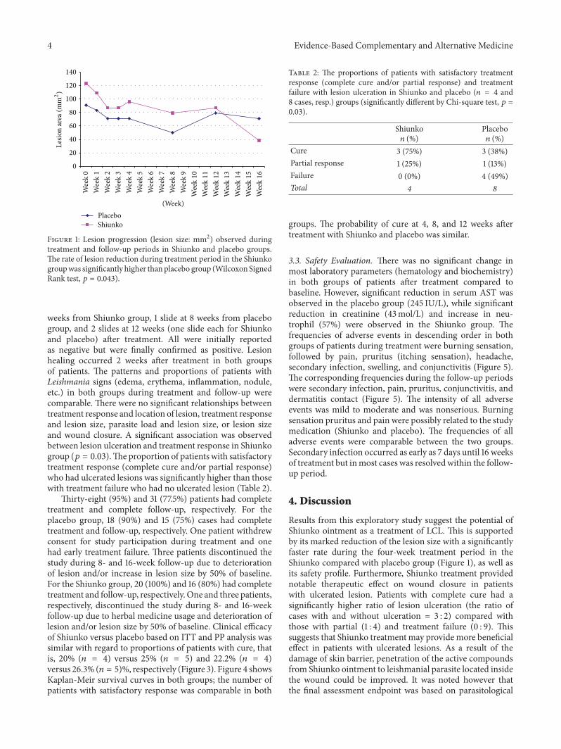
3.2. Efficacy Evaluation. Following the start of treatmentwithShiunko and placebo, lesion size gradually reduced until theend of follow-up (week 16) (Figure 1).The lesion size on week16 was significantly reduced compared with baseline (𝑝 =0.03) in Shiunko group. Figure 2 shows representative photosof the lesions in a patient with cure after treatment withShiunko ointment (before treatment, at the end of treatment,and 4, 8, and 12 weeks after the end of treatment). Themedian lesion size on week 16 was about 31% of baselinein Shiunko group, while it was 78% in placebo group. Inaddition, the overall rate of lesion reduction during thefour-week treatment period was significantly faster in theShiunko compared with placebo (𝑝 = 0.043). A total of 178parasitological smears collected before treatment, at the endof treatment, and at 4, 8, and 12 weeks after treatment wereconfirmed by an expert at NEKKEN laboratory. Discrepancyof results was found only in four slides (2.25%) collectedfrom four patients on different occasions, that is, 1 slide at 4
4 Evidence-Based Complementary and Alternative Medicine
0
20
40
60
80
100
120
140
(Week)PlaceboShiunko
Lesio
n ar
ea (m
m2)
Wee
k0
Wee
k1
Wee
k2
Wee
k3
Wee
k4
Wee
k5
Wee
k6
Wee
k7
Wee
k8
Wee
k9
Wee
k10
Wee
k11
Wee
k12
Wee
k13
Wee
k14
Wee
k15
Wee
k16
Figure 1: Lesion progression (lesion size: mm2) observed duringtreatment and follow-up periods in Shiunko and placebo groups.The rate of lesion reduction during treatment period in the Shiunkogroupwas significantly higher than placebo group (Wilcoxon SignedRank test, 𝑝 = 0.043).
weeks from Shiunko group, 1 slide at 8 weeks from placebogroup, and 2 slides at 12 weeks (one slide each for Shiunkoand placebo) after treatment. All were initially reportedas negative but were finally confirmed as positive. Lesionhealing occurred 2 weeks after treatment in both groupsof patients. The patterns and proportions of patients withLeishmania signs (edema, erythema, inflammation, nodule,etc.) in both groups during treatment and follow-up werecomparable. There were no significant relationships betweentreatment response and location of lesion, treatment responseand lesion size, parasite load and lesion size, or lesion sizeand wound closure. A significant association was observedbetween lesion ulceration and treatment response in Shiunkogroup (𝑝 = 0.03).The proportion of patients with satisfactorytreatment response (complete cure and/or partial response)who had ulcerated lesions was significantly higher than thosewith treatment failure who had no ulcerated lesion (Table 2).
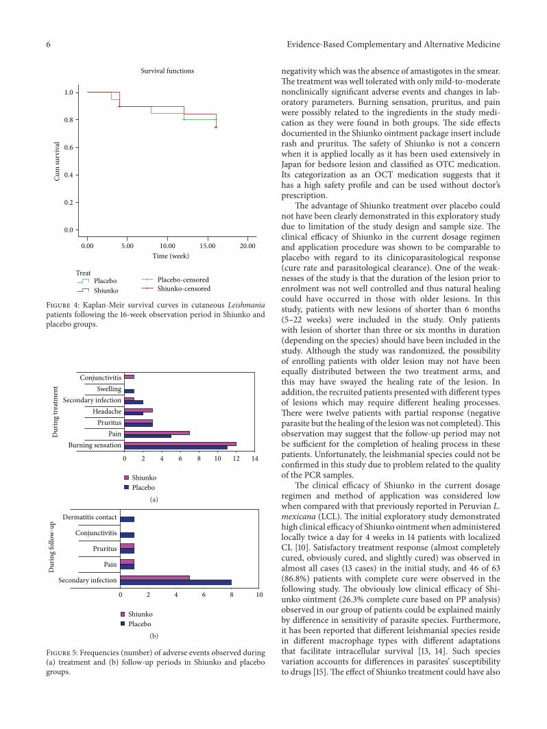
Thirty-eight (95%) and 31 (77.5%) patients had completetreatment and complete follow-up, respectively. For theplacebo group, 18 (90%) and 15 (75%) cases had completetreatment and follow-up, respectively. One patient withdrewconsent for study participation during treatment and onehad early treatment failure. Three patients discontinued thestudy during 8- and 16-week follow-up due to deteriorationof lesion and/or increase in lesion size by 50% of baseline.For the Shiunko group, 20 (100%) and 16 (80%) had completetreatment and follow-up, respectively. One and three patients,respectively, discontinued the study during 8- and 16-weekfollow-up due to herbal medicine usage and deterioration oflesion and/or lesion size by 50% of baseline. Clinical efficacyof Shiunko versus placebo based on ITT and PP analysis wassimilar with regard to proportions of patients with cure, thatis, 20% (𝑛 = 4) versus 25% (𝑛 = 5) and 22.2% (𝑛 = 4)versus 26.3% (𝑛 = 5)%, respectively (Figure 3). Figure 4 showsKaplan-Meir survival curves in both groups; the number ofpatients with satisfactory response was comparable in both
Table 2: The proportions of patients with satisfactory treatmentresponse (complete cure and/or partial response) and treatmentfailure with lesion ulceration in Shiunko and placebo (𝑛 = 4 and8 cases, resp.) groups (significantly different by Chi-square test, 𝑝 =0.03).
Shiunko𝑛 (%)
Placebo𝑛 (%)
Cure 3 (75%) 3 (38%)Partial response 1 (25%) 1 (13%)Failure 0 (0%) 4 (49%)Total 4 8
groups. The probability of cure at 4, 8, and 12 weeks aftertreatment with Shiunko and placebo was similar.
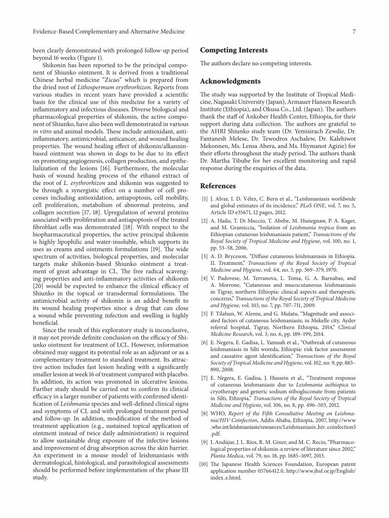
3.3. Safety Evaluation. There was no significant change inmost laboratory parameters (hematology and biochemistry)in both groups of patients after treatment compared tobaseline. However, significant reduction in serum AST wasobserved in the placebo group (245 IU/L), while significantreduction in creatinine (43mol/L) and increase in neu-trophil (57%) were observed in the Shiunko group. Thefrequencies of adverse events in descending order in bothgroups of patients during treatment were burning sensation,followed by pain, pruritus (itching sensation), headache,secondary infection, swelling, and conjunctivitis (Figure 5).The corresponding frequencies during the follow-up periodswere secondary infection, pain, pruritus, conjunctivitis, anddermatitis contact (Figure 5). The intensity of all adverseevents was mild to moderate and was nonserious. Burningsensation pruritus and pain were possibly related to the studymedication (Shiunko and placebo). The frequencies of alladverse events were comparable between the two groups.Secondary infection occurred as early as 7 days until 16 weeksof treatment but inmost cases was resolvedwithin the follow-up period.
4. Discussion
Results from this exploratory study suggest the potential ofShiunko ointment as a treatment of LCL. This is supportedby its marked reduction of the lesion size with a significantlyfaster rate during the four-week treatment period in theShiunko compared with placebo group (Figure 1), as well asits safety profile. Furthermore, Shiunko treatment providednotable therapeutic effect on wound closure in patientswith ulcerated lesion. Patients with complete cure had asignificantly higher ratio of lesion ulceration (the ratio ofcases with and without ulceration = 3 : 2) compared withthose with partial (1 : 4) and treatment failure (0 : 9). Thissuggests that Shiunko treatment may provide more beneficialeffect in patients with ulcerated lesions. As a result of thedamage of skin barrier, penetration of the active compoundsfrom Shiunko ointment to leishmanial parasite located insidethe wound could be improved. It was noted however thatthe final assessment endpoint was based on parasitological

Evidence-Based Complementary and Alternative Medicine 5
(a) Before treatment (b) At the end of treatment (4 weeks) (c) At 4 weeks after end of treatment
(d) At 8 weeks after end of treatment (e) At 12 weeks after end of treatment
Figure 2: Representative photos of the lesions in a patient with cure following treatment with Shiunko ointment: (a) before treatment, (b) atthe end of treatment, (c) at 4 weeks after end of treatment, (d) at 8 weeks, and (e) at 12 weeks of treatment.
Randomization
Withdrew consent (n = 1)Early treatment failure (n = 1)
Use of herbal medicine (n = 1)
Complete follow-up (n = 16)
Complete treatment (n = 20)
Complete follow-up (n = 15)
Complete treatment (n = 18)
Shiunko (n = 20)Placebo (n = 20)
Patients with LCL (n = 40)
Partial response (n = 6; 95% CI = 0.12, 0.54)Treatment failure (n = 9; 95% CI = 0.23, 0.69)
ITT∗ (n = 20): Cure (n = 4; 95% CI = 0.06, 0.44)
PP∗∗ (n = 18): Cure (n = 4; 95% CI = 0.06, 0.48)
Treatment failure (n = 8; 95% CI = 0.22, 0.69)Partial response (n = 6; 95% CI = 0.13, 0.59)
PP∗∗ (n = 19): Cure (n = 5; 95% CI = 0.09, 0.51)
Treatment failure (n = 8; 95% CI = 0.20, 0.67)Partial response (n = 6; 95% CI = 0.13, 0.57)
ITT∗ (n = 20): Cure (n = 5; 95% CI = 0.09, 0.49)
Treatment failure (n = 8; 95% CI = 0.19, 0.64)Partial response (n = 6; 95% CI = 0.12, 0.54)
∗Completion of at least one dose of the treatment regimen∗∗Completion of at least 85% of the treatment regimen
Treatment discontinued (nosignificant reduction of lesion size) (n = 3) Treatment discontinued (no
significant reduction of lesion size) (n = 3)
Figure 3: Efficacy assessment (based on ITT and PP analysis) of CL patients following treatment with Shiunko and placebo.
6 Evidence-Based Complementary and Alternative Medicine
1.0
0.8
0.6
0.4
0.2
0.0
Cum
surv
ival
0.00 5.00 10.00 15.00 20.00
Time (week)
TreatPlaceboShiunko
Placebo-censoredShiunko-censored
Survival functions
Figure 4: Kaplan-Meir survival curves in cutaneous Leishmaniapatients following the 16-week observation period in Shiunko andplacebo groups.
0 2 4 6 8 10 12 14
Burning sensationPain
PruritusHeadache
Secondary infectionSwelling
Conjunctivitis
Dur
ing
treat
men
t
ShiunkoPlacebo
(a)
0 2 4 6 8 10
Secondary infection
Pain
Pruritus
Conjunctivitis
Dermatitis contact
Dur
ing
follo
w-u
p
ShiunkoPlacebo
(b)
Figure 5: Frequencies (number) of adverse events observed during(a) treatment and (b) follow-up periods in Shiunko and placebogroups.
negativity which was the absence of amastigotes in the smear.The treatment was well tolerated with only mild-to-moderatenonclinically significant adverse events and changes in lab-oratory parameters. Burning sensation, pruritus, and painwere possibly related to the ingredients in the study medi-cation as they were found in both groups. The side effectsdocumented in the Shiunko ointment package insert includerash and pruritus. The safety of Shiunko is not a concernwhen it is applied locally as it has been used extensively inJapan for bedsore lesion and classified as OTC medication.Its categorization as an OCT medication suggests that ithas a high safety profile and can be used without doctor’sprescription.
The advantage of Shiunko treatment over placebo couldnot have been clearly demonstrated in this exploratory studydue to limitation of the study design and sample size. Theclinical efficacy of Shiunko in the current dosage regimenand application procedure was shown to be comparable toplacebo with regard to its clinicoparasitological response(cure rate and parasitological clearance). One of the weak-nesses of the study is that the duration of the lesion prior toenrolment was not well controlled and thus natural healingcould have occurred in those with older lesions. In thisstudy, patients with new lesions of shorter than 6 months(5–22 weeks) were included in the study. Only patientswith lesion of shorter than three or six months in duration(depending on the species) should have been included in thestudy. Although the study was randomized, the possibilityof enrolling patients with older lesion may not have beenequally distributed between the two treatment arms, andthis may have swayed the healing rate of the lesion. Inaddition, the recruited patients presented with different typesof lesions which may require different healing processes.There were twelve patients with partial response (negativeparasite but the healing of the lesion was not completed).Thisobservation may suggest that the follow-up period may notbe sufficient for the completion of healing process in thesepatients. Unfortunately, the leishmanial species could not beconfirmed in this study due to problem related to the qualityof the PCR samples.
The clinical efficacy of Shiunko in the current dosageregimen and method of application was considered lowwhen compared with that previously reported in Peruvian L.mexicana (LCL). The initial exploratory study demonstratedhigh clinical efficacy of Shiunko ointment when administeredlocally twice a day for 4 weeks in 14 patients with localizedCL [10]. Satisfactory treatment response (almost completelycured, obviously cured, and slightly cured) was observed inalmost all cases (13 cases) in the initial study, and 46 of 63(86.8%) patients with complete cure were observed in thefollowing study. The obviously low clinical efficacy of Shi-unko ointment (26.3% complete cure based on PP analysis)observed in our group of patients could be explained mainlyby difference in sensitivity of parasite species. Furthermore,it has been reported that different leishmanial species residein different macrophage types with different adaptationsthat facilitate intracellular survival [13, 14]. Such speciesvariation accounts for differences in parasites’ susceptibilityto drugs [15].The effect of Shiunko treatment could have also
Evidence-Based Complementary and Alternative Medicine 7
been clearly demonstrated with prolonged follow-up periodbeyond 16 weeks (Figure 1).
Shikonin has been reported to be the principal compo-nent of Shiunko ointment. It is derived from a traditionalChinese herbal medicine “Zicao” which is prepared fromthe dried root of Lithospermum erythrorhizon. Reports fromvarious studies in recent years have provided a scientificbasis for the clinical use of this medicine for a variety ofinflammatory and infectious diseases. Diverse biological andpharmacological properties of shikonin, the active compo-nent of Shiunko, have also been well demonstrated in variousin vitro and animal models. These include antioxidant, anti-inflammatory, antimicrobial, anticancer, and wound healingproperties. The wound healing effect of shikonin/alkannin-based ointment was shown in dogs to be due to its effecton promoting angiogenesis, collagen production, and epithe-lialization of the lesions [16]. Furthermore, the molecularbasis of wound healing process of the ethanol extract ofthe root of L. erythrorhizon and shikonin was suggested tobe through a synergistic effect on a number of cell pro-cesses including antioxidation, antiapoptosis, cell mobility,cell proliferation, metabolism of abnormal proteins, andcollagen secretion [17, 18]. Upregulation of several proteinsassociated with proliferation and antiapoptosis of the treatedfibroblast cells was demonstrated [18]. With respect to thebiopharmaceutical properties, the active principal shikoninis highly lipophilic and water-insoluble, which supports itsuses as creams and ointments formulations [19]. The widespectrum of activities, biological properties, and moleculartargets make shikonin-based Shiunko ointment a treat-ment of great advantage in CL. The free radical scaveng-ing properties and anti-inflammatory activities of shikonin[20] would be expected to enhance the clinical efficacy ofShiunko in the topical or transdermal formulations. Theantimicrobial activity of shikonin is an added benefit toits wound healing properties since a drug that can closea wound while preventing infection and swelling is highlybeneficial.
Since the result of this exploratory study is inconclusive,it may not provide definite conclusion on the efficacy of Shi-unko ointment for treatment of LCL. However, informationobtained may suggest its potential role as an adjuvant or as acomplementary treatment to standard treatment. Its attrac-tive action includes fast lesion healing with a significantlysmaller lesion at week 16 of treatment comparedwith placebo.In addition, its action was promoted in ulcerative lesions.Further study should be carried out to confirm its clinicalefficacy in a larger number of patients with confirmed identi-fication of Leishmania species and well-defined clinical signsand symptoms of CL and with prolonged treatment periodand follow-up. In addition, modification of the method oftreatment application (e.g., sustained topical application ofointment instead of twice daily administration) is requiredto allow sustainable drug exposure of the infective lesionsand improvement of drug absorption across the skin barrier.An experiment in a mouse model of leishmaniasis withdermatological, histological, and parasitological assessmentsshould be performed before implementation of the phase IIIstudy.
Competing Interests
The authors declare no competing interests.
Acknowledgments
The study was supported by the Institute of Tropical Medi-cine, Nagasaki University (Japan), ArmauerHansenResearchInstitute (Ethiopia), and Okusa Co., Ltd. (Japan).The authorsthank the staff of Ankober Health Center, Ethiopia, for theirsupport during data collection. The authors are grateful tothe AHRI Shiunko study team (Dr. Yemisirach Zewdie, Dr.Fantanesh Melese, Dr. Tewodros Aschalew, Dr. KalehiwotMekonnen, Ms. Lensa Abera, and Ms. Hiymanot Agizie) fortheir efforts throughout the study period. The authors thankDr. Martha Tibube for her excellent monitoring and rapidresponse during the enquiries of the data.
References
[1] J. Alvar, I. D. Velez, C. Bern et al., “Leishmaniasis worldwideand global estimates of its incidence,” PLoS ONE, vol. 7, no. 5,Article ID e35671, 12 pages, 2012.
[2] A. Hailu, T. Di Muccio, T. Abebe, M. Hunegnaw, P. A. Kager,and M. Gramiccia, “Isolation of Leishmania tropica from anEthiopian cutaneous leishmaniasis patient,” Transactions of theRoyal Society of Tropical Medicine and Hygiene, vol. 100, no. 1,pp. 53–58, 2006.
[3] A. D. Bryceson, “Diffuse cutaneous leishmaniasis in Ethiopia.II. Treatment,” Transactions of the Royal Society of TropicalMedicine and Hygiene, vol. 64, no. 3, pp. 369–379, 1970.
[4] V. Padovese, M. Terranova, L. Toma, G. A. Barnabas, andA. Morrone, “Cutaneous and mucocutaneous leishmaniasisin Tigray, northern Ethiopia: clinical aspects and therapeuticconcerns,” Transactions of the Royal Society of Tropical Medicineand Hygiene, vol. 103, no. 7, pp. 707–711, 2009.
[5] F. Tilahun, W. Alemu, and G. Mulatu, “Magnitude and associ-ated factors of cutaneous leishmaniasis; in Mekelle city, Ayderreferral hospital, Tigray, Northern Ethiopia, 2014,” ClinicalMedicine Research, vol. 3, no. 6, pp. 189–199, 2014.
[6] E. Negera, E. Gadisa, L. Yamuah et al., “Outbreak of cutaneousleishmaniasis in Silti woreda, Ethiopia: risk factor assessmentand causative agent identification,” Transactions of the RoyalSociety of TropicalMedicine andHygiene, vol. 102, no. 9, pp. 883–890, 2008.
[7] E. Negera, E. Gadisa, J. Hussein et al., “Treatment responseof cutaneous leishmaniasis due to Leishmania aethiopica tocryotherapy and generic sodium stibogluconate from patientsin Silti, Ethiopia,” Transactions of the Royal Society of TropicalMedicine and Hygiene, vol. 106, no. 8, pp. 496–503, 2012.
[8] WHO, Report of the Fifth Consultative Meeting on Leishma-nia/HIV Coinfection, Addis Ababa, Ethiopia, 2007, http://www.who.int/leishmaniasis/resources/Leishmaniasis hiv coinfection5.pdf.
[9] I. Andujar, J. L. Rıos, R. M. Giner, andM. C. Recio, “Pharmaco-logical properties of shikonin-a review of literature since 2002,”Planta Medica, vol. 79, no. 18, pp. 1685–1697, 2013.
[10] The Japanese Health Sciences Foundation, European patentapplication number 05766412.0, http://www.jhsf.or.jp/English/index e.html.
8 Evidence-Based Complementary and Alternative Medicine
[11] U. Gonzalez, M. Pinart, L. Reveiz, and J. Alvar, “InterventionsforOldWorld cutaneous leishmaniasis,”TheCochraneDatabaseof Systematic Reviews, no. 4, Article ID CD005067, 2008.
[12] National Institute ofHealth andNationalCancer Institute.USA,Common Terminology Criteria for Adverse Events (CTCAE)Version 4, 2009, (v4.03, 2010).
[13] M. Utaile, A. Kassahun, T. Abebe, and A. Hailu, “Susceptibilityof clinical isolates of Leishmania aethiopica to miltefosine,paromomycin, amphotericin B and sodium stibogluconateusing amastigote-macrophage in vitro model,” ExperimentalParasitology, vol. 134, no. 1, pp. 68–75, 2013.
[14] R. J. S. Burchmore andM. P. Barrett, “Life in vacuoles—nutrientacquisition byLeishmania amastigotes,” International Journal forParasitology, vol. 31, no. 12, pp. 1311–1320, 2001.
[15] S. Sundar, “Drug resistance in Indian visceral leishmaniasis,”Tropical Medicine and International Health, vol. 6, no. 11, pp.849–854, 2001.
[16] M. Karayannopoulou, V. Tsioli, P. Loukopoulos et al., “Evalu-ation of the effectiveness of an ointment based on alkannins/shikonins on second intention wound healing in the dog,”Canadian Journal of Veterinary Research, vol. 75, no. 1, pp. 42–48, 2011.
[17] C.-Y. Hsiao, T.-H. Tsai, and K.-F. Chak, “The molecular basisof wound healing processes induced by lithospermi radix: aproteomics and biochemical analysis,” Evidence-Based Comple-mentary and Alternative Medicine, vol. 2012, Article ID 508972,15 pages, 2012.
[18] K. F. Chak, C. Y. Hsiao, and T. Y. Chen, “A study of the effect ofshiunko, a traditional Chinese herbal medicine, on fibroblastsand its implication on wound healing processes,” Advances inWound Care, vol. 2, no. 8, pp. 448–455, 2013.
[19] A. Albreht, I. Vovk, and B. Simonovska, “Addition of 𝛽-lactoglobulin produces water-soluble shikonin,” Journal of Agri-cultural and Food Chemistry, vol. 60, no. 43, pp. 10834–10843,2012.
[20] M. Nishizawa, M. Kohno, M. Nishimura, A. Kitagawa, and Y.Niwano, “Presence of peroxyradicals in cigarette smoke andthe scavenging effect of shikonin, a naphthoquinone pigment,”Chemical and Pharmaceutical Bulletin, vol. 53, no. 7, pp. 796–799, 2005.
Submit your manuscripts athttp://www.hindawi.com
Stem CellsInternational
Hindawi Publishing Corporationhttp://www.hindawi.com Volume 2014
Hindawi Publishing Corporationhttp://www.hindawi.com Volume 2014
MEDIATORSINFLAMMATION
of
Hindawi Publishing Corporationhttp://www.hindawi.com Volume 2014
Behavioural Neurology
EndocrinologyInternational Journal of
Hindawi Publishing Corporationhttp://www.hindawi.com Volume 2014
Hindawi Publishing Corporationhttp://www.hindawi.com Volume 2014
Disease Markers
Hindawi Publishing Corporationhttp://www.hindawi.com Volume 2014
BioMed Research International
OncologyJournal of
Hindawi Publishing Corporationhttp://www.hindawi.com Volume 2014
Hindawi Publishing Corporationhttp://www.hindawi.com Volume 2014
Oxidative Medicine and Cellular Longevity
Hindawi Publishing Corporationhttp://www.hindawi.com Volume 2014
PPAR Research
The Scientific World JournalHindawi Publishing Corporation http://www.hindawi.com Volume 2014
Immunology ResearchHindawi Publishing Corporationhttp://www.hindawi.com Volume 2014
Journal of
ObesityJournal of
Hindawi Publishing Corporationhttp://www.hindawi.com Volume 2014
Hindawi Publishing Corporationhttp://www.hindawi.com Volume 2014
Computational and Mathematical Methods in Medicine
OphthalmologyJournal of
Hindawi Publishing Corporationhttp://www.hindawi.com Volume 2014
Diabetes ResearchJournal of
Hindawi Publishing Corporationhttp://www.hindawi.com Volume 2014
Hindawi Publishing Corporationhttp://www.hindawi.com Volume 2014
Research and TreatmentAIDS
Hindawi Publishing Corporationhttp://www.hindawi.com Volume 2014
Gastroenterology Research and Practice
Hindawi Publishing Corporationhttp://www.hindawi.com Volume 2014
Parkinson’s Disease
Evidence-Based Complementary and Alternative Medicine
Volume 2014Hindawi Publishing Corporationhttp://www.hindawi.com


![Exploratory Clinical Trial of (4S F]fl C Transporter Using Positron … · Imaging, Diagnosis, Prognosis Exploratory Clinical Trial of (4S)-4-(3-[18F]fluoropropyl)-L-glutamate for](https://img.pdfslide.us/doc/110x75/60f90642e71e4a0af4493803/exploratory-clinical-trial-of-4s-fi-c-transporter-using-positron-imaging-diagnosis.jpg)