Embed Size (px)
Citation preview
Research ArticleEr:YAG Laser for Brackets Bonding:A SEM Study after Debonding
G. Ierardo,1 G. Di Carlo,1 F. Petrillo,1 V. Luzzi,1 I. Vozza,1 G. Migliau,1
R. Kornblit,1 J. P. Rocca,2 and A. Polimeni1
1 Department of Oral and Maxillofacial Science, “Sapienza” University of Rome, Via Caserta 6, 00161 Rome, Italy2 Faculty of Odontology, University Hospital “St. Roch”, University of Nice-Sophia Antipolis, 5 rue Pierre Devoluy, 06006 Nice, France
Correspondence should be addressed to G. Di Carlo; gdc [email protected]
Received 4 August 2014; Revised 23 September 2014; Accepted 23 September 2014; Published 22 October 2014
Academic Editor: Romeo Umberto
Copyright © 2014 G. Ierardo et al. This is an open access article distributed under the Creative Commons Attribution License,which permits unrestricted use, distribution, and reproduction in any medium, provided the original work is properly cited.
Background. The introduction of Er:YAG laser in dentistry for ablation of hard tissues advocated an alternative method of enameletching for orthodontics purpose. Materials and Methods. 55 extracted human third molars were inserted in acrylic resin blocksand divided into five groups of 11 teeth. Group 1 was treated with 37% orthophosphoric acid for 30 seconds. Group 2 was treatedwith laser irradiation (Er:YAG Fidelius III, Fotona, Slovenia) at 80mJ and 4Hz. Group 3 underwent laser treatment (80mJ, 4Hz),followed by 37% orthophosphoric acid for 30 seconds.The teeth in Group 4 were treated with laser at 40mJ and 10Hz.The teeth inGroup 5 were treated with laser (40mJ, 10Hz), followed by 37% orthophosphoric acid for 30 seconds. The adhesive remnant indexwas determined after debonding. Results. Kruskas-Wallis test showed that location parameters (median andmean) are significantlydifferent betweenGroups 2 and 4when comparedwith control group; on the contrary no significant differencewas detected betweenGroups 3 and 5 with the controls.Conclusion.The use of Er:YAG laser alone, as in Groups 2 and 4, showed no significant advantagesover phosphoric acid in the bonding procedure for orthodontics brackets.
1. Introduction
Phosphoric acid etching is the gold standard method ofenamel preparation before application of bonding resinsfor orthodontic brackets [1]. Enamel etching changes thetooth surface from being of low-energy and hydrophobic tobeing of high-energy and hydrophilic, increasing the surfacearea for bonding [2]. Studies have demonstrated that thiskind of attachment can have disadvantages, such as enameldecalcification, which leaves the enamel surface susceptibleto acid attack (cavity formation) under orthodontic brackets[3–6]. One of the most important challenges in orthodontictreatment, however, is the frequent debonding of brackets,with the consequent lengthening of treatment duration.
With the recent introduction of erbium-doped yttriumaluminum garnet (Er:YAG) laser in dentistry for the ablationof hard tissues, including enamel and dentin, laser enamel
preparation has been proposed as an alternative to phos-phoric acid etching [7–9]. The Er:YAG laser can effectivelymodify enamel and dentin surfaces because of its 2.94mmwavelength emission, which coincides with the main absorp-tion band of water and OH− groups in hydroxyapatite [10].
In dentistry, the Er:YAG laser is primarily used to ablatehard tissues (enamel, dentin, and bone), but also to treatsoft tissues [11–14]. Many papers [15–17] have reported thatEr:YAG laser ablation of enamel and dentin is effectiveand efficient without causing heat damage to the pulp andwithout carbonization or cracks in the irradiated enamel anddentin. Moreover, use of the Er:YAG laser for dental hardtissue treatment, such as caries removal, cavity preparation,and enamel etching within certain parameters, is both safeand effective [18–21]. Additionally, the surface created bylaser etching is reportedly resistant to carious attacks [22].One study reported that the ultrastructural morphological
Hindawi Publishing Corporatione Scientific World JournalVolume 2014, Article ID 935946, 5 pageshttp://dx.doi.org/10.1155/2014/935946

2 The Scientific World Journal
changes in the surface enamel of permanent teeth after irra-diation with Er:YAG laser were similar to lava flow, with anopened prism core and modification of the prism form [23].To evaluate the advantages of the Er:YAG laser for enamelsurface preparation before orthodontic bracket bonding, thisstudy compared the adhesive remnant index (ARI) scores ofteeth treated with different bonding procedures.
2. Materials and Methods
Our study included 55 intact human third mandibular andmaxillary molars, extracted for orthodontic reasons. Theinclusion criteria were noncarious lesions or enamel defects.The teeth were stored in saline solution at 4∘C for no morethan 28 days before insertion into acrylic resin blocks. Theteethwere then divided in five groups of 11 teeth each.Thefirstgroup (control group) was treated with 37% orthophosphoricacid (etching solution, ORMCO, USA) for 30 seconds. Thesecond group was treated with laser irradiation (Er:YAGFidelius III, Fotona, Slovenia) at 80mJ and 4Hz. The thirdgroup underwent laser treatment (80mJ and 4Hz), followedby 37% orthophosphoric acid for 30 seconds. The teeth inthe fourth group were treated with laser at 40mJ and 10Hz.The fifth group underwent laser treatment (40mJ and 10Hz),followed by 37% orthophosphoric acid for 30 seconds.
To limit the area of enamel treated, a ceramic windowwas prepared with the exact dimensions of an orthodonticbracket.The ceramicwindowwas held on the tooth surface byone operator while a second one applied the acid or laser lighttreatment only to the area within the window (Figure 1). TheEr:YAG laser was used with the following parameters: VSPmode (pulse length, 100𝜇s) with the noncontact handpiece(mirror) in a focus mode (theoretical distance from thetooth surface, 10mm) using water/air spray in a continuousmovement on a theoretical spot 0.8mm in diameter (onespot next to another). The same operator (R. Kornblit)performed all laser enamel conditioning under 2.5 × 350magnification (Univet medical eyewear). Immediately afterenamel surface preparation, a bracket (Damon MX3-UR3,ORMCO) was attached by an experienced orthodontist (G.Ierardo) to each tooth following the different procedurefor each single group as explained above. All teeth weredried before bonding placement.The bondingwas performedusing the same bonding adhesive (ORTHOSOLO, ORMCO)and a composite material (GRENGLOO, ORMCO) (Figures2, 3, and 4). A microbrush was used to apply adhesivefor 10 seconds on each surface, followed immediately bya thin layer of composite resin and a bracket. Teeth werecured for 30 seconds with a Coolbeam Orthodontic CuringLight (ORMCO). The bonded teeth were then kept in salinesolution in five different plastic boxes at room temperaturefor 48 hours to allow complete polymerization. After 48hours, all brackets were manually removed from the 55 teethby the same experienced orthodontist (G. Ierardo), using adebonding plier (AEZ 8664008, ORMCO) designed for thisprocedure and exerting continuous rotational force towardthe cervical part of the tooth (Figure 5). All 55 teeth were thensectioned in vertical (mesiodistal) and horizontal (cervical)
Figure 1: Ceramic mask equipped with the central hole of the sizecorresponding to the bracket base.
Figure 2: Phosphoric acid, bonding and composite resin.
Figure 3: Enamel surface after conditioning with Er:YAG laser.
directions with an abrasive disc (COD: Yellow-Flex 220) bythe same operator who performed the laser preparation (R.Kornblit).
All 55 sampleswere dipped for 30 seconds in an ultrasonicbath at 30∘C to remove any residual powder left aftersectioning. The samples were then kept in an oven at 40∘Cfor 24 hours to remove all moisture, which can interfere withthe vacuum needed for metallization. All samples were thenconventionally metallized (Gold sputtering JEOL JFC 1100E)and observed under scanning electron microscope (SEM)(JEOL, JSM 5310 LV).
The ARI score was recorded by a senior student who wasnot informed regarding the different procedure applied foreach tooth using a stereoscope (Nikon, Tokyo, Japan) at 10xmagnification to determine the amount of residual adhesive
The Scientific World Journal 3
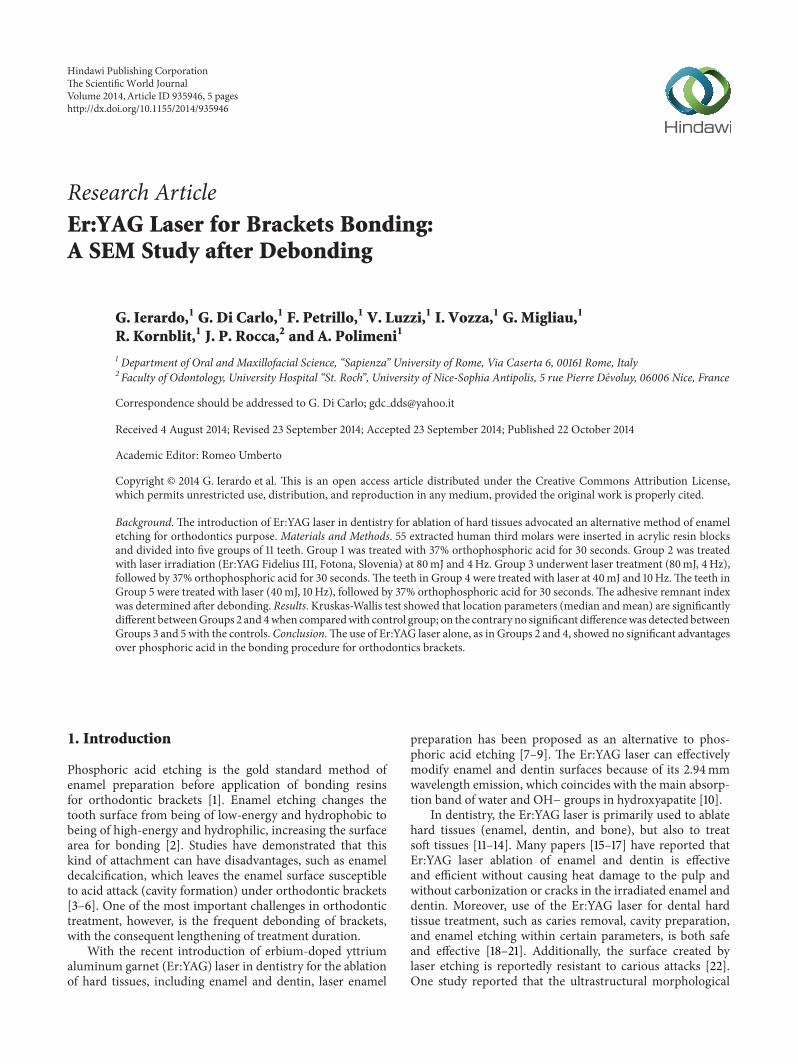
Figure 4: Bracket bonded on the enamel.
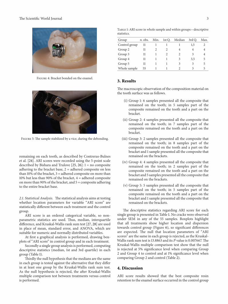
Figure 5: The sample stabilized by a vice, during the debonding.
remaining on each tooth, as described by Contreras-Bulneset al. [24]. ARI scores were recorded using the 5-point scaledescribed by Bishara and Trulove [25, 26]: 1 = no compositeadhering to the bracket base, 2 = adhered composite on lessthan 10% of the bracket, 3 = adhered composite onmore than10% but less than 90% of the bracket, 4 = adhered compositeonmore than 90% of the bracket, and 5 = composite adheringto the entire bracket base.
2.1. Statistical Analysis. The statistical analysis aims at testingwhether location parameters for variable “ARI score” arestatistically different between each treatment and the controlgroup.
ARI score is an ordered categorical variable, so non-parametric statistics are used. Thus, median, interquartiledifference, and Kruskal-Wallis rank sum test [27, 28] are usedin place of mean, standard error, and ANOVA, which aresuitable for numeric and normally distributed variables.
At first a graphical analysis is performed, drawing box-plots of “ARI score” in control group and in each treatment.
Secondly a single group analysis is performed, computingdescriptive statistics (median, 1st and 3rd quartile) in eachgroup (Table 1).
Thirdly the null hypothesis that the medians are the samein each group is tested against the alternative that they differin at least one group by the Kruskal-Wallis rank sum test.As the null hypothesis is rejected, the after Kruskal-Wallismultiple comparison test between treatments versus controlis performed.
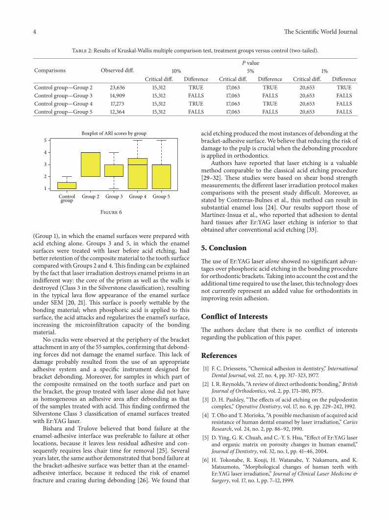
Table 1: ARI score in whole sample and within groups—descriptivestatistics.
Group 𝑛. obs. Min. 1st Q. Median 3rdQ. Max.Control group 11 1 1 1 1,5 2Group 2 11 2 2 4 4 4Group 3 11 1 2 2 3 4Group 4 11 1 1 3 3,5 5Group 5 11 1 1 3 3 5Whole sample 55 1 1 2 3 5
3. Results
Themacroscopic observation of the composition material onthe tooth surface was as follows.
(i) Group 1: 6 samples presented all the composite thatremained on the tooth; in 5 samples part of thecomposite remained on the tooth and a part on thebracket.
(ii) Group 2: 4 samples presented all the composite thatremained on the tooth; in 7 samples part of thecomposite remained on the tooth and a part on thebracket.
(iii) Group 3: 2 samples presented all the composite thatremained on the tooth; in 8 samples part of thecomposite remained on the tooth and a part on thebracket and 1 sample presented all the composite thatremained on the brackets.
(iv) Group 4: 4 samples presented all the composite thatremained on the tooth; in 2 samples part of thecomposite remained on the tooth and a part on thebracket and 5 samples presented all the composite thatremained on the brackets.
(v) Group 5: 5 samples presented all the composite thatremained on the tooth; in 5 samples part of thecomposite remained on the tooth and a part on thebracket and 1 sample presented all the composite thatremained on the brackets.
The descriptive statistics regarding ARI score for eachsingle group is presented in Table 1. No cracks were observedunder SEM in any of the 55 samples. Boxplots highlightthat all treatments show higher location and dispersiontowards control group (Figure 6), so significant differencesare expected. The null that location parameters of “ARIscores” are the same in each group is rejected, as the Kruskal-Wallis rank sum test is 13.8863 and its 𝑃 value is 0.007667.TheKruskal-Wallis multiple comparison test show that the nullis rejected at 5% significance level when comparing Group2 and Group 4 to control and at 1% significance level whencomparing Group 2 and control (Table 2).
4. Discussion
ARI score results showed that the best composite resinretention to the enamel surface occurred in the control group
4 The Scientific World Journal
Table 2: Results of Kruskal-Wallis multiple comparison test, treatment groups versus control (two-tailed).
Comparisons Observed diff.𝑃 value
10% 5% 1%Critical diff. Difference Critical diff. Difference Critical diff. Difference
Control group—Group 2 23,636 15,312 TRUE 17,063 TRUE 20,653 TRUEControl group—Group 3 14,909 15,312 FALLS 17,063 FALLS 20,653 FALLSControl group—Group 4 17,273 15,312 TRUE 17,063 TRUE 20,653 FALLSControl group—Group 5 12,364 15,312 FALLS 17,063 FALLS 20,653 FALLS
1
2
3
4
5
Controlgroup
Group 2 Group 3 Group 4 Group 5
Boxplot of ARI scores by group
Figure 6
(Group 1), in which the enamel surfaces were prepared withacid etching alone. Groups 3 and 5, in which the enamelsurfaces were treated with laser before acid etching, hadbetter retention of the compositematerial to the tooth surfacecompared with Groups 2 and 4.This finding can be explainedby the fact that laser irradiation destroys enamel prisms in anindifferent way: the core of the prism as well as the walls isdestroyed (Class 3 in the Silverstone classification), resultingin the typical lava flow appearance of the enamel surfaceunder SEM [20, 21]. This surface is poorly wettable by thebonding material; when phosphoric acid is applied to thissurface, the acid attacks and regularizes the enamel’s surface,increasing the microinfiltration capacity of the bondingmaterial.
No cracks were observed at the periphery of the bracketattachment in any of the 55 samples, confirming that debond-ing forces did not damage the enamel surface. This lack ofdamage probably resulted from the use of an appropriateadhesive system and a specific instrument designed forbracket debonding. Moreover, for samples in which part ofthe composite remained on the tooth surface and part onthe bracket, the group treated with laser alone did not haveas homogeneous an adhesive area after debonding as thatof the samples treated with acid. This finding confirmed theSilverstone Class 3 classification of enamel surfaces treatedwith Er:YAG laser.
Bishara and Trulove believed that bond failure at theenamel-adhesive interface was preferable to failure at otherlocations, because it leaves less residual adhesive and con-sequently requires less chair time for removal [25]. Severalyears later, the same author demonstrated that bond failure atthe bracket-adhesive surface was better than at the enamel-adhesive interface, because it reduced the risk of enamelfracture and crazing during debonding [26]. We found that
acid etching produced themost instances of debonding at thebracket-adhesive surface. We believe that reducing the risk ofdamage to the pulp is crucial when the debonding procedureis applied in orthodontics.
Authors have reported that laser etching is a valuablemethod comparable to the classical acid etching procedure[29–32]. These studies were based on shear bond strengthmeasurements; the different laser irradiation protocol makescomparisons with the present study difficult. Moreover, asstated by Contreras-Bulnes et al., this method can result insubstantial enamel loss [24]. Our results support those ofMartınez-Insua et al., who reported that adhesion to dentalhard tissues after Er:YAG laser etching is inferior to thatobtained after conventional acid etching [33].
5. Conclusion
The use of Er:YAG laser alone showed no significant advan-tages over phosphoric acid etching in the bonding procedurefor orthodontic brackets. Taking into account the cost and theadditional time required to use the laser, this technology doesnot currently represent an added value for orthodontists inimproving resin adhesion.
Conflict of Interests
The authors declare that there is no conflict of interestsregarding the publication of this paper.
References
[1] F. C. Driessens, “Chemical adhesion in dentistry,” InternationalDental Journal, vol. 27, no. 4, pp. 317–323, 1977.
[2] I. R. Reynolds, “A review of direct orthodontic bonding,” BritishJournal of Orthodontics, vol. 2, pp. 171–180, 1975.
[3] D. H. Pashley, “The effects of acid etching on the pulpodentincomplex,” Operative Dentistry, vol. 17, no. 6, pp. 229–242, 1992.
[4] T. Oho and T.Morioka, “A possible mechanism of acquired acidresistance of human dental enamel by laser irradiation,” CariesResearch, vol. 24, no. 2, pp. 86–92, 1990.
[5] D. Ying, G. K. Chuah, and C.-Y. S. Hsu, “Effect of Er:YAG laserand organic matrix on porosity changes in human enamel,”Journal of Dentistry, vol. 32, no. 1, pp. 41–46, 2004.
[6] H. Tokonabe, R. Kouji, H. Watanabe, Y. Nakamura, and K.Matsumoto, “Morphological changes of human teeth withEr:YAG laser irradiation,” Journal of Clinical Laser Medicine &Surgery, vol. 17, no. 1, pp. 7–12, 1999.
The Scientific World Journal 5
[7] T. Ozer, G. Basaran, and N. Berk, “Laser etching of enamel fororthodontic bonding,” American Journal of Orthodontics andDentofacial Orthopedics, vol. 134, no. 2, pp. 193–197, 2008.
[8] S. Usumez, M. Orhan, and A. Usumez, “Laser etching ofenamel for direct bonding with an Er,Cr:YSGG hydrokineticlaser system,”American Journal of Orthodontics and DentofacialOrthopedics, vol. 122, no. 6, pp. 649–656, 2002.
[9] G. Basaran, T. Ozer, N. Berk, and O. Hamamci, “Etchingenamel for orthodontics with an erbium, chromium:yttrium-scandium- gallium-garnet laser system,” Angle Orthodontist,vol. 77, no. 1, pp. 117–123, 2007.
[10] E. Firat, S. Gurgan, and N. Gutknecht, “Microtensile bondstrength of an etch-and-rinse adhesive to enamel and dentinafter Er:YAG laser pretreatment with different pulse durations,”Lasers in Medical Science, vol. 27, no. 1, pp. 15–21, 2012.
[11] U. Keller and R. Hibst, “Experimental studies of the applicationof the Er:YAG laser on dental hard substances: II. Lightmicroscopic and SEM investigations,” Lasers in Surgery andMedicine, vol. 9, no. 4, pp. 345–351, 1989.
[12] A. Stabholz, R. Zeltser, M. Sela, B. Peretz, J. Moshonov, andD. Ziskind, “The use of lasers in dentistry: principles ofoperation and clinical applications,”Compendium of ContinuingEducation in Dentistry, vol. 24, no. 12, pp. 935–949, 2003.
[13] C. M. Cobb, “Lasers in periodontics: a review of the literature,”Journal of Periodontology, vol. 77, no. 4, pp. 545–564, 2006.
[14] M. D. Genovese and G. Olivi, “Use of laser technology inorthodontics: hard and soft tissue laser treatments,” EuropeanJournal of Paediatric Dentistry, vol. 11, no. 1, pp. 44–48, 2010.
[15] C. Cozean, C. J. Arcoria, J. Pelagalli, andG. L. Powell, “Dentistryfor the 21st century? Erbium:YAG laser for teeth,”The Journal ofthe American Dental Association, vol. 128, no. 8, pp. 1080–1087,1997.
[16] L. H. Sasaki, P. D. C. Lobo, Y. Moriyama et al., “Tensile bondstrength and SEM analysis of enamel etched with Er:YAG laserand phosphoric acid: a comparative study in vitro,” BrazilianDental Journal, vol. 19, no. 1, pp. 57–61, 2008.
[17] V. Colucci, F. L. B. do Amaral, J. D. Pecora, R. G. Palma-Dibb, and S. A. Milori Corona, “Water flow on erbium:yttrium-aluminum-garnet laser irradiation: effects on dental tissues,”Lasers in Medical Science, vol. 24, no. 5, pp. 811–818, 2009.
[18] R. Kornblit, D. Trapani, M. Bossu, M. Muller-Bolla, J. P. Rocca,and A. Polimeni, “The use of Erbium:YAG laser for cariesremoval in paediatric patients following minimally invasivedentistry concepts,” European Journal of Paediatric Dentistry,vol. 9, no. 2, pp. 81–87, 2008.
[19] B. N. Cavalcanti, J. L. Lage-Marques, and S. M. Rode, “Pulpaltemperature increases with Er:YAG laser and high-speed hand-pieces,” Journal of Prosthetic Dentistry, vol. 90, no. 5, pp. 447–451, 2003.
[20] V. R. Geraldo-Martins, F. R. P. Robles, and A. B. Matos,“Chlorhexidine’s effect on sealing ability of composite restora-tions following Er:YAG laser cavity preparation,” Journal ofContemporary Dental Practice, vol. 8, no. 5, pp. 26–33, 2007.
[21] A. L. L. Klein, L. K. A. Rodrigues, C. P. Eduardo, M. N. dosSantos, and J. A. Cury, “Caries inhibition around compositerestorations by pulsed carbon dioxide laser application,” Euro-pean Journal of Oral Sciences, vol. 113, no. 3, pp. 239–244, 2005.
[22] J. A. Hoke, E. J. Burkes Jr., E. D. Gomes, and M. L. Wolbarsht,“Erbium:YAG (2.94 mum) laser effects on dental tissues,”Journal of Laser Applications, vol. 2, no. 3-4, pp. 61–65, 1990.
[23] E. B. Groth, C. E. Mercer, and P. Anderson, “Microtomographicanalysis of subsurface enamel and dentine following Er:YAGlaser and acid etching,”The European Journal of Prosthodonticsand Restorative Dentistry, vol. 9, no. 2, pp. 73–79, 2001.
[24] R. Contreras-Bulnes, R. J. Scougall-Vilchis, L. E. Rodrıguez-Vilchis, C. Centeno-Pedraza, O. F. Olea-Mejıa, and M. D.C. Z. Alcantara-Galena, “Evaluation of self-etching adhesiveand Er:YAG laser conditioning on the shear bond strength oforthodontic brackets,” The Scientific World Journal, vol. 2013,Article ID 719182, 5 pages, 2013.
[25] S. E. Bishara and T. S. Trulove, “Comparisons of differentdebonding techniques for ceramic brackets: an in vitro study.Part I. Background and methods,” The American Journal ofOrthodontics and Dentofacial Orthopedics, vol. 98, no. 2, pp.145–153, 1990.
[26] S. E. Bishara and T. S. Trulove, “Comparisons of differentdebonding techniques for ceramic brackets: an in vitro study:part II. Findings and clinical implications,” American Journalof Orthodontics and Dentofacial Orthopedics, vol. 98, no. 3, pp.263–273, 1990.
[27] M. Hollander and D. A. Wolfe, Nonparametric Statistical Meth-ods, John Wiley & Sons, New York, NY, USA, 1973.
[28] S. Siegel and N. J. Castellan, Nonparametric Statistics for theBehavioral Sciences, McGraw-Hill, New York, NY, USA, 1988.
[29] G. Basaran, N. Hamamci, and A. Akkurt, “Shear bond strengthof bonding to enamel with different laser irradiation distances,”Lasers in Medical Science, vol. 26, no. 2, pp. 149–156, 2011.
[30] A. Gokcelik, Y. Ozel, E. Ozel et al., “The influence of Er:YAGlaser conditioning versus self-etching adhesives with acidetching on the shear bond strength of orthodontic brackets,”Photomedicine and Laser Surgery, vol. 25, no. 6, pp. 508–512,2007.
[31] J.-H. Kim, O.-W. Kwon, H.-I. Kim, and Y. H. Kwon, “Effec-tiveness of an Er:YAG laser in etching the enamel surface fororthodontic bracket retention,” Dental Materials Journal, vol.24, no. 4, pp. 596–602, 2005.
[32] B.-S. Lee, T.-T. Hsieh, Y.-L. Lee et al., “Bond strengths oforthodontic bracket after acid-etched, Er:YAG laser-irradiatedand combined treatment on enamel surface,” Angle Orthodon-tist, vol. 73, no. 5, pp. 565–570, 2003.
[33] A. Martınez-Insua, L. D. S. Dominguez, F. G. Rivera, and U.A. Santana-Penın, “Differences in bonding to acid-etched orEr:YAG-laser-treated enamel and dentin surfaces,” Journal ofProsthetic Dentistry, vol. 84, no. 3, pp. 280–288, 2000.

Submit your manuscripts athttp://www.hindawi.com
Hindawi Publishing Corporationhttp://www.hindawi.com Volume 2014
Oral OncologyJournal of
DentistryInternational Journal of
Hindawi Publishing Corporationhttp://www.hindawi.com Volume 2014
Hindawi Publishing Corporationhttp://www.hindawi.com Volume 2014
International Journal of
Biomaterials
Hindawi Publishing Corporationhttp://www.hindawi.com Volume 2014
BioMed Research International
Hindawi Publishing Corporationhttp://www.hindawi.com Volume 2014
Case Reports in Dentistry
Hindawi Publishing Corporationhttp://www.hindawi.com Volume 2014
Oral ImplantsJournal of
Hindawi Publishing Corporationhttp://www.hindawi.com Volume 2014
Anesthesiology Research and Practice
Hindawi Publishing Corporationhttp://www.hindawi.com Volume 2014
Radiology Research and Practice
Environmental and Public Health
Journal of
Hindawi Publishing Corporationhttp://www.hindawi.com Volume 2014
The Scientific World JournalHindawi Publishing Corporation http://www.hindawi.com Volume 2014
Hindawi Publishing Corporationhttp://www.hindawi.com Volume 2014
Dental SurgeryJournal of
Drug DeliveryJournal of
Hindawi Publishing Corporationhttp://www.hindawi.com Volume 2014
Hindawi Publishing Corporationhttp://www.hindawi.com Volume 2014
Oral DiseasesJournal of
Hindawi Publishing Corporationhttp://www.hindawi.com Volume 2014
Computational and Mathematical Methods in Medicine
ScientificaHindawi Publishing Corporationhttp://www.hindawi.com Volume 2014
PainResearch and TreatmentHindawi Publishing Corporationhttp://www.hindawi.com Volume 2014
Preventive MedicineAdvances in
Hindawi Publishing Corporationhttp://www.hindawi.com Volume 2014
EndocrinologyInternational Journal of
Hindawi Publishing Corporationhttp://www.hindawi.com Volume 2014
Hindawi Publishing Corporationhttp://www.hindawi.com Volume 2014
OrthopedicsAdvances in


![REVIEW Comparison of Er:YAG and Er,Cr:YSGG lasers used in ... · commonly used in dentistry; the Er:YAG (2940 nm) laser and the Er,Cr:YSGG (2790 nm) laser [1]. They exhibit the highest](https://img.pdfslide.us/doc/110x75/5f5c5b19e029dd1783396fd5/review-comparison-of-eryag-and-ercrysgg-lasers-used-in-commonly-used-in-dentistry.jpg)