Embed Size (px)
Citation preview

Research ArticleEffects of Alendronate Sodium Content on the InterfaceStrengths of Composite Acrylic Bone Cement
De-Ye Song, Xin-Zhan Mao, Mu-liang Ding, and Jiang-Dong Ni
Department of Orthopaedics, 2nd Xiangya Hospital, Central South University, Changsha, Hunan 410011, China
Correspondence should be addressed to Jiang-Dong Ni; [email protected]
Received 20 November 2014; Revised 15 January 2015; Accepted 21 January 2015
Academic Editor: Yebin Jiang
Copyright © 2015 De-Ye Song et al. This is an open access article distributed under the Creative Commons Attribution License,which permits unrestricted use, distribution, and reproduction in any medium, provided the original work is properly cited.
Objective. Aim to study how the content of alendronate affected shear strengths at bone-bone cement-metal interfaces. Methods.All samples were divided into 6 groups, G
0–G5. On the 1st and 60th day after surgery, bone-bone cement interface shear strengths
and bone densities were examined. Interface strengths of metal-bone cement specimens were studied before immersion and 4weeks after immersion. Results. On the 60th day, bone-bone cement interface shear strengths and bone densities showed significantdifferences (𝑃 < 0.05), and compared with G
0, G2–G5values increased significantly (𝑃 < 0.05), and the peak value was met in G
3.
Compared with the 1st day, on the 60th postoperative day both factors decreased significantly in G0and G
1(𝑃 < 0.05). Four weeks
after immersion, with the increasing dose of alendronate, the shear strengths decreased gradually and in G5decreased significantly
(𝑃 < 0.05). Compared with before immersion, the metal-bone cement interface strengths decreased significantly 4 weeks afterimmersion (𝑃 < 0.05). Conclusions. 50–500mg alendronate in 50 g cement powders could prevent the decrease of shear strengthsat bone-bone cement interfaces and had no effect on metal-bone cement interface strengths. While the addition dose was 100mg,bone cement showed the best strengths.
1. Introduction
With the growing aging population, the number of osteo-porotic elderly hip fracture patients has been graduallyincreasing [1]. Bone cement prosthesis replacement hasbecome a very effective method to treat these fractures [2].Due to the continual friction between the joint prosthesis,aseptic loosening induced by wear particles has become themain reason of the failure in long-term joint replacement [3].Therefore, how to prevent bone loss and the aseptic looseningafter joint prosthesis replacement has become a researchfocus.
As a class of synthetic analogs of pyrophosphate, bispho-sphonate is a potent new drug to inhibit bone resorption [4].Experiment researches had shown that the drug could inhibitbone loss after joint prosthesis arthroplasty [5], continuouslyincreasing bone densities around the prostheses [6], inhibit-ing the release of osteolytic factors [7], inhibiting osteolysisinduced by wear particles [8], promoting the proliferationand differentiation of osteoblasts [9], enhancing osteoblastactivity, inhibiting apoptosis of osteoblasts [10] and bone
absorption of osteoclasts, and accelerating apoptosis of osteo-clasts [11]. It is supposed that the drugmay be an ideal drug toprevent and cure aseptic loosening after prosthesis replace-ment.
Oral taking is amajor administration of bisphosphonates.If these drugs are taken orally for a long time, they have a lot ofside effects, such as low bioavailability, high treatment costs,and upper gastrointestinal ulcers [12, 13]. To avoid these sideeffects, the topical use added in acrylic bone cement may bea better way of administration. Alendronate is a third gener-ation of bisphosphonate and a regular drug used in the treat-ment of osteoporosis and osteoporotic fractures. In the formof powder, it has some advantages of being mixed easilyin bone cement powders, high temperature resistance andremaining drug efficacy in bone cement, and so forth. Liter-atures reported that acrylic bone cement compounded withalendronate had a favorable biocompatibility [14], and certaincontents of alendronate showed no detrimental effects on thefatigue life of composite acrylic bone cement [15].
Bone-bone cement andmetal-bone cement interfaces arecommon sites of aseptic loosening after bone cement joint
Hindawi Publishing CorporationInternational Journal of EndocrinologyVolume 2015, Article ID 502820, 6 pageshttp://dx.doi.org/10.1155/2015/502820
2 International Journal of Endocrinology
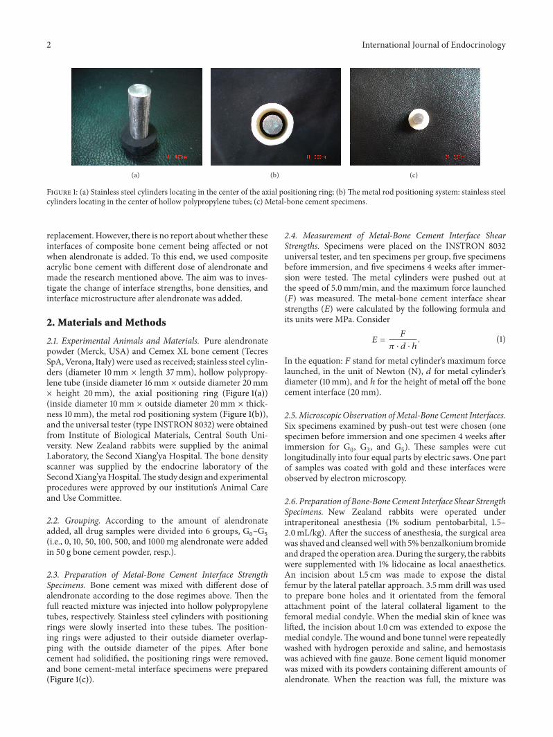
(a) (b) (c)
Figure 1: (a) Stainless steel cylinders locating in the center of the axial positioning ring; (b) The metal rod positioning system: stainless steelcylinders locating in the center of hollow polypropylene tubes; (c) Metal-bone cement specimens.
replacement. However, there is no report about whether theseinterfaces of composite bone cement being affected or notwhen alendronate is added. To this end, we used compositeacrylic bone cement with different dose of alendronate andmade the research mentioned above. The aim was to inves-tigate the change of interface strengths, bone densities, andinterface microstructure after alendronate was added.
2. Materials and Methods
2.1. Experimental Animals and Materials. Pure alendronatepowder (Merck, USA) and Cemex XL bone cement (TecresSpA, Verona, Italy) were used as received; stainless steel cylin-ders (diameter 10mm × length 37mm), hollow polypropy-lene tube (inside diameter 16mm × outside diameter 20mm× height 20mm), the axial positioning ring (Figure 1(a))(inside diameter 10mm × outside diameter 20mm × thick-ness 10mm), the metal rod positioning system (Figure 1(b)),and the universal tester (type INSTRON 8032) were obtainedfrom Institute of Biological Materials, Central South Uni-versity. New Zealand rabbits were supplied by the animalLaboratory, the Second Xiang’ya Hospital. The bone densityscanner was supplied by the endocrine laboratory of theSecondXiang’yaHospital.The study design and experimentalprocedures were approved by our institution’s Animal Careand Use Committee.
2.2. Grouping. According to the amount of alendronateadded, all drug samples were divided into 6 groups, G
0–G5
(i.e., 0, 10, 50, 100, 500, and 1000mg alendronate were addedin 50 g bone cement powder, resp.).
2.3. Preparation of Metal-Bone Cement Interface StrengthSpecimens. Bone cement was mixed with different dose ofalendronate according to the dose regimes above. Then thefull reacted mixture was injected into hollow polypropylenetubes, respectively. Stainless steel cylinders with positioningrings were slowly inserted into these tubes. The position-ing rings were adjusted to their outside diameter overlap-ping with the outside diameter of the pipes. After bonecement had solidified, the positioning rings were removed,and bone cement-metal interface specimens were prepared(Figure 1(c)).
2.4. Measurement of Metal-Bone Cement Interface ShearStrengths. Specimens were placed on the INSTRON 8032universal tester, and ten specimens per group, five specimensbefore immersion, and five specimens 4 weeks after immer-sion were tested. The metal cylinders were pushed out atthe speed of 5.0mm/min, and the maximum force launched(𝐹) was measured. The metal-bone cement interface shearstrengths (𝐸) were calculated by the following formula andits units were MPa. Consider
𝐸 =𝐹
𝜋 ⋅ 𝑑 ⋅ ℎ. (1)
In the equation: 𝐹 stand for metal cylinder’s maximum forcelaunched, in the unit of Newton (N), 𝑑 for metal cylinder’sdiameter (10mm), and ℎ for the height of metal off the bonecement interface (20mm).
2.5.Microscopic Observation ofMetal-Bone Cement Interfaces.Six specimens examined by push-out test were chosen (onespecimen before immersion and one specimen 4 weeks afterimmersion for G
0, G3, and G
5). These samples were cut
longitudinally into four equal parts by electric saws. One partof samples was coated with gold and these interfaces wereobserved by electron microscopy.
2.6. Preparation of Bone-BoneCement Interface Shear StrengthSpecimens. New Zealand rabbits were operated underintraperitoneal anesthesia (1% sodium pentobarbital, 1.5–2.0mL/kg). After the success of anesthesia, the surgical areawas shaved and cleansedwell with 5%benzalkoniumbromideand draped the operation area.During the surgery, the rabbitswere supplemented with 1% lidocaine as local anaesthetics.An incision about 1.5 cm was made to expose the distalfemur by the lateral patellar approach. 3.5mm drill was usedto prepare bone holes and it orientated from the femoralattachment point of the lateral collateral ligament to thefemoral medial condyle. When the medial skin of knee waslifted, the incision about 1.0 cm was extended to expose themedial condyle.The wound and bone tunnel were repeatedlywashed with hydrogen peroxide and saline, and hemostasiswas achieved with fine gauze. Bone cement liquid monomerwas mixed with its powders containing different amounts ofalendronate. When the reaction was full, the mixture was
International Journal of Endocrinology 3
filled into a volume of 20mL injector and injected into thebone tunnels in the bilateral distal femurs. Moderate pressurewas applied to both ends of the tunnel until the bone cementsolidified. The wound was washed twice and closed layer bylayer. Penicillin (800,000U) was injected by intramuscularinjection every day after surgery for 7 days. During theobservation period, the rabbits were fed under a standarddiet and raised in separate cages.
2.7. Preparation of the Bone-Bone Cement Interface Specimens.Twelve rabbits were assigned to each group. Six rabbits weresacrificed on the 1st day, and the other six rabbits on 60th dayafter surgery. The lower ends of the femurs, which containedthe specimens, were removed. The left specimens were usedto test the bone-bone cement interface shear strengths, whilethe right ones were used to scan bone densities surroundingthe interfaces.
2.8. Test of Shear Strength at the Bone-Bone Cement Interfaces.Bilateral femur condyles of the rabbits were trimmed to bone-bone cement interface samples with 9.0mm lengths withscalpel. Then the specimens were loaded on an INSTRON8032 universal tester, with a loading speed of 5mm/min. Thetester was halted until the load began to decline gradually.The maximum load was recorded, and the interface shearstrengths (𝐸), in the unit of MPa, were calculated by thefollowing equation:
𝐸 =𝐹
𝜋 ⋅ 𝑑 ⋅ 𝑙. (2)
In the equation:𝐹 stand formaximum load force launched, inthe unit ofNewton (N),𝑑 for bone cement cylinder’s diameter(3.5mm), and 𝑙 for the length of bone-bone cement interface(9.0mm).
2.9. Bone Densities Surrounding the Bone-Bone Cement Inter-faces. Specimens were trimmed to bone-bone cement inter-face with 3.0mm thickness with scalpel. Then cut unneces-sary bone and make these samples of a standard size (length6.0mm ×width 6.0mm × thickness 3.0mm) and at the sametimemake sure the bone cement cylinders locate in the centerof the specimens. Specimens were scanned using a bonedensity scanner and the bone densities were calculated by thetester’s software.
2.10. Statistical Analysis. SPSS 13.0 for Windows softwarewas used for the statistical analysis. Each set of data wereexpressed as mean ± standard deviation (SD) and one-wayanalysis of variance was performed. If there was a significantdifference, pairwise comparison was carried out betweengroups using Scheffe post hoc test. Paired 𝑡-tests were usedfor the shear strengths and the bone densities at bone-bonecement interfaces between the 1st day and 60th day aftersurgery, and metal-bone cement interface strengths betweenbefore immersion and 4 weeks after immersion. Test levelbilateral 𝛼 = 0.05 and 𝑃 < 0.05 was considered statisticallysignificant.
Table 1: Shear strengths of bone-bone cement interfaces (MPa) onthe 1st day and 60th day after surgery in each group.
Group 1st day 60th day 𝑃#
G0 5.5372 ± 0.2516 3.6700 ± 0.1341 <0.05G1 5.5868 ± 0.1729 3.7600 ± 0.1707 <0.05G2 5.5573 ± 0.2041 5.6625 ± 0.2906∗ >0.05G3 5.5630 ± 0.2708 5.6967 ± 0.2170∗ >0.05G4 5.6450 ± 0.2843 5.6100 ± 0.2184∗ >0.05G5 5.5330 ± 0.1787 5.6300 ± 0.1975∗ >0.05𝑃## 0.981 <0.05
Note: ##indicates one-way analysis of variance (ANOVA); #indicates 𝑡-test,𝑃 < 0.05; ∗indicates Scheffe’s post hoc test, 𝑃 < 0.05 compared with G0.
Table 2: Bone densities surrounding the bone-bone cement inter-faces (g/cm2) on the 1st day and 60th day after surgery in each group.
Group 1st day 60th day 𝑃#
G0 0.2396 ± 0.0527 0.1356 ± 0.0274 <0.05G1 0.2455 ± 0.0427 0.1313 ± 0.0095 <0.05G2 0.2512 ± 0.0108 0.2509 ± 0.0275∗ >0.05G3 0.2525 ± 0.0121 0.2584 ± 0.0206∗ >0.05G4 0.2546 ± 0.0111 0.2554 ± 0.0245∗ >0.05G5 0.2546 ± 0.0138 0.2512 ± 0.0139∗ >0.05𝑃## 0.957 <0.05
Note: ##indicates one-way analysis of variance (ANOVA); #indicates 𝑡-test,𝑃 < 0.05; ∗indicates Scheffe’s post hoc test, 𝑃 < 0.05 compared with G0.
3. Results
3.1. Shear Strengths of the Bone-Bone Cement Interfaces.Table 1 showed that on the 1st day after surgery, bone-bone cement interface shear strengths showed no significantdifferences in all groups (𝑃 > 0.05). However, on the 60th dayafter surgery, they showed significant differences (𝑃 < 0.05),and compared with G
0, bone-bone cement interface shear
strengths in G1showed no significant differences (𝑃 > 0.05),
but in the other groups their values increased significantly(𝑃 < 0.05), and the peak value wasmet in G
3. Comparedwith
the 1st postoperative day, on the 60th postoperative day bone-bone cement interface shear strengths significantly decreasedin G0and G
1(𝑃 < 0.05), not in G
2to G5(𝑃 > 0.05).
3.2. Bone Densities Surrounding the Bone-Bone Cement Inter-faces. Table 2 showed that on the 1st day after surgery, bonedensities surrounding bone-bone cement interfaces showedno significant differences in all groups (𝑃 > 0.05). How-ever, on the 60th day after surgery, they showed significantdifferences (𝑃 < 0.05), and compared with G
0, bone-bone
cement interface shear strengths in G1showed no significant
differences (𝑃 > 0.05), but in the other groups their valuesincreased significantly (𝑃 < 0.05), and the peak value wasmet in G
3. Compared with the 1st postoperative day, on the
60th postoperative day bone densities surrounding bone-bone cement interfaces significantly decreased in G
0and G
1
(𝑃 < 0.05), not in G2to G5(𝑃 > 0.05).

4 International Journal of Endocrinology
(a) (b) (c)
(d) (e) (f)
Figure 2: Electron microscopy of the metal-bone cement interfaces: (a) and (d) before immersion and 4 weeks after immersion in G0,
respectively; (b) and (e) before immersion and 4 weeks after immersion in G3, respectively; and (c) and (f) before immersion and 4 weeks
after immersion in G5, respectively.
Table 3: Shear strengths of themetal-bone cement interfaces (MPa)before immersion and 4 weeks after immersion in each group.
Group Before immersion 4 weeks after immersion 𝑃#
G0 5.746 ± 0.7701 4.244 ± 0.0709 <0.05G1 5.668 ± 0.0864 4.200 ± 0.0632 <0.05G2 5.652 ± 0.0834 4.178 ± 0.0581 <0.05G3 5.598 ± 0.1188 4.172 ± 0.0286 <0.05G4 5.564 ± 0.1250 4.138 ± 0.0835 <0.05G5 5.534 ± 0.1043 3.530 ± 0.0418∗ <0.05𝑃## 0.053 <0.05
Note: ##indicates one-way analysis of variance (ANOVA); #indicates 𝑡-test,𝑃 < 0.05; ∗indicates Scheffe’s post hoc test, 𝑃 < 0.05 compared with G0.
3.3. Shear Strengths of Metal-Bone Cement Interfaces. Table 3showed that before immersion, metal-bone cement interfacestrengths in all groups showed no significant difference (𝑃 >0.05), while 4 weeks after immersion, with the increasingdose of alendronate, the shear strengths decreased gradually,and in G
5the decrease showed significant difference (𝑃 <
0.05). Compared with that before immersion, the metal-bone cement interface strengths significantly decreased in allgroups 4 weeks after immersion (𝑃 < 0.05).
3.4. Electron Microscopy Observation of the Metal-BoneCement Interfaces. Figure 2 showed that the porosity of bonecement specimens was similar before immersion, or 4 weeksafter immersion in G
0, G3, or G
5. But compared with before
immersion, the porosity in the same group increased obvi-ously 4 weeks after immersion.
4. Discussion
The interfaces between the femoral component and bonecement were known to be a weak area of bone-bone cementprosthesis complex [16]. Previously, Harris and Jasty foundthat the mainmechanism of aseptic loosening on the femoralside was the debonding of the femoral component-bonecement by analyzing the prosthesis removed. Finite elementanalysis showed that shear stress was a major stress factorfor joint prosthesis failure [17]. Interface shear strengths wereinfluenced by a variety of factors, including surface roughnessof the femoral stem component, preheating or precoating ofthe stem component [18], precooling of bone cement mono-mer, the type of bone cement, the type of prosthesis metal,and the load rate. As a part of this study, we investigatedthe effects of alendronate on metal-bone cement interfaceshear strengths. The results showed that before immer-sion, there is no significant difference in the metal-cementinterface strengths in all groups, while 4 weeks afterimmersion, with the increasing dose of alendronate, theshear strengths decreased gradually, and in group G
5the
decrease showed significant difference. Meanwhile, electronmicroscopy showed that no significant difference was foundwith regard to the interface porosity before immersion or4 weeks after immersion in groups G
0, G3, and G
5. These
results indicated that the decrease of shear strength of metal-bone cement, was more attributed to decrease of the bonecement bonding capacity than the interface porosity. Thesestudies also found that, compared with that before immer-sion, 4 weeks after immersion the metal-bone cement inter-face strengths decreased significantly in the same group.Therefore, it was proposed that the main factor decreasing

International Journal of Endocrinology 5
bone cement bonding capacitymight be related to immersionin saline.
Bone-bone cement interfaces were another commonsite of aseptic loosening after joint prosthesis replacement.According to published reports, aseptic loosening aboutapproximately 50–79% was found 15 years after total hiparthroplasty for young active patients, and 16% of thesepatients needed revision arthroplasty [19]. Because of theimpossible bonding between hydrophobic bone cement andhydrophilic bone tissues, bone cement was used as fillersinstead of binders [19]. Instead, interfaces between bonecement and bone tissue become stable fixation by a mechan-ical intercourse locking. Some studies showed that the shearstrengths of bone-bone cement interfaces could be increasedby enhancing microlocking between bone cement and bone,and precoating with a layer of an amphiphilic substance onthe bone surface. There are many factors that can influencethe interface shear strengths, including bone porosity [20],trabecular orientation [20], continuous pressures on thecement [20], preparation of bone surface, and viscosity ofbone cement.Moran et al. found that shear strengths of bone-bone cement interfaces were not influenced by gentamicin(0.5 g, 1.0 g, 2.0 g, or 4.0 g) added in 40 g bone cement pow-ders [21]. Moreover, the shear strengths in bone cement werehigher than that at bone-bone cement interfaces.Therefore, itis obvious that shear strengths of bone-bone cement interfaceare a key factor for joint prosthesis service life.
This study found that on the 1st day after surgery shearstrengths of bone-bone cement interface in all groups showedno significant difference. However, significant differenceswere observed on the 60th day after operation. Comparedwith the 1st day, the interface strengths decreased significantlyafter 60 days after surgery in G
0and G
1, but no obvious
changes were shown in G2–G5. To investigate the reason
of shear strengths’ changes, we scanned the bone densitiessurrounding the bone-bone cement interfaces on the 1st and60th day after surgery. The results showed that there weresimilar changes between bone densities and shear strengths atbone-bone cement interfaces. According to the phenomenonabove, we inferred that the bone densities might be animportant factor to decide the shear strengths at bone-bonecement interfaces. The reason was that bone cement had bet-ter mechanical strengths than trabecular bone tissue. Mean-while, in G
0and G
1, bone densities had significant reduction
after 60 days after operation. It might be related to surgicaltrauma, thermal damage from bone cement, and activitiesreduction of rabbits. However, in G
2–G5, bone densities at
bone-bone cement interfaces showed no significant change.This might result frommineralization capacity enhancementof osteoblast and function inhibition of osteoclast, which wascaused by the topical release of alendronate and offset of thenegative effects on bone densities.
The advantages and disadvantages of this study alsodeserved discussion: (1) In this study, we used distal femurs ofNew Zealand rabbits to prepare the bone-bone cement inter-faces. Compared with diseased femoral heads used byMoran[21], the advantages included convenience of obtaining spec-imens, and an increase in sample volume, and avoidingnegative impact from structure differences in diseased bone.
However, using healthy tissue also had some drawbacks.These specimens obtained were smaller and more difficult toprepare bone-bone cement interfaces. When alive specimenssubjects were used, the bleeding at the interfaces could affectthe study results. (2) As artificial femoral stem substitute,stainless steel cylinder had different morphology and surfacefriction coefficient, which resulted in different shear strengthvalues. However, we had already homogenized these factorsthat might affect the interface strengths in our experiments,therefore the study results were reliable.
In conclusion, these results showed that a certain amountof alendronate in bone cement had a remarkable effect oninterface strengths of composite acrylic bone cement andinterfacial bone densities. A dose of 50–500mg alendronatein 50 g bone cement powder could prevent the decrease ofinterface strengths at bone-bone cement interfaces, and it hada similar effect on bone densities around these interfaces.However, the same doses of alendronate showed no effecton the interface strengths of metal-bone cement interfacesbefore immersion and 4 weeks after immersion. While theaddition dose was 100mg, bone cement showed the beststrengths.The results of this study indicated that alendronate-loaded bone cement could be made, but alendronate amountmust be controlled to below a certain level which hadno effecton the shear strengths at metal-bone cement-bone interfaces.
Conflict of Interests
The authors declare that there is no conflict of interestsregarding the publication of this paper.
Acknowledgments
This study was supported in part by a grant from the Devel-opment and Reform Commission Plan of Hunan Province((2013) no. 1199). The authors would like to extend theirappreciation to Dr. Junjie Wang and Dr. Jun Wang for theirconstructive suggestions.
References
[1] S. R. Cummings and L. J. Melton III, “Epidemiology and out-comes of osteoporotic fractures,”The Lancet, vol. 359, no. 9319,pp. 1761–1767, 2002.
[2] W. H. Harris, “The problem is osteolysis,” Clinical Orthopaedicsand Related Research, no. 311, pp. 46–53, 1995.
[3] J. E. Dowd, L. J. Schwendeman, W. Macaulay et al., “Asepticloosening in uncemented total hip arthroplasty in a caninemodel,” Clinical Orthopaedics and Related Research, no. 319, pp.106–121, 1995.
[4] B. Jobke, P. Milovanovic, M. Amling, and B. Busse, “Bisphos-phonate-osteoclasts: changes in osteoclast morphology andfunction induced by antiresorptive nitrogen-containing bispho-sphonate treatment in osteoporosis patients,” Bone, vol. 6, no.59, pp. 37–43, 2013.
[5] A. Nehme, G. Maalouf, J.-L. Tricoire, G. Giordano, P. Chiron,and J. Puget, “Effect of alendronate on periprosthetic bone lossafter cemented primary total hip arthroplasty: a prospective

6 International Journal of Endocrinology
randomized study,” Revue de Chirurgie Orthopedique et Repara-trice de l’Appareil Moteur, vol. 89, no. 7, pp. 593–598, 2003.
[6] J. K. Lee, C. H. Choi, and C.-N. Kang, “Quantitative computedtomography assessment of bone mineral density after 2 years’oral bisphosphonate treatment in postmenopausal osteoarthri-tis patients who underwent total knee arthroplasty,” Journal ofInternational Medical Research, vol. 41, no. 3, pp. 878–888, 2013.
[7] O. L. Huk, D. J. Zukor, J. Antoniou, and A. Petit, “Effect ofpamidronate on the stimulation of macrophage TNF-𝛼 releaseby ultra-high-molecular-weight polyethylene particles: a rolefor apoptosis,” Journal of Orthopaedic Research, vol. 21, no. 1, pp.81–87, 2003.
[8] C. Trevisan, V. Nava, M. Mattavelli, and C. G. Parra, “Bisphos-phonate treatment for osteolysis in total hip arthroplasty. Areport of four cases,” Clinical Cases in Mineral and Bone Metab-olism, vol. 10, no. 1, pp. 61–64, 2013.
[9] G.-I. Im, S. A. Qureshi, J. Kenney, H. E. Rubash, and A. S.Shanbhag, “Osteoblast proliferation and maturation by bispho-sphonates,” Biomaterials, vol. 25, no. 18, pp. 4105–4115, 2004.
[10] L. I. Plotkin, R. S. Weinstein, A. M. Parfitt, P. K. Roberson,S. C. Manolagas, and T. Bellido, “Prevention of osteocyte andosteoblast apoptosis by bisphosphonates and calcitonin,” Jour-nal of Clinical Investigation, vol. 104, no. 10, pp. 1363–1374, 1999.
[11] M. Kellinsalmi, H. Monkkonen, J. Monkkonen et al., “In vitrocomparison of clodronate, pamidronate and zoledronic acideffects on rat osteoclasts and human stem cell-derived osteo-blasts,” Basic and Clinical Pharmacology and Toxicology, vol. 97,no. 6, pp. 382–391, 2005.
[12] M. A. Gonzalez-Moles and J. V. Bagan-Sebastian, “Alendronate-related oral mucosa ulcerations,” Journal of Oral Pathology andMedicine, vol. 29, no. 10, pp. 514–518, 2000.
[13] P. C. de Groen, D. F. Lubbe, L. J. Hirsch et al., “Esophagitisassociated with the use of alendronate,” New England Journalof Medicine, vol. 335, no. 14, pp. 1016–1021, 1996.
[14] T. Calvo-Fernandez, J. Parra, M. Fernandez-Gutierrez et al.,“Biocompatibility of alendronate-loaded acrylic cement for ver-tebroplasty,” European Cells and Materials, vol. 20, pp. 260–273,2010.
[15] G. Lewis and S. Janna, “Alendronate in bone cement: fatigue lifedegraded by liquid, not by powder,” Clinical Orthopaedics andRelated Research, no. 445, pp. 233–238, 2006.
[16] T. P. Harrigan, J. A. Kareh, D. O. O’Connor, D.W. Burke, andW.H. Harris, “A finite element study of the initiation of failure offixation in cemented femoral total hip components,” Journal ofOrthopaedic Research, vol. 10, no. 1, pp. 134–144, 1992.
[17] N. Verdonschot and R. Huiskes, “The effects of cement-stemdebonding in THA on the long-term failure probability ofcement,” Journal of Biomechanics, vol. 30, no. 8, pp. 795–802,1997.
[18] R. T. Muller and N. Schurmann, “Shear strength of the cementmetal interface—an experimental study,” Archives of Orthopae-dic and Trauma Surgery, vol. 119, no. 3-4, pp. 133–138, 1999.
[19] H. J. Erli, R. Marx, O. Paar, F. U. Niethard, M. Weber, andD. C. Wirtz, “Surface pretreatments for medical applicationof adhesion,” BioMedical Engineering Online, vol. 2, article 15,2003.
[20] J. Graham,M. Ries, and L. Pruitt, “Effect of bone porosity on themechanical integrity of the bone-cement interface,”The Journalof Bone and Joint Surgery. American Volume, vol. 85, no. 10, pp.1901–1908, 2003.
[21] J. M. Moran, A. S. Greenwald, and M. B. Matejczyk, “Effect ofgentamicin on shear and interface strengths of bone cement,”Clinical Orthopaedics and Related Research, vol. 141, pp. 96–101,1979.

Submit your manuscripts athttp://www.hindawi.com
Stem CellsInternational
Hindawi Publishing Corporationhttp://www.hindawi.com Volume 2014
Hindawi Publishing Corporationhttp://www.hindawi.com Volume 2014
MEDIATORSINFLAMMATION
of
Hindawi Publishing Corporationhttp://www.hindawi.com Volume 2014
Behavioural Neurology
EndocrinologyInternational Journal of
Hindawi Publishing Corporationhttp://www.hindawi.com Volume 2014
Hindawi Publishing Corporationhttp://www.hindawi.com Volume 2014
Disease Markers
Hindawi Publishing Corporationhttp://www.hindawi.com Volume 2014
BioMed Research International
OncologyJournal of
Hindawi Publishing Corporationhttp://www.hindawi.com Volume 2014
Hindawi Publishing Corporationhttp://www.hindawi.com Volume 2014
Oxidative Medicine and Cellular Longevity
Hindawi Publishing Corporationhttp://www.hindawi.com Volume 2014
PPAR Research
The Scientific World JournalHindawi Publishing Corporation http://www.hindawi.com Volume 2014
Immunology ResearchHindawi Publishing Corporationhttp://www.hindawi.com Volume 2014
Journal of
ObesityJournal of
Hindawi Publishing Corporationhttp://www.hindawi.com Volume 2014
Hindawi Publishing Corporationhttp://www.hindawi.com Volume 2014
Computational and Mathematical Methods in Medicine
OphthalmologyJournal of
Hindawi Publishing Corporationhttp://www.hindawi.com Volume 2014
Diabetes ResearchJournal of
Hindawi Publishing Corporationhttp://www.hindawi.com Volume 2014
Hindawi Publishing Corporationhttp://www.hindawi.com Volume 2014
Research and TreatmentAIDS
Hindawi Publishing Corporationhttp://www.hindawi.com Volume 2014
Gastroenterology Research and Practice
Hindawi Publishing Corporationhttp://www.hindawi.com Volume 2014
Parkinson’s Disease
Evidence-Based Complementary and Alternative Medicine
Volume 2014Hindawi Publishing Corporationhttp://www.hindawi.com