Embed Size (px)
Citation preview

38
SAR Journal of Pathology and Microbiology
Abbreviated Key Title: SAR J Pathol Microbiol
Home page: https://sarmedjournals.com/sarjpm/
ISSN 2707-7756 (P)
Research Art ic le
Effect of fruits methanolic extracts on Tamarindus indica against some
bacterial isolates causing urinary tract infection among pregnant
women
Abdallah M.S1, Chilariye, I. A
2 and Aliyu, I. S
3
1 Desert research monitoring and control centre, Yobe State University, Damaturu, Nigeria.
2 Department of Biology, Yobe State University, Damaturu, Nigeria.
3 Kano University of Science and technology, Wudil, Kano State, Nigeria
*Corresponding Author
Abdallah, M.S
Article History: | Received: 12.03.2020 | Accepted: 22.04.2020 | Published: 28.04.2020 | Abstract: The present study aimed to determine the sensitivity pattern of the isolates against the Tamarindus indica fruit methanolic extracts as well
as to detect the mode of action of the extract. Phytoconstituents were obtained from the crude extracts through the process of qualitative mode of
screening and antibacterial activity was evaluated by agar well diffusion method against the gram negative bacteria. The bioactive ingredients found were mainly; the alkaloid, flavonoid, tannin, saponins, phenol, and phytosterols were found in the extracts of methanolic leaves which showed sound
activities against the tested organisms; E. coli and Shigella. The package used for the data analysis was (SAS) version 8.0. Keywords: Medicinal plants, microbes, traditional medicine, isolates tamarind.
Copyright @ 2020: This is an open-access article distributed under the terms of the Creative Commons Attribution license which permits unrestricted
use, distribution, and reproduction in any medium for non commercial use (NonCommercial, or CC-BY-NC) provided the original author and source
are credited.
INTRODUCTION Tamarindus indica being an indigenous to
tropical Africa, but has been cultivated for so long on
the Indian subcontinent that it is some time reported to
be indigenous there, where its known as (Imli) in Hindu
Urdu. It grows wild in Africa in local as diverse to
Sudan, Cameroon, Nigeria and Tanzania. In Arabia it is
found growing wild in Owon, especially Dhafur, where
it grows on the sea-facing slope of mountain. It reaches
south Asia likely through human transportation and
cultivation several thousand years and the America,
especially Mexico (Doughari et al., 2013).
Nevertheless, the young green leaves and the isolated
pule are component of a drink in Nigeria. Prepared by
infusing T. indica dried pule. In some part s of West
Africa non cereal plant contribute to the diets of local
resident mainly during time of grain shortage.
Moreover, fruit of T. indica (Tsamiya) contained a
moderate portion of protein WHO, 2002. T. indica
fruits also contained a reasonable amount of fatty acids
which served as a supplier of some diets for a wellbeing
of locals. In western Mali the nutritional importance of
green leaves and fruits from T. indica were used in
different season. Preferentially in rural region wild also
gathered the foods are used as much as fresh cultivated
food due to the presence of some important metabolites.
In Nigeria T. indica was applied against worm
infection, Trypanasomiasis in domestic as well as
against guinea worm. (Chung et al., 2005., Garba et
al.,2005 M et al., 2003, and Nassereddin.,
2005).However, T. indica reported to have a wide
spectrum of antibacterial activity. Some parts of it like
leaves and fruits methanolic extracts revealed a wide
potencies against the enteric bacterial isolates such as;
Klebsiella pneumoniae and E. coli using Agar well
diffusion method as well as compared with standard
antibiotic disks Amikacin and piperacillin (Vaghasiya et
al., 2009). The methanolic and aqueous of T. indica
revealed highest inhibition zones against isolates from
both gram positive and gram negative bacteria
(Doughari et al., 2006). Other studies have suggested
that T. indica has shown potential antibacterial activity.
Ethanolic extracts of T. indica ripe fruits were
pinpointed for the antibacterial potentialities against the
bacterial isolates (Warda et al., 2007). T. indica fruits
extracts when soaked in water consumed by Fulani
people in Nigeria, it cures diarrhoea (Lockett, et al.,
2000). It has also been reported that ethanolic, aqueous
and methanolic fruits of T. indica revealed a sound
inhibition zones against some clinical isolates;
Escherichia coli, Klebsiela pneumonia, Salmonella

Abdallah M.S et al., SAR J Pathol Microbiol; Vol-1, Iss- 2 (Mar-Apr, 2020): 38-43
39
paratyphi but resistant to some Pseudomonas
aeruginosa and Staphylococcus aureus, which was in
line with the presence of some metabolites that allow
the inhibitory actions perfectly well; alkaloids, saponin,
flavonoid, and tannins (Daniyan et al., 2008). The fruits
part of T. indica possessed vitamin B, carotene and
Vitamin C in a higher quantity. Subsequently, tannins
and some minerals (P, K, Ca and Mg) were also found
in a higher quantity. It served as an antioxidants, anti-
inflammatory, antimicrobial and antifungal, it was in
line with the properties possessed, recommended to be
utilised traditionally as medicine for many ailments in
many parts of Africa and beyond (Caluwe et al., 2010).
MATERIALS AND METHODS Preparation of leaves and fruits extracts
The fresh leaves of Tamarindus indica were
rinsed thoroughly in running tap water, chopped to tiny
pieces and air dried at room temperature for a period of
14 days, and subsequently pulverised with a pestle and
mortar. The fresh or pulp covering the seed was
removed and dried as below. Approximately 60.0 gram
of powered leaves pulp were each macerated in 500ml
of distilled water and methanol for period of 24 hour at
room temperature. The distilled water extraction and
methanol of each of the two plant part was described by
(Okoli et al., 2000). Also 60.0g fruits of Tamarindus
indica, pulp were each macerated in 500ml of hot water
for period of 24 hours. The hot water extraction and
methanol of each of the two plant part. Each preparation
was filtered through a Whatman filter paper and filtrate
evaporated to dryness in a steady air current after with
all extract were stored in a sterile container and store at
a room temperature (Azoro et al., 2000).
Phytochemical screening
The phytochemical screening purposed at
standardized extraction procedure for crude drugs
(Medicinal plant part) is to attain the therapeutically
desired portion and to eliminate unwanted materials by
treatment with a selective solvent known as menstrum.
The extract medicinal agent as such in the form of
tincture or fluid extracts or further processed to be
incorporated in any dosage form such as tablet and
capsules. The product contain complex mixture of many
medicinal plant metabolite such as alkaloid, flavonoid,
phytosterols, tannin, saponins and phenols (Prashant et
al., 2011).
Phytochemical screening on leaves of Tamarindus
Indica
Detection of flavonoids
Alkaline reagent test: Extract were treated with few
drops of sodium hydroxide solution. Formation of
intense yellow colour, which becomes colourless on
addition of diluted acid, indicates the presence of
flavonoids. (Ncube et al., 2008).
Detection of saponin
From the Test: The extracts were diluted with
distilled water to 20ml and this was shaken in a
graduated cylinder for 15minutes. Formation of 1cm
layer of foam indicate the presence of saponins. (Handa
et al., 2008).
Detection of tannins
Braymers Test: 2ml of the extracts plus few
drops (2-5) of 10% alcoholic ferric chloride solution.
Formation of brown-reddish precipitate indicate the
presence of tannin (Roy et al., 2005).
Detection of alkaloid
Wagner’s test: Filtrate were wagners reagent
(Iodine in potassium iodide). Formation of brown
reddish precipitate indicates the presence of alkaloids
(Parekh et al., 2010).
Detection of phenol
Ferric chloride test: Extract was treated with 3-
4 drops of ferric chloride solution. Formation of bluish
black color indicate the presence of phenol and the
phenol is absent (Parekh et al., 2009).
Detection of phytosterols
Salkawskis test: Extracts were treated with chloroform
and filtered. The filtrates were treated with few drops of
concentration sulphuric acid, shaken and allowed to
stand, golden yellow colour appeared indicates the
presence of phytosterols (Kumar et al., 2010).
Collection of sample for cultivation of test organisms
Stool samples were collected from
microbiology laboratory unit, General hospital
Potiskum, Yobe State. In sterile universal containers,
preserved and transported to Yobe State University,
Biology research laboratory under aseptic condition.
Media preparation/ XLD agar (Xylose, Lysine and
Deoxycholate)
28g of the XLD agar was weighed into a conical flask
and transferred into a conical flask containing 500ml of
distilled water. The mixture was heated on hot plate at
75 for 1 hour. The media was allowed to cool, and
poured into 20 petri-plates and allowed to solidify. The
stool samples were inoculated on the media using sterile
wire loop, incubated at 37 for 24 hours and observe
the growth of Shigella (Abdallah and Ali, 2018).
MacConkey agar
28g of the macConkey was weighed into a
conical flask and transferred into a conical flask
containing 500ml of distilled water. Heated boiled and
dissolved completely as well as Sterilized by
autoclaving for 15 minute, at 121 . The media was
allowed to cool and transferred into a sterile petri
dished up to the mark and to solidify (Abdallah and Ali,
2018).
Abdallah M.S et al., SAR J Pathol Microbiol; Vol-1, Iss- 2 (Mar-Apr, 2020): 38-43
40
Gram staining techniques
Thin smear of about 200mm in diameter were
formed on grease free slides which were also fixed over
a burning flame. A crystal violet solution was used to
cover the smear for 60 seconds and after that, distilled
water was applied to decolorized the stain and acetone
was applied, lastly the safranin solution was applied for
counter stain on the surface for a minute, washed and
allowed to dry at room temperature, then the stains were
observed under microscope with oil immersion (Mada
et al., 2012).
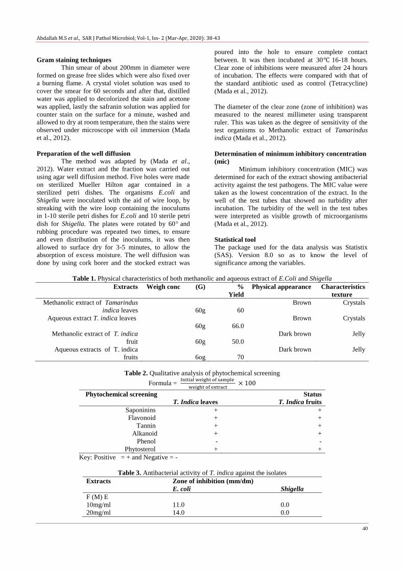
Preparation of the well diffusion
The method was adapted by (Mada et al.,
2012). Water extract and the fraction was carried out
using agar well diffusion method. Five holes were made
on sterilized Mueller Hilton agar contained in a
sterilized petri dishes. The organisms E.coli and
Shigella were inoculated with the aid of wire loop, by
streaking with the wire loop containing the inoculums
in 1-10 sterile petri dishes for E.coli and 10 sterile petri
dish for Shigella. The plates were rotated by 60 and
rubbing procedure was repeated two times, to ensure
and even distribution of the inoculums, it was then
allowed to surface dry for 3-5 minutes, to allow the
absorption of excess moisture. The well diffusion was
done by using cork borer and the stocked extract was
poured into the hole to ensure complete contact
between. It was then incubated at 30 16-18 hours.
Clear zone of inhibitions were measured after 24 hours
of incubation. The effects were compared with that of
the standard antibiotic used as control (Tetracycline)
(Mada et al., 2012).
The diameter of the clear zone (zone of inhibition) was
measured to the nearest millimeter using transparent
ruler. This was taken as the degree of sensitivity of the
test organisms to Methanolic extract of Tamarindus
indica (Mada et al., 2012).
Determination of minimum inhibitory concentration
(mic)
Minimum inhibitory concentration (MIC) was
determined for each of the extract showing antibacterial
activity against the test pathogens. The MIC value were
taken as the lowest concentration of the extract. In the
well of the test tubes that showed no turbidity after
incubation. The turbidity of the well in the test tubes
were interpreted as visible growth of microorganisms
(Mada et al., 2012).
Statistical tool
The package used for the data analysis was Statistix
(SAS). Version 8.0 so as to know the level of
significance among the variables.
Table 1. Physical characteristics of both methanolic and aqueous extract of E.Coli and Shigella
Extracts Weigh conc (G) %
Yield
Physical appearance Characteristics
texture
Methanolic extract of Tamarindus
indica leaves
60g
60
Brown Crystals
Aqueous extract T. indica leaves
60g
66.0
Brown Crystals
Methanolic extract of T. indica
fruit
60g
50.0
Dark brown Jelly
Aqueous extracts of T. indica
fruits
6og
70
Dark brown Jelly
Table 2. Qualitative analysis of phytochemical screening
Formula =
Phytochemical screening Status
T. Indica leaves T. Indica fruits
Saponinins + +
Flavonoid + +
Tannin + +
Alkanoid + +
Phenol - -
Phytosterol + +
Key: Positive = + and Negative = -
Table 3. Antibacterial activity of T. indica against the isolates
Extracts Zone of inhibition (mm/dm)
E. coli Shigella
F (M) E
10mg/ml
20mg/ml
11.0
14.0
0.0
0.0

Abdallah M.S et al., SAR J Pathol Microbiol; Vol-1, Iss- 2 (Mar-Apr, 2020): 38-43
41
30mg/ml
40mg/ml
50mg/ml
9.0
20.0
13.0
15.0
28.0
10.0
Table 4. Showing the zone of inhibitions in various extracts against the test organisms
Treatment EC SGL
Extract (ml)
L(m)E 12.800a 5.800
ab
L(m)E 3.6000b 3.600
ab
F(m)E 13.400a 10.600
ab
F(m)E 3.600b 1.2000
b
S.E 3.8678 4.4045
Sig. * NS
Conc. Levels (mg)
10 4.0000a 3.0000
a
20 7.2500a 2.2500
a
30 6.0000a 6.2500
a
40 10.250a 9.5000
a
50 14.250a 5.5000
a
S.E 5.1454 5.4268
Sig. NS NS
Means within a column followed by the same letters are statistically not significant at 5%
Level of probability using Duncan’s multiple range test (DMRT)
** = Significant at 1%, * = Significant only at 5% and Ns = Not significant at 5%. EC = E-Coli, SGL = Shigella
Table 5. Showing the minimum inhibitory concentration of methanolic extracts of fruit of Tamarindus indica on test
organisms
Treatment Cex
Test Organism
EC 0.2960a
SHG 0.1060a
S.E 0.1605
Sig. NS
Conc. Levels (mg)
10 0.0550a
20 495.07a
30 0.5250a
40 0.2400a
50 0.1150a
S.E 313.02
Sig. NS
Means within a column followed by the same letters are statistically not significant at 5% level of probability using
Duncan’s multiple range test (DMRT)
** = Significant at 1%, * = Significant only at 5% and Ns = Not significant at 5%. EC = E-Coli, SGL = Shigella, Cex
= Concentration of Extract
Table 6. Minimum inhibitory concentration (MIC) of aqueous extract fruits of Tamarindus Indica on test organisms.
Test organisms Concentration of extracts (mg/ml)
10 20 30 40 50
E. coli 0.0 0.0 0.0 0.0 0.13
Shigella spp 0.0 0.0 0.5 0.0 0.1
Formula: MIC =
Abdallah M.S et al., SAR J Pathol Microbiol; Vol-1, Iss- 2 (Mar-Apr, 2020): 38-43
42
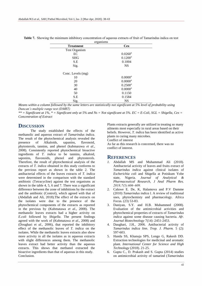
Table 7. Showing the minimum inhibitory concentration of aqueous extracts of fruit of Tamarindus indica on test
organisms
Treatment Cex
Test Organism
EC 0.0260a
SHG 0.1200a
S.E 0.1004
Sig. NS
Conc. Levels (mg)
10 0.0000a
20 0.0000a
30 0.2500a
40 0.0000a
50 0.1150
S.E 0.1584
Sig. NS
Means within a column followed by the same letters are statistically not significant at 5% level of probability using
Duncan’s multiple range test (DMRT)
** = Significant at 1%, * = Significant only at 5% and Ns = Not significant at 5%. EC = E-Coli, SGL = Shigella, Cex =
Concentration of Extract
DISCUSSION The study established the effects of the
methanolic and aqueous extract of Tamarindus indica.
The result of the phytochemical analysis revealed the
presence of Alkaloids, sapanins, flavonoid,
phytosterols, tannins, and phenol (kubmarawa et al.,
2008). Consistently reported phytochemical bioactive
ingredients of T. indica to be tannins, alkaloid,
saponins, flavonoids, phenol and phytosterols.
Therefore, the result of phytochemical analysis of the
extracts of T. indica obtained in this study conforms to
the previous report as shown in the table 2. The
antibacterial effects of the leaves extracts of T. indica
were determined in the comparison with the standard
antibiotic (Tetracycline) against the test organisms as
shown in the table 4, 5, 6 and 7. There was a significant
difference between the zone of inhibitions by the extract
and the antibiotic (Control), which agreed with that of
(Abdallah and Ali, 2018).The effect of the extracts on
the isolates were due to the presence of the
phytochemical components of the extracts as reported
in the previous by (Kubmatawa et al., 2008). The
methanolic leaves extracts had a higher activity on
E.coli followed by Shigella. The present findings
agreed with the work of (Kubmatawa et al., 2008) and
(Doughari et al., 2006), that reported the inhibitory
effect of the methanolic leaves of T. indica on the
isolates. While the methanolic leaves extracts also show
more activity in all the isolates as in aqueous extracts
with slight differences among them. The methanolic
leaves extract had better activity than the aqueous
extracts. This shows that methanol extracted the
bioactive ingredients than that of aqueous in this study.
Conclusion.
Plants extracts generally are utilized in treating so many
ailments most especially in rural areas based on their
beliefs. However, T. indica has been identified as active
plants in curing many microbes.
Conflict of interest
As far as this research is concerned, there was no
conflict of interest.
REFERENCES 1. Abdallah MS and Muhammad Ali (2018).
Antibacterial activity of leaves and fruits extract of
Tamarindus indica against clinical isolates of
Escherichia coli and Shigella at Potiskum Yobe
state, Nigeria. Journal of Analytical &
Pharmaceutical Research, J Anal Pharm Res.
2018;7(5):606‒609.
2. Caluwe E. De, K, Halamova and P.V Damme
(2010) Tamarindus indica l. A review of traditional
uses, phytochemisty and pharmacology. Africa
Focus. (23) 53-83.
3. Daniyan, S.Y. and H.B. Muhammed (2008).
Evaluation of the antimicrobial activities and
phytochemical properties of extracts of Tamarindus
indica against some disease causing bacteria. Afr.
Journal Biotechnology 7(14) :2451-2453.
4. Daughari, J.H., 2006. Antibacterial activity of
Tamarindus indica linn. Trop. J. Pharm. 5 (2):
597-603.
5. Handa SS, Khanuja SPS, Longo G, Rakesh DD.
Extraction technologies for medicinal and aromatic
plant. International Center for Science and High
Technology (2018). 21-25.
6. Gupta C., D. Prakash and S. Gupta (2014) studies
on antimicrobial activity of tamarind (Tamarindus
Abdallah M.S et al., SAR J Pathol Microbiol; Vol-1, Iss- 2 (Mar-Apr, 2020): 38-43
43
indica) and its potential as food bio-preservative.
International Food Research Journal. 21 (6) 2437-
2441.
7. Kubmarawa, D., Khan, M.E., Punah, A.M., and
Hassan, M., (2008), phytochemical screening and
antimicrobial efficacy of extracts of Khaya
senegalensis against human pathogenic bacteria
Afric.J. Biotechnol. 7 (24): 4563-4566.
8. Kumar R, Sharma RJ, Bairwa K, Roy RK, kumar
A. Pharmacological review on natural diarrhea
agents. Der pharma chemical (2010). 2(2): 66-93.
9. Lockett CY, Grivetti LE, Food-related behaviours
during drought: a Study o rural Fulani, northen
eastern Nigeria. International Journal Food
science Nutrition . 2000; 51; 91-107.
10. Mada ., Phytochemical and Antimicrobial Efficacy
of Mangiferaindica Stem Bark.World journal Life
sci. and Research. (2012):2; (2): 82.
11. Ncube NS, Afolayan HJ, Okoh AI. Assessment
techniques of antimicrobial properties of natural
compound of plant origin. Current method and
future trend. Africam Journal of Biotechnology
(2008). 7, (12): 1797-1806.
12. Kokwaro, O., 1976. Medicinal plant for west
Africa. East Africa literature Bureau, kampala,
Nairobi, Dar es salaam.
13. Prashant Tiwari, (Research scholar) Department of
pharmaceutical science (2011). (Jan-march
2011/vol 1/ issue 1.
14. Vaghasiya Y, chanda S. Screening of some
traditionally uses Indian plants for antibacterial
activity against klebsiella pneumonia. Journal
Herbs Medicine Toxicol. 2009; 3:161-4.
15. Warda SAG, Fathia M, Amel O. Antibacterial
activity of Tamaarindus Indica Fruits. Research
Journal of Microbiology. 2007; 2(11): 824- 30.


![REVIEW OF SUDANESE MEDICINAL PLANTS TESTED FOR THEIR ... · 100% mortality at concentration 1000 g/ml after 216 hours. [23] The methanolic and petroleum ether extracts of the seeds](https://img.pdfslide.us/doc/110x75/5f46c6834469056b7f739ad1/review-of-sudanese-medicinal-plants-tested-for-their-100-mortality-at-concentration.jpg)

![Mechanism of antidiabetic effects of Plicosepalus Acaciae ......their efficacy as antidiabetic drugs [6]. Bamane et al. [7] reported that methanolic extracts of Plicosepalus acacia](https://img.pdfslide.us/doc/110x75/60db669939dd7a66f77f8583/mechanism-of-antidiabetic-effects-of-plicosepalus-acaciae-their-efficacy.jpg)

![Journal of Plant Pathology & Microbiology...spp., Bipolaris sorokiniana, Curvularia lunata, Fusarium spp. of wheat to other extracts followed by ginger and neem [17,18]. Methanolic](https://img.pdfslide.us/doc/110x75/5e723483c7f1860d17507174/journal-of-plant-pathology-microbiology-spp-bipolaris-sorokiniana-curvularia.jpg)