Embed Size (px)
Citation preview
Research ArticleComputational Biophysical, Biochemical, and EvolutionarySignature of Human R-Spondin Family Proteins, the Member ofCanonical Wnt/𝛽-Catenin Signaling Pathway
Ashish Ranjan Sharma,1,2 Chiranjib Chakraborty,1,3 Sang-Soo Lee,1 Garima Sharma,1
Jeong Kyo Yoon,4 C. George Priya Doss,5 Dong-Keun Song,1 and Ju-Suk Nam1
1 Institute for Skeletal Aging & Orthopedic Surgery, Hallym University-Chuncheon Sacred Heart Hospital,Chuncheon 200704, Republic of Korea
2 Institute for Skeletal Aging & Orthopedic Surgery, Hallym University Hospital, College of Medicine, Chuncheon-si,Gangwon-do 200-704, Republic of Korea
3 Department of Bioinformatics, School of Computer Sciences, Galgotias University, Greater Noida 203201, India4Center for Molecular Medicine, Maine Medial Center Research Institute, 81 Research Drive, Scarborough, ME 04074, USA5Medical Biotechnology Division, School of Biosciences and Technology, VIT University, Vellore, Tamil Nadu 632014, India
Correspondence should be addressed to Chiranjib Chakraborty; [email protected] and Ju-Suk Nam; [email protected]
Received 4 April 2014; Revised 12 July 2014; Accepted 12 July 2014; Published 8 September 2014
Academic Editor: Tun-Wen Pai
Copyright © 2014 Ashish Ranjan Sharma et al. This is an open access article distributed under the Creative Commons AttributionLicense, which permits unrestricted use, distribution, and reproduction in any medium, provided the original work is properlycited.
In human,Wnt/𝛽-catenin signaling pathway plays a significant role in cell growth, cell development, and disease pathogenesis. Fourhuman (Rspo)s are known to activate canonical Wnt/𝛽-catenin signaling pathway. Presently, (Rspo)s serve as therapeutic target forseveral human diseases. Henceforth, basic understanding about themolecular properties of (Rspo)s is essential.We approached thisissue by interpreting the biochemical and biophysical properties alongwithmolecular evolution of (Rspo)s thorough computationalalgorithm methods. Our analysis shows that signal peptide length is roughly similar in (Rspo)s family along with similarity in aadistribution pattern. In Rspo3, four N-glycosylation sites were noted. All members are hydrophilic in nature and showed alikeGRAVY values, approximately. Conversely, Rspo3 contains the maximum positively charged residues while Rspo4 includes thelowest. Four highly aligned blocks were recorded through Gblocks. Phylogenetic analysis shows Rspo4 is being rooted with Rspo2and similarly Rspo3 and Rspo1 have the common point of origin. Through phylogenomics study, we developed a phylogenetictree of sixty proteins (𝑛 = 60) with the orthologs and paralogs seed sequences. Protein-protein network was also illustrated. Resultsdemonstrated in our studymay help the future researchers to unfold significant physiological and therapeutic properties of (Rspo)sin various disease models.
1. Introduction
R-spondins (Rspo)s are a recently discovered family of genesthat encodes cysteine-rich secretory proteins containing athrombospondin type 1 domain/repeat-1 [1]. The (Rspo)sfamily includes four conserved proteins (Rspo1, Rspo2,Rspo3, and Rspo4), showing overall similarity of 40–60%sequence homology and domain organization [2]. Besidesthe existence of TSR-1 domain, all four (Rspo)s can berecognized by the existence of a carboxy-terminal regionwith
positively charged amino acids and two furin-like cysteine-rich repeats adjacent to the amino terminus of the matureprotein. Numerous studies have implicated (Rspo)s for actingsynergistically with extracellular components of the Wntsignaling pathway (Figure 1) [3–5]. Studies showed closeor overlapped gene expression of Wnt and (Rspo)s duringdevelopmental events, implying a possible coupling of the(Rspo)s with Wnt signaling [6–8]. Consistent with this, asignificant reduction in mRNA expression of Rspo1 wasobserved in a Wnt1/3a double knockout mouse [1]. Rspo1
Hindawi Publishing CorporationBioMed Research InternationalVolume 2014, Article ID 974316, 22 pageshttp://dx.doi.org/10.1155/2014/974316

2 BioMed Research International
𝛽-catenin
𝛽-catenin
𝛽-catenin𝛽-catenin 𝛽-catenin
FrizzledWnt
R-spondin
Lrp
Lgr
Nucleus Nucleus
Tcf
Tcf
Wnt target geneWnt target gene
(a)
Sig. peptide furin-like CR1 furin-like CR2 TSR BR
(b)
Figure 1: The role of (Rspo)s in canonical Wnt signaling pathway and the general architecture of (Rspo)s. (a) Schematic diagram of Wntand R-spondin signaling models. In absence of Wnt ligand, constitutively synthesized cytoplasmic 𝛽-catenin is destroyed by the 𝛽-catenindestruction complex causing no 𝛽-catenin complex formation with T-cell transcription factor (Tcf)/Lef transcription factors for an activetranscriptional response. Canonical Wnt signaling is instigated by the binding of Wnt ligands to the frizzled/LRP receptor complex whichin turn deactivates the 𝛽-catenin destruction complex increasing its concentration in cytoplasm. The Wnt-frizzled/LRP complex-inducedcytoplasmic buildup of 𝛽-catenin leads to its import into the nucleus and binding to (Tcf)/Lef transcription factors initiating transcriptionof Wnt targeted genes. (Rspo)s also act in similar manner but induce this unique property of enhancing Wnt activity by binding to recentlydiscovered seven transmembraneG protein coupled receptors, Lgr (4, 5, and 6). (b) Schematic diagram shows the general domain architectureof human (Rspo)s. The architecture shows (i) signal peptide at N terminal end, (ii) two cysteine-rich furin-like repeats/domains, (iii) a singlethrombospondin domain, and (iv) a basic amino-acid-rich domain at C-terminal basic region.
has been shown to augment Wnt signaling by interactingwith the low-density lipoprotein receptor related protein 5or 6 (LRP5/6) coreceptor and inhibiting Dickkopf-1 (Dkk-1) mediated receptor internalization [9]. Rspo2 deficientmice show death at early stages and have limb patterningdefects associated with altered Wnt signaling [10, 11]. Rspo3interacts with Frizzled 8 and LRP-6 and enhancesWnt ligandsignaling [3, 4]. In addition to interaction with Wnt/𝛽-catenin signaling, (Rspo)s can also regulate noncanonicalWnt signaling [12]. It was found that furin domain repeatsare essential and sufficient for (Rspo)s to mediate Wnt-potentiating effects [13, 14]. Most recently, several studiesconclusively determined that the (Rspo)s are the ligandsfor the leucine-rich repeat containing G protein-coupledreceptor 4/5/6 (LGR4/5/6 receptors) [15–18].
Wnt signaling plays a fundamental role during fatedetermination steps of embryonic development and has beenshown to govern process like cell differentiation, cell prolif-eration, and stem cell maintenance [19, 20]. Due to (Rspo)sability to function as regulators of Wnt signaling pathways,various potential roles of (Rspo)s have been proposed andhave been suggested as novel therapeutic targets [17, 21].Rspo1 has been shown to control sex phenotypes betweenindividuals. A study by Parma et al. [22] observed sex reversaldue to the homozygous Rspo1 gene mutations in affectedindividuals. In addition, palmoplantar hyperkeratosis andpredisposition to squamous cell carcinoma of the skin werealso observed in these individuals. Rspo1 has also beenrecognized as a potent and specific mitogen for the gas-trointestinal epithelium [13, 23]. Various studies have also

BioMed Research International 3
implicated the importance of Rspo1 in skeletal biology. Rspo1has been shown to synergize with Wnt3a to promote theprocess of osteoblast differentiation and inhibit the process ofosteoclastogenesis by inducing expression of osteoprotegerin(OPG) [24–26]. Expression of Rspo2 has been shown to pro-motemyogenesis via theWnt/𝛽-catenin signaling pathway inXenopus [6]. A study with Rspo2 gene-targeted mutant miceobserved that Rspo2 is requisite for normal developmentof several tissues, including craniofacial structures, lung,kidney, and limbs [27]. Moreover, study reported that Rspo2is required for the maintenance of apical ectoderm ridge inthe hind limbs of the mice. In other studies on Rspo2 mutantmice, hypoplasia and branching defects within the lungswere also being reported [11, 28]. It was observed that Lrp6-mediated Rspo2 signaling via the canonical Wnt pathwayis essential for normal morphogenesis of the respiratorytract and for limbs as well [11]. Investigation into the genesresponsible for coat features in domestic dogs revealed thatRspo2 is also supported in the Wnt-mediated hair folliclegrowth [29]. More recently, the role of recurrent Rspo2 genefusion exclusively with APC mutations has been linked tothe activation ofWnt signaling and colon tumorigenesis [30].Like Rspo2 gene, recurrent Rspo3 gene fusions were alsofound to be associated with human colon tumors [30]. Inrecent time, it was proposed that Rspo3 gene may functionalong with Rspo2 gene in hind limb development, since theknockout of both Rspo2 and Rspo3 in limb mesenchymalcells caused more severe hind limb defects than those ofRspo2 mutant mice [31]. Rspo2 and Rspo3 genes were alsoidentified for their oncogenic potential in mouse mammarytumor virus associatedwithmammary tumorigenesis inmice[32, 33]. Expression of Rspo4 has been shown to play akey role during nail development and mutations in Rspo4gene results into absence of the nails in humans termed asanonychia/hyponychia congenita [34].
Given the diverse role of (Rspo)s in dynamic processesof life, like embryogenesis, tumor progression, angiogenesis,myogenesis, development of skeletal system, and so forth, wecan expect (Rspo)s as vital therapeutic targets for a numberof disabilities. Therefore, we tried to decipher biochemical,biophysical, molecular evolution, and protein-protein inter-action characteristics of (Rspo)s by a series of computer basedanalysis. It may help us to understand the basic molecularproperties of these molecules and thus their participation incritical events regulating essential life processes.
2. Materials and Methods
2.1. Data Mining for Human R-Spondin Protein FamilySequences and Their Feature of the Different Regions. Wegathered the information on the sequences of human (Rspo)sfamily members based on searches in the National Centre forBiotechnology Information database (http://www.ncbi.nlm.nih.gov/protein) [35] and UniProt (http://www.uniprot.org/) [36, 37]. The FASTA formats of the sequences werefurther retrieved for analysis. To investigate the features of theprimary structure such as the signal peptide in the proteinchain and the chain other than the signal peptide portion,
we usedUniProt server (http://www.uniprot.org/), a databasefor information on proteins [36–38]. To understand signalpeptide with “C-score” (predicted cleavage site value), “S-score” (the predicted signal peptide value), and “Y-score”(a combination of C- and S-scores), SignalP 4.0 server wasused [39]. In addition, different repeats and domain in the R-spondin family members have been analysed using UniProtserver.
2.2. Investigation of Amino Acid Distribution, Amino AcidComposition, and Some Parameters Related to the Pri-mary Structure Such as Charge Distribution Analysis, Repet-itive Structures, Cysteine Positions, and Disulphide Bondsof Human R-Spondin Family Proteins. To understand theamino acid distribution in the investigated proteins, we usedprotein calculator (http://spin.niddk.nih.gov/clore/Software/A205.html) [40]. In order to examine the amino acid pro-totype and protein sequence properties, such as amino acidcomposition percentage, high scoring hydrophobic segments,and tandem and periodic repeats of structure data of thehuman (Rspo)s, we used the statistical analysis of proteinsequences (SAPS) [41], which is one of the most significanttools to bring out the details about protein sequence proper-ties.
For the study of the secondary structural aspect of R-spondin family members such as cysteine positions anddisulphide bond topology prediction, we used “SCRATCHprotein predictor” for cysteine positions [42] as well asUniProt (http://www.uniprot.org/) [36, 37] server.
2.3. Structural Prediction of Thrombospondin-1 Domain Type1 (TSP1) Repeats and Its Molecular Dynamics and Geom-etry. To understand the thrombospondin-1 domain type1 repeats, we used the PDB file (1LSL.pdb) extractedfrom the protein data bank (http://www.rcsb.org); for fur-ther analysis see [43, 44]. The structure was visualizedusing Jmol Applet. We used InterPro, a database for pro-tein families, domains, and functional sites, to understanddomain structure [45]. The geometry of thrombospondin-1 domain type 1 repeats such as B factor plot, Omega plot,and FDS (fold deviation score) plot was developed usingPDB server. Furthermore, we also developed Ramachan-dran plot for thrombospondin-1 domain type 1 repeatsusing PROCHECK server (http://www.ebi.ac.uk/thornton-srv/software/PROCHECK/).
2.4. Prediction of Glycosylation Sites. Analyses of the seque-nce location of the posttranslational modifications assistto determine the functional characteristics of the proteins.Glycosylation is a type of posttranslational modification(PTM) that assists in protein structural folding, transport,and different types of functions. We predicted the two kindsof glycosylation such as O-glycosylation and N-glycosylationsites by using the NetNGlyc andNetOGlyc servers of the fourhuman (Rspo)s [46–48].
2.5. Prediction of R-Spondin Family Proteins Instability Index,Grand Average of Hydrophobicity (GRAVY), Aliphatic Index,

4 BioMed Research International
and Total Number of Positively/Negatively Charged Residues.A comparison of the various biophysical and biochemicalparameters of the proteins coded by the human (Rspo)s wascarried out using the ProtParam tool from the ExPASy portal(http://web.expasy.org/protparam/) [49]. The different com-puted parameters for the (Rspo)s includes instability index,aliphatic index, grand average of hydrophobicity (GRAVY),total number of negative charged residues (Asp, Glu), and thetotal number of positive charged residues (Arg, Lys).
2.6. Prediction of Globularity in the R-Spondin Protein Fam-ily. Globular (globe-like) domain of the protein is havingspherical domain. The ability to discover the functional sitesof domains in proteins is becoming increasingly important.GlobPlot was used to predict the globularity in the domains[17]. The algorithm was as follows:
Ω(𝑎𝑖) =𝑖=1
∑𝑗=1
Ω(𝑎𝑗) + ln (𝑖 + 1) ⋅ 𝑃 (𝑎𝑖) for 𝑖 = 1, . . . , 𝐿.
(1)
For the protein sequencewhich is used for analysis, the lengthof the sequence is 𝐿; Linding et al. [50] defined the sumfunction Ω as 𝑃(𝑎
𝑖) ∈ 𝑅. 𝑃(𝑎
𝑖) is the propensity of the 𝑖th
amino acid and ln is the natural logarithm.The globularity inthe domains of the regulatory subunit p85𝛼 was determinedusing the GlobPlot Web server.
2.7. Multiple Sequences Alignment (MSA) Analysis among R-Spondin Family Proteins. Four sequences of R-spondin fam-ily proteinswere used to understand the sequences similarityand alignment positions using MSA analysis. For that, weused clustal-omega to understand the sequence similaritiesand to elucidate the respective pairwise alignment scores.Clustal-omega has a graphical interface that is easy to use[51]. The clustal-omega server was organized on the basisof “progressive algorithm” [52] and the scoring system ofthe pairwise alignment algorithm is possibly the powerfulcomponent of the progressive algorithm. During the bestalignment between𝑁 sequences, a computational complexityis found (𝐿𝑁) for 𝑁 sequences of length 𝐿. The basicalgorithm to elucidate respective pairwise alignment scoresis based on Needleman and Wunsch’s algorithm [53].
Additionally, other MSA tools were used known as “mul-tiple sequence comparison by log-expectation” (MUSCLE) tolocate the conserved pattern across R-spondin protein family[54]. MUSCLE uses a function that can be described as thefollowing log-expectation (LE) score function:
LE𝑥𝑦 = (1 − 𝑓𝑥𝐺) (1 − 𝑓
𝑦
𝐺) log∑
𝑖
∑𝑗
𝑓𝑥𝑖
𝑓𝑦
𝑗𝑝𝑖𝑗
𝑝𝑖𝑝𝑗
. (2)
This function is a modified version of the log-average func-tion expressed as follows:
LA𝑥𝑦 = log∑𝑖
∑𝑗
𝑓𝑥𝑖
𝑓𝑦
𝑗𝑝𝑖𝑗
𝑝𝑖𝑝𝑗
, (3)
where 𝑖 and 𝑗 are amino acid types; 𝑝𝑖is the background
probability of 𝐼; 𝑝𝑖𝑗is the joint probability of 𝑖 and 𝑗 being
aligned to each other; 𝑓𝑥𝑖is the observed frequency of 𝑖 in
column 𝑥 of the first profile; and𝑓𝑥𝐺is the observed frequency
of gaps in that column at position 𝑥 in the family andlikewise for position 𝑦 in the second profile.The approximateprobability 𝛼𝑥
𝑖of experimental amino acid 𝑖 in location 𝑥
can be derived from 𝑓𝑥. The graphical yield of MUSCLEwas visualised through JalView. Finally, Gblocks server wasused to observe the aligned blocks of the sequences, whichdescribes a set of conserved blocks from an MSA accordingto a set of simple requirements [55].
2.8. Multiple Sequences Alignment (MSA) Analysis of R-Spondin Family Proteins with Other Species. To understandthe sequence similarity of human four (Rspo)s with otherspecies, we used PhylomeDB server [56, 57]. This server per-formed homology searches bymeans of the Smith-Watermanalgorithm [58] and ultimately filtered the sequences accord-ing to specific 𝑒-value and overlap cut-offs.
2.9. Analysis of Molecular Phylogenetics of Human R-SpondinFamily Proteins. For the molecular phylogenetics, we usedthree servers to develop two phylogenetic trees. First we usedaccessible computer software and constructed the phyloge-netic tree using Phylogeny.fr and performed computationalbiology [59]. This software uses several kinds of software forthe workflow such as MUSCLE multiple alignment, Gblocksfor the alignment curation, PhyML for the construction ofthe phylogenetic tree, and TreeDyn for the visualisation ofphylogenetic tree. We have developed two types of the phy-logenetic tree, namely, phylogram and cladogram (withoutbranch distance). The phylogram depicted distances amongprotein sequences within the(Rspo)s. Then, another treeknown as the “circular alpha phylogenetic tree” has beendeveloped using MAFFT (version 7) [60]. Again using thefour family sequences, we used clustal-omega to developanother phylogenetic tree [51]. The servers implementedeither a neighbour-joining method or the bottom-up clus-tering method developed by Saitou and Nei [61] and thealgorithm used a distance matrix to specify the distancebetween each pair of taxa. In this case, the matrix had amagnitude which is 𝑁 × 𝑁. In this case, 𝑁 is the number ofpoints or nodes.
2.10. Prediction of Phylogenomics of Human R-Spondin FamilyProteins Using Molecular Phylogenetics to Understand theFramework Topology of Other Related Species. To understandthe phylogenomics of four human (Rspo)s and frameworktopology of other related species, we developed anotherphylogenetic tree using the sequence similarity of four human(Rspo)s with other species. For this analysis, we use Phy-lomeDB server, one of the largest phylogenetic repository[56, 57]. This server performed homology searches by meansof the Smith-Waterman algorithm [58] and ultimately filteredthe sequences according to specific 𝑒-value and overlapcut-offs. The server is a resulting collection of trees whichcharacterize the full complement of evolutionary histories

BioMed Research International 5
of all genes determined in a given genome. This has beenentitled with the term phylome [59]. For phylogenomicsanalysis, themethod used in this study is more closely a gene-centered method. And it is computationally more extensivecompared to developing a family-based approach.
2.11. Understanding the Protein-Protein Interaction Networkof R-Spondin Family Proteins. We have developed protein-protein interaction network using STRING server to under-stand the possible protein interactions with (Rspo)s [62,63]. We developed four interaction networks, one for each(Rspo). Finally, we also developed scores to understand theinteraction among possible interacting proteins with (Rspo)s.
3. Results
3.1. Searched Data for Corresponding Proteins and TheirFeatures Such as Signal Peptide, Repeats, and Domains. Sup-plementary Table S1 (available online at http://dx.doi.org/10.1155/2014/974316) shows the protein sequence informationrelated to the human (Rspo)s analysed in this study, whilethe genes and proteins information related to human (Rspo)shave been displayed in Supplementary Table S2.The sequencelengths of Rspo1, Rspo2, Rspo3, and Rspo4 have been plottedin Figure 2(a). The figure shows that Rspo3 contains thehighest sequence length of amino acids (aa) (272 aa), whileRspo4 contains the lowest sequence length (234 aa). Next, weplotted the sequence of amino acid number in the scattereddistribution (𝑅2 = 0.1824) (Figure 2(b)). The features ofthe primary structure such as the signal peptide in theprotein chain and the chain other than the signal peptide andinformation of different regions such as repeat and domainof human R-spondin family members were analysed. Wedepicted the position of regions, length, and graphical view ofsuch regions in Figures 2(c), 2(d), 2(e), and 2(f). Thereafter,we compared the amino acid length of the signal peptidein the protein chains and the chain other than the signalpeptide of these four proteins (Figure 2(g)). We observedthat the length of the signal peptide portions is more orless similar (19 to 21 aa length) among the four proteins.Conversely, differences in the amino acid length have beennoted in the chain other than the signal peptide portionwhere Rspo3 comprises the highest sequence length (251aa) while Rspo4 contains the lowest sequence length (215aa). Furthermore, we have analyzed the signal peptides offour human (Rspo)s and depicted their “C-score” (predictedcleavage site value), “S-score” (the predicted signal peptidevalue), and “Y-score” (a combination of C- and S-scores)(Figure 3).
3.2. Investigation of Amino Acid Distribution, Amino AcidComposition, and Some Parameters Related to the PrimaryStructure Such as Charge Distribution Analysis, Repeti-tive Structures, Cysteine Positions, and Disulphide Bonds ofHuman R-Spondin Protein Family. Amino acid distributionsof human R-spondin protein family have been reprinted inFigures 4(a), 4(b), 4(c), and 4(d). Furthermore, we exposedthe four amino acid distributions at a time to understand
the distribution pattern of these proteins (Figure 4(e)). Thecomposition analysis of the amino acids of human (Rspo)shas been represented in Supplementary Table S3 and Supple-mentary Figure S1. From the calculated distribution of aminoacids as well as the composition of the amino acids of human(Rspo)s, we found the following data: Rspo1 with highest Arg27 (10.3%) and lowest Trp 3 (1.1%) and Tyr 3 (1.1%) both,Rspo2 with highest Arg 38 (11.1%) and lowest Trp 4 (1.6%),Rspo3 with highest Cys 22 (8.1%) and Ser 22 (8.1%) both andlowest Trp 3 (1.1%), and Rspo4 with highest Gly 27 (11.5%)and Arg 27 (11.5%) both and lowest Trp 3 (1.3%), respectively.From the distribution and composition of amino acid, it wasnoted that the highest of number of Arg residue was notedin the three proteins (Rspo1, Rspo2, and Rspo4), and thelowest number of residue was Trp in all of the R-spondinfamily proteins. The charge distribution analysis, repetitivestructures, and cysteine positions of human (Rspo)s hasbeen noted in Supplementary Table S3. Total numbers ofcysteine and disulphide bonds present among (Rspo)s havebeen illustrated in Figure 4(f) showing maximum number ofcysteine residues in Rspo2 protein (twenty-four). However,disulphide bonds are same in all of the human (Rspo)s(eleven).
3.3. Structural Prediction of Thrombospondin-1 Type 1 (TSP1)Repeats and Its Geometry. Human R-spondin family protein-scontain a thrombospondin type 1 domain type 1 repeats[1] (Figure 5(a)). The structure of monomeric assembly ofthe thrombospondin type 1 domain type 1 repeats has beendepicted in Figure 5(b).This domain structure has been illus-trated through the CATH and Pfam database and describedin Figures 5(c) and 5(d).The surface structure of this domainhas beendevelopedwith atomic properties described throughdifferent colours (Figure 5(e)). TSP1 domain(s) has beenidentified in a number of proteins, but generally in multiplecopies. From this aspect, R-spondin is very unique since ithas only one copy and predicted structure of this domainis hinge-like structure. This specific hinge-like structure ofTSP1 domain may play a vital role in binding activity withthe receptors. It has been found that TSR motifs especiallythe WSGWSSCSVSCG sequence are most significant for dif-ferent neuronal responses such as neurite extension, neuronalsurvival, neuronal aggregation, and so forth [64].
B factors plot signifies the convolution of static anddynamic disorder in the crystal structure. While, dynamicdisorder present in a crystal can be recognized through thelocal motions of individual atoms. Conversely, static disordersignifies the different atomic positions in a particular proteinmolecule [65]. Omega plot is helpful to understand theproper residue. Fold Deviation Score (FDS) plot is impor-tant to understand the structural geometry of the protein[32]. Ramachandran plot is also significant to comprehendresidues in a generously allowed region [66]. Therefore,we developed the geometry of the thrombospondin type 1domain type 1 repeats and the associated different geometryof these domain, such as B factor Plot, Omega plot, FoldDeviation Score (FDS) plot and Ramachandran plot and

6 BioMed Research International
Num
ber o
f am
ino
acid
s in
R-sp
ondi
n
210R-spondin-1 R-spondin-2 R-spondin-3 R-spondin-4
220
230
240
250
260
270
280
(a)
Num
ber o
f am
ino
acid
s in
R-sp
ondi
n
R-spondin-1
R-spondin-2
R-spondin-3
R-spondin-4
275270265260255250245240235230
0 1 2R-spondin family members
3 4 5
y = −5.8x + 267.5
R2 = 0.1824
(b)
FeatureSignal peptide
Chain
Graphical view
R-spondin-1
(c)
FeatureSignal peptide
Chain
Graphical view
R-spondin-2
(d)
FeatureSignal peptide
Chain
Graphical view
R-spondin-3
(e)
FeatureSignal peptide
Chain
Graphical viewR-spondin-4
(f)
Num
ber o
f am
ino
acid
s
0
ChainSignal peptide
50
100
150
200
250
300
R-sp
ondi
n-1
R-sp
ondi
n-2
R-sp
ondi
n-3
R-sp
ondi
n-4
(g)
Figure 2: General architecture of human (Rspo)s in respect to amino acid sequence. (a) Comparison between the numbers of amino acidsin all of the R-spondin family proteins. (b) Plot showing the scattered distribution of amino acid numbers along with the (Rspo)s and theircorrelations. ((c), (d), (e), (f)) Graphical overview of signal peptide and other parts of the amino acid chain-Rspo1, Rspo2, Rspo3, and Rspo4.(g) Comparison between number of amino acids in signal peptide and other parts of the protein in four human (Rspo)s.
recorded in Supplementary Figures S2(a), S2(B), S2(C), andS2(D), respectively.
3.4. Prediction of Glycosylation Sites. Similar to phospho-rylation, in some eukaryotic proteins, glycosylation plays asignificant role in protein function and interaction during thesignalling process [67]. In biophysical and biochemical point
of view, N-glycosylation sites and O-glycosylation sites areimportant for functionality of the protein. In reviewing thepresence of N-glycosylation sites (Supplementary Table S5)among (Rspo)s, we found the following: Rspo1 with 1 site (atthe residue position of 137), Rspo2 with 1 site (at the residueposition of 160), Rspo3 with 4 sites (at the residue position of23, 36, 137 and 194) and Rspo4with 1 site.The results of Rspo3

BioMed Research International 7
SignalP-4.1 prediction (euk networks): R spondin 1
SignalP-4.1 prediction (euk networks): R spondin 3 SignalP-4.1 prediction (euk networks): R spondin 4
SignalP-4.1 prediction (euk networks): R spondin 2
Scor
e
1.0
0.8
0.6
0.4
0.2
0.0
Scor
e
1.0
0.8
0.6
0.4
0.2
0.0
0
MRLGLCVVALVLSWTHLTISSRGIKGKRQRRISAEGSQACAKGCELCSEVNGCLKCSPKLFILLERNDIF
MHLRLISWLFIILNFMEYIGSQNASRGRRQRRMHPNVSQGCQGGCATCSDYNGCLSCKPRLFFALERIGMMRAPLCLLLLVAHAVDMLALNRRKKQVGTGLGGNCTGCIICSEENGCSTCQQRLFLFIRREGIRQYGKCL
MQFRLFSFALIILNCMDYSHCQGNRWRRSKRASYVSNPICKGCLSCSKDNGCSRCQQKLFFFLRREGMRQ
10 20 30Position
(a) (b)
(c) (d)
40 50 60 70
Scor
e
1.0
0.8
0.6
0.4
0.2
0.0
0 10 20 30Position
40 50 60 70
Scor
e
1.0
0.8
0.6
0.4
0.2
0.0
0 10 20 30Position
40 50 60 70
0 10 20 30Position
40 50 60 70
Sig. peptide furin-like CR1 furin-like CR2 TSR BR
C-score
Y-scoreS-score
C-score
Y-scoreS-score
C-score
Y-scoreS-score
C-score
Y-scoreS-score
Figure 3: Predicted architecture of signal peptide of different human (Rspo)s with “C-score,” “S-score,” and “Y-score” (C-score representspredicted cleavage site value; S-score represents the predicted signal peptide value; Y-score represents a combination of C- and S-scores). (a)Rspo1, (b) Rspo2, (c) Rspo3, and (d) Rspo4 (the schematic diagram shows the location of signal peptide in the general domain architectureof human (Rspo)s and our region of analysis).
showed highest N-glycosylation sites.While reviewing theO-glycosylation potentiality and location (Supplementary TableS6), we found that only Rspo1 has one site. No other (Rspo)shave O-glycosylation sites. However, several O-glycosylationsites potentialities were recorded among (Rspo)s; although,the values of these sites were below the threshold limit(Figure 6).
3.5. Prediction of R-Spondin Family Proteins Instability Index,Grand Average of Hydrophobicity (GRAVY), Aliphatic Index,and Total Number of Positively/Negatively Charged Residues.
The protein stability is associated with different structuralproperties and functionality of the proteins such asmetabolicstability [68], protein-protein interactions [69], and so forth.An instability index provides the knowledge about a pro-tein’s stability, in particular in an in vitro environment. Theinstability index value greater than 40 designates an unstableprotein, and one less than 40 designates a stable protein.Several factors such as the arrangement of amino acids ina sequence and some peptide bonds make in vivo proteinsstable [70]. The results of our instability index analysis ofthe R-spondin family proteins are shown in Figure 7(a). TheRspo1was found to have the highest instability index, whereas

8 BioMed Research International
R-spondin-1A C
DE
F
G
H
I
KLMN
P
Q
R
S
T
VW
Y
05
1015202530
(a)
R-spondin-2
A CD
E
F
G
H
I
KLMN
P
Q
R
S
T
VW
Y
0
10
20
3040
(b)
R-spondin-3A C
D
E
F
G
H
I
KLMN
P
Q
R
S
T
VW
Y 35302520151050
(c)
R-spondin-4A C
DE
F
G
H
I
KLMN
P
Q
R
S
T
VW
Y
05
1015202530
(d)
A CD
E
F
G
H
I
KLMN
P
Q
R
S
T
VW
Y
01020
40
30
Rspo-1Rspo-2
Rspo-3Rspo-4
(e)
R-sp
ondi
n-2
R-sp
ondi
n-3
R-sp
ondi
n-4
R-sp
ondi
n-1
30
Number of cystinesNumber of disulphide bonds
25
20
15
10
5
0
(f)
Figure 4: General amino acid distribution of amino acids in human (Rspo)s. (a) Rspo1, (b) Rspo2, (c) Rspo3, (d) Rspo4, (e) a general trendof amino acid distribution of amino acids for all human (Rspo)s where we have exposed the four protein’s amino acid distribution at a time,and (f) comparison between number of cystine residue and disulphide bond in four human (Rspo)s.
Rspo3 was found to have the lowest. Every R-spondin proteinwas found to be unstable as per their instability index,since the values are greater than 40. The changes in aminoacid composition and hydrophobicity may have caused theobserved distinct stability of the protein.
Kyte and Doolittle have formulated the scale of hydropa-thy in which the hydrophilic and hydrophobic possessionsof amino acid chain are assessed in a protein [71]. Grandaverage of hydrophobicity (GRAVY) score can be computedas the sum of the hydropathy values for all the amino acidsin a protein that can be divided by the total number ofresidues in the protein. Grand average of hydrophobicity(GRAVY) is associated with protein solubility. It has beennoted that the positive GRAVY value is positively associ-ated with hydrophobicity and negatively associated with the
hydrophilicity. Because a more hydrophilic protein formsa larger amount of hydrogen bonds with water, therefore,the solubility is more. A ProtParam GRAVY study predictedgrand average of hydrophobicity in the (Rspo)s (Figure 7(b)).Our analysis revealed that all (Rspo)s were hydrophilic innature, Rspo3 being the most hydrophilic.The GRAVY valueshows approximate similar values for the Rspo1, Rspo2, andRspo4 (−0.717, −0.769, and −0.701, resp.).
The aliphatic index (AI) is very significant for under-standing a protein, as it describes the relative volume occu-pied by aliphatic side chains such as alanine, valine, isoleucineand leucine. Aliphatic hydrophobicity is amplified with a risein temperature and is, therefore, a positive factor enhancingthe thermal stability of globular proteins [72]. Our analyses(Figure 7(c)) showed that, Rspo4 have the highest aliphatic

BioMed Research International 9
Color code
Monomeric assembly Pfam domains
CATH domains Surface coloured by atomic properties
Carbon, nitrogen, oxygen, sulphur, phosphorus, selenium.
Positively and negatively-charged atoms of standard residues in proteinsand polynucleotides are blue and red, respectively
sig. peptide furin-like CR1 furin-like CR2 TSR BR
(a) (b)
(c) (d)
Figure 5: Unique backbone structure of thrombospondin-1 type 1 repeats/domain (TSR). (a) Monomeric assembly structure of TSR, (b)structure of TSR domain generated through Pfam domain database, (c) structure of TSR domain generated through CATH domain database,and (d) surface structure of TSR domain shows the atomic properties (the schematic diagram shows the location of thrombospondin-1 type1 repeats/domain peptide in the general domain architecture of human (Rspo)s).
index among (Rspo)s and the Rspo2 have the lowest. The AIvalue of Rspo1 (54.94) was approximately closer to the valueof Rspo3 (51.58).
It has been reported that AI value is directly proportionalto the structural stability of the protein. The procedure isgenerally used to calculate the AI of a protein [72, 73], whichis as follows:
AI = 𝑋𝐴+ 𝑎𝑋𝑉+ 𝑏 (𝑋
𝐼+ 𝑋𝐿) , (4)
where, 𝑋𝐴, 𝑋𝑉, 𝑋𝐼, and 𝑋
𝐿represent the mole percentage of
the four residues in a protein which are Ala, Val, Ile, andLeu, respectively. The notation “𝑎” and “𝑏” are coefficientsrepresenting the relative volumes of aliphatic side chains andthe values are (𝑎 = 2.9 ± 0.1 and 𝑏 = 3.9 ± 0.1), calculatedfrom the volume occupied by the aliphatic amino acids in aprotein.
Positively charged residues (PCR) and negatively chargedresidues (NCR) control several cell properties such as PCR
controlled ribosomal velocity [74], NCR controlled K+ chan-nels [75]. These two parameters are helpful to determinethe topology of protein [76, 77]. A sum of Arg and Lys arecalculated for the presence of the total number of positivelycharged residues in a protein. Conversely, totality of Aspand Glu are used to calculate the total number of negativelycharged residues. Our analysis revealed that, Rspo3 containsthe maximum number of positively charged residues whileRspo4 consisted of lowest number. Similarly, Rspo3 consistedof the highest number of negatively charged residues whileRspo4 had the lowest number (Figure 7(d)). The resultssignify that total numbers of positively charged residues aremore than the total number of negatively charged residues forall (Rspo)s.
3.6. Prediction ofGlobularity in the R-Spondin Family Proteins.From globular domains, several conventional concepts ofprotein science were initially developed and it challenge byessentially disordered domains [78]. It is frequently analysed

10 BioMed Research International
NetNGlyc 1.0: predicted N-glycosylation sites in R-spondin-1 N
-gly
cosy
latio
n po
tent
ial
1
0.75
0.5
0.25
00 50 100 150 200 250
R-spondin-1
Sequence position
(a)
N-g
lyco
syla
tion
pote
ntia
l
1
0.75
0.5
0.25
0
NetNGlyc 1.0: predicted N-glycosylation sites in R-spondin-2R-spondin-2
0 50 100 150 200
Sequence position
(b)
NetNGlyc 1.0: predicted N-glycosylation sites in R-spondin-3
N-g
lyco
syla
tion
pote
ntia
l
1
0.75
0.5
0.25
0
R-spondin-3
0 50 100 150 200 250
Sequence position
(c)
NetNGlyc 1.0: predicted N-glycosylation sites in R-spondin-4
N-g
lyco
syla
tion
pote
ntia
l
1
0.75
0.5
0.25
0
R-spondin-4
0 50 100 150 200
Sequence position
(d)
0
1
O-g
lyco
syla
tion
pote
ntia
l
R-spondin-1NetOGlyc 3.1: predicted O-glycosylation sites in R.spondin.1
0 50 100 150 200 250
Sequence position
(e)
0
1
O-g
lyco
syla
tion
pote
ntia
l
R-spondin-2NetOGlyc 3.1: predicted O-glycosylation sites in R.spondin.2
0 50 100 150 200
Sequence position
(f)
0
1
O-g
lyco
syla
tion
pote
ntia
l
R-spondin-3NetOGlyc 3.1: predicted O-glycosylation sites in R.spondin.3
0 50 100 150 200 250
Sequence position
PotentialThreshold
(g)
0
1
O-g
lyco
syla
tion
pote
ntia
l
R-spondin-4NetOGlyc 3.1: predicted O-glycosylation sites in R.spondin.4
0 50 100 150 200
Sequence position
PotentialThreshold
(h)
Figure 6: Continued.

BioMed Research International 11
R-spondin-2 R-spondin-3 R-spondin-4R-spondin-1
1
2
3
4
Num
ber o
f site
s
5
0
Number of N-glycosylation sitesNumber of O-glycosylation sites
(i)
Figure 6: Predicted N-glycosylation and O-glycosylation potentialities and their positions in the different human (Rspo)s. (a) N-glycosylation potentialities of Rspo1, (b) N-glycosylation potentialities of Rspo2, (c) N-glycosylation potentialities of Rspo3, (d) N-glycosylation potentialities of Rspo4, (e) O-glycosylation potentialities of Rspo1, (f) O-glycosylation potentialities of Rspo2, (g) O-glycosylation potentialities of Rspo3, (h) O-glycosylation potentialities of Rspo4, and (i) comparison of predicted N-glycosylation and O-glycosylation sites for four human (Rspo)s.
R-spondin-2 R-spondin-3 R-spondin-4R-spondin-1
80
70
60
50
40
30
20
10
Inst
abili
ty in
dex
0
(a)
R-spondin-2 R-spondin-3 R-spondin-4R-spondin-1G
rand
aver
age o
f hyd
ropa
thic
ity (G
RAV
Y) 0
−1
−0.9
−0.8
−0.7
−0.6
−0.5
−0.4
−0.3
−0.2
−0.1
(b)
R-spondin-2 R-spondin-3 R-spondin-4R-spondin-1
70
60
50
40
30
20
10
Alip
hatic
inde
x
0
(c)
R-sp
ondi
n-2
R-sp
ondi
n-3
R-sp
ondi
n-4
R-sp
ondi
n-1
60
50
40
30
20
10
Num
ber o
f res
idue
s
0
Total number of negatively charged residues (Asp + Glu)Total number of positively charged residues (Arg + Lys)
(d)
Figure 7: Comparison of biophysical and biochemical properties of four human (Rspo)s. (a) Comparison of instability index, (b) comparisonof grand average of hydrophobicity (GRAVY), (c) comparison of aliphatic index, and (d) comparison of total number of positively/negativelycharged residues.
12 BioMed Research International
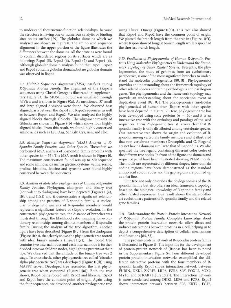
to understand thestructure-function relationships, becausethe structure is having one or numerous catalytic or bindingsites on its surface [79]. The globular domains which weanalysed are shown in Figure 8. The amino acid sequencealignment in the upper portion of the figure illustrates thedifferences between the domains. All the proteins were foundto contain disordered regions on its surfaces which are asfollowing: Rspo1 (5), Rspo2 (6), Rspo3 (7) and Rspo4 (6).Although globular domain analysis found that Rspo1, Rspo2andRspo3 contains globular domain, but no globular domainwas observed in Rspo4.
3.7. Multiple Sequences Alignment (MSA) Analysis amongR-Spondin Protein Family. The alignment of the (Rspo)ssequences using Clustal Omega is illustrated in supplemen-tary Figure S3. The MUSCLE output was visualised throughJalView and is shown in Figure 9(a). As mentioned, 37 smalland large aligned divisions were found. We observed bestaligned parts between theRspo4 andRspo2 sequences, aswellas between Rspo4 and Rspo2. We also analysed the highlyaligned blocks through Gblocks. The alignment results ofGblocks are shown in Figure 9(b) which shows four highlyaligned blocks. From this result, we found highly conservedamino acids such as Leu, Arg, Ser, Gly, Cys, Asn, and Phe.
3.8. Multiple Sequences Alignment (MSA) Analysis of R-Spondin Family Proteins with Other Species. Thereafter, weperformed MSA analysis of R-spondin family proteins withother species (𝑛 = 53). TheMSA result is shown in Figure 10.The maximum conservation found was up to 270 sequenceand some amino acids such as glycine, cysteine, valine, serine,proline, histidine, leucine and tyrosine were found highlyconserved between the sequences.
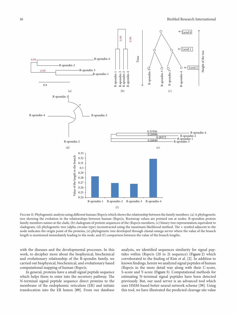
3.9. Analysis of Molecular Phylogenetics of Human R-SpondinFamily Proteins. Phylogram, cladogram and binary tree(equivalent to cladogram) have been depicted (Figures 11(a),11(b), and 11(c)) and it demonstrates a significant relation-ship among the proteins of R-spondin family. A molec-ular phylogenetic analysis of R-spondin members wouldrepresent a significant feature of (Rspo)s evolution. In theconstructed phylogenetic tree, the distance of branches wasillustrated through the likelihood ratio mapping for evolu-tionary relationships among distinct members of R-spondinfamily. During the analysis of the tree algorithm, anotherfigure have been described (Figure 11(c)) from the cladogram(Figure 11(b)), that clearly shows the phylogenetic tree rootedwith ideal binary numbers (Figure 11(c)). The rooted treecontains two internal nodes and each internal node is furtherdivided into two children nodes, highlighting proteins at theirtips. We observed that the altitude of the binary tree was 2stage. To cross check, other phylogenetic tree called “circularalpha phylogenetic tree”, was developed (Figure 11(d)) usingMAFFT server. Developed tree resembled the first phylo-genetic tree when compared (Figure 11(a)). Both the treeshows, Rspo4 being rooted with Rspo2 and likewise, Rspo3and Rspo1 have the common point of origin. Again usingthe four sequences, we developed another phylogenetic tree
using Clustal Omega (Figure 11(e)). This tree also showedthat Rspo4 and Rspo2 have the common point of origin.We plotted the branch length from the tree in (Figure 11(f)),where Rspo4 showed longest branch length while Rspo3 hadthe shortest branch length.
3.10. Prediction of Phylogenomics of Human R-Spondin Pro-teins Using Molecular Phylogenetics to Understand the Frame-work Topology of Other Related Species. Presently, the phy-logenomics, the study of genomes from an evolutionaryperspective, is one of the most significant branches to under-stand the molecular phylogenetics [80, 81]. Phylogenomicsprovides an understanding about the framework topology ofother related species containing orthologous and paralogousgenes. The phylogenomics and the framework topology mayprovide an understanding about the speciation event orduplication event [82, 83]. The phylogenomics (molecularphylogenetics) of human four (Rspo)s with other specieshave been depicted in Figure 12. Here, phylogenetic tree hasbeen developed using sixty proteins (𝑛 = 60) and it is aninteractive tree with the orthologs and paralogs of the seedsequences. Form Phylogenetic tree, it is very clear that R-spondin family is only distributed among vertebrate species.Our interactive tree shows the origin and evolution of R-spondin among vertebrate family members and it illustratedthat non-vertebrate members (Drosophila and C. Elegans)are not having domains similar to that of R-spondins.We alsospecified the tree legend containing different color codes ofthe different tree nodes. In front of the figure, the domain andsequence panel have been illustrated showing PFAM motifs.The motifs are represented by different shapes. Inter-domaincoding regions have been demonstrated as the standardamino acid colour codes and the gap regions are pointed upas a flat line.
Our tree not only describes the phylogenomics of the R-spondin family but also offers an ideal framework topologybased on the biological knowledge of R-spondin family andother related sequences. Our result shows the state-of-the-art evolutionary patterns of R-spondin family and the relatedgene families.
3.11. Understanding the Protein-Protein Interaction Networkof R-Spondin Protein Family. Complete knowledge aboutthe protein-protein interaction networks offers direct andindirect interactions between proteins in a cell, helping us todepict a comprehensive description of cellular mechanismsand functions [84, 85].
The protein-protein network of R-spondin protein familyis illustrated in Figure 13. The input file for the developmentof protein-protein network of (Rspo)s has been is notedin the Supplementary Figure S4. Four different developedprotein-protein interaction networks exemplified the dif-ferent interactive proteins with the four members of R-spondin family. Rspo1 shows interaction network betweenFURIN, DKK1, ZNRF3, LRP6, FZR8, SRY, FOXL2, SOX9,MYF5, and STRA8 (Figure 13(a)). The interaction networkis more condensed among DKK1, LRP6 and FZR8. Rspo2shows interaction network between SP8, KRT71, FGF5,

BioMed Research International 13
0.8
0 20 40 60Residue
RusseIICurves
Disordered by RusseII/Linding definition
mrlglcvval vlswthltis srgikgkrqr risaegsqac akgcelcsev ngclkcspkl fillerndir qvGVCLPSCP
mrlglcvval vlswthltis srgikgkrqr risaegsqac akgcelcsev ngclkcspkl fillerndir qvgvclpscp
PGYFDARNPD mnkcikckie hceacfshnf ctkckeglyl hkGRCYPACP EGSSAANGTM ECsspaqcEMSEWSPWGPCS KKQqlcgfrr gseertrrvl hapvgdhaac sdtketrrct vrrvpcPEGQ KRRKGGQGrrenanrnlark eskeagagsr rrkgqqqqqq qGTVGPLTSA Gpa
sewspwgpcs kkqQLCGFRR GSEERTRRVL HAPVGDHAAC SDTKETRRCT VRRVPCPEGQ KRRKGGQGRR ENANRNLARK ESKEAGAGSR RRKGQQQQQQ QGTVGPLTSA GPA
Potential globular domains (GlobDoms) by RusseII/Linding definition
pgyfdarnpd mnkcikckie hceacfshnf ctkckeglyl hkgrcypacp egssaangtm ecsspaqcem
Linding
Low complexity
DisorderGlobdom
FU 34-85
FU 91-135TSP1 150-207
80 100 120 140 160 180 200 220 240 260−11.2
−8.8
−6.4
−4.0
−1.6
Diso
rder
pro
pens
ity su
m
>R-spondin-1 Disorder
>R-spondin-1 GlobDoms
73–90, 123–142, 149–163, 207–218, 252–261
164–263
(a)
Disordered by RusseII/Linding definition
mqfrlfsfal iilncmdysh cqgnrwrrsk rasyvsnplC KGCLSCSKDN Gcsrcqqklf fflrregmrq ygeCLHSCPS GYYGHRAPdmnrcarcrien cdscfskdfc tkckvgfylh rgrCFDECPD GFapleetme cvegcevghw SEWGTCSRNN RTCgfkwgle trtrqivkkpvkdtilcpti aesrrckmtM RHCPGGKRTP KAkekrnkkk krklieraqe qhsvflATDR Anq
mqfrlfsfal iilncmdysh cqgnrwrrsk rasyvsnpic kgclscskdn gcsrcqqklf fflrregmrq ygeclhscps gyyghrapDMNRCARCRIEN CDSCFSKDFC TKCKVGFYLH RGRCFDECPD GFAPLEETME CVEGCEVGHW SEWGTCSRNN RTCGFKWGLE TRTRQIVKKP VKDTILCPTI AESRRCKMTM RHCPGGKRTP KAKEKRNKKK KRKLIERAQE QHSVFLATDR ANQ
Potential globular domains (GlobDoms) by RusseII/Linding definition
Low complexity
DisorderGlobdom
FU 37-84
FU 90-134TSP1 147-204
0.8
0 20 40 60Residue
80 100 120 140 160 180 200 220 240−13.8
−10.9
−8.0
−5.1
−2.1
Diso
rder
pro
pens
ity su
m
>R-spondin-2 Disorder
>R-spondin-2 GlobDoms
RusseIICurves
Linding
39–51, 74–88, 124–132, 151–163, 200–212, 237–241
89–243
(b)Disordered by RusseII/Linding definition
Potential globular domains (GlobDoms) by RusseII/Linding definition
mhlrliswlf iilnfmeyig sqnasrgrrq rrmHPNVSQG CQGGCATCSD YNGCLsckpr lffalerigm kqigvcLSSCPSGYYGTRYP DINKCtkcka dcdtcfnknf ctkcksgfyl hlGKCLDNCP EGLEannhtm ecvsivhcev sewNPWSPCKKGKTCGfkr gtetrvreii qhpSAKGNLC PPTNEtrkct vqrkkcqkge rgkkgRERKR KKPNKGE SKE Alpdskslesskeipeqren kqqqkkrkvq dkqKSVSVSt vh
0.8
0 20 40 60Residue
Low complexity
DisorderGlobdom
FU92-135
EGF 97-126TSP1 150-207
80 100 120 140 160 180 200 220 240 260−10.7
−8.4
−6.1
−3.8
−1.5
Diso
rder
pro
pens
ity su
m
>R-spondin-3 Disorder
>R-spondin-3 GlobDomsmhlrliswlf iilnfmeyig sqnasrgrrq rrmhpnvsqg cqggcatcsd yngclsckpr lffalerigm kqigvclsscpsgyygtryp dinkctkcka dcdtcfnknf ctkcksgfyl hlgkcldncp egleannhtm ecvsivhcev sewnpwspctkkgktcgFKR GTETRVREII QHPSAKGNLC PPTNETRKCT VQRKKCQKGE RGKKGRERKR KKPNKGE SKEAIPDSKSLES SKEIPEQREN KQQQKKRKVQ DKQKSVSVST VH
FU35-86
RusseIICurves
Linding
34–55, 77–95, 123–134, 154–167, 184–195, 216–232, 264–269
168–272
(c)
Disordered by RusseII/Linding definition
Potential globular domains (GlobDoms) by RusseII/Linding definition
mrapicllll vahavdmial nrrkkqVGTG LGGNCTGCII Cseengcstc qqrlflfirr egirqygKCL HDCPPGYFGI Rgqevnrckk cgatcescfsqdfcirckrq fylykGKCLP TCPPGTLAHQ ntrecqGECE LGPWGGWSPC THNGKTCGSa wglesrvrea gragheeaat cqvlsesrkCPIQRPCPGER SPGQKKGRKD Rrprkdrkid rridvRPRQP GLQP
0.9
0 20 40 60Residue
Low complexityDisorder
FU92-135
EGF 97-126TSP1 141-197
80 100 120 140 160 180 200 220
−1.1
−3.1
−5.0
−7.0
−9.0
Diso
rder
pro
pens
ity su
m
>R-spondin-4 Disorder
> R-spondin 4 GlobDoms none
mrapicllll vahavdmial nrrkkqvgtg lggnctgcii cseengcstc qqrlflfirr egirqygkcl hdcppgyfgi rgqevnrckk cgatcescfsqdfcirckrq fylykgkclp tcppgtlahq ntrecqgece lgpwggwspc thngktcgsa wglesrvrea gragheeaat cqvlsesrkc piqrpcpger spgqkkgrkd rrprkdrkid rridvrprqp glqp
RusseIICurves
Linding
27–41, 68–81, 116–130, 137–159, 190–211, 226–234
(d)
Figure 8: Globular domain gain/loss as a function of the variation between the four human (Rspo)s. The disorder propensity of the proteinstretch was calculated using GlobPlot analyses to identify the disorder region (blue).The upper portion in the figure illustrates the differencesbetween the amino acid sequence alignments among the domains.The tool uses a simple peak-finder algorithm to select the putative globularand disorder segments. (a) Rspo1, (b) Rspo2, (c) Rspo3, and (d) Rspo4.

14 BioMed Research International
R-spondin-4/1-234R-spondin-2/1-243R-spondin-1/1-263R-spondin-3/1-272
R-spondin-4/1-234R-spondin-2/1-243R-spondin-1/1-263R-spondin-3/1-272
Conservation
Conservation
Quality
10 20
140 150 160 170 180 190 200 210 220 230 240 250 260 270
30 40 50 60 70 80 90 100 110 120 130
Consensus
Consensus
Quality
(a)
R-spondin-4R-spondin-1R-spondin-2R-spondin-3
R-spondin-4R-spondin-1R-spondin-2R-spondin-3
R-spondin-4R-spondin-1R-spondin-2R-spondin-3
10
130 140 150
250 260 270
160 170Block-1 Block-2
Block-4Block-3Block-2
180 190 200 210 220 230 240
20 30 40 50 60 70 80 90 100 110 120
(b)
Figure 9:Multiple sequence alignment (MSA) of the different human (Rspo)s. (a)MSAoutput visualised through JalView and (b) theGblocksresults of human (Rspo)s show blocks from the alignments. The results show highly aligned four blocks.
GORAB, PTPRK, KIAA1804, PDIK1L, GUCY2F, MYLK2,and WNT3A (Figure 13(b)). In this network, no condensedpart was found. Rspo3 shows interaction network betweenFZD8, SDC4, MYF5, FURIN, FAM70A, WNT1, LRP6, KRE-MEN2, DVL1, and CTNNB1 (Figure 13(c)). The interactionnetwork is more condensed among the proteins which arelocated in the upper portion of the network such as FZD8,SDC4, WNT1, LRP6, KREMEN2, DVL1, and CTNNB1.Rspo4 shows network between only one protein that is,FURIN (Figure 13(d)) and it is the shortest network amongR-spondin protein family.
4. Discussion
R-spondin protein family is an immensely important proteinfamily, which acts as a key regulator factor during vertebrate
development and several signalling pathways, especially asagonists for the canonical Wnt/𝛽-catenin signalling pathway[17]. Association with different diseases has been foundwith R-spondin family proteins. (Rspo)s are associated withvarious developmental stages as an essential regulator. Forexample, Rspo1 has been found to be associated with sexdetermination and skin differentiation [22]; Rspo2 is a crucialprotein for development of limbs; lungs and hair follicles[11, 27, 86]; Rspo3 is essential for placental development [10]and Rspo4 is a significant protein for nail deployment [17].(Rspo)s have therapeutic potential for various diseases suchas skeletal diseases [87], inflammatory bowel disease andchemotherapy-induced mucositis [23], cancer [21], and dia-betes [88].Therefore, basic understanding about the biophys-ical, biochemical properties of (Rspo)s may provide moreunderstanding about their functional mechanism associated

BioMed Research International 15
Phy00022R0 BOVINPhy000281R BOVINPhy0003F9Y CANFA
Phy0006HC5 DANREPhy0006HDY DANREPhy0007CXK CHICKPhy0007DVX CHICK
Phy0008BNO HUMANPhy0008HIP HUMANPhy0008JNN HUMAN
Phy0009PRL MOUSEPhy00099UG MOUSEPhy000A62P MOUSE
Phy000A9VR MACMUPhy000BVOA PANTR
Phy000COXD RATPhy000W2FO NEMVE
Phy000XI7D BRAFLPhy001QA47 BOVIN
Phy001R7O6 HUMANPhy001R9A1 MOUSE
Phy0019JXD RATPhy002V2Q6 ORNANPhy002VGXB ORNANPhy002VI9U ORNANPhy002VK87 ORNAN
Phy002WDMH PHYPAPhy0031N8X TRIVAPhy0031Q7V TRIVAPhy0031UJ3 TRIVAPhy0036P86 CIOIN
Phy00372V9 DANRE
Phy003F24K RATPhy003F2Y3 RATPhy003F858 RAT
Phy003FE0C MONDOPhy003FFK4 MONDOPhy003FJEL MONDOPhy003G0TW TAKRUPhy003G1UB TAKRUPhy003G51M TAKRU
Phy003HQPO DANREPhy003 I2DW XENTRPhy003I42W XENTRPhy003IBRY CHICK
Phy003IHGD MACMUPhy003II0O MACMU
Phy003IPNO MACMUPhy003IRLW PANTRPhy003I99N PANTR
Phy003IWH2 PANTRPhy003J7WO CANFA
Phy003J9BF CANFA
Phy00022R0 BOVINPhy000281R BOVINPhy0003F9Y CANFA
Phy0006HC5 DANREPhy0006HDY DANREPhy0007CXK CHICKPhy0007DVX CHICK
Phy0008BNO HUMANPhy0008HIP HUMANPhy0008JNN HUMAN
Phy0009PRL MOUSEPhy00099UG MOUSEPhy000A62P MOUSE
Phy000A9VR MACMUPhy000BVOA PANTR
Phy000COXD RATPhy000W2F0 NEMVE
Phy000XI7D BRAFLPhy001QA47 BOVIN
Phy001R7O6 HUMANPhy001R9A1 MOUSE
Phy0019JXD RATPhy002V2Q6 ORNANPhy002VGXB ORNANPhy002VI9U ORNANPhy002VK87 ORNAN
Phy002WDMH PHYPAPhy0031NBX TRIVAPhy0031Q7V TRIVAPhy0031UJ3 TRIVAPhy0036P86 CIOIN
Phy00372V9 DANREPhy003F24K RATPhy003F2Y3 RATPhy003F858 RAT
Phy003FE0C MONDOPhy003FFK4 MONDOPhy003FJEL MONDOPhy003G0TW TAKRUPhy003G1UB TAKRUPhy003G51M TAKRU
Phy003HQPO DANREPhy003I2DW XENTRPhy003I42W XENTRPhy003IBRY CHICK
Phy003IHGD MACMUPhy003II0O MACMU
Phy003IPNO MACMUPhy003IRLW PANTRPhy003I99N PANTR
Phy003IWH2 PANTRPhy003J7WO CANFA
Phy003J98F CANFA
10 20 30 40 50 60 70 80 90 100
110 120 130 140 150 160 170 180 190 200
n = 53
Figure 10: Multiple sequence alignment (MSA) of the different human (Rspo)s with other species (𝑛 = 53) which are having sequencesimilarity.
16 BioMed Research International
R-spondin-4
R-spondin-2
0.99
0.99
0.4
R-spondin-3R-spondin-1
(a)
0.99
0.99
R-sp
ondi
n-4
R-sp
ondi
n-2
R-sp
ondi
n-3
R-sp
ondi
n-1
(b)
Tim
e
Level 0
Level 1
Level 2
Hei
ght o
f the
tree
R-sp
ondi
n-4
R-sp
ondi
n-2
R-sp
ondi
n-3
R-sp
ondi
n-1
(c)
R-spondin-4
R-spondin-2
R-spondin-3
R-spondin-1
∗
(d)
R-spondin-4R-spondin-2
R-spondin-3R-spondin-1
0.319560.26996
0.283720.26840
(e)
R-spondin-4R-spondin-2 R-spondin-3R-spondin-1
0.330.320.31
0.30.290.280.270.260.250.24Va
lue o
f the
leng
th to
the b
ranc
h
(f)
Figure 11: Phylogenetic analysis using different human (Rspo)s which shows the relationship between the familymembers. (a) A phylogenetictree showing the evolution in the relationships between human (Rspo)s. Bootstrap values are pointed out at nodes. R-spondins proteinfamily members names at the clade, (b) cladogram of protein sequences of the (Rspo)s members, (c) binary tree representation equivalent tocladogram, (d) phylogenetic tree (alpha circular type) reconstructed using the maximum likelihood method. The ∗ symbol adjacent to thenode indicates the origin point of the proteins, (e) phylogenetic tree developed through clustal-omega server where the value of the branchlength is mentioned immediately leading to the node, and (f) comparison between the value of the branch lengths.
with the diseases and the developmental processes. In thiswork, to decipher more about the biophysical, biochemicaland evolutionary relationship of the R-spondin family, wecarried out biophysical, biochemical, and evolutionary basedcomputational mapping of human (Rspo)s.
In general, proteins have a small signal peptide sequencewhich helps them to enter into the secretory pathway. TheN-terminal signal peptide sequence direct proteins to themembrane of the endoplasmic reticulum (ER) and initiatetranslocation into the ER lumen [89]. From our database
analysis, we identified sequences similarity for signal pep-tides within (Rspo)s (20 to 21 sequence) (Figure 2) whichcorroborated to the finding of Kim et al. [2]. In addition toknown findings, hereinwe analyzed signal peptides of human(Rspo)s in the more detail way along with their C-score,S-score and Y-score (Figure 3). Computational methods forestimating N-terminal signal peptides have been detectedpreviously. But, our used server is an advanced tool whichuses HMM-based better neural network scheme [39]. Usingthis tool, we have illustrated the predicted cleavage site value

BioMed Research International 17
FBpp0074042K04F10.4d
RSPO20
0.720.87
0.930.86
0.99
0.084
0.87
0.96
0.69 0.850.90.86
0.96
0.15
0
0.4
0.99
0.780.19
0.880.94
0.940.99
0.220.93
0.320.52
0.970.96
0.29
0.950.94
0.83
0.86
0.93
0.98
ENSMMUP00000014076H2QWK5RSPO2
ENSCAFP00000000995ENSBTAP00000038814
ENSMODP00000006466RSPO2
RSPO2
NEWSINFRUP00000128517RSPO4
ENSPTRP00000022516RSPO4ENSRNOP00000012951
ENSCAFP00000010193ENSBTAP00000039099
ENSMODP00000024370ENSDARP00000056339
ENSRNOP00000002814FRAS1
FRAS1ENSMODP00000026029
ENSGALP00000016778ENSXETP00000000809
Q4RPQ1
NEWSINFRUP00000153383RSPO1
RSPO1B4XH82
RSPO1RSPO1
ENSP00000362150ENSPTRP00000000970
ENSMMUP00000011037ENSGALP00000034443
ENSP00000357300ENSPTRP00000031716
F6SP05ENSBTAP00000010680RSPO3ENSRNOP00000015395RSPO3
ENSMODP00000022041ENSGALP00000023893
ENSXETP00000023282RSPO3RSPO3
NEWSINFRUP00000136059Q4RFD9
ENSDARP00000049063PCSK5
ENSMODP00000002357ENSGALP00000024430
ENSXETP00000014601ENSP00000305056ENSMMUP00000015521
Speciation events
Duplication events
Target sequence
Node inconsistencySeed sequence
NEWSINFRUP00000144951
Caenorhabditis elegansDrosophila melanogaster
Mus musculusMacaca mulatta
Macaca mulatta
Macaca mulatta
Macaca mulatta
Pan troglodytesHomo sapiensCanis familiaris
Bos taurusMonodelphis domestica
Gallus gallus
Gallus gallus
Gallus gallus
Gallus gallus
Gallus gallus
Xenopus tropicalis
Xenopus tropicalis
Xenopus tropicalis
Xenopus tropicalis
Tetraodon nigroviridis
Tetraodon nigroviridis
Danio rerio
Homo sapiens
Danio rerio
Danio rerio
Danio rerio
Danio rerioDanio rerio
Takifugu rubripes
Takifugu rubripesTakifugu rubripes
Takifugu rubripes
Pan troglodytes
Pan troglodytes
Pan troglodytes
Mus musculus
Mus musculus
Rattus norvegicusCanis familiaris
Canis familiaris
Canis familiaris
Bos taurus
Bos taurus
Bos taurus
Monodelphis domestica
Monodelphis domestica
Rattus norvegicus
Rattus norvegicus
Rattus norvegicus
Mus musculus
Mus musculus
Mus musculus
Homo sapiens
Homo sapiens
Homo sapiens
Homo sapiens
Monodelphis domestica
Monodelphis domestica
N = 60
SI : CH211-149P10.1
Figure 12: Phylogenomics of human four (Rspo)s and similar proteins from other species (𝑛 = 60) which are having sequence similarity. Infront of the figure, the domain and sequence panel have been depicted which uses PFAMmotifs, and the motifs are represented by differentshapes.
(C-score) in the signal peptide of human (Rspo)s where pos-sible two signals are noted in a single signal peptidase cleavagesite (Rspo2 and Rspo3) (Figure 3). Hiss and Schneider [89]revealed that long signal peptides mingle two or more signalsof signal peptidase cleavage site.
From the amino acids distributed pattern of human(Rspo)s especially from the exposed distribution analysis ata time (Figure 4(e)), we observed the similarity of the aminoacids distributed pattern is more or less same. However,Rspo1, Rspo2 and Rspo4 showed more similarity in thedistribution pattern. At the same time, our analysis revealedidentical amino acid composition pattern in the Rspo1, Rspo2and Rspo4 (Figures 4(a), 4(b), 4(c) and 4(d)). Recently, itwas reported that there is an association between amino acidcomposition and distribution with mutation. Researchershave shown the correlation between the amino acids dis-tribution pattern; missense mutations and genetic disorders[90]. Conversely, amino acid composition was linked withthe deleterious impact of mutations [91]. Therefore, aminoacid composition and distributed pattern of human (Rspo)smay help to the future researcher to understand the impactand association with genetic disorders. Further analysis withCys residues revealed that all these four (Rspo)s are Cys rich
protein. Also, the Cys architecture and the disulphide bondpattern show a common architecture and may be necessaryfor the stability of these proteins (Figure 4(f)). Recent in vitrostudy with mass spectrometry documented the pattern ofdisulfide bonds between the 15 available Cys residues presentin furin domains in (Rspo)s [14]. However, they found fivefree cysteine residues in Rspo2.
Our analysis found some glycosylation sites for (Rspo)swhich may be necessary for their functionality and signallingprocess (Figure 6). Previously, Kamata et al. [1] has indicatedthe N-linked glycosylation sites for (Rspo)s. Our previoussimilar kind of computational analysis shows that the N-glycosylation sites and O-glycosylation sites are vital for thefunctionality of the proteins in the insulin signalling pathwayproteins such as IRS andGLUT4 [67, 92]. However, identifiedO- and N-glycosylation sites by our analysis with (Rspo)sneeds to be confirmed with molecular and biochemicalexperiments.
Previously, Kim et al. [2] and Nam et al. [4] performedmultiple sequence analysis with four (Rspo)s. We also per-formed MSA among four (Rspo)s as well as with severalother species proteins using different computational server(Figure 10). Compared to the previous analysis, our MSA

18 BioMed Research International
LRP6
Predicted functional partners:
DKK1
FOXL2
FZD8
STRA8
MYF5
FURIN
ZNRF3
SRY
SOX9
LRP6
DKK1
FOXL2
FZD8
STRA8
MYF5
FURIN
ZNRF3
SRY
SOX9
RSPO1
Nei
ghbo
rhoo
d
Gen
e fus
ion
Coe
xpre
ssio
n
Coo
ccur
renc
e
Dat
abas
es
Text
min
ing
[Hom
olog
y]
Scor
e
0.989
0.939
0.900
0.896
0.854
0.800
0.657
0.619
0.573
0.540
Expe
rimen
ts
Dickkopf homolog 1 (Xenopus laevis); inhibitor of Wnt signaling pathway (266 aa)
Forkhead box L2; probable transcriptional regulator (376 aa)
Zinc and ring finger 3 (836 aa)
Low density lipoprotein receptor-related protein 6; essential for the Wnt/beta catenin signalin [ · · · ] (1613 aa)
Frizzled homolog 8 (drosophila); receptor for wnt proteins. most of frizzled receptors are coup [· · · ] (694 aa)
Stimulated by retinoic acid gene 8 homolog (mouse); required for the transition into meiosis fo [· · · ] (330 aa)
Myogenic factor 5; involved in muscle differentiation (myogenic factor). Induces fibroblasts to [· · · ] (255 aa)
Furin (paired basic amino acid cleaving enzyme); furin is likely to represent the ubiquitous en [ · · · ] (794 aa)
Sex determining region Y; transcriptional regulator which control a genetic switch in male deve [· · · ] (204 aa)
SRY (sex determining region Y)-box 9; plays an important role in the normal skeletal development [· · · ] (509 aa)
(a)
0.835
0.796
0.764
0.554
0.550
0.530
0.510
0.500
0.430
0.429
SP8
KRT71
FGF5
GORAB
PTPRK
KIAA1804
PDIK1L
GUCY2F
MYLK2
WNT3A
Predicted functional partners:
Neigh
borh
ood
Gene
fusio
n
Coex
press
ion
cooc
curre
nce
Datab
ases
Textm
inin
g
[hom
ology
]
Scor
e
Expe
rimen
ts
Sp8 transcription factor (508 aa)
Golgin, RAB6-interacting (394 aa)
PDLIM1 interacting kinase 1 like (341 aa)
Keratin 71; plays a central role in hair formation. Essential component of keratin intermediate [· · · ] (523 aa)
Fibroblast: growth factor 5; functions as an inhibitor of hair elongation by promoting progressi [· · · ] (268 aa)
Protein tyrosine phosphatase, receptor type, K; regulation of processes involving cell contact [· · · ] (1446 aa)
Mitogen-activated protein kinase kinase kinase (EC 2.7.11.25)(mixed lineage kinase 4); activate [· · · ] (1036 aa)
Guanylate cyclase 2F, retinal; probably plays a specific functional role in the rods and/or con [· · · ] (1108 aa)
Myosin light chain kinase 2; implicated in the level of global muscle contraction and cardiac f [· · · ] (596 aa)
Wingless-type MMTV integration site family, member3A; ligand for members of the frizzled famil [ · · · ] (352 aa)
RSPO2
SP8
KRT71
FGF5
GORAB
PTPRK
KIAA1804 PDIK1L
GUCY2F
MYLK2
WNT3A
(b)
FZD8
SDC4MYF5
FURIN
FAM70A
WNT1
LRP6KREMEN2DVL1
RSPO3
FZD8
SDC4
MYF5
FURIN
FAM70A
WNT1
LRP6
KREMEN2
DVL1
CTNNB1
CTNNB1
0905
0.800
0.800
0.675
0.507
0.462
0.423
0.411
0.402
0.402
Predicted functional partners:
Neig
hbor
hood
Gen
e fus
ion
Coex
pres
sion
cooc
curr
ence
Dat
abas
es
Text
min
ing
[hom
olog
y]
Scor
e
Expe
rimen
ts
Syndecan 4; cell surface proteoglycan that bears heparan sulfate (198aa)
Myogenic factor 5; involved in muscle differentiation (myogenic factor). induces 2 fibroblasts to [· · · ] (255 aa)
Furin (paired basic amino acid cleaving enzyme); furin is likely to represent the ubiquitous en [ · · · ] (794 aa)
Family with sequence similarity 70, member A (349aa)
Wingless-type MMTV integration site family, member 1; ligand for members of the frizzled family [· · · ] (370 aa)
Low density lipoprotein receptor-related protein 6; essential for the Wnt/beta catenin signalin [· · · ] (1613 aa)Kringle containing transmembrane protein 2; receptor for dickkopf protein. Cooperates with dick [ · · · ] (462 aa)Dishevelled, dsh homolog1(Drosophila); may play a role in the signal transduction pathway med [ · · · ] (670 aa)
Catenin (cadherin-associated protein), beta 1,88kDa; involved in the regulation of cell adhesi [· · · ] (781 aa)
Frizzled homolog 8 (Drosophila); receptor for Wnt proteins. Most of frizzled receptors are coup [· · · ] (694 aa)
(c)
FURIN
FURINRSPO4
Furin (paired basic amino acid cleaving enzyme); furin is likely to respresent the ubiquitous en[· · · ] (794 aa) 0.722
Predicted functional partners:
Nei
ghbo
rhoo
d
Gen
e fu
sion
Coe
xpre
ssio
n
cooc
curr
ence
Dat
abas
es
Text
min
ing
[hom
olog
y]
Scor
e
Exp
erim
ents
(d)
Figure 13: Protein-protein interaction network of R-spondin family proteins using STRING server. (a) Rspo1, (b) Rspo2, (c) Rspo3, and (d)Rspo4.
investigation provides a very clear picture about the alignedand conserved residues with different colour codes visualisedthrough JalView. We then analysed through Gblocks serverto understand the conserve blocks within the R-spondinfamily. Our data showed four highly conserved blocks withindepicted Gblocks (Figure 9). Furthermore, another MSAanalysis of (Rspo)s with other species (𝑛 = 53) was performedto understand more conserved residues among different
species where we found several small conserved blocks andresidues such as glycine, cysteine, valine, serine, proline,histidine, leucine and tyrosine (Figure 10).
The evolutionary history of R-spondin family and thephylogenetic relationships prototype can be investigatedthrough the molecular approach involving amino acidsequencing. Utilizing similar approach, we developed phylo-genetic relationships among the members of the R-spondin

BioMed Research International 19
family, and we found that Rspo4 and Rspo2 were siblingsin 99% bootstrap replications and likewise, Rspo3 andRspo1 were siblings in 99% bootstrap replications (Figure 11).Previously, de lau et al. [17] and our group also [4] anal-ysed phylogenetic relationships. Here, we performed moreadvanced two types of phylogenetic analyses: (i) phylogeneticrelationships pattern of R-spondin family (Figure 11) and (ii)phylogenetic relationships usingR-spondin family using sixtyspecies (𝑛 = 60) (Figure 12). Second one is the interactivetree with the orthologs and paralogs of the seed sequenceswhich describe the phylogenomics of the R-spondin familyand also determines evolutionary relationship of differentspecies (Figure 12). This analysis directs the study towardsnext generation phylogenomics [93]whichmay be robust andalignment-free.
From our protein-protein interaction network analysis,we noted an interaction among the Rspo1 with the LRP6and FZR8 receptor confirming them as candidate proteinfor Wnt signaling pathway (Figure 13(a)). Hao et al. [94]reported that LRP6 and FZD receptors are present on themembrane and these receptors permit the Wnt ligands togenerate much stronger signals. The network of ZNRF3 withRspo1 confirms that ZNRF3 is associated with Wnt receptoryield in an R-spondin sensitive manner [94]. The networkalso shows Rspo1 interaction with DKK1 (an antagonist ofWnt signaling). Binnerts et al. [9] reported that Rspo1 bindsto the Kremen family of transmembrane proteins and itnegatively regulates the LRP6 receptor through the DKK1-associated endocytosis. Due to the controlling property ofindividual’s sex phenotype, Rspo1 networks with SRY andSOX9 protein [95]. The network of Rspo2 with FGF showsthat damage Wnt signal directs to defective expression ofthe important apical ectodermal ridge maintenance factors,FGF4 and FGF8, which is related with the lung and limbdevelopment (Figure 13(b)). Similar to Rspo1, we observed astrong association between Rspo3 and the LRP6/FZR8 recep-tor as well as DVL for Wnt signaling pathway (Figure 13(c)).Rspo4 shows an interaction between FURIN proteins. Ithas been known that FURIN like domain is necessary forthe activity of Rspo4. Blaydon et al. [96] demonstratedthat mutations interrupting furin-like domains in Rspo4may affect its signaling activity. Recent studies showed that(Rspo)s are the ligands for the leucine-rich repeat containingG protein-coupled receptor 4/5/6 (LGR4/5/6) receptors [15–18]. However, in our analysis we have not found any networkbetween the (Rspo)s with LGR4/5/6.Thismight be due to thelack of updated data in server database (STRING database)containing information about the LGR4/5/6.
In summary, through computational analysis, we per-formed biophysical, biochemical, and evolutionary topologyof human R-spondin family proteins. In this work, we haveapplied innovative and rapid approach to study the structuralbased biophysical, biochemical, and evolutionary relation-ship among (Rspo)s.Thedifficult and time-consumingnatureof the experimental analysis led us to attempt to developa cost-effective computational research of biophysical, bio-chemical and evolutionary topology of human R-spondinfamily. In this study, we have tried to highlight the possiblepotent sites for O- and N-glycosylation, distribution and
conservation of amino acids and to predict phylogenetic andprotein-protein interaction among (Rspo)s with the availabledata base. However, experimental biochemical and func-tional studies are required to further establish these finding.Our attempt to decipher the biophysical and biochemicalproperties of (Rspo)s may provide useful platform and astarting point for scientists to unfold significant physiologicaland therapeutic properties of R-spondin protein family invarious disease models.
Conflict of Interests
The authors declare that there is no conflict of interestsregarding the publication of this paper.
Authors’ Contribution
Ashish Ranjan Sharma, Chiranjib Chakraborty, and Sang-Soo Lee contributed equally to this work.
Acknowledgments
This research was supported by Basic Science ResearchProgram through the National Research Foundation ofKorea (NRF) funded by the Ministry of Education (NRF-2014R1A1A4A03009388 and 2011-001-4792) and by a grantof the Korea Health Technology R&D Project throughthe Korea Health Industry Development Institute (KHIDI),funded by the Ministry of Health & Welfare, Republic ofKorea (HI12C1265).The authors also take this opportunity tothank the management of VIT and Galgotias University forproviding the facilities and encouragement to carry out thiswork.
References
[1] T. Kamata, K.-I. Katsube,M.Michikawa,M. Yamada, S. Takada,and H. Mizusawa, “R-spondin, a novel gene with throm-bospondin type 1 domain, was expressed in the dorsal neuraltube and affected in Wnts mutants,” Biochimica et BiophysicaActa—Gene Structure and Expression, vol. 1676, no. 1, pp. 51–62,2004.
[2] K.-A. Kim, J. Zhao, S. Andarmani et al., “R-spondin proteins: anovel link to 𝛽-catenin activation,” Cell Cycle, vol. 5, no. 1, pp.23–26, 2006.
[3] K.-A. Kim, M. Wagle, K. Tran et al., “R-Spondin familymembers regulate theWnt pathway by a commonmechanism,”Molecular Biology of the Cell, vol. 19, no. 6, pp. 2588–2596, 2008.
[4] J.-S. Nam, T. J. Turcotte, P. F. Smith, S. Choi, and K. Y.Jeong, “Mouse cristin/R-spondin family proteins are novelligands for the frizzled 8 and LRP6 receptors and activate 𝛽-catenin-dependent gene expression,” The Journal of BiologicalChemistry, vol. 281, no. 19, pp. 13247–13257, 2006.
[5] Q.Wei, C. Yokota, M. V. Semenov, B. Doble, J.Woodgett, and X.He, “R-spondin1 is a high affinity ligand for LRP6 and inducesLRP6 phosphorylation and 𝛽-catenin signaling,”The Journal ofBiological Chemistry, vol. 282, no. 21, pp. 15903–15911, 2007.
[6] O. Kazanskaya, A. Glinka, I. del Barco Barrantes, P. Stannek,C. Niehrs, and W. Wu, “R-Spondin2 is a secreted activator of

20 BioMed Research International
Wnt/𝛽-catenin signaling and is required for Xenopus myogen-esis,” Developmental Cell, vol. 7, no. 4, pp. 525–534, 2004.
[7] J.-S. Nam, T. J. Turcotte, and J. K. Yoon, “Dynamic expression ofR-spondin family genes in mouse development,” Gene Expres-sion Patterns, vol. 7, no. 3, pp. 306–312, 2007.
[8] B. A. Parr, M. J. Shea, G. Vassileva, and A. P. McMahon, “MouseWnt genes exhibit discrete domains of expression in the earlyembryonic CNS and limb buds,”Development, vol. 119, no. 1, pp.247–261, 1993.
[9] M. E. Binnerts, K.-A. Kim, J. M. Bright et al., “R-Spondin1regulates Wnt signaling by inhibiting internalization of LRP6,”Proceedings of the National Academy of Sciences of the UnitedStates of America, vol. 104, no. 37, pp. 14700–14705, 2007.
[10] M. Aoki, M. Mieda, T. Ikeda, Y. Hamada, H. Nakamura, andH. Okamoto, “R-spondin3 is required for mouse placentaldevelopment,” Developmental Biology, vol. 301, no. 1, pp. 218–226, 2007.
[11] S. M. Bell, C. M. Schreiner, S. E. Wert, M. L. Mucenski,W. J. Scott, and J. A. Whitsett, “R-spondin 2 is requiredfor normal laryngeal-tracheal, lung and limb morphogenesis,”Development, vol. 135, no. 6, pp. 1049–1058, 2008.
[12] B. Ohkawara, A. Glinka, and C. Niehrs, “Rspo3 binds syndecan4 and induces Wnt/PCP signaling via clathrin-mediated endo-cytosis to promotemorphogenesis,”Developmental Cell, vol. 20,no. 3, pp. 303–314, 2011.
[13] K.-A. Kim, M. Kakitani, J. Zhao et al., “Mitogenic influence ofhuman R-spondin1 on the intestinal epithelium,” Science, vol.309, no. 5738, pp. 1256–1259, 2005.
[14] S.-J. Li, T.-Y. Yen, Y. Endo et al., “Loss-of-function pointmutations and two-furin domain derivatives provide insightsabout R-spondin2 structure and function,” Cellular Signalling,vol. 21, no. 6, pp. 916–925, 2009.
[15] K. S. Carmon, X. Gong, Q. Lin, A. Thomas, and Q. Liu, “R-spondins function as ligands of the orphan receptors LGR4 andLGR5 to regulate Wnt/𝛽-catenin signaling,” Proceedings of theNational Academy of Sciences of the United States of America,vol. 108, no. 28, pp. 11452–11457, 2011.
[16] K. S. Carmon, Q. Lin, X. Gong, A. Thomas, and Q. Liu, “LGR5interacts and cointernalizes with Wnt receptors to modulateWnt/beta-catenin signaling,” Molecular and Cellular Biology,vol. 32, no. 11, pp. 2054–2064, 2012.
[17] W. B. de Lau, B. Snel, andH. C. Clevers, “TheR-spondin proteinfamily,” Genome Biology, vol. 13, no. 3, article 242, 2012.
[18] A. Glinka, C. Dolde, N. Kirsch et al., “LGR4 and LGR5 areR-spondin receptors mediating Wnt/𝛽-catenin and Wnt/PCPsignalling,” EMBO Reports, vol. 12, no. 10, pp. 1055–1061, 2011.
[19] T. Grigoryan, P.Wend, A. Klaus, andW. Birchmeier, “Decipher-ing the function of canonical Wnt signals in development anddisease: conditional loss- and gain-of-function mutations of 𝛽-catenin in mice,” Genes and Development, vol. 22, no. 17, pp.2308–2341, 2008.
[20] R. Nusse, “Wnt signaling in disease and in development,” CellResearch, vol. 15, no. 1, pp. 28–32, 2005.
[21] Y. R. Jin and J. K. Yoon, “The R-spondin family of proteins:emerging regulators of WNT signaling,” The InternationalJournal of Biochemistry and Cell Biology, vol. 44, no. 12, pp.2278–2287, 2012.
[22] P. Parma, O. Radi, V. Vidal et al., “R-spondin1 is essential insex determination, skin differentiation andmalignancy,”NatureGenetics, vol. 38, no. 11, pp. 1304–1309, 2006.
[23] J. Zhao, J. de Vera, S. Narushima et al., “R-spondin1, a novelintestinotrophic mitogen, ameliorates experimental colitis inmice,” Gastroenterology, vol. 132, no. 4, pp. 1331–1343, 2007.
[24] G. Kronke, S. Uderhardt, K.-A. Kim et al., “R-spondin 1 protectsagainst inflammatory bone damage during murine arthritis bymodulating theWnt pathway,” Arthritis & Rheumatism, vol. 62,no. 8, pp. 2303–2312, 2010.
[25] W. Lu, K.-A. Kim, J. Liu et al., “R-spondin1 synergizes withWnt3A in inducing osteoblast differentiation and osteoprote-gerin expression,” FEBS Letters, vol. 582, no. 5, pp. 643–650,2008.
[26] A. R. Sharma, B. S. Choi, J. M. Park et al., “Rspo 1 promotesosteoblast differentiation via Wnt signaling pathway,” IndianJournal of Biochemistry and Biophysics, vol. 50, no. 1, pp. 19–25,2013.
[27] J.-S. Nam, E. Park, T. J. Turcotte et al., “Mouse R-spondin2is required for apical ectodermal ridge maintenance in thehindlimb,” Developmental Biology, vol. 311, no. 1, pp. 124–135,2007.
[28] W. Yamada, K. Nagao, K. Horikoshi et al., “Craniofacial mal-formation in R-spondin2 knockout mice,” Biochemical andBiophysical Research Communications, vol. 381, no. 3, pp. 453–458, 2009.
[29] E. Cadieu, M. W. Neff, P. Quignon et al., “Coat variation in thedomestic dog is governed by variants in three genes,” Science,vol. 326, no. 5949, pp. 150–153, 2009.
[30] S. Seshagiri, E. W. Stawiski, S. Durinck et al., “Recurrent R-spondin fusions in colon cancer,” Nature, vol. 488, no. 7413, pp.660–664, 2012.
[31] S. Neufeld, J.M. Rosin, A. Ambasta et al., “A conditional allele ofRspo3 reveals redundant function of R-spondins during mouselimb development,” Genesis, vol. 50, no. 10, pp. 741–749, 2012.
[32] M. Klauzinska, B. Baljinnyam, A. Raafat et al., “Rspo2/Int7regulates invasiveness and tumorigenic properties of mammaryepithelial cells,” Journal of Cellular Physiology, vol. 227, no. 5, pp.1960–1971, 2012.
[33] W. Lowther, K. Wiley, G. H. Smith, and R. Callahan, “A newcommon integration site, Int7, for the mouse mammary tumorvirus in mouse mammary tumors identifies a gene whoseproduct has furin-like and thrombospondin-like sequences,”Journal of Virology, vol. 79, no. 15, pp. 10093–10096, 2005.
[34] Y. Ishii, M. Wajid, H. Bazzi et al., “Mutations in R-spondin 4(RSPO4) underlie inherited anonychia,” Journal of InvestigativeDermatology, vol. 128, no. 4, pp. 867–870, 2008.
[35] E.W. Sayers, T. Barrett, D. A. Benson et al., “Database resourcesof the national center for biotechnology information,” NucleicAcids Research, vol. 39, no. 1, pp. D38–D51, 2011.
[36] UniProt Consortium, “Update on activities at the UniversalProtein Resource (UniProt) in 2013,”Nucleic Acids Research, vol.41, pp. D43–D47, 2013.
[37] UniProt Consortium, “The universal protein resource(UniProt) in 2010,” Nucleic Acids Research, vol. 38, pp.D142–D148, 2010.
[38] UniProt Consortium, “Ongoing and future developments at theUniversal Protein Resource,”Nucleic Acids Research, vol. 39, pp.D214–D219, 2011.
[39] T. N. Petersen, S. Brunak, G. von Heijne, and H. Nielsen, “Sig-nalP 4.0: discriminating signal peptides from transmembraneregions,” Nature Methods, vol. 8, no. 10, pp. 785–786, 2011.
[40] N. J. Anthis and G. M. Clore, “Sequence-specific determinationof protein andpeptide concentrations by absorbance at 205 nm,”Protein Science, vol. 22, no. 6, pp. 851–858, 2013.

BioMed Research International 21
[41] V. Brendel, P. Bucher, I. R. Nourbakhsh, B. E. Blaisdell, andS. Karlin, “Methods and algorithms for statistical analysis ofprotein sequences,” Proceedings of the National Academy ofSciences of the United States of America, vol. 89, no. 6, pp. 2002–2006, 1992.
[42] J. Cheng, A. Z. Randall, M. J. Sweredoski, and P. Baldi,“SCRATCH: a protein structure and structural feature predic-tion server,”Nucleic Acids Research, vol. 33, no. 2, pp.W72–W76,2005.
[43] H. M. Berman, J. Westbrook, Z. Feng et al., “The protein databank,” Nucleic Acids Research, vol. 28, no. 1, pp. 235–242, 2000.
[44] R. J. Read, P. D. Adams, W. B. Arendall III et al., “A newgeneration of crystallographic validation tools for the proteindata bank,” Structure, vol. 19, no. 10, pp. 1395–1412, 2011.
[45] S. Hunter, P. Jones, A. Mitchell et al., “InterPro in 2011: newdevelopments in the family and domain prediction database,”Nucleic Acids Research, vol. 40, no. 1, pp. D306–D312, 2012.
[46] S. E. Hamby and J. D. Hirst, “Prediction of glycosylation sitesusing random forests,” BMC Bioinformatics, vol. 9, article 500,2008.
[47] K. Julenius, A. Mølgaard, R. Gupta, and S. Brunak, “Prediction,conservation analysis, and structural characterization of mam-malian mucin-type O-glycosylation sites,” Glycobiology, vol. 15,no. 2, pp. 153–164, 2005.
[48] J. E. Hansen, O. Lund, N. Tolstrup, A. A. Gooley, K. L.Williams, and S. Brunak, “NetOglyc: prediction of mucin typeO-glycosylation sites based on sequence context and surfaceaccessibility,” Glycoconjugate Journal, vol. 15, no. 2, pp. 115–130,1998.
[49] E. Gasteiger, A. Gattiker, C. Hoogland, I. Ivanyi, R. D. Appel,and A. Bairoch, “ExPASy: the proteomics server for in-depthprotein knowledge and analysis,”Nucleic Acids Research, vol. 31,no. 13, pp. 3784–3788, 2003.
[50] R. Linding, R. B. Russell, V. Neduva, and T. J. Gibson, “GlobPlot:exploring protein sequences for globularity and disorder,”Nucleic Acids Research, vol. 31, no. 13, pp. 3701–3708, 2003.
[51] F. Sievers, A. Wilm, D. Dineen et al., “Fast, scalable generationof high-quality protein multiple sequence alignments usingClustal Omega,” Molecular Systems Biology, vol. 7, article 539,2011.
[52] P. Hogeweg and B. Hesper, “The alignment of sets of sequencesand the construction of phyletic trees: an integrated method,”Journal of Molecular Evolution, vol. 20, no. 2, pp. 175–186, 1984.
[53] S. B. Needleman and C. D. Wunsch, “A general method appli-cable to the search for similarities in the amino acid sequenceof two proteins,” Journal of Molecular Biology, vol. 48, no. 3, pp.443–453, 1970.
[54] R. C. Edgar, “MUSCLE: multiple sequence alignment with highaccuracy and high throughput,” Nucleic Acids Research, vol. 32,no. 5, pp. 1792–1797, 2004.
[55] C. Chakraborty, S. S. Roy, M. J. Hsu, and G. Agoramoorthy,“Can computational biology improve the phylogenetic analysisof insulin?” Computer Methods and Programs in Biomedicine,vol. 108, no. 2, pp. 860–872, 2012.
[56] J. Huerta-Cepas, S. Capella-Gutierrez, L. P. Pryszcz et al.,“PhylomeDB v3.0: an expanding repository of genome-widecollections of trees, alignments and phylogeny-based orthologyand paralogy predictions,” Nucleic Acids Research, vol. 39, no. 1,pp. D556–D560, 2011.
[57] J. Huerta-Cepas, A. Bueno, J. Dopazo, and T. Gabaldon,“PhylomeDB: a database for genome-wide collections of gene
phylogenies,” Nucleic Acids Research, vol. 36, no. 1, pp. D491–D496, 2008.
[58] T. F. Smith and M. S. Waterman, “Identification of commonmolecular subsequences,” Journal of Molecular Biology, vol. 147,no. 1, pp. 195–197, 1981.
[59] A. Dereeper, V. Guignon, G. Blanc et al., “Phylogeny.fr: robustphylogenetic analysis for the non-specialist,” Nucleic AcidsResearch, vol. 36, pp. W465–W469, 2008.
[60] K. Katoh, K.Misawa, K. Kuma, and T.Miyata, “MAFFT: a novelmethod for rapid multiple sequence alignment based on fastFourier transform,” Nucleic Acids Research, vol. 30, no. 14, pp.3059–3066, 2002.
[61] N. Saitou and M. Nei, “The neighbor-joining method: a newmethod for reconstructing phylogenetic trees,” Molecular Biol-ogy and Evolution, vol. 4, no. 4, pp. 406–425, 1987.
[62] L. J. Jensen, M. Kuhn, M. Stark et al., “STRING—a global viewon proteins and their functional interactions in 630 organisms,”Nucleic Acids Research, vol. 37, no. 1, pp. D412–D416, 2009.
[63] D. Szklarczyk, A. Franceschini, M. Kuhn et al., “The STRINGdatabase in 2011: functional interaction networks of proteins,globally integrated and scored,” Nucleic Acids Research, vol. 39,no. 1, pp. D561–D568, 2011.
[64] A. Meiniel, R. Meiniel, N. Goncalves-Mendes, I. Creveaux, R.Didier, and B. Dastugue, “The thrombospondin type 1 repeat(TSR) and neuronal differentiation: roles of SCO- spondinoligopeptides on neuronal cell types and cell lines,” Interna-tional Review of Cytology, vol. 230, pp. 1–39, 2003.
[65] T. L. Rodgers, P. D. Townsend, D. Burnell et al., “Modulationof global low-frequencymotions underlies allosteric regulation:demonstration in CRP/FNR family transcription factors,” PLoSBiology, vol. 11, no. 9, Article ID e1001651, 2013.
[66] C. G. P. Doss, C. Chakraborty, B. Rajith, and N. Nagasundaram,“In silico discrimination of nsSNPs in hTERT gene by meansof local DNA sequence context and regularity,” Journal ofMolecular Modeling, vol. 19, no. 9, pp. 3517–3527, 2013.
[67] C. Chakraborty, G. Agoramoorthy, and M. J. Hsu, “Exploringthe evolutionary relationship of insulin receptor substratefamily using computational biology,” PLoS ONE, vol. 6, no. 2,Article ID e16580, 2011.
[68] T. Huang, X.-H. Shi, P. Wang et al., “Analysis and predictionof the metabolic stability of proteins based on their sequentialfeatures, subcellular locations and interaction networks,” PLoSONE, vol. 5, no. 6, Article ID e10972, 2010.
[69] H. Jubb, A. P. Higueruelo, A. Winter, and T. L. Blundell,“Structural biology and drug discovery for protein-proteininteractions,” Trends in Pharmacological Sciences, vol. 33, no. 5,pp. 241–248, 2012.
[70] K. Guruprasad, B. V. B. Reddy, and M. W. Pandit, “Correlationbetween stability of a protein and its dipeptide composition: anovel approach for predicting in vivo stability of a protein fromits primary sequence,” Protein Engineering, vol. 4, no. 2, pp. 155–161, 1990.
[71] J. Kyte and R. F. Doolittle, “A simple method for displayingthe hydropathic character of a protein,” Journal of MolecularBiology, vol. 157, no. 1, pp. 105–132, 1982.
[72] A. Ikai, “Thermostability and aliphatic index of globular pro-teins,”The Journal of Biochemistry, vol. 88, no. 6, pp. 1895–1898,1980.
[73] P. Argos, M. G. Rossmann, U. M. Grau, H. Zuber, G. Frank,and J. D. Tratschin, “Thermal stability and protein structure,”Biochemistry, vol. 18, no. 25, pp. 5698–5703, 1979.
22 BioMed Research International
[74] C. A. Charneski and L. D. Hurst, “Positively charged residuesare themajor determinants of ribosomal velocity,” PLoS Biology,vol. 11, no. 3, Article ID e1001508, 2013.
[75] B. A. Wible, M. Taglialatela, E. Ficker, and A. M. Brown,“Gating of inwardly rectifying K+ channels localized to a singlenegatively charged residue,” Nature, vol. 371, no. 6494, pp. 246–249, 1994.
[76] H. Nakashima and K. Nishikawa, “Discrimination of intracel-lular and extracellular proteins using amino acid compositionand residue-pair frequencies,” Journal of Molecular Biology, vol.238, no. 1, pp. 54–61, 1994.
[77] A. Krogh, B. Larsson, G. von Heijne, and E. L. L. Sonnhammer,“Predicting transmembrane protein topology with a hiddenMarkov model: application to complete genomes,” Journal ofMolecular Biology, vol. 305, no. 3, pp. 567–580, 2001.
[78] V. N. Uversky, “Intrinsically disordered proteins from A to Z,”The International Journal of Biochemistry and Cell Biology, vol.43, no. 8, pp. 1090–1103, 2011.
[79] L. B. Chemes, J. Glavina, L. G. Alonso, C. Marino-Buslje, G.de Prat-Gay, and I. E. Sanchez, “Sequence evolution of theintrinsically disordered and globular domains of a model viraloncoprotein,” PLoS ONE, vol. 7, no. 10, Article ID e47661, 2012.
[80] J. A. Eisen and C. M. Fraser, “Phylogenomics: intersection ofevolution and genomics,” Science, vol. 300, no. 5626, pp. 1706–1707, 2003.
[81] J. Huerta-Cepas, S. Capella-Gutierrez, L. P. Pryszcz, M. Marcet-Houben, and T. Gabaldon, “PhylomeDB v4: zooming into theplurality of evolutionary histories of a genome,” Nucleic AcidsResearch, vol. 42, pp. D897–D902, 2014.
[82] B. Boeckmann, M. Robinson-rechavi, I. Xenarios, and C.Dessimoz, “Conceptual framework and pilot study to bench-mark phylogenomic databases based on reference gene trees,”Briefings in Bioinformatics, vol. 12, no. 5, pp. 423–435, 2011.
[83] K. Sjolander, “Phylogenomic inference of protein molecularfunction: advances and challenges,” Bioinformatics, vol. 20, no.2, pp. 170–179, 2004.
[84] A. Franceschini, D. Szklarczyk, S. Frankild et al., “STRING v9.1:protein-protein interaction networks, with increased coverageand integration,”Nucleic Acids Research, vol. 41, no. 1, pp. D808–D815, 2013.
[85] A. Pottekat, S. Becker, K. Spencer et al., “Insulin biosyntheticinteraction network component, TMEM24, facilitates insulinreserve pool release,”Cell Reports, vol. 4, no. 5, pp. 921–930, 2013.
[86] R. DasGupta and E. Fuchs, “Multiple roles for activatedLEF/TCF transcription complexes during hair follicle devel-opment and differentiation,” Development, vol. 126, no. 20, pp.4557–4568, 1999.
[87] J. M. Kerkhof, A. G. Uitterlinden, A. M. Valdes et al., “Radio-graphic osteoarthritis at three joint sites and FRZB, LRP5,and LRP6 polymorphisms in two population-based cohorts,”Osteoarthritis and Cartilage, vol. 16, no. 10, pp. 1141–1149, 2008.
[88] V. S. C. Wong, A. Yeung, W. Schultz, and P. L. Brubaker, “R-spondin-1 is a novel 𝛽-cell growth factor and insulin secreta-gogue,”The Journal of Biological Chemistry, vol. 285, no. 28, pp.21292–21302, 2010.
[89] J. A. Hiss and G. Schneider, “Architecture, function and predic-tion of long signal peptides,” Briefings in Bioinformatics, vol. 10,no. 5, pp. 569–578, 2009.
[90] S. Khan and M. Vihinen, “Spectrum of disease-causing muta-tions in protein secondary structures,” BMC Structural Biology,vol. 7, article no. 56, 2007.
[91] S. Hormoz, “Amino acid composition of proteins reducesdeleterious impact ofmutations,” Scientific Reports, vol. 3, article2919, 2013.
[92] C. Chakraborty, S. Bandyopadhyay, U. Maulik, and G. Ago-ramoorthy, “Topology mapping of insulin-regulated glucosetransporter GLUT4 using computational biology,” Cell Bio-chemistry and Biophysics, vol. 67, no. 3, pp. 1261–1274, 2013.
[93] C. X. Chan andM.A. Ragan, “Next-generation phylogenomics,”Biology Direct, vol. 8, no. 1, article 3, 2013.
[94] H.-X. Hao, Y. Xie, Y. Zhang et al., “ZNRF3 promotes Wntreceptor turnover in an R-spondin-sensitive manner,” Nature,vol. 484, no. 7397, pp. 195–200, 2012.
[95] K. Kashimada and P. Koopman, “Sry: the master switch inmammalian sex determination,” Development, vol. 137, no. 23,pp. 3921–3930, 2010.
[96] D. C. Blaydon, Y. Ishii, E. A. O’Toole, H. C. Unsworth, and M.T. Teh, “The gene encoding R-spondin 4 (RSPO4), a secretedprotein implicated in Wnt signaling, is mutated in inheritedanonychia,”Nature Genetics, vol. 38, no. 11, pp. 1245–1247, 2006.

Submit your manuscripts athttp://www.hindawi.com
Hindawi Publishing Corporationhttp://www.hindawi.com Volume 2014
Anatomy Research International
PeptidesInternational Journal of
Hindawi Publishing Corporationhttp://www.hindawi.com Volume 2014
Hindawi Publishing Corporation http://www.hindawi.com
International Journal of
Volume 2014
Zoology
Hindawi Publishing Corporationhttp://www.hindawi.com Volume 2014
Molecular Biology International
GenomicsInternational Journal of
Hindawi Publishing Corporationhttp://www.hindawi.com Volume 2014
The Scientific World JournalHindawi Publishing Corporation http://www.hindawi.com Volume 2014
Hindawi Publishing Corporationhttp://www.hindawi.com Volume 2014
BioinformaticsAdvances in
Marine BiologyJournal of
Hindawi Publishing Corporationhttp://www.hindawi.com Volume 2014
Hindawi Publishing Corporationhttp://www.hindawi.com Volume 2014
Signal TransductionJournal of
Hindawi Publishing Corporationhttp://www.hindawi.com Volume 2014
BioMed Research International
Evolutionary BiologyInternational Journal of
Hindawi Publishing Corporationhttp://www.hindawi.com Volume 2014
Hindawi Publishing Corporationhttp://www.hindawi.com Volume 2014
Biochemistry Research International
ArchaeaHindawi Publishing Corporationhttp://www.hindawi.com Volume 2014
Hindawi Publishing Corporationhttp://www.hindawi.com Volume 2014
Genetics Research International
Hindawi Publishing Corporationhttp://www.hindawi.com Volume 2014
Advances in
Virolog y
Hindawi Publishing Corporationhttp://www.hindawi.com
Nucleic AcidsJournal of
Volume 2014
Stem CellsInternational
Hindawi Publishing Corporationhttp://www.hindawi.com Volume 2014
Hindawi Publishing Corporationhttp://www.hindawi.com Volume 2014
Enzyme Research
Hindawi Publishing Corporationhttp://www.hindawi.com Volume 2014
International Journal of
Microbiology