Embed Size (px)
Citation preview
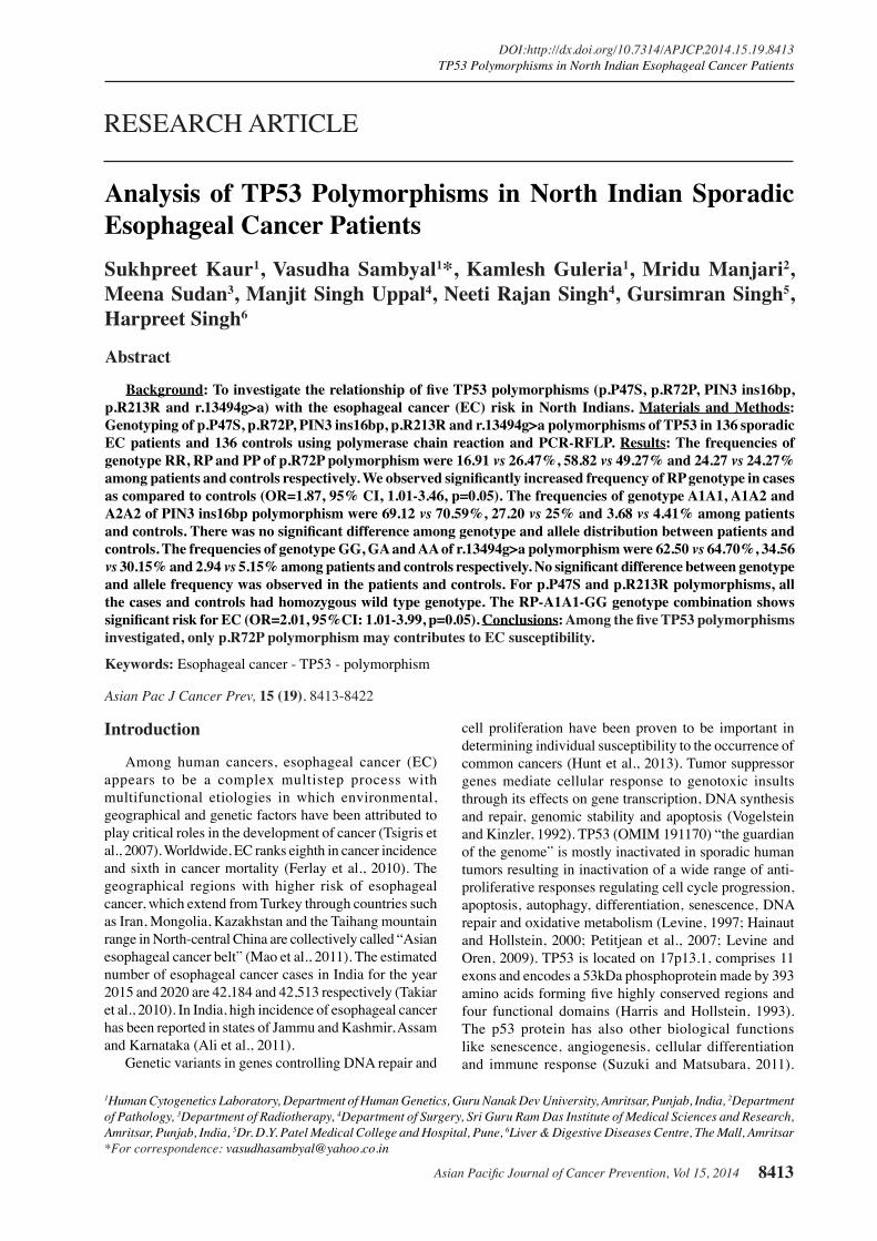
Asian Pacific Journal of Cancer Prevention, Vol 15, 2014 8413
DOI:http://dx.doi.org/10.7314/APJCP.2014.15.19.8413TP53 Polymorphisms in North Indian Esophageal Cancer Patients
Asian Pac J Cancer Prev, 15 (19), 8413-8422
Introduction
Among human cancers, esophageal cancer (EC) appears to be a complex multistep process with multifunctional etiologies in which environmental, geographical and genetic factors have been attributed to play critical roles in the development of cancer (Tsigris et al., 2007). Worldwide, EC ranks eighth in cancer incidence and sixth in cancer mortality (Ferlay et al., 2010). The geographical regions with higher risk of esophageal cancer, which extend from Turkey through countries such as Iran, Mongolia, Kazakhstan and the Taihang mountain range in North-central China are collectively called “Asian esophageal cancer belt” (Mao et al., 2011). The estimated number of esophageal cancer cases in India for the year 2015 and 2020 are 42,184 and 42,513 respectively (Takiar et al., 2010). In India, high incidence of esophageal cancer has been reported in states of Jammu and Kashmir, Assam and Karnataka (Ali et al., 2011).
Genetic variants in genes controlling DNA repair and
1Human Cytogenetics Laboratory, Department of Human Genetics, Guru Nanak Dev University, Amritsar, Punjab, India, 2Department of Pathology, 3Department of Radiotherapy, 4Department of Surgery, Sri Guru Ram Das Institute of Medical Sciences and Research, Amritsar, Punjab, India, 5Dr. D.Y. Patel Medical College and Hospital, Pune, 6Liver & Digestive Diseases Centre, The Mall, Amritsar *For correspondence: [email protected]
Abstract
Background: To investigate the relationship of five TP53 polymorphisms (p.P47S, p.R72P, PIN3 ins16bp, p.R213R and r.13494g>a) with the esophageal cancer (EC) risk in North Indians. Materials and Methods: Genotyping of p.P47S, p.R72P, PIN3 ins16bp, p.R213R and r.13494g>a polymorphisms of TP53 in 136 sporadic EC patients and 136 controls using polymerase chain reaction and PCR-RFLP. Results: The frequencies of genotype RR, RP and PP of p.R72P polymorphism were 16.91 vs 26.47%, 58.82 vs 49.27% and 24.27 vs 24.27% among patients and controls respectively. We observed significantly increased frequency of RP genotype in cases as compared to controls (OR=1.87, 95% CI, 1.01-3.46, p=0.05). The frequencies of genotype A1A1, A1A2 and A2A2 of PIN3 ins16bp polymorphism were 69.12 vs 70.59%, 27.20 vs 25% and 3.68 vs 4.41% among patients and controls. There was no significant difference among genotype and allele distribution between patients and controls. The frequencies of genotype GG, GA and AA of r.13494g>a polymorphism were 62.50 vs 64.70%, 34.56 vs 30.15% and 2.94 vs 5.15% among patients and controls respectively. No significant difference between genotype and allele frequency was observed in the patients and controls. For p.P47S and p.R213R polymorphisms, all the cases and controls had homozygous wild type genotype. The RP-A1A1-GG genotype combination shows significant risk for EC (OR=2.01, 95%CI: 1.01-3.99, p=0.05). Conclusions: Among the five TP53 polymorphisms investigated, only p.R72P polymorphism may contributes to EC susceptibility. Keywords: Esophageal cancer - TP53 - polymorphism
RESEARCH ARTICLE
Analysis of TP53 Polymorphisms in North Indian Sporadic Esophageal Cancer PatientsSukhpreet Kaur1, Vasudha Sambyal1*, Kamlesh Guleria1, Mridu Manjari2, Meena Sudan3, Manjit Singh Uppal4, Neeti Rajan Singh4, Gursimran Singh5, Harpreet Singh6
cell proliferation have been proven to be important in determining individual susceptibility to the occurrence of common cancers (Hunt et al., 2013). Tumor suppressor genes mediate cellular response to genotoxic insults through its effects on gene transcription, DNA synthesis and repair, genomic stability and apoptosis (Vogelstein and Kinzler, 1992). TP53 (OMIM 191170) “the guardian of the genome” is mostly inactivated in sporadic human tumors resulting in inactivation of a wide range of anti-proliferative responses regulating cell cycle progression, apoptosis, autophagy, differentiation, senescence, DNA repair and oxidative metabolism (Levine, 1997; Hainaut and Hollstein, 2000; Petitjean et al., 2007; Levine and Oren, 2009). TP53 is located on 17p13.1, comprises 11 exons and encodes a 53kDa phosphoprotein made by 393 amino acids forming five highly conserved regions and four functional domains (Harris and Hollstein, 1993). The p53 protein has also other biological functions like senescence, angiogenesis, cellular differentiation and immune response (Suzuki and Matsubara, 2011).
Sukhpreet Kaur et al
Asian Pacific Journal of Cancer Prevention, Vol 15, 20148414
The TP53 pathway is well known for maintaining genomic integrity and preventing cells from undergoing oncogenic transformation (Hamroun et al., 2006). Genetic polymorphisms can contribute to differences between individuals in susceptibility to various cancers by affecting the regulation of gene expression (Wade et al., 2013). Analysis of polymorphisms in a variety of genes has revealed a correlation between specific allele variants and cancer predisposition (Rogler et al., 2011). The loss of p53 function also mediates resistance to chemotherapy induced apoptosis, which is often associated with poor clinical outcome (Owen et al., 1997).
In TP53 gene, polymorphisms have been identified in both coding and non-coding regions (Murphy, 2006; Bojesen and Nordestgaard, 2008; Costa et al., 2008; Whibley et al., 2009). About 200 genetic polymorphisms have been identified in TP53 (http://p53.iarc.fr), many of which show geographic and population variations, but their effects on cancer risk appear to be inconsistent across studies (Whibley et al., 2009; Stacey et al., 2011).
The p.R72P and p.P47S are functionally important polymorphisms of TP53. The p.P47S is present in the N-terminal domain of TP53 that leads to non-synonymous amino acid substitution from Proline to Serine. The p.P47S polymorphism has been investigated in various cancers including breast (Alawadi et al., 2011), colorectal (Sameer et al., 2010), gliomas (Pinto et al., 2008), bladder (Santos et al., 2011), brain (Almeida et al., 2009) and urinary bladder (Jaiswal et al., 2011) cancer. Significant association of S47 has been reported in Caucasian lung cancer (Felley-Bosco et al., 1993) and South Indian colorectal cancer (Singamsetty et al., 2014) patients. So far there is no published report on p.P47S polymorphism in EC.
The p.R72P is non-conservative change of the arginine to proline located within the prolin-rich domain of TP53 which is the critical site for apoptosis signaling (Sakamuro et al., 1997). The two isoforms of this polymorphism (R72 and P72) differ in their biochemical and biological properties and behave differently (Thomas et al., 1999). The R72 form has been reported to induce apoptosis more effectively than P72 form (Dumont et al., 2003). Several studies have focused on the association between TP53 p.R72P polymorphism and esophageal cancer susceptibility. However, contradictory data is available, where few studies reported an association while several studies found no association. Positive association between p.R72P polymorphism and EC have previously reported in European and Asian (Kawaguchi et al., 2000), Chinese (Lee et al., 2000; Li et al., 2002; Lu et al., 2004; He et al., 2005; Hong et al., 2005; Cai et al., 2006; Shao et al., 2008; Yang et al., 2008; Ma et al., 2012; Yang et al., 2013), South African (Vos et al., 2003), German (Pantelis et al., 2007), Caucasian (Cescon et al., 2009; Renouf et al., 2013) and Korean (Piao et al., 2011) population. Still other studies which have failed to demonstrate any association between codon 72 variants of TP53 and EC cancer risk have been in Chinese (Guimaraes et al., 2001; Hu et al., 2003), Japanese (Hamajima et al., 2002) and Caucasian (Liu et al., 2010) population.
A rare polymorphism p.R213R localized in exon 6
resulted from alteration of CGA to CGG at codon 213. Though, published studies substantially lack information on p.R213R polymorphism in EC. No association of p.R213R polymorphism with cancer risk has been observed in Brazilian Barretts esophagus patients (Pilger et al., 2007).
Intronic polymorphisms of TP53 gene may influence coding-region sequence alterations that results in increase of a deleterious phenotype (Malkinson and You, 1994). PIN3 Ins16bp polymorphism (rs17878362) a 16 base pair duplication in intron 3 has been implicated in regulation of gene expression and DNA protein interactions (Mattick 1994, 2004).
Several case-control studies have reported an increased risk of various cancer types associated with PIN3 Ins16bp polymorphism, with the most consistent association reported for breast (Wang-Gohrke et al., 2002; Costa et al., 2008) and colorectal cancers (Gemignani et al., 2004; Perfumo et al., 2006). To date, there are three reports examining the association between PIN3 ins16bp polymorphism and the risk for EC. Two of these reports found a positive association between A2A2 genotype and EC (Vos et al., 2003; Malik et al., 2011), whereas one study from North India failed to find an association (Umar et al., 2012).
The r.13494g>a is a rare polymorphism in the intron 6 of TP53 resulting from G > A transition at 61bp downstream of exon 6. The r.13494g>a has been studied in various cancers including ovarian (Wang-Gohrke et al., 1999; Yair et al., 2000), head and neck (Mitra et al., 2003; Chen et al., 2007), esophageal (Pilger et al., 2007), breast (Peller et al., 1995; Sjalander et al., 1996; Weston et al., 1997; Akkiprik et al., 2009; Singh et al., 2008; Hrstka et al., 2009), cervical (Mitra et al., 2004), colorectal (Mammano et al., 2009), lung (Biros et al., 2001; Wang et al., 2007), prostate cancer (Mittal et al., 2011) and yielded inconsistent results for association.
The previous published studies showed that these polymorphisms vary in different ethnic and population groups. In Punjab state in North West part of India, Population Based Cancer Registry (PBCR) has reported prevalence of cancer as 90 patients per one lakh of population with maximum incidence varying in different districts, i.e. in Mutktsar (136.3 per lakh), Mansa (134.8 per lakh), Bathinda (125.8 per lakh) to lower incidence in Taran Taran district (40.9 per lakh). Amritsar district has highest incidence of cancer (81.2 per lakh) in Majha region of Punjab. (http://www.downtoearth.org.in/content/punjab-cancer-capital-india). In Amritsar city, the third largest city of Punjab state, increased incidence of esophageal cancer is being reported (personal Communication, SGRD Rotary Cancer Hospital, Vallah, Sri Amritsar). There is no published report on TP53 polymorphisms in the esophageal cancer patients from this region. The present case-control study was conducted to investigate the relationship of five TP53 polymorphisms (p.P47S, p.R72P, PIN3 ins16bp, p.R213R and r.13494g>a) with the esophageal cancer risk in North Indian sporadic esophageal cancer patients. The identification of susceptibility factors that predispose individuals to esophageal cancer will give further insight

Asian Pacific Journal of Cancer Prevention, Vol 15, 2014 8415
DOI:http://dx.doi.org/10.7314/APJCP.2014.15.19.8413TP53 Polymorphisms in North Indian Esophageal Cancer Patients
into the etiology of this cancer and provide targets for the future development of therapeutic approaches.
Materials and Methods
Clinical evaluation and selection of subjectsThis study was approved by the ethical committee
of Guru Nanak Dev University, Amritsar, Punjab, India. The patients were recruited from Sri Guru Ram Das Institute of Medical Sciences and Research, Vallah, Amritsar, Punjab. Patients included were those who had no prior history of any cancer and had not undergone chemotherapy, radiotherapy or blood transfusion. The controls were age and gender matched unrelated healthy individuals from the same geographical region as that of patients. The individuals who had family history for any type of cancer or chronic diseases or on regular medication were not included in the study. Relevant information including self reported personal history and disease history of each subject was recorded on a pre-tested structured questionnaire by interview and from medical records. After informed consent, 5ml venous blood was collected from each subject.
DNA extraction and genotypingGenomic DNA was extracted from peripheral blood
leucocytes using standard phenol chloroform method (Adeli and Ogbonna, 1990). The DNA fragment harboring p.P47S, p.R72P, PIN3, p.R213R and r.13494g>a
polymorphisms were amplified using the published primer sequences (Table 1). Amplification was performed in 15μl reaction volume containing 0.4μl of dNTPs, 6pmoles of each oligonucleotide primer and 0.9U of Taq DNA polymerase (Bangalore GeNei). A negative control without DNA template was included in each reaction. Details of reaction conditions have been mentioned in Table 1. The amplified PCR products were analyzed on ethidium bromide stained agarose gel. The PCR products were digested with appropriate restriction enzyme (Table 1) using the manufacturer instructions (New England Biolabs, Beverly, MA), followed by agarose gel electrophoresis. The genotype were categorized as wild type, heterozygous and homozygous variant based on band sizes as mentioned in Table 1 and Figures 1-4. Genotyping was performed without knowledge of case/control status to ensure quality control.
Statistical analysis Continuous variables were analyzed by t-test and
presented as means ± standard deviation (SD). Categorical variables were presented as percentages and were compared by chi-square test. Hardy Weinberg equilibrium (HWE) was tested by comparing the observed to expected genotype frequencies in controls using a χ2 test. The odds ratios (ORs), 95%CI ranges and corresponding p-values were calculated using the Web-Assotest program (http://www.ekstroem.com) for measuring the association between different genotypes and esophageal
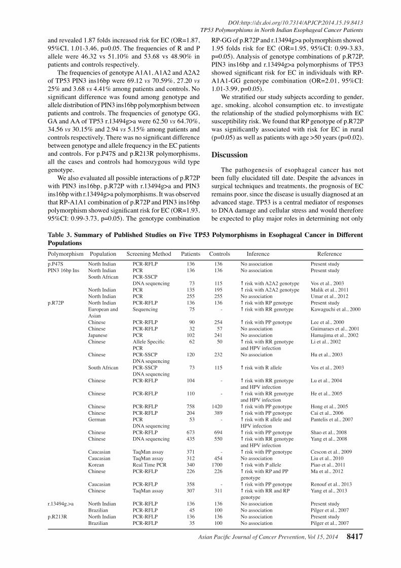
Figure 1. Restriction Digestion of PCR Products Demonstrating the Patterns of Digestion in Different Genotypes of p.P47S Polymorphism of TP53. Lane 1-5=Wild type homozygous genotype (PP)
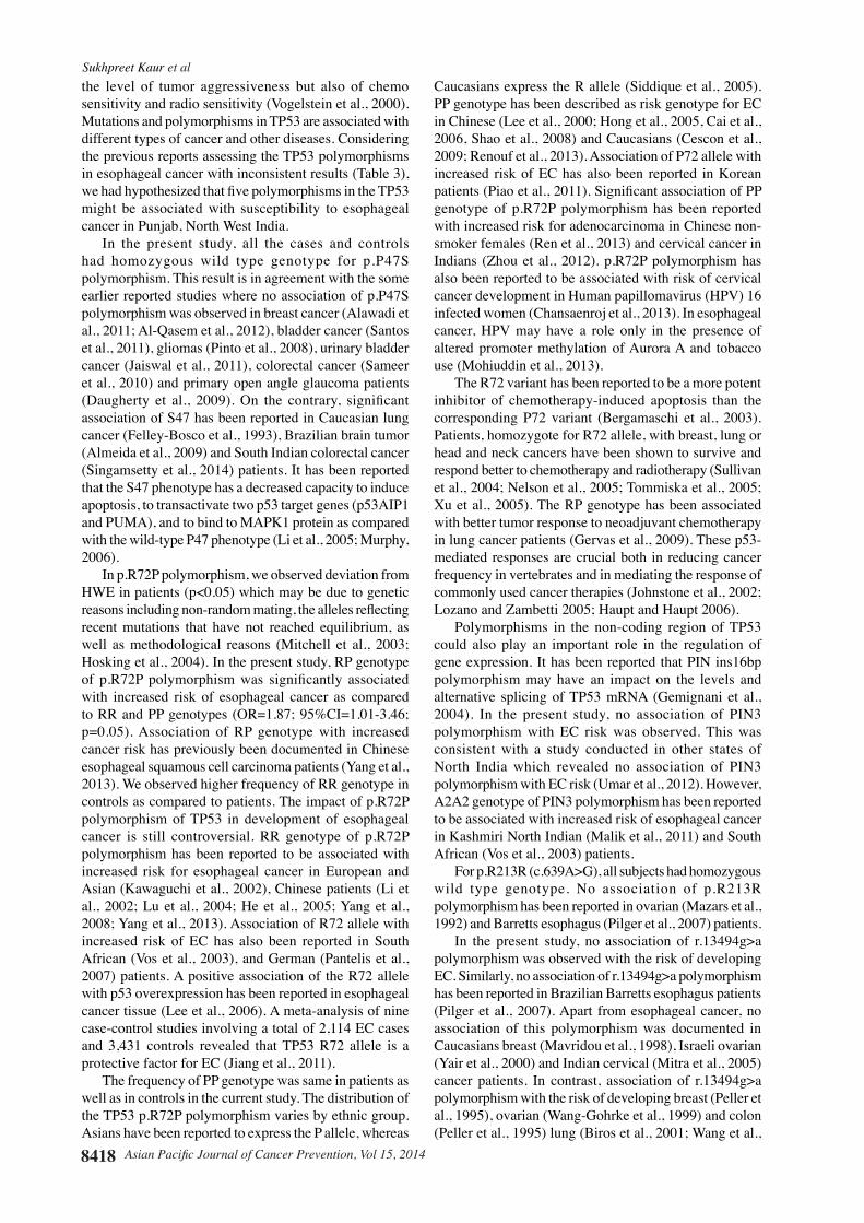
Figure 2. Restriction Digestion of PCR Products Demonstrating the Patterns of Digestion in Different Genotypes of p.R72P Polymorphism of TP53. Lane 1, 6 and 8=PP genotype; Lane 2-5=Heterozygous genotype (RP); Lane 7=RR genotype
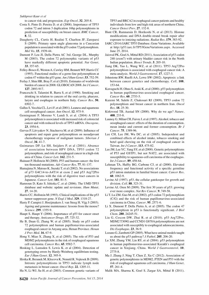
Figure 3. A photograph of Gel Demonstrating the Different Genotypes of PIN3 Polymorphism of TP53. Lane 1 and 2=A1A1 genotype, Lane 3=A1A2 genotype; Lane 4=A2A2 genotype
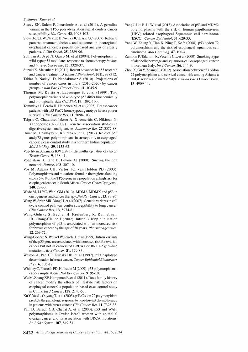
Figure 4. Restriction Digestion of PCR Products Demonstrating the Patterns of Digestion in Different Genotypes of p.R213R and r.13494g>a Polymorphisms of TP53. Lane 1-7=Wild type homozygous genotype (AA). Lane 8, 12 and 13=GG genotype; Lane 9 and 10=GA genotype; Lane 11=AA genotype

Sukhpreet Kaur et al
Asian Pacific Journal of Cancer Prevention, Vol 15, 20148416
Table 2. Genotype and Allele Frequencies of TP53 Polymorphisms in Esophageal Cancer Patients and Healthy Controls Variant Genotype Allele Patients Controls OR (95% CI) p value(SNP No.) n(%) n(%)
p.P47S PP 136(100) 136(100) (rs1800371) PS - - SS - - P 272(100) 272(100) S - - p.R72P RR 23(16.91) 36(26.47) 1(Reference) (rs1042522) RP 80(58.82) 67(49.26) 1.87(1.01-3.46) 0.05 PP 33(24.27) 33(24.27) 1.57(0.77-3.19) 0.22 R 126(46.32) 139(51.10) 1(Reference) P 146(53.68) 133(48.90) 1.21(0.87-1.70) 0.27PIN3Ins16bp A1A1 94(69.12) 96(70.59) 1(Reference) (rs17878362) A1A2 37(27.20) 34(25.00) 1.11(0.64-1.92) 0.7 A2A2 5(3.68) 6(4.41) 0.85(0.25-2.88) 0.79 A1 225(82.72) 226(83.09) 1(Reference) A2 47(17.28) 46(16.91) 1.03(0.66-1.60) 0.92p.R213R AA 136(100) 136(100) 136(100) (rs1800372) AG - - GG - - A 272(100) 272(100) G - - r.13494g>a GG 85(62.50) 88(64.70) 1(Reference) (rs1625895) GA 47(34.56) 41(30.15) 1.19(0.71-1.98) 0.51 AA 4(2.94) 7(5.15) 0.59(0.17-2.09) 0.41 G 217(79.78) 217(79.78) 1(Reference) A 55(20.22) 55(20.22) 1.00(0.66-1.52) 1
Statistically significant p values (p<0.05) are displayed in bold; OR- Odds ratio; CI- confidence interval
0
25.0
50.0
75.0
100.0
New
ly d
iagn
osed
with
out
trea
tmen
t
New
ly d
iagn
osed
with
tre
atm
ent
Pers
iste
nce
or r
ecur
renc
e
Rem
issi
on
Non
e
Chem
othe
rapy
Radi
othe
rapy
Conc
urre
nt c
hem
orad
iatio
n
10.3
0
12.8
30.025.0
20.310.16.3
51.7
75.051.1
30.031.354.2
46.856.3
27.625.033.130.031.3
23.738.0
31.3
Table 1. Detail of TP53 Polymorphisms and Reaction Conditions Used for Screening Variant Location Screening PCR Annealing Restriction Restriction digestion Primers References(SNP No.) Method product Temperature, enzyme patterns for size (bp) MgCl2 different Alleles
p.P47S Exon 4 PCR-RFLP 201/185* 59℃, 1mM MspI S allele-201/185 Pinto et al., 2008(rs1800371) P allele-156/140 and 45p.R72P Exon 4 PCR-RFLP 279 59℃, 1mM BstUI P allele 279 Kazemi et al., 2009(rs1042522) R allele-160 and 119PIN3 Intron 3 PCR - 61℃, 1mM - A1 allele-119 Costa et al., 2008(rs17878362) A2 allele-135p.R213R Exon 6 PCR-RFLP 1621 59℃, 1.5mM TaqI A allele-312, 383 and 926 Pilger et al., 2008(rs1800372) G allele-695 and 926r.13494g>a Intron 6 PCR-RFLP 1621 59℃, 1.5mM MspI G allele-356, 277, 277, Pilger et al., 2008(rs1625895) 299, 168, 124 and120 A allele - 633, 299, 277, 168, 124 and 120
*Size divergence is due to 16bp ins/del polymorphism in intron 3
cancer risk. The ORs, 95%CI ranges and p-value for genotypic combinations were calculated using online Vassar Stats Calculator (http://www.faculty.vassar.edu/lowry/VassarStats.html). The cut off p-value adopted for statistical analysis was 0.05.
Results
Characteristics of subjectsA total of 136 sporadic esophageal cancer patients (47
males and 89 females) and 136 age and gender matched unrelated healthy individuals (47 males and 89 females) were analyzed in this study. The mean age of patients and controls was 55.34±12.54 years and 53.08±12.27 years respectively. There were 111 rural and 25 urban cases. EC incidence was higher among the individuals more than 50 years of age as compared to those less than 50
years. When the incidence was compared between males and females, a preponderance of EC was observed among females (65%).
Genotypic frequencies of TP53 polymorphisms and esophageal cancer risk
The genotype and allele frequencies of TP53 polymorphisms in esophageal cancer patients and controls individuals are shown in Table 2. The observed genotypes frequencies of two polymorphisms (PIN3 ins16bp and r.13494g>a) were in HWE (p>0.05). In p.R72P polymorphism, we observed deviation from HWE in patients (p<0.05). The frequencies of genotype RR, RP and PP of p.R72P polymorphism were 16.91 vs 26.47%, 58.82 vs 49.27% and 24.27 vs 24.27% among patients and controls respectively. We observed significantly increased frequency of RP genotype in cases as compared to controls
Asian Pacific Journal of Cancer Prevention, Vol 15, 2014 8417
DOI:http://dx.doi.org/10.7314/APJCP.2014.15.19.8413TP53 Polymorphisms in North Indian Esophageal Cancer Patients
and revealed 1.87 folds increased risk for EC (OR=1.87, 95%CI, 1.01-3.46, p=0.05. The frequencies of R and P allele were 46.32 vs 51.10% and 53.68 vs 48.90% in patients and controls respectively.
The frequencies of genotype A1A1, A1A2 and A2A2 of TP53 PIN3 ins16bp were 69.12 vs 70.59%, 27.20 vs 25% and 3.68 vs 4.41% among patients and controls. No significant difference was found among genotype and allele distribution of PIN3 ins16bp polymorphism between patients and controls. The frequencies of genotype GG, GA and AA of TP53 r.13494g>a were 62.50 vs 64.70%, 34.56 vs 30.15% and 2.94 vs 5.15% among patients and controls respectively. There was no significant difference between genotype and allele frequency in the EC patients and controls. For p.P47S and p.R213R polymorphisms, all the cases and controls had homozygous wild type genotype.
We also evaluated all possible interactions of p.R72P with PIN3 ins16bp, p.R72P with r.13494g>a and PIN3 ins16bp with r.13494g>a polymorphisms. It was observed that RP-A1A1 combination of p.R72P and PIN3 ins16bp polymorphism showed significant risk for EC (OR=1.93, 95%CI: 0.99-3.73, p=0.05). The genotype combination
RP-GG of p.R72P and r.13494g>a polymorphism showed 1.95 folds risk for EC (OR=1.95, 95%CI: 0.99-3.83, p=0.05). Analysis of genotype combinations of p.R72P, PIN3 ins16bp and r.13494g>a polymorphisms of TP53 showed significant risk for EC in individuals with RP-A1A1-GG genotype combination (OR=2.01, 95%CI: 1.01-3.99, p=0.05).
We stratified our study subjects according to gender, age, smoking, alcohol consumption etc. to investigate the relationship of the studied polymorphisms with EC susceptibility risk. We found that RP genotype of p.R72P was significantly associated with risk for EC in rural (p=0.05) as well as patients with age >50 years (p=0.02).
Discussion
The pathogenesis of esophageal cancer has not been fully elucidated till date. Despite the advances in surgical techniques and treatments, the prognosis of EC remains poor, since the disease is usually diagnosed at an advanced stage. TP53 is a central mediator of responses to DNA damage and cellular stress and would therefore be expected to play major roles in determining not only
Table 3. Summary of Published Studies on Five TP53 Polymorphisms in Esophageal Cancer in Different PopulationsPolymorphism Population Screening Method Patients Controls Inference Referencep.P47S North Indian PCR-RFLP 136 136 No association Present studyPIN3 16bp Ins North Indian PCR 136 136 No association Present study South African PCR-SSCP DNA sequencing 73 115 # risk with A2A2 genotype Vos et al., 2003 North Indian PCR 135 195 # risk with A2A2 genotype Malik et al., 2011 North Indian PCR 255 255 No association Umar et al., 2012p.R72P North Indian PCR-RFLP 136 136 # risk with RP genotype Present study European and Sequencing 75 - # risk with RR genotype Kawaguchi et al., 2000 Asian Chinese PCR-RFLP 90 254 # risk with PP genotype Lee et al., 2000 Chinese PCR-RFLP 32 57 No association Guimaraes et al., 2001 Japanese PCR 102 241 No association Hamajima et al., 2002 Chinese Allele Specific 62 50 # risk with RR genotype Li et al., 2002 PCR and HPV infection Chinese PCR-SSCP 120 232 No association Hu et al., 2003 DNA sequencing South African PCR-SSCP 73 115 # risk with R allele Vos et al., 2003 DNA sequencing Chinese PCR-RFLP 104 - # risk with RR genotype Lu et al., 2004 and HPV infection Chinese PCR-RFLP 110 - # risk with RR genotype He et al., 2005 and HPV infection Chinese PCR-RFLP 758 1420 # risk with PP genotype Hong et al., 2005 Chinese PCR-RFLP 204 389 # risk with PP genotype Cai et al., 2006 German PCR 53 - # risk with R allele and Pantelis et al., 2007 DNA sequencing HPV infection Chinese PCR-RFLP 673 694 # risk with PP genotype Shao et al., 2008 Chinese DNA sequencing 435 550 # risk with RR genotype Yang et al., 2008 and HPV infection Caucasian TaqMan assay 371 - # risk with PP genotype Cescon et al., 2009 Caucasian TaqMan assay 312 454 No association Liu et al., 2010 Korean Real Time PCR 340 1700 # risk with P allele Piao et al., 2011 Chinese PCR-RFLP 226 226 # risk with RP and PP Ma et al., 2012 genotype Caucasian PCR-RFLP 358 - # risk with PP genotype Renouf et al., 2013 Chinese TaqMan assay 307 311 # risk with RR and RP Yang et al., 2013 genotyper.13494g.>a North Indian PCR-RFLP 136 136 No association Present study Brazilian PCR-RFLP 45 100 No association Pilger et al., 2007p.R213R North Indian PCR-RFLP 136 136 No association Present study Brazilian PCR-RFLP 35 100 No association Pilger et al., 2007
Sukhpreet Kaur et al
Asian Pacific Journal of Cancer Prevention, Vol 15, 20148418
the level of tumor aggressiveness but also of chemo sensitivity and radio sensitivity (Vogelstein et al., 2000). Mutations and polymorphisms in TP53 are associated with different types of cancer and other diseases. Considering the previous reports assessing the TP53 polymorphisms in esophageal cancer with inconsistent results (Table 3), we had hypothesized that five polymorphisms in the TP53 might be associated with susceptibility to esophageal cancer in Punjab, North West India.
In the present study, all the cases and controls had homozygous wild type genotype for p.P47S polymorphism. This result is in agreement with the some earlier reported studies where no association of p.P47S polymorphism was observed in breast cancer (Alawadi et al., 2011; Al-Qasem et al., 2012), bladder cancer (Santos et al., 2011), gliomas (Pinto et al., 2008), urinary bladder cancer (Jaiswal et al., 2011), colorectal cancer (Sameer et al., 2010) and primary open angle glaucoma patients (Daugherty et al., 2009). On the contrary, significant association of S47 has been reported in Caucasian lung cancer (Felley-Bosco et al., 1993), Brazilian brain tumor (Almeida et al., 2009) and South Indian colorectal cancer (Singamsetty et al., 2014) patients. It has been reported that the S47 phenotype has a decreased capacity to induce apoptosis, to transactivate two p53 target genes (p53AIP1 and PUMA), and to bind to MAPK1 protein as compared with the wild-type P47 phenotype (Li et al., 2005; Murphy, 2006).
In p.R72P polymorphism, we observed deviation from HWE in patients (p<0.05) which may be due to genetic reasons including non-random mating, the alleles reflecting recent mutations that have not reached equilibrium, as well as methodological reasons (Mitchell et al., 2003; Hosking et al., 2004). In the present study, RP genotype of p.R72P polymorphism was significantly associated with increased risk of esophageal cancer as compared to RR and PP genotypes (OR=1.87; 95%CI=1.01-3.46; p=0.05). Association of RP genotype with increased cancer risk has previously been documented in Chinese esophageal squamous cell carcinoma patients (Yang et al., 2013). We observed higher frequency of RR genotype in controls as compared to patients. The impact of p.R72P polymorphism of TP53 in development of esophageal cancer is still controversial. RR genotype of p.R72P polymorphism has been reported to be associated with increased risk for esophageal cancer in European and Asian (Kawaguchi et al., 2002), Chinese patients (Li et al., 2002; Lu et al., 2004; He et al., 2005; Yang et al., 2008; Yang et al., 2013). Association of R72 allele with increased risk of EC has also been reported in South African (Vos et al., 2003), and German (Pantelis et al., 2007) patients. A positive association of the R72 allele with p53 overexpression has been reported in esophageal cancer tissue (Lee et al., 2006). A meta-analysis of nine case-control studies involving a total of 2,114 EC cases and 3,431 controls revealed that TP53 R72 allele is a protective factor for EC (Jiang et al., 2011).
The frequency of PP genotype was same in patients as well as in controls in the current study. The distribution of the TP53 p.R72P polymorphism varies by ethnic group. Asians have been reported to express the P allele, whereas
Caucasians express the R allele (Siddique et al., 2005). PP genotype has been described as risk genotype for EC in Chinese (Lee et al., 2000; Hong et al., 2005, Cai et al., 2006, Shao et al., 2008) and Caucasians (Cescon et al., 2009; Renouf et al., 2013). Association of P72 allele with increased risk of EC has also been reported in Korean patients (Piao et al., 2011). Significant association of PP genotype of p.R72P polymorphism has been reported with increased risk for adenocarcinoma in Chinese non-smoker females (Ren et al., 2013) and cervical cancer in Indians (Zhou et al., 2012). p.R72P polymorphism has also been reported to be associated with risk of cervical cancer development in Human papillomavirus (HPV) 16 infected women (Chansaenroj et al., 2013). In esophageal cancer, HPV may have a role only in the presence of altered promoter methylation of Aurora A and tobacco use (Mohiuddin et al., 2013).
The R72 variant has been reported to be a more potent inhibitor of chemotherapy-induced apoptosis than the corresponding P72 variant (Bergamaschi et al., 2003). Patients, homozygote for R72 allele, with breast, lung or head and neck cancers have been shown to survive and respond better to chemotherapy and radiotherapy (Sullivan et al., 2004; Nelson et al., 2005; Tommiska et al., 2005; Xu et al., 2005). The RP genotype has been associated with better tumor response to neoadjuvant chemotherapy in lung cancer patients (Gervas et al., 2009). These p53-mediated responses are crucial both in reducing cancer frequency in vertebrates and in mediating the response of commonly used cancer therapies (Johnstone et al., 2002; Lozano and Zambetti 2005; Haupt and Haupt 2006).
Polymorphisms in the non-coding region of TP53 could also play an important role in the regulation of gene expression. It has been reported that PIN ins16bp polymorphism may have an impact on the levels and alternative splicing of TP53 mRNA (Gemignani et al., 2004). In the present study, no association of PIN3 polymorphism with EC risk was observed. This was consistent with a study conducted in other states of North India which revealed no association of PIN3 polymorphism with EC risk (Umar et al., 2012). However, A2A2 genotype of PIN3 polymorphism has been reported to be associated with increased risk of esophageal cancer in Kashmiri North Indian (Malik et al., 2011) and South African (Vos et al., 2003) patients.
For p.R213R (c.639A>G), all subjects had homozygous wild type genotype. No association of p.R213R polymorphism has been reported in ovarian (Mazars et al., 1992) and Barretts esophagus (Pilger et al., 2007) patients.
In the present study, no association of r.13494g>a polymorphism was observed with the risk of developing EC. Similarly, no association of r.13494g>a polymorphism has been reported in Brazilian Barretts esophagus patients (Pilger et al., 2007). Apart from esophageal cancer, no association of this polymorphism was documented in Caucasians breast (Mavridou et al., 1998), Israeli ovarian (Yair et al., 2000) and Indian cervical (Mitra et al., 2005) cancer patients. In contrast, association of r.13494g>a polymorphism with the risk of developing breast (Peller et al., 1995), ovarian (Wang-Gohrke et al., 1999) and colon (Peller et al., 1995) lung (Biros et al., 2001; Wang et al.,

Asian Pacific Journal of Cancer Prevention, Vol 15, 2014 8419
DOI:http://dx.doi.org/10.7314/APJCP.2014.15.19.8413TP53 Polymorphisms in North Indian Esophageal Cancer Patients
2007; Cherdyntseva et al ., 2010), prostrate (Mittal et al., 2011) cancer has also been reported. Functional analysis using an in vitro cell survival assay demonstrated that lymphoblastoid cell lines derived from patients with the r.13494g>a variant exhibited a reduced level of apoptosis after chemotherapy and prolonged cell survival following DNA damage (Lehman et al., 2000).
We observed that PR-A1A1 genotype combination of p.R72P and PIN3 ins16bp and RP-GG combination of p.R72P and r.13494g>a polymorphism showed significant risk of EC (OR=1.93, 95%CI: 0.99-3.73, p=0.05; OR=1.95, 95%CI: 0.99-3.83, p=0.05). The RP-A1A1-GG genotype combination of p.R72P and PIN3 Ins16bp and r.13494g>a polymorphism showed significant risk for EC in the current study (OR=2.01, 95%CI: 1.01-3.99, p=0.05).
In the present study, 44.12% of the patients were underweight. A low body mass index reportedly increases the risk for esophageal squamous cell carcinoma (Ryan et al., 2006). Leanness has been associated with increased risk for squamous cell carcinoma of esophagus in North Americans (Gallus et al., 2001; Smith et al., 2008).
We observed that 60.29% of EC patients were of age>50 years. It has been reported that people between the age of 50 and 70 years have a greater risk of developing esophageal cancer with 3-4 times higher risk in men as compared to women (Steyerberg et al., 2007; Ferlay et al., 2010; Mao et al., 2011) though in the present study more females were affected compared to males. Ageing is associated with the ability to maintain and repair somatic cells (Kirkwood et al., 2000). It has been proposed that age associated tissue dysfunction is caused by the accumulation of molecular and cellular damage (Hasty et al., 2003). In patients, 13.97% subjects were smokers, 29.41% were alcoholic while 11.77% were smokers and alcoholic. Several studies investigating the effects of smoking and alcohol intake on the risk of esophageal cancer have demonstrated that long duration, high consumption and the interaction of these habits may increase the risk of EC (Franceschi et al., 1990; Launoy et al., 1997; Zambon et al., 2000; Castellsague et al., 2004; Lee et al., 2005; Muwonge et al., 2008; Chen et al., 2010; Wu et al., 2011).
About 81.62% of patients were from rural background with agriculture as family occupation and 50.74% of them were exposed to pesticides and other agriculture chemicals. Many pesticides are carcinogenic in nature and may increase the risk of cancer through a variety of mechanisms including genotoxicity, tumor promotion, hormonal action and immunotoxicity. In a study from Brazil, it has been reported that the agricultural workers were at a higher risk for esophageal cancer as compared to non-agricultural workers (Meyer et al., 2011).
In conclusion, our data suggest that among the five TP53 polymorphisms investigated, only p.R72P polymorphism and the RP-A1A1-GG genotype combination may contributes to EC susceptibility. The discrepancy among results of the present and previous studies may be due to ethnic and geographic differences, environmental factors and life style among various populations. Further comprehensive studies on other genes of various pathways involved in esophageal cancer along with TP53 are needed
to elucidate accurate EC risk in Punjab, North-West India.
Acknowledgements
We are thankful to the patients and controls for taking part in this study. The present study was supported by the DST grant SR/S0/HS/0089/2010 sanctioned to VS and KG. Research fellowship to SK under University with Potential for Excellence Scheme of UGC is duly acknowledged. We are thankful to Dr. Geeta Sharma, Principal, Sri Guru Ram Das Institute of Medical Sciences and Research, Vallah, Amritsar, Punjab for her help in providing access to patients and facilities for execution of research work.
References
Adeli K, Ogbonna G (1990). Rapid purification of human DNA from whole blood for potential application in clinical chemistry laboratories. Clin Chem, 36, 261-4.
Akkiprik M, Sonmez O, Gulluoglu BM, et al (2009). Analysis of p53 gene polymorphisms and protein over-expression in patients with breast cancer. Pathol Oncol Res, 15, 359-68.
Alawadi S, Ghabreau L, Alsaleh M, et al (2011). P53 gene polymorphisms and breast cancer risk in Arab women. Med Oncol, 28, 709-15.
Ali I, Wani WA, Saleem K (2011). Cancer scenario in India with future perspectives. Cancer Therapy, 8, 56-70.
Almeida LO, Custodio AC, Pinto GR, et al (2009). Polymorphisms and DNA methylation of gene TP53 associated with extra-axial brain tumors. Genet Mol Res, 8, 8-18.
Al-Qasem A, Toulimat M, Tulbah A, et al (2012). The p53 codon 72 polymorphism is associated with the risk and early onset of breast cancer among Saudi women. Oncol Lett, 3, 875-8.
Bergamaschi D, Gasco M, Hiller L, et al (2003). p53 polymorphism influences response in cancer chemotherapy via modulation of p73-dependent apoptosis. Cancer Cell, 3, 387-402.
Biros E, Kalina I, Kohut A, Stubna J, Salagovic J (2001). Germ line polymorphisms of the tumor suppressor gene p53 and lung cancer. Lung Cancer, 31, 157-62.
Bojesen SE, Nordestgaard BG (2008). The common germline Arg72Pro polymorphism of p53 and increased longevity in humans. Cell Cycle, 7, 158-63.
Cai L, Mu LN, Lu H, et al (2006). Dietary selenium intake and genetic polymorphisms of the GSTP1 and p53 genes on the risk of esophageal squamous cell carcinoma. Cancer Epidemiol Biomarkers Prev, 15, 294-300.
Castellsaque X, Quintana MJ, Martinez MC, et al (2004). The role of type of tobacco and type of alcoholic beverage in oral carcinogenesis. Int J Cancer, 108, 741-9.
Cescon DW, Bradbury PA, Asomaning K, et al (2009). p53 Arg72Pro and MDM2 T309G polymorphisms, histology and esophageal cancer prognosis. Clin Cancer Res, 15, 3103-9.
Chansaenroj J, Theamboonlers A, Junyangdikul P, et al (2013). Polymorphisms in TP53 (rs1042522), p16 (rs11515 and rs3088440) and NQO1 (rs1800566) genes in Thai cervical cancer patients with HPV 16 infection. Asian Pac J Cancer Prev, 14, 341-6.
Chen K, Hu Z, Wang LE, et al (2007). Polymorphic TP53BP1 and TP53 gene interactions associated with risk of squamous cell carcinoma of the head and neck. Clin Cancer Res, 13, 4300-5.
Cherdyntseva NV, Gervas PA, Litvyakov NV, et al (2010). Age-related function of tumor suppressor gene TP53: contribution
Sukhpreet Kaur et al
Asian Pacific Journal of Cancer Prevention, Vol 15, 20148420
to cancer risk and progression. Exp Oncol, 32, 205-8.Costa S, Pinto D, Pereira D, et al (2008). Importance of TP53
codon 72 and intron 3 duplication 16bp polymorphisms in prediction of susceptibility on breast cancer. BMC Cancer, 8, 32.
Daugherty CL, Curtis H, Realini T, Charlton JF, Zareparsi S (2009). Primary open angle glaucoma in a Caucasian population is associated with the p53 codon 72 polymorphism. Mol Vis, 15, 1939-44.
Dumont P, Leu JI, Della Pietra AC 3rd, George DL, Murphy M (2003). The codon 72 polymorphic variants of p53 have markedly different apoptotic potential. Nat Genet, 33, 357-65.
Felley-Bosco E, Weston A, Cawley HM, Bennett WP, Harris CC (1993). Functional studies of a germ-line polymorphism at codon 47 within the p53 gene. Am J Hum Genet, 53, 752-59.
Ferlay J, Shin HR, Bray F, et al (2010). Estimates of worldwide burden of cancer in 2008: GLOBOCAN 2008. Int J Cancer, 127, 2893-917.
Franceschi S, Talamini R, Barra S, et al (1990). Smoking and drinking in relation to cancers of the oral cavity, pharynx, larynx and esophagus in northern Italy. Cancer Res, 50, 6502-7.
Gallus S, Vecchia CL, Levi F, et al (2001). Leaness and squamous cell oesophageal cancer. Ann Oncol, 12, 975-9.
Gemingnani F, Moreno V, Landi S, et al. (2004) A TP53 polymorphism is associated with increased risk of colorectal cancer and with reduced levels of TP53 mRNA. Oncogene, 23, 1954-6.
Gervas P, Litvyakov N, Stacheeva M, et al (2009). Influence of apoptosis and repair gene polymorphism on neoadjuvant chemotherapy response of malignant tumors. Siberian J Oncol, 34, 41-7.
Guimaraes DP, Lu SH, Snijders P, et al (2001). Absence of association between HPV DNA, TP53 codon 72 polymorphism, and risk of oesophageal cancer in a high-risk area of China. Cancer Lett, 162, 231-5.
Hainaut P, Hollstein M (2000). P53 and human cancer: the first ten thousand mutations. Adv Cancer Res, 77, 81-137.
Hamajima N, Matsuo K, Suzuki T, et al (2002). No associations of p73 G4C14-to-A4T14 at exon 2 and p53 Arg72Pro polymorphisms with the risk of digestive tract cancers in Japanese. Cancer Lett, 181, 81-5.
Hamroun D, Kato S, Ishioka C, et al (2006). The UMD TP53 database and website: update and revisions. Hum Mutat, 27, 14-20.
Harris CC, Hollstein M (1993). Clinical implications of the p53 tumor-suppressor gene. N Engl J Med, 329, 1318-27.
Hasty P, Campisi J, Hoeijmakers J, van Steeg H, Vijg J (2003). Ageing and genome maintenance: lessons from the mouse? Science, 299, 1355-9.
Haupt S, Haupt Y (2006). Importance of p53 for cancer onset and therapy. Anticancer Drugs, 17, 725-32.
He B, Duan G, Zhang W, et al (2005). Study on p53 codon 72 polymorphism and human papillomavirus-associated esophageal cancer in Anyang area, Henan Province. Henan J Prev Med, 16, 67-9.
Hong Y, Miao X, Zhang X, et al (2005). The role of P53 and MDM2 polymorphisms in the risk of esophageal squamous cell carcinoma. Cancer Res, 65, 9582-7.
Hosking L, Lumsden S, Lewis K, et al (2004). Detection of genotyping errors by Hardy-Weinberg equilibrium testing. Eur J Hum Genet, 12, 395-9.
Hrstka R, Beranek M, Klocova K, Nenutil R, Vojtesek B (2009). Intronic polymorphisms in TP53 indicate lymph node metastasis in breast cancer. Oncol Rep, 22, 1205-11.
Hu N, Li WJ, Su H, et al (2003). Common genetic variants of
TP53 and BRCA2 in esophageal cancer patients and healthy individuals from low and high risk areas of northern China. Cancer Detect Prev, 27, 132-8.
Hunt CR, Ramnarain D, Horikoshi N, et al (2013). Histone modifications and DNA double-strand break repair after exposure to ionizing radiations. Radiat Res, 179, 383-92.
IARC (2014) IARC TP53 Database, Gene Variations. Available at http://p53.iarc.fr/TP53GeneVariations.aspx. Accessed June 25, 2014.
Jaiswal PK, Goel A, Mittal RD (2011). Association of p53 codon 248 (exon7) with urinary bladder cancer risk in the North Indian population. Biosci Trends, 5, 205-10.
Jiang DK, Yao L, Wang WZ, et al (2011). TP53 Arg72Pro polymorphism is associated with esophageal cancer risk: A meta-analysis. World J Gastroenterol, 17, 1227-3.
Johnstone RW, Ruefli AA, Lowe SW (2002). Apoptosis: a link between cancer genetics and chemotherapy. Cell, 108, 153-64.
Kawaguchi H, Ohno S, Araki K, et al (2000). p53 polymorphism in human papillomavirus-associated esophageal cancer. Cancer Res, 60, 2753-5.
Kazemi M, Salehi Z, Chakosari RJ (2009). TP53 codon 72 polymorphism and breast cancer in northern Iran. Oncol Res, 18, 25-30.
Kirkwood TB, Austad SN (2000). Why do we age? Nature, 408, 233-8.
Launoy G, Milan CH, Faivre J, et al (1997). Alcohol, tobacco and oesophageal cancer: effects of the duration of consumption, mean intake and current and former consumption. Br J Cancer, 75, 1389-96.
Lee CH, Lee JM, Wu DC, et al (2005). Independent and combined effects of alcohol intake, tobacco smoking and betel quid chewing on the risk of esophageal cancer in Taiwan. Int J Cancer, 113, 475-82.
Lee JM, Lee YC, Yang SY, et al (2000). Genetic polymorphisms of P53 and GSTP1, but not NAT2, are associated with susceptibility to squamous-cell carcinoma of the esophagus. Int J Cancer, 89, 458-64.
Lehman TA, Haffty BG, Carbone CJ, et al (2000). Elevated frequency and functional activity of a specific germline p53 intron mutation in familial breast cancer. Cancer Res, 60, 1062-9.
Levine AJ (1997). p53, the cellular gatekeeper for growth and division. Cell, 88, 323-31.
Levine AJ, Oren M (2009). The first 30 years of p53: growing ever more complex. Nat Rev Cancer, 9, 749-58.
Li T, Lu ZM, Guo M, et al (2002). p53 codon 72 polymorphism (C/G) and the risk of human papillomavirus-associated carcinoma in China. Cancer, 95, 2571-6.
Li X, Dumont P, Della Pietra A, et al (2005). The codon 47 polymorphism in p53 is functionally significant. J Biol Chem, 280, 24245-51.
Liu G, Cescon DW, Zhai R, et al (2010). p53 Arg72Pro, MDM2 T309G and CCND1 G870A polymorphisms are not associated with susceptibly to esophageal adenocarcinoma. Dis Esophagus, 23, 36-9.
Lozano G, Zambetti GP (2005). What have animal models taught us about the p53 pathway? J Pathol, 205, 206-20.
Lu XM, Zhang YM, Lin RY, et al (2004). p53 polymorphism in human papillomavirus-associated Kazakh’s esophageal cancer in Xinjiang, China. World J Gastroenterol, 10, 2775-8.
Ma J, Zhang J, Ning T, Chen Z, Xu C (2012). Association of genetic polymorphisms in MDM2, PTEN and P53 with the risk of esophageal squamous cell carcinoma. J Hum Genet, 57, 261-4.
Malik MA, Sharma K, Goel S, Zargar SA, Mittal B (2011).

Asian Pacific Journal of Cancer Prevention, Vol 15, 2014 8421
DOI:http://dx.doi.org/10.7314/APJCP.2014.15.19.8413TP53 Polymorphisms in North Indian Esophageal Cancer Patients
Association of TP53 intron 3, 16bp duplication polymorphism with esophageal and gastric cancer susceptibility in Kashmir Valley. Oncol Res, 19, 165-9.
Malkinson AM, You M (1994). The intronic structure of cancer-related genes regulates susceptibility to cancer. Mol Carcinog, 10, 61-5.
Mammano E, Belluco C, Bonafe M, et al (2009). Association of p53 polymorphisms and colorectal cancer: modulation of risk and progression. Eur J Surg Oncol, 35, 415-9.
Mao WM, Zheng WH, Ling ZQ (2011). Epidemiology risk factors for esophageal cancer development. Asian Pac J Cancer Prev, 12, 2461-6.
Mattick JS (1994). Introns: evolution and function. Curr Opin Genet Dev, 4, 823-31.
Mattick JS (2004). RNA regulation: a new genetics? Nat Rev Genet, 5, 316-23.
Mavridou D, Gornall R, Campbell IG, Eccles DM (1998). TP53 intron 6 polymorphism and the risk of ovarian and breast cancer. Br J Cancer, 77, 676-7.
Mazars GR, Jeanteur P, Lynch HT, Lenoir G, Theillet C (1992). Nucleotide sequence polymorphism in a hotspot mutation region of the p53 gene. Oncogene, 7, 781-2.
Meyer A, Alexandre PC, Chrisman Jde R, et al (2011). Esophageal cancer among Brazilian agricultural workers: case-control study based on death certificates. Int J Hyg Environ Health, 214, 151-5.
Mitchell AA, Cutler DJ, Chakravarti A (2003). Undetected genotyping errors cause apparent overtransmission of common alleles in the transmission/disequilibrium test. Am J Hum Genet, 72, 598-610.
Mitra S, Chatterjee S, Panda CK, et al (2003). Haplotype structure of TP53 locus in Indian population and possible association with head and neck cancer. Ann Hum Genet, 67, 26-34.
Mitra S, Misra C, Singh RK, Panda CK, Roychoudhury S (2005). Association of specific genotype and haplotype of p53 gene with cervical cancer in India. J Clin Pathol, 58, 26-31.
Mittal RD, George GP, Mishra J, Mittal T, Kapoor R (2011). Role of functional polymorphisms of P53 and P73 genes with the risk of prostate cancer in a case-control study from Northern India. Arch Med Res, 42, 122-7.
Mohiuddin MK, Chava S, Upendrum P, et al (2013). Role of Human papilloma virus infection and altered methylation of specific genes in esophageal cancer. Asian Pac J Cancer Prev, 14, 4187-93.
Murphy ME (2006). Polymorphic variants in the p53 pathway. Cell Death Differ, 13, 916-20.
Muwonge R, Ramadas K, Sankila R, et al (2008). Role of tobacco smoking, chewing and alcohol drinking in the risk of oral cancer in Trivandrum, India: a nested case-control design using incident cancer cases. Oral Oncol, 44, 446-54.
Nelson HH, Wilkojmen M, Marsit CJ, Kelsey KT (2005). TP53 mutation, allelism and survival in non-small cell lung cancer. Carcinogenesis, 26, 1770-3.
Owen RG, Davis SA, Randerson J, et al (1997). p53 gene mutations in multiple myeloma. Mol Pathol, 50, 18-20.
Pantelis A, Pantelis D, Ruemmele P, et al (2007). p53 codon 72 polymophism, loss of heterozygosity and high-risk human papillomavirus infection in a low-incidence German esophageal squamous cell carcinoma patient cohort. Oncol Rep, 17, 1243-8.
Peller S, Kopilova Y, Slutzki S, et al (1995). A novel polymorphism in intron 6 of the human p53 gene: a possible association with cancer predisposition and susceptibility. DNA Cell Biol, 14, 983-90.
Perfumo C, Bonelli L, Menichini P, et al (2006). Increased risk of colorectal adenomas in Italian subjects carrying the p53
PIN3 A2-Pro72 haplotype. Digestion, 74, 228-35. Petitjean A, Mathe E, Kato S, et al (2007). Impact of mutant p53
functional properties on TP53 mutation patterns and tumor phenotype: lessons from recent developments in the IARC TP53 database. Hum Mutat, 28, 622-9.
Piao JM, Kim HN, Song HR, et al (2011). p53 codon 72 polymorphism and the risk of esophageal cancer: a Korean case-control study. Dis Esophagus, 24, 596-600.
Pilger DA, Lopez PL, Segal F, Leistner-Segal S (2007). Analysis of R213R and 13494 g>a polymorphisms of the p53 in individuals with esophagitis, intestinal metaplasia of the cardia and Barrett’s Esophagus compared with a control group. Genomic Med, 1, 57-63.
Pinto GR, Yoshioka FK, Silva RL, et al (2008). Prognostic value of TP53 Pro47Ser and Arg72Pro single nucleotide polymorphisms and susceptibility to gliomas in individuals from Southeast Brazil. Genet Mol Res, 7, 207-16.
Ren YW, Yin ZH, Wan Y, et al (2013). P53 Arg72Pro and MDM2 SNP309 polymorphisms cooperate to increase lung adenocarcinoma risk in Chinese female non-smokers: a case control study. Asian Pac J Cancer Prev, 14, 5415-20.
Renouf DJ, Zhai R, Sun B, et al (2013). Association of MDM2 T309G and p53 Arg72Pro polymorphisms and gastroesophageal reflux disease with survival in esophageal adenocarcinoma. J Gastroenterol Hepatol, 28, 1482-8.
Rogler A, Rogenhofer M, Borchardt A, et al (2011). P53 codon 72 (Arg72Pro) polymorphism and prostate cancer risk: association between disease onset and proline genotype. Pathobiology, 78, 193-200.
Ryan AM, Rowley SP, Fitegerald AP, Ravi N, Reynolds JV (2006). Adenocarcinoma of the oesophagus and gastric cardia: male preponderance in association with obesity. Eur J Cancer, 42, 1151-8.
Sakamuro D, Sabbatini P, White E, Prendergast GC (1997). The polyproline region of p53 is required to activate apoptosis but not growth arrest. Oncogene, 15, 887-98.
Sameer AS, Shah ZA, Syeed N, et al (2010). TP53 Pro47Ser and Arg72Pro polymorphisms and colorectal cancer predisposition in an ethnic Kashmiri population. Genet Mol Res, 9, 651-60.
Santos LE, Guilhen AC, de Andrade RA, Sumi LG, Ward LS (2011). The role of TP53 Pro47Ser and Arg72Pro single nucleotide polymorphisms in the susceptibility to bladder cancer. Urol Oncol, 29, 291-4.
Shao Y, Tan W, Zhang S (2008). P53 gene codon 72 polymorphism and risk of esophageal squamous cell carcinoma: a case/control study in a Chinese Population. Dis Esophagus, 21, 139-43.
Siddique MM, Balram C, Fiszer-MalisZewska L, et al (2005). Evidence for selective expression of the p53 codon 72 polymorphs: implications in cancer development. Cancer Epidemiol Biomarkers Prev, 14, 2245-52.
Singamsetty GK, Malempati S, Bhogadhi S, et al (2014). TP53 alterations and colorectal cancer predisposition in south Indian population: a case-control study. Tumor Biol, 35, 2303-11.
Singh V, Rastogi N, Mathur N, Singh K, Singh MP (2008). Association of polymorphism in MDM-2 and p53 genes with breast cancer risk in Indian women. Ann Epidemiol, 18, 48-57.
Sjalander A, Birgander R, Hallmans G, et al (1996). p53 polymorphisms and haplotypes in breast cancer. Carcinogenesis, 17, 1313-16.
Smith M, Zhou M, Whitlock G, et al (2008). Esophageal cancer and body mass index: results from prospective study of 220,000 men in China and meta-analysis of published studies. Int J Cancer, 122, 1604-10.
Sukhpreet Kaur et al
Asian Pacific Journal of Cancer Prevention, Vol 15, 20148422
0
25.0
50.0
75.0
100.0
New
ly d
iagn
osed
with
out
trea
tmen
t
New
ly d
iagn
osed
with
tre
atm
ent
Pers
iste
nce
or r
ecur
renc
e
Rem
issi
on
Non
e
Chem
othe
rapy
Radi
othe
rapy
Conc
urre
nt c
hem
orad
iatio
n
10.3
0
12.8
30.025.0
20.310.16.3
51.7
75.051.1
30.031.354.2
46.856.3
27.625.033.130.031.3
23.738.0
31.3
0
25.0
50.0
75.0
100.0
New
ly d
iagn
osed
with
out
trea
tmen
t
New
ly d
iagn
osed
with
tre
atm
ent
Pers
iste
nce
or r
ecur
renc
e
Rem
issi
on
Non
e
Chem
othe
rapy
Radi
othe
rapy
Conc
urre
nt c
hem
orad
iatio
n
10.3
0
12.8
30.025.0
20.310.16.3
51.7
75.051.1
30.031.354.2
46.856.3
27.625.033.130.031.3
23.738.0
31.3
Stacey SN, Sulem P, Jonasdottir A, et al (2011). A germline variant in the TP53 polyadenylation signal confers cancer susceptibility. Nat Genet, 43, 1098-103.
Steyerberg EW, Neville B, Weeks JC, Earle CC (2007). Referral patterns, treatment choices, and outcomes in locoregional esophageal cancer: a population-based analysis of elderly patients. J Clin Oncol, 25, 2389-96.
Sullivan A, Syed N, Gasco M, et al (2004). Polymorphism in wild-type p53 modulates response to chemotherapy in vitro and in vivo. Oncogene, 23, 3328-37.
Suzuki K, Matsubara H (2011). Recent advances in p53 research and cancer treatment. J Biomed Biotechnol, 2011, 978312.
Takiar R, Nadayil D, Nandakumar A (2010). Projections of number of cancer cases in India (2010-2020) by cancer groups. Asian Pac J Cancer Prev, 11, 1045-9.
Thomas M, Kalita A, Labrecque S, et al (1999). Two polymorphic variants of wild-type p53 differ biochemically and biologically. Mol Cell Biol, 19, 1092-100.
Tommiska J, Eerola H, Heinonen M, et al (2005). Breast cancer patients with p53 Pro72 homozygous genotype have a poorer survival. Clin Cancer Res, 11, 5098-103.
Tsigris C, Chatzitheoflaktou A, Xiromeritis C, Nikiteas N, Yannopoulos A (2007). Genetic association studies in digestive system malignancies. Anticancer Res, 27, 3577-88.
Umar M, Upadhyay R, Khurana R, et al (2012). Role of p53 and p73 genes polymorphisms in susceptibily to esophageal cancer: a case control study in a northern Indian population. Mol Biol Rep, 39, 1153-62.
Vogelstein B, Kinzler KW (1993). The multistep nature of cancer. Trends Genet, 9, 138-41.
Vogelstein B, Lane D, Levine AJ (2000). Surfing the p53 network. Nature, 408, 307-10.
Vos M, Adams CH, Victor TC, van Helden PD (2003). Polymorphisms and mutations found in the regions flanking exons 5 to 8 of the TP53 gene in a population at high risk for esophageal cancer in South Africa. Cancer Genet Cytogenet, 140, 23-30.
Wade M, Li YC, Wahl GM (2013). MDM2, MDMX and p53 in oncogenesis and cancer therapy. Nat Rev Cancer, 13, 83-96.
Wang W, Spitz MR, Yang H, et al (2007). Genetic variants in cell cycle control pathway confer susceptibility to lung cancer. Clin Cancer Res, 13, 5974-81.
Wang-Gohrke S, Becher H, Kreienberg R, Runnebaum IB, Chang-Claude J (2002). Intron 3 16bp duplication polymorphism of p53 is associated with an increased risk for breast cancer by the age of 50 years. Pharmacogenetics, 12, 269-72.
Wang-Gohrke S, Weikel W, Risch H, et al (1999). Intron variants of the p53 gene are associated with increased risk for ovarian cancer but not in carriers of BRCA1 or BRCA2 germline mutations. Br J Cancer, 81, 179-83.
Weston A, Pan CF, Ksieski HB, et al (1997). p53 haplotype determination in breast cancer. Cancer Epidemiol Biomarkers Prev, 6, 105-12.
Whibley C, Pharoah PD, Hollstein M (2009). p53 polymorphisms: cancer implications. Nat Rev Cancer, 9, 95-107.
Wu M, Zhang ZF, Kampman E, et al (2011). Does family history of cancer modify the effects of lifestyle risk factors on esophageal cancer? a population-based case–control study in China. Int J Cancer, 128, 2147-57.
Xu Y, Yao L, Ouyang T, et al (2005). p53 Codon 72 polymorphism predicts the pathologic response to neoadjuvant chemotherapy in patients with breast cancer. Clin Cancer Res, 11, 7328-33.
Yair D, Baruch GB, Chetrit A, et al (2000). p53 and WAFl polymorphisms in Jewish-Israeli women with epithelial ovarian cancer and its association with BRCA mutations. Br J Obs Gynae, 107, 849-54.
Yang J, Liu B, Li W, et al (2013). Association of p53 and MDM2 polymorphisms with the risk of human papillomavirus (HPV)-related esophageal Squamous cell carcinoma (ESCC). Cancer Epidemiol, 37, 629-33.
Yang W, Zhang Y, Tian X, Ning T, Ke Y (2008). p53 codon 72 polymorphism and the risk of esophageal squamous cell carcinoma. Mol Carcinog, 47, 100-4.
Zambon P, Talamini R, Vecchia CL, et al (2000). Smoking, type of alcoholic beverage and squamous-cell oesophageal cancer in northern Italy. Int J Cancer, 86, 144-9.
Zhou X, Gu Y, Zhang SL (2012). Association between p53 codon 72 polymorphism and cervical cancer risk among Asians: a HuGE review and meta-analysis. Asian Pac J Cancer Prev, 13, 4909-14.