Embed Size (px)
Citation preview

Bioscience Reports (2019) 39 BSR20181662https://doi.org/10.1042/BSR20181662
Received: 19 September 2018Revised: 26 April 2019Accepted: 20 May 2019
Accepted Manuscript Online:21 May 2019Version of Record published:04 June 2019
Research Article
As a downstream target of the AKT pathway, NPTX1inhibits proliferation and promotes apoptosis inhepatocellular carcinoma
Yue Zhao1, Yaqi Yu1, Wenxiu Zhao1, Song You2, Min Feng1, Chengrong Xie1, Xiaoqin Chi1, Yi Zhang2 andXiaomin Wang1
1Department of Hepatobiliary Surgery, Zhongshan Hospital, Xiamen University, Fujian Provincial Key Laboratory of Chronic Liver Disease and Hepatocellular Carcinoma, Xiamen,Fujian, P.R. China; 2Faculty of Clinical Medicine, Fujian Medical University, Fuzhou, Fujian, P.R. China
Correspondence: Xiaomin Wang ([email protected])
Hepatocellular carcinoma (HCC) is correlated with a poor prognosis and high mortalityworldwide. Neuronal pentraxin 1 (NPTX1) has been reported to play an oncogenic role inseveral types of tumors. However, its expression and function in HCC is not yet fully under-stood. In the present study, we aimed to investigate the clinicopathological significance ofNPTX1 in HCC and the underlying mechanisms. We observed that the expression of NPTX1was decreased significantly in HCC and was associated with tumor size and metastasis inpatients. Gain-of-function approaches revealed that NPTX1 suppressed the growth abilityof HCC cells and contributed to mitochondria- related apoptosis. Furthermore, mechanisticinvestigations showed that the AKT (AKT serine/threonine kinase) pathway can regulate theeffects of NPTX1 in HCC cells. After blocking the AKT pathway, the action of NPTX1 wasgreatly increased. In summary, we demonstrated that NPTX1 inhibited growth and promotedapoptosis in HCC via an AKT-mediated signaling mechanism. These findings indicate thatNPTX1 is a potential clinical therapeutic target.
IntroductionHepatocellular carcinoma (HCC) is the fifth most common cancer and ranked third among causes ofcancer-related mortality worldwide [1,2]. Despite the rapid development of HCC treatments, such assurgery, chemotherapy, radiotherapy, and targeted therapy, the prognosis of patients with HCC remainsunsatisfactory because of the high rate of intrahepatic recurrence and the lack of effective therapy [3].Thus, it is urgent to clarify the molecular mechanisms underlying HCC development to identify noveldiagnostic markers and promising treatment strategies for improving the prognosis of HCC patients.
As a member of the long pentraxin family of proteins, neuronal pentraxin 1 (NPTX1) is mainly ex-pressed in central neurons and plays roles in promoting neurite outgrowth and modulating cellular prop-erties [4,5]. The long pentraxin family has two other members: neuronal pentraxin 2 (NPTX2) and neu-ronal pentraxin receptor (NPTXR) [6,7]. Previous studies have reported that NPTX1 acts as a media-tor of mitochondria-mediated hypoxic–ischemic neuronal injury via a glycogen synthase kinase 3α/β(GSK3α/β)-dependent mechanism [8–10]. In recent years, an increasing number of studies have shownthat NPTX1 may be involved in the progression of cancers, including lung cancer [11], colon cancer [12],and pancreatic cancer [13,14]. For example, the expression of NPTX1 is down-regulated in colon can-cer and inhibits cell proliferation via the down-regulation of Cyclin A2 and cyclin-dependent kinase 2(CDK2) expression. However, the expression pattern and biological function of NPTX1 in HCC remainunclear.
© 2019 The Author(s). This is an open access article published by Portland Press Limited on behalf of the Biochemical Society and distributed under the Creative Commons AttributionLicense 4.0 (CC BY).
1
Dow
nloaded from https://portlandpress.com
/HTTPH
andlers/ArticlePdfHandler.ashx?partialdoi=BSR
20181662&journal=bioscirep by guest on 29 Decem
ber 2019

Bioscience Reports (2019) 39 BSR20181662https://doi.org/10.1042/BSR20181662
As an oncoprotein in human cancers, the AKT serine/threonine kinase (AKT) plays a key role in cell-to-cell sig-naling during tumorigenesis [15]. There are numerous downstream targets in the AKT signaling pathway, includingGSK-3 and FoxO, which significantly enhance the functionality of AKT. For instance, the activation of AKT causesthe phosphorylation of the downstream target GSK-3β [16], thereby decreasing the transcriptional repression of Snailand reducing the degradation ofβ-catenin [17]. This dynamic and complex network of hyperactivated AKT signalingpathways contributes to cancer cell proliferation, survival and migration. There are three AKT isoforms (AKT1–3),which are encoded by separate genes and share more than 80% identity at the amino acid level. Previous geneticstudies in mice demonstrated that AKT1 deletion inhibited cancer development in various mouse models [18–20],whereas AKT2 deletion had limited effects on cancer development in mouse models [21,22]. Interestingly, AKT ishighly mutated and activated in various cancers, especially HCC [23]. Previous studies have shown that the AKTcascade participates in HCC progression and is associated with poor prognosis and survival in HCC patients [23,24].Therefore, further understanding of the mechanisms of the AKT signaling pathway could provide new insight intothe etiology of HCC.
Herein, we provide the first report that NPTX1 is down-regulated in HCC and that NPTX1 expression is correlatedwith tumor size and metastasis. Ectopic expression of NPTX1 suppressed cell proliferation and promoted apoptosis.As a downstream target of AKT signaling, the effects of NPTX1 on HCC are regulated by the AKT pathway. Ourinvestigations revealed the role of NPTX1 in HCC progression.
Materials and methodsHuman HCC samplesAll HCC and corresponding peritumor tissue samples and follow-up information were provided by the Chronic LiverDisease Biological Sample Bank, Department of Hepatobiliary Surgery, Zhongshan Hospital Xiamen University. Noneof the patients had received any preoperative treatment before undergoing hepatectomy. All the procedures for samplecollection were approved by the Ethics Committee of the Zhongshan Hospital of Xiamen University, and writteninformed consent was obtained from all patients. In this experiment, we used 53 pairs of paraffin-embedded HCCtissue and matched adjacent normal tissue, and we used mRNA from an additional 74 pairs of matched tissues fromHCC patients.
Cell cultureHepG2, BEL-7402, QGY-7701, SMMC-7721, PLC/PRF/5, SK-Hep-1, Huh-7, and LO2 cells were obtained from theCell Bank of the Chinese Academy of Sciences, and MHCC-97h and HCC-LM3 cells were obtained from ZhongshanHospital of Fudan University. The cells were cultured in DMEM (HyClone) supplemented with 10% fetal bovineserum (FBS; Gibco) at 37◦C in 5% CO2.
ImmunohistochemistryHCC and adjacent normal tissues were fixed with 10% formalin, and embedded in paraffin, and 3-μm-thick sectionswere made. The sections were deparaffinized, hydrated and soaked in 3% H2O2 at room temperature for 1 h. Afterblocking nonspecific binding proteins, the slides were incubated with an NPTX1 polyclonal antibody at 4◦C in amoist chamber overnight. The slides were sequentially incubated with a biotinylated secondary antibody and thenstreptavidin–peroxidase conjugate, each for 30 min at room temperature. Finally, 3,5-diaminobenzidine (DAB) wasused for color development, which was followed by Hematoxylin counterstaining.
Plasmid construction and lentivirus preparationFor NPTX1 overexpression, a 1299-bp genomic sequence of the NPTX1 coding region was inserted into thepLV-puro plasmid (Inovogen Tech, Beijing, China). For NPTX1 knockdown, shRNA targeting NPTX1 was in-serted into the pLV-shRNA-puro plasmid (Inovogen Tech, Beijing, China). The target sequence of NPTX1shRNA is 5′-GATCCGCAAACTTTGCAATCGCTC AACTCGAGTTGAGCGATTGCAAAGTTTGCTTTTTG-3′.HEK 293T cells were transfected with the plasmids mentioned above and the virus packing plasmids by using Tur-boFect Transfection Reagent (Thermo Scientific). The virus-containing medium was harvested and filtered to re-move cell debris after 48 h. HCC cell lines were plated in six-well plates. Twenty-four hours later, the cell lines weretransduced with virus-containing medium supplemented with Polybrene (10 μg/ml). Infected cells were cultured forselection with puromycin (InvivoGen) after two infections to establish stably infected cell lines.
2 © 2019 The Author(s). This is an open access article published by Portland Press Limited on behalf of the Biochemical Society and distributed under the Creative Commons AttributionLicense 4.0 (CC BY).
Dow
nloaded from https://portlandpress.com
/HTTPH
andlers/ArticlePdfHandler.ashx?partialdoi=BSR
20181662&journal=bioscirep by guest on 29 Decem
ber 2019

Bioscience Reports (2019) 39 BSR20181662https://doi.org/10.1042/BSR20181662
Western blot analysisProteins were extracted using RIPA lysis buffer containing a protease inhibitor cocktail (Sigma, St. Louis, MO, U.S.A.)and a phosphatase inhibitor cocktail (Roche, Mannheim, Germany). After quantitation using the Bradford method,protein lysates were subjected to sodium dodecyl sulfate polyacrylamide gel electrophoresis and transferred to apolyvinylidene fluoride membrane (Millipore). The membranes were blocked with nonfat milk for 1 h and incubatedwith primary antibodies, which are listed in Supplementary Table S1, overnight at 4◦C. They were then incubatedwith goat anti-rabbit (111-035-003, Jackson) or anti-mouse (115-035-003, Jackson) HRP–conjugated secondary an-tibodies. Protein bands were visualized by enhanced chemiluminescence (Millipore). β-actin was used as a loadingcontrol.
Real-time PCR analysisTotal RNA from the different cell lines, HCC tissues and adjacent normal liver tissues was extracted with TRIzolreagent (Invitrogen) according to the manufacturer’s instructions. cDNA was synthesized using the GoScript™ Re-verse Transcription System Kit (Promega, Madison, WI). Quantitative real-time PCR (qRT-PCR) was performed withthe Lightcycle 96 Real-Time PCR System (Roche) using FastStart Universal SYBR Green Master (Rox) (Roche). Thefollowing primers were used for amplification of NPTX1: sense primer, 5′- GAGACAAGTTCCAGCTCACA-3′, andantisense primer, 5′- CAGACAGTGAAGGCGTACAT-3′. Glyceraldehyde-3-phosphate dehydrogenase (GAPDH)was amplified as an internal control using sense primer, 5′-CGACCACTTTGTCAAGCTCA-3′, and antisense primer,5′-GGAGAGTCAACGGGCATATAG-3′. Comparative quantitation was determined using the 2−��C
t method.
Cell proliferation and colony formation assayFor the cell proliferation assay, 1 × 10³ cells per well were seeded in 96-well culture plates. Cell counting kit-8 (CCK-8)(Dojindo) was added to each well at the indicated time points and incubated at 37◦C for 1 h. The absorbance valueswere measured using a spectrophotometer (Bio-Rad) at a wavelength of 450 nm. For the colony formation assay,2 × 103 cells per well were seeded in six-well culture plates. After 14 days of culture, the cells were fixed in 4%paraformaldehyde and stained with Crystal Violet.
Flow-cytometry assayControl or pLV-NPTX1 SMMC-7721 cells were harvested at the exponential growth phase, and single-cell suspen-sions were fixed with 70% ethanol. The cell cycle was monitored using propidium iodide (PI) staining and measuredwith a flow cytometer, and the results were analyzed with ModFit 3.0 software (Verity Software House, Topsham, ME,U.S.A.). Cells were treated with cisplatin (10 μg/ml) 24 h prior to analysis. Apoptosis was detected by using an Apop-tosis Detection Kit (Dojindo) according to the manufacturer’s instructions. Stained cells were then analyzed with aflow cytometer, and Kaluza software was used to analyze the data.
Terminal deoxynucleotidyl transferase-mediated dUTP-biotin nick endlabeling assayFor terminal deoxynucleotidyl transferase-mediated dUTP-biotin nick end labeling (TUNEL) analysis, cells werefixed in 4% paraformaldehyde and then analyzed using a DeadEnd colorimetric TUNEL analysis kit (Promega, Madi-son, WI) according to the manufacturer’s instructions.
Tumor xenografts in nude miceA xenograft mouse model was developed using 5- to 6-week-old male BALB/c nude mice. Control or pLV-NPTX1SMMC-7721 cells were trypsinized and harvested in serum-free DMEM, and 0.15 ml serum-free DMEM containing1 × 106 cells was injected subcutaneously into the right flank of each nude mouse. Tumor size was measured after 10days and every 4 days thereafter. Tumor-carrying nude mice were killed 30 days after injecting the cells, and tumorswere removed for further analysis. All animal experiments were approved by the Animal Care and Use Committee ofXiamen University.
Statistical analysisAll experiments were performed in triplicate. Statistical analyses were performed by using SPSS software (version19.0). Data are shown as the mean +− S.E.M. Differences between groups were analyzed by Student’s t test or chi-squaretest. The Kaplan–Meier method was performed for the analysis of patient overall survival and recurrence-free sur-vival. P<0.05 was considered to be statistically significant.
© 2019 The Author(s). This is an open access article published by Portland Press Limited on behalf of the Biochemical Society and distributed under the Creative Commons AttributionLicense 4.0 (CC BY).
3
Dow
nloaded from https://portlandpress.com
/HTTPH
andlers/ArticlePdfHandler.ashx?partialdoi=BSR
20181662&journal=bioscirep by guest on 29 Decem
ber 2019

Bioscience Reports (2019) 39 BSR20181662https://doi.org/10.1042/BSR20181662
Table 1 Correlation of NPTX1 protein expression with clinicopathological factors in HCC
Clinicopathologicalfactors NPTX1 expression X2 P-value
High Low
Age (years)
<55 11 15
≥55 8 19 0.9257 0.3360
Gender
Male 14 27
Female 5 7 0.2283 0.6328
AFP (μg/l)
<400 13 15
≥400 6 19 2.889 0.0892
HBV DNA copies (cps/ml)
<1000 7 11
1000 12 23 0.1095 0.7407
Tumor size (cm)
<5 10 8
≥5 9 26 4.603 0.0319*
Differentiation
Well 1 0
Moderate 17 27
Poor 1 7 3.835 0.1470
Metastasis
Yes 9 31
No 10 3 12.64 0.0004†
*P<0.05.†P<0.001.
ResultsLow expression of NPTX1 in HCC and clinical correlation of NPTX1expression with poor prognosisTo investigate the role of NPTX1 in HCC progression, we first examined the expression of NPTX1 in HCC tissuesand adjacent nontumor liver tissues by performing immunohistochemical staining. We found that 34 (64.15%) out of53 HCC specimens showed low expression levels relative to expression in adjacent nontumor tissues, whereas only 19(35.85%) specimens showed high relative expression levels (Figure 1A). These findings were confirmed by Westernblot analysis and real-time RT-PCR (Figure 1B,C). We also detected the expression of NPTX1 in different HCC celllines and a normal liver cell line (Figure 1D). The cells of the LO2 line are normal liver cells, and they expressed moreNPTX1 than did most of the HCC cell lines (QGY-7701, SMMC-7721, PLC/PRF/5, MHCC-97h, HCC-LM3). Theseresults revealed that NPTX1 is down-regulated in HCC.
Then, we analyzed the clinical significance of NPTX1 expression based on clinical data from 53 HCC patients.Statistical analysis revealed that low NPTX1 expression strongly correlated with tumor size (P=0.0319) and metastasis(P=0.0004), whereas no significant correlations were observed between NPTX1 expression and other factors, such asgender, age, α-fetoprotein (AFP), hepatitis B virus (HBV) DNA copies and differentiation (Table 1). Furthermore, wefound by Kaplan–Meier analysis that patients whose tumors showed low NPTX1 expression levels had significantlyshorter overall survival and tumor-free survival times than did patients whose tumors showed high NPTX1 expressionlevels (Figure 1E,F, P=0.0335 and 0.0368, respectively). Taken together, these results indicated that down-regulatedNPTX1 expression is associated with the clinical progression of human HCC.
NPTX1 inhibits tumor cell proliferation and induces cell cycle arrestTo investigate the biological role of NPTX1 in HCC, we established stable NPTX1-overexpressing SMMC-7721 andMHCC-97h model cells (Figure 2A), which exhibit relatively low expression of NPTX1 among HCC cell lines basedon our previous results (Figure 1D). To analyze the effects of NPTX1 on HCC cell proliferation, we performed CCK-8assays and found that overexpression of NPTX1 suppressed the growth ability of HCC cells relative to that of control
4 © 2019 The Author(s). This is an open access article published by Portland Press Limited on behalf of the Biochemical Society and distributed under the Creative Commons AttributionLicense 4.0 (CC BY).
Dow
nloaded from https://portlandpress.com
/HTTPH
andlers/ArticlePdfHandler.ashx?partialdoi=BSR
20181662&journal=bioscirep by guest on 29 Decem
ber 2019
Bioscience Reports (2019) 39 BSR20181662https://doi.org/10.1042/BSR20181662
Figure 1. Low expression of NPTX1 is closely correlated with the poor prognosis of patients with HCC
(A) Immunohistochemistry staining analysis of NPTX1 expression in 53 pairs of cancer tissues and matched adjacent normal tissues.
(B) Western blot analysis of NPTX1 protein expression in 15 representative HCC tissues (C) and adjacent normal tissues (N). β-actin
was used as a loading control. (C) NPTX1 mRNA levels were decreased in HCCs relative to the levels in control tissues. (D) NPTX1
expression was evaluated in HCC cell lines by Western blot. β-actin was used as a loading control. (E,F) Kaplan–Meier analysis of
the correlation between NPTX1 expression and overall survival or tumor-free survival of HCC patients.
© 2019 The Author(s). This is an open access article published by Portland Press Limited on behalf of the Biochemical Society and distributed under the Creative Commons AttributionLicense 4.0 (CC BY).
5
Dow
nloaded from https://portlandpress.com
/HTTPH
andlers/ArticlePdfHandler.ashx?partialdoi=BSR
20181662&journal=bioscirep by guest on 29 Decem
ber 2019
Bioscience Reports (2019) 39 BSR20181662https://doi.org/10.1042/BSR20181662
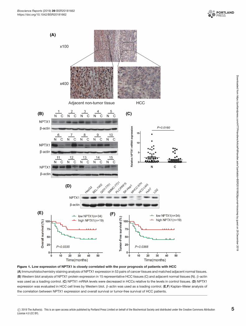
Figure 2. NPTX1 inhibits HCC cell proliferation in vitro
(A) The mRNA and protein levels of NPTX1 in SMMC-7721 and MHCC-97h cells after transfection with lentiviruses expressing
NPTX1. (B) The proliferation abilities of control and NPTX1-overexpressing cells were assessed by performing a CCK-8 assay. (C)
Colony formation assays were performed using control and NPTX1-overexpressing SMMC-7721 and MHCC-97h cells. (D) The
percentage of cells in different phases of the cell cycle was determined by FACS analysis of control and NPTX1-overexpressing
cells. (E) The protein levels of CDK2, CDK4, CDK6, Cyclin A2 and Cyclin D2 in control and NPTX1-overexpressing cells were
measured by Western blot. Data are shown as the mean +− SD; ***P<0.001 (Student’s t test). Abbreviation: CDK, cyclin-dependent
kinase.
cells (Figure 2B). Similarly, NPTX1-overexpressing cells showed a significantly lower colony formation rate thancontrol cells (Figure 2C).
To further reveal the mechanism by which NPTX1 contributes to proliferation, we detected the influence of NPTX1on cell cycle distribution by performing flow cytometry. NPTX1 overexpression was associated with an increase in thenumber of HCC cells in the G0/G1 phase and a decrease in the number of cells entering S phase (Figure 2D), suggestingthat NPTX1 could induce G0/G1 phase arrest in HCC cells. Western blot analysis of CDK2, cyclin-dependent kinase4 (CDK4), cyclin-dependent kinase 6 (CDK6), Cyclin A2 and Cyclin D2, which are cycle-related proteins, revealeddecreases relative to control levels in the expression of CDK2 and Cyclin A2 proteins in NPTX1-overexpressing cells(Figure 2E), whereas there were no significant changes in CDK4, CDK6 and Cyclin D2 levels relative to control levelsin either NPTX1-overexpressing cell line. These findings suggest that NPTX1 inhibits cell proliferation by inducingG0/G1 cell cycle arrest in HCC.
6 © 2019 The Author(s). This is an open access article published by Portland Press Limited on behalf of the Biochemical Society and distributed under the Creative Commons AttributionLicense 4.0 (CC BY).
Dow
nloaded from https://portlandpress.com
/HTTPH
andlers/ArticlePdfHandler.ashx?partialdoi=BSR
20181662&journal=bioscirep by guest on 29 Decem
ber 2019

Bioscience Reports (2019) 39 BSR20181662https://doi.org/10.1042/BSR20181662
Figure 3. Up-regulated NPTX1 expression induces mitochondria-related apoptosis in HCC cells
(A) Control and NPTX1-overexpressing SMMC-7721 and MHCC-97h cells were treated with cisplatin (10 μg/ml) for 24 h. Af-
ter treatment, the cells were analyzed by flow cytometry for Annexin V and PI dual labeling. Annexin V-positive cells were
designated as apoptotic cells. The percentage of apoptotic cells is shown. (B) Before performing the TUNEL assay, control
and NPTX1-overexpressing HCC cells were treated with cisplatin (10 μg/ml) for 24 h. The cells were observed by microscopy
at 200× magnification. (C) Western blot analysis of BAD, BAX, Mcl-1, Bcl-2, Cyt c, Caspase 3 and PARP 1 in control and
NPTX1-overexpressing SMMC-7721 and MHCC-97h cells treated with cisplatin (10 μg/ml) for 24 h was performed. Data are shown
as the mean +− SD; *P<0.05, ***P<0.001 (Student’s t test).
NPTX1 promotes mitochondria-related apoptosis in HCC cellsWe then analyzed the effects of NPTX1 on apoptosis in HCC cells by performing flow cytometry with Annexin Vand PI staining. We observed that NPTX1-overexpressing SMMC-7721 and MHCC-97h cells showed higher pro-portions of Annexin V-positive cells than did control cells (Figure 3A). This result was further verified by TUNELassay, which revealed a higher percentage of TUNEL-positive cells among NPTX1-overexpressing HCC cells thanamong control cells (Figure 3B). Previous reports have shown that, as a mediator of hypoxic injury in the brain,NPTX1 plays a critical role in regulating mitochondria-driven neuron death [8]. We speculated that NPTX1 mightcontribute to HCC cell apoptosis in a mitochondria-related manner. To test our hypothesis, Western blot analysis ofwell-known mitochondria-related proteins was performed. We found that the protein levels of BCL2-associated ago-nist of cell death (BAD) and BCL2-associated X protein (BAX) were increased in NPTX1-overexpressing SMMC-7721and MHCC-97h cells relative to control cells. In contrast, decreased levels of myeloid cell leukemia sequence 1 (Mcl-1)
© 2019 The Author(s). This is an open access article published by Portland Press Limited on behalf of the Biochemical Society and distributed under the Creative Commons AttributionLicense 4.0 (CC BY).
7
Dow
nloaded from https://portlandpress.com
/HTTPH
andlers/ArticlePdfHandler.ashx?partialdoi=BSR
20181662&journal=bioscirep by guest on 29 Decem
ber 2019

Bioscience Reports (2019) 39 BSR20181662https://doi.org/10.1042/BSR20181662
Figure 4. Effects of NPTX1 overexpression on HCC growth and apoptosis in vivo
(A) Control and NPTX1-overexpressing SMMC-7721 cells were injected subcutaneously into the right posterior flank of nude mice.
Representative images and tumor weights at 31 days postinjection are displayed. (B) Tumor sizes were measured and growth
curves were generated (n=6). (C) TUNEL assays were performed to identify the apoptotic cells in tumor xenografts of nude mice.
The results were observed by microscopy at 200× magnification. (D) Immunohistochemical staining for NPTX1, Ki67, Cyclin A2
and BAD proteins in tumor xenografts of nude mice. The results were observed by microscopy at 400× magnification. Data are
shown as the mean +− SD; *P<0.05 (Student’s t test).
and B-cell lymphoma-2 (Bcl-2) were found in NPTX1-overexpressing SMMC-7721 and MHCC-97h cells relative tocontrol cells (Figure 3C). Consistently, cytochrome c was released from mitochondria to the cytoplasm, and cleavageof caspase 3 and poly ADP-ribose polymerase 1 (PARP1) increased in NPTX1-overexpressing cells, indicating thatNPTX1 promotes mitochondria-related apoptosis in HCC cells.
Ectopic expression of NPTX1 suppresses HCC cell growth andcontributes to apoptosis in vivoTo verify the function of NPTX1 in HCC progression, we established xenograft mouse models by subcutaneously in-jecting control and NPTX1-overexpressing SMMC-7721 cells into nude mice. As shown in Figure 4A, compared withcontrol cells, NPTX1-overexpressing SMMC-7721 cells generated smaller volumes and weights of xenografts in nudemice. We also observed that xenografted tumors derived from NPTX1-overexpressing SMMC-7721 cells grew slowerthan did tumors derived from control cells (Figure 4B). To examine the effects of NPTX1 on apoptosis in xenograftedtumors, a TUNEL assay was performed to detect the apoptosis rates in control and NPTX1-overexpressing xenografts.We found more TUNEL-positive cells in NPTX1-overexpressing xenografts than in control xenografts (Figure4C). Subsequently, immunohistochemistry staining analysis revealed weaker staining for Ki67 and Cyclin A2 inNPTX1-overexpressing xenografts than in control xenografts (Figure 4D). In contrast, stronger BAD expression was
8 © 2019 The Author(s). This is an open access article published by Portland Press Limited on behalf of the Biochemical Society and distributed under the Creative Commons AttributionLicense 4.0 (CC BY).
Dow
nloaded from https://portlandpress.com
/HTTPH
andlers/ArticlePdfHandler.ashx?partialdoi=BSR
20181662&journal=bioscirep by guest on 29 Decem
ber 2019

Bioscience Reports (2019) 39 BSR20181662https://doi.org/10.1042/BSR20181662
Figure 5. The AKT pathway is involved in the NPTX1-mediated functions in HCC cells
(A,B) The protein levels of p-AKT, total AKT, p-GSK3α/β, total GSK3α/β, and NPTX1 in SMMC-7721 and MHCC-97h cells were
measured by Western blot analysis after incubation with LY294002 for 1 h and with GSK2141795 for 4 h at the indicated doses. (C,D)
Western blot analysis of NPTX1, Cyclin A2 and BAD in sh-NPTX1 SMMC-7721 and MHCC-97h cells after treatment with LY294002
(20 μM) for 1 h and GSK2141795 (5 μM) for 4 h was performed. (E) Western blot analysis of control and NPTX1-overexpressing
HCC cells after incubation with SC79 (10 μg/ml) for 2 h was performed. β-actin was used as a loading control. ImageJ software
was used to quantitate the protein bands. Data are shown as the mean +− SD; *P<0.05, **P<0.01, ***P<0.001 (Student’s t test).
observed in NPTX1-overexpressing xenografts than in control xenografts, consistent with our previous results. Col-lectively, our findings suggest that NPTX1 inhibits tumor growth and promotes apoptosis in vivo.
AKT acts as an upstream factor of NPTX1 and inhibits the effects ofNPTX1 in HCC cellsAs an oncogene reported to play a critical role in HCC progression, AKT regulates various cellular functions, includ-ing proliferation, apoptosis and invasion [25,26]. To investigate the potential molecular mechanisms linking NPTX1and the AKT pathway, we treated SMMC-7721 and MHCC-97h cells with the phosphoinositide-3-kinase inhibitorLY294002. We found that the levels of phosphorylated AKT and phosphorylated GSK3α/β were significantly re-duced after treatment with LY294002, whereas the expression of NPTX1 was enhanced by LY294002 treatment in adose-dependent manner (Figure 5A). We also observed a similar dose-dependent NPTX1 regulation trend in HCCcells treated with the AKT inhibitor GSK2141795 (Figure 5B), which indicate that the expression of NPTX1 mightbe modulated by the AKT signaling pathway in HCC.
To determine whether AKT functions upstream of NPTX1 to regulate cell cycle-related proteins andmitochondria-related proteins in HCC cells, we treated NPTX1 knockdown HCC cells with the inhibitors LY294002and GSK2141795. LY294002 and GSK2141795 rescued the decreased NPTX1 expression in NPTX1 knockdown cells,
© 2019 The Author(s). This is an open access article published by Portland Press Limited on behalf of the Biochemical Society and distributed under the Creative Commons AttributionLicense 4.0 (CC BY).
9
Dow
nloaded from https://portlandpress.com
/HTTPH
andlers/ArticlePdfHandler.ashx?partialdoi=BSR
20181662&journal=bioscirep by guest on 29 Decem
ber 2019

Bioscience Reports (2019) 39 BSR20181662https://doi.org/10.1042/BSR20181662
thereby inhibiting the up-regulated expression of Cyclin A2 induced by NPTX1 knockdown and rescuing the sup-pression of BAD induced by NPTX1 knockdown (Figure 5C,D). In contrast, SC79, an AKT phosphorylation activa-tor, inhibited the up-regulated NPTX1 expression in NPTX1-overexpressing cells, thereby reversing Cyclin A2 andBAD expression levels in NPTX1-overexpressing cells (Figure 5E). These results indicate that AKT signaling pathwaymodulated downstream targets, such as Cyclin A2 and BAD, via regulating the expression of NPTX1 in HCC cells.
DiscussionThe result of the present study showed that the expression of NPTX1 was decreased in HCC specimens. A similarresult was reported in colon cancer [12]. However, the present study is the first to show that NPTX1 expression levelcorrelates with clinicopathological factors (tumor size and metastasis) in HCC and is associated with survival time inHCC patients. Previous studies have identified NPTX1 as an epigenetic target and showed that it acted as a methy-lation marker in human pancreatic cancer [27], and consistent results were also have been found in cervical cancer[28] and colorectal cancer [29]. In 2015, Zhou et al. [11] demonstrated that promoter hypermethylation contributesto lower NPTX1 expression in lung cancer, which may lead to cancer pathogenesis. In addition, NPTX1 was foundto be an epigenetic target in regulating HDAC3-mediated neurotoxicity [30]. All these findings indicated that epige-netic regulation may be an important reason why NPTX1 shows low expression in various cancers, and ultimatelycontributes to the poor prognosis.
The high proliferation ability and resistance to death of cancer cells are worldwide obstacles in cancer therapy, andthese features contribute to cancer recurrence [31]. In our study, we found that NPTX1 inhibits cell proliferation byinducing cell cycle arrest and down-regulating cycle-related proteins (Cyclin A2 and CDK2), these findings are con-sistent with findings in colon cancer [12]. Cyclins play a critical role in modulating the cell cycle through binding andactivating cyclin-dependent kinase (CDK) enzymes [32,33]. As the regulatory subunit of CDK2, Cyclin A2 is essen-tial for cycle phase transitions and could be an independent prognostic factor for the relapse of human HCC [34,35].NPTX1 inhibits the level of Cyclin A2 in HCC, implicating an anti-cancer role of NPTX1 in cancer progression. Un-expectedly, we did not observe any changes in the levels of CDK4, CDK6 and Cyclin D2 proteins, which indicatedthat NPTX1 did not modulate cell proliferation via the CDK4 or CDK6/Cyclin D complex pathway.
Previous work showed that after exposure to hypoxia, NPTX1 was induced in a time-dependent manner and thatthis induction preceded neuronal death in the neonatal brain, suggesting that NPTX1 acts as an apoptosis mediatorduring brain injury [9,10]. Interestingly, subsequent studies reported that NPTX1 was involved in regulating apopto-sis in various cells, such as pancreatic β-cells [36] and human endometrial endothelial cells [37]. However, the effectsof NPTX1 on apoptosis in HCC remain unclear. Our present study revealed that NPTX1 could contribute to apop-tosis in HCC. Moreover, we found that NPTX1 promotes apoptosis in the mitochondria-related apoptotic pathway.Mitochondrial impairment or dysfunction could rapidly induce the inhibition of cell survival, and mitochondrialactivity-related therapies often achieve satisfying results [38,39]. Thus, NPTX1 represents a potential therapeutic tar-get in clinical treatment.
AKT plays indispensable roles in cellular proliferation, apoptosis, and invasion in various kinds of tumors, promot-ing cancer progression as an oncogene [25,26]. In HCC, AKT signaling has been identified as an important mediatorof multiple functions [24]. Our present study demonstrated that AKT signaling can down-regulate the expression ofNPTX1 and modulate its function, thereby being responsible for proliferation and apoptosis in HCC. The balancebetween growth and apoptosis is usually controlled by the AKT pathway. AKT promotes cell survival by inhibitingapoptosis through its ability to activate or inactivate several targets, including BAD and caspase 3 [40]. The presentresults showed that AKT signaling could abolish the effects of NPTX1 on Cyclin A2 and BAD, confirming the regula-tory role of AKT in HCC. AKT is significantly activated in HCC specimens [23], which explains why NPTX1 showslow expression in HCC. The AKT signaling pathway mediates the expression and stabilization of methyltransferasein various cancers and promotes tumor progression [41,42]. Based on previous studies that identified NPTX1 as anepigenetic target, we speculate that in HCC, the AKT signaling pathway down-regulates NPTX1 by modulating thepotential methyltransferases that catalyze the methylation of NPTX1. This hypothesis requires further investigation.Although down-regulated expression of NPTX1 occurs in diverse tumor types, the function and molecular mech-anisms are not fully understood. Whether NPTX1 performs its function in other malignancies through the AKTsignaling pathway warrants exploration.
ConclusionIn summary, we revealed that NPTX1 is down-regulated in HCC and that its expression is associated with clini-copathological factors. Furthermore, we identified NPTX1 as an important regulator of HCC cell proliferation and
10 © 2019 The Author(s). This is an open access article published by Portland Press Limited on behalf of the Biochemical Society and distributed under the Creative CommonsAttribution License 4.0 (CC BY).
Dow
nloaded from https://portlandpress.com
/HTTPH
andlers/ArticlePdfHandler.ashx?partialdoi=BSR
20181662&journal=bioscirep by guest on 29 Decem
ber 2019

Bioscience Reports (2019) 39 BSR20181662https://doi.org/10.1042/BSR20181662
apoptosis via AKT signaling. These findings indicate that NPTX1 can be utilized as a prognostic marker and potentialtherapeutic target.
FundingThis work was supported by the National Natural Science Foundation of China [grant numbers 81871963, 81572335].
Competing InterestsThe authors declare that there are no competing interests associated with the manuscript.
Author ContributionAll the authors participated in performing the experiments. Y.Z. and X.W. designed the study. Y.Z. and Y.Y. performed data analy-sis. Y.Z. and X.W. wrote the manuscript. All authors read and approved the final manuscript.
AbbreviationsAKT, AKT serine/threonine kinase; BAD, BCL2-associated agonist of cell death; CCK-8, cell counting kit-8; CDK2,cyclin-dependent kinase 2; CDK4, cyclin-dependent kinase 4; CDK6, cyclin-dependent kinase 6; FoxO, forkhead transcriptionfactors of the O class; GSK3α/β, glycogen synthase kinase 3α/β; HCC, hepatocellular carcinoma; HDAC3, histone deacetylase3; HRP, horseradish peroxidase; NPTX1, neuronal pentraxin 1; NPTX2, neuronal pentraxin 2; PARP1, poly ADP-ribose poly-merase 1; PI, propidium iodide; RIPA, radio immunoprecipitation assay; TUNEL, terminal deoxynucleotidyl transferase-mediateddUTP-biotin nick end labeling.
References1 Siegel, R.L., Miller, K.D. and Jemal, A. (2017) Cancer statistics, 2017. CA Cancer J. Clin. 67, 7–30, https://doi.org/10.3322/caac.213872 Lafaro, K.J., Demirjian, A.N. and Pawlik, T.M. (2015) Epidemiology of hepatocellular carcinoma. Surg. Oncol. Clin. N. Am. 24, 1–17,
https://doi.org/10.1016/j.soc.2014.09.0013 Poon, R.T. (2011) Prevention of recurrence after resection of hepatocellular carcinoma: a daunting challenge. Hepatology 54, 757–759,
https://doi.org/10.1002/hep.245694 Schlimgen, A.K. et al. (1995) Neuronal pentraxin, a secreted protein with homology to acute phase proteins of the immune system. Neuron 14,
519–526, https://doi.org/10.1016/0896-6273(95)90308-95 Tsui, C.C. et al. (1996) Narp, a novel member of the pentraxin family, promotes neurite outgrowth and is dynamically regulated by neuronal activity. J.
Neurosci. 16, 2463–2478, https://doi.org/10.1523/JNEUROSCI.16-08-02463.19966 Hsu, Y.C. and Perin, M.S. (1995) Human neuronal pentraxin II (NPTX2): conservation, genomic structure, and chromosomal localization. Genomics 28,
220–227, https://doi.org/10.1006/geno.1995.11347 Dodds, D.C. et al. (1997) Neuronal pentraxin receptor, a novel putative integral membrane pentraxin that interacts with neuronal pentraxin 1 and 2 and
taipoxin-associated calcium-binding protein 49. J. Biol. Chem. 272, 21488–21494, https://doi.org/10.1074/jbc.272.34.214888 Al Rahim, M., Thatipamula, S. and Hossain, M.A. (2013) Critical role of neuronal pentraxin 1 in mitochondria-mediated hypoxic-ischemic neuronal
injury. Neurobiol. Dis. 50, 59–68, https://doi.org/10.1016/j.nbd.2012.10.0039 Hossain, M.A. et al. (2004) Neuronal pentraxin 1: a novel mediator of hypoxic-ischemic injury in neonatal brain. J. Neurosci. 24, 4187–4196,
https://doi.org/10.1523/JNEUROSCI.0347-04.200410 Russell, J.C. et al. (2011) Neuronal pentraxin 1 induction in hypoxic-ischemic neuronal death is regulated via a glycogen synthase kinase-3alpha/beta
dependent mechanism. Cell. Signal. 23, 673–682, https://doi.org/10.1016/j.cellsig.2010.11.02111 Zhou, C. et al. (2015) NPTX1 is a novel epigenetic regulation gene and associated with prognosis in lung cancer. Biochem. Biophys. Res. Commun.
458, 381–386, https://doi.org/10.1016/j.bbrc.2015.01.12412 Peng, X. et al. (2018) NPTX1 inhibits colon cancer cell proliferation through down-regulating cyclin A2 and CDK2 expression. Cell Biol. Int. 42,
589–597, https://doi.org/10.1002/cbin.1093513 Yue, W. et al. (2015) Transcriptomic analysis of pancreatic cancer cells in response to metformin and aspirin: an implication of synergy. Sci. Rep. 5,
13390, https://doi.org/10.1038/srep1339014 Yue, W. et al. (2014) Repurposing of metformin and aspirin by targeting AMPK-mTOR and inflammation for pancreatic cancer prevention and treatment.
Cancer Prev. Res. 7, 388–397, https://doi.org/10.1158/1940-6207.CAPR-13-033715 Vivanco, I. and Sawyers, C.L. (2002) The phosphatidylinositol 3-Kinase AKT pathway in human cancer. Nat. Rev. Cancer 2, 489–501,
https://doi.org/10.1038/nrc83916 Fukumoto, S. et al. (2001) Akt participation in the Wnt signaling pathway through Dishevelled. J. Biol. Chem. 276, 17479–17483,
https://doi.org/10.1074/jbc.C00088020017 Zhou, B.P. et al. (2004) Dual regulation of Snail by GSK-3beta-mediated phosphorylation in control of epithelial-mesenchymal transition. Nat. Cell Biol.
6, 931–940, https://doi.org/10.1038/ncb117318 Chen, M.L. et al. (2006) The deficiency of Akt1 is sufficient to suppress tumor development in Pten+/- mice. Genes Dev. 20, 1569–1574,
https://doi.org/10.1101/gad.1395006
© 2019 The Author(s). This is an open access article published by Portland Press Limited on behalf of the Biochemical Society and distributed under the Creative CommonsAttribution License 4.0 (CC BY).
11
Dow
nloaded from https://portlandpress.com
/HTTPH
andlers/ArticlePdfHandler.ashx?partialdoi=BSR
20181662&journal=bioscirep by guest on 29 Decem
ber 2019

Bioscience Reports (2019) 39 BSR20181662https://doi.org/10.1042/BSR20181662
19 Hollander, M.C. et al. (2011) Akt1 deletion prevents lung tumorigenesis by mutant K-ras. Oncogene 30, 1812–1821,https://doi.org/10.1038/onc.2010.556
20 Skeen, J.E. et al. (2006) Akt deficiency impairs normal cell proliferation and suppresses oncogenesis in a p53-independent and mTORC1-dependentmanner. Cancer Cell 10, 269–280, https://doi.org/10.1016/j.ccr.2006.08.022
21 Xu, P.Z. et al. (2012) The effect Akt2 deletion on tumor development in Pten(+/-) mice. Oncogene 31, 518–526, https://doi.org/10.1038/onc.2011.24322 Maroulakou, I.G. et al. (2007) Akt1 ablation inhibits, whereas Akt2 ablation accelerates, the development of mammary adenocarcinomas in mouse
mammary tumor virus (MMTV)-ErbB2/neu and MMTV-polyoma middle T transgenic mice. Cancer Res. 67, 167–177,https://doi.org/10.1158/0008-5472.CAN-06-3782
23 Zhou, L. et al. (2010) The mTOR pathway is associated with the poor prognosis of human hepatocellular carcinoma. Med. Oncol. 27, 255–261,https://doi.org/10.1007/s12032-009-9201-4
24 Zhou, Q., Lui, V.W. and Yeo, W. (2011) Targeting the PI3K/Akt/mTOR pathway in hepatocellular carcinoma. Future Oncol. 7, 1149–1167,https://doi.org/10.2217/fon.11.95
25 Cheng, G.Z. et al. (2008) Advances of AKT pathway in human oncogenesis and as a target for anti-cancer drug discovery. Curr. Cancer Drug Targets 8,2–6, https://doi.org/10.2174/156800908783497159
26 Tommasi, S. et al. (2007) Molecular pathways and related target therapies in liver carcinoma. Curr. Pharm. Des. 13, 3279–3287,https://doi.org/10.2174/138161207782360663
27 Hagihara, A. et al. (2004) Identification of 27 5′ CpG islands aberrantly methylated and 13 genes silenced in human pancreatic cancers. Oncogene 23,8705–8710, https://doi.org/10.1038/sj.onc.1207783
28 Ongenaert, M. et al. (2008) Discovery of DNA methylation markers in cervical cancer using relaxation ranking. BMC Med. Genomics 1, 57,https://doi.org/10.1186/1755-8794-1-57
29 Mori, Y. et al. (2011) Novel candidate colorectal cancer biomarkers identified by methylation microarray-based scanning. Endocr. Relat. Cancer 18,465–478, https://doi.org/10.1530/ERC-11-0083
30 Qu, Z. and D’Mello, S.R. (2018) Proteomic analysis identifies NPTX1 and HIP1R as potential targets of histone deacetylase-3-mediatedneurodegeneration. Exp. Biol. Med. (Maywood) 243, 627–638, https://doi.org/10.1177/1535370218761149
31 Teufel, A., Marquardt, J.U. and Galle, P.R. (2012) Novel insights in the genetics of HCC recurrence and advances in transcriptomic data integration. J.Hepatol. 56, 279–281, https://doi.org/10.1016/j.jhep.2011.05.035
32 Deshpande, A., Sicinski, P. and Hinds, P.W. (2005) Cyclins and cdks in development and cancer: a perspective. Oncogene 24, 2909–2915,https://doi.org/10.1038/sj.onc.1208618
33 Arellano, M. and Moreno, S. (1997) Regulation of CDK/cyclin complexes during the cell cycle. Int. J. Biochem. Cell Biol. 29, 559–573,https://doi.org/10.1016/S1357-2725(96)00178-1
34 Chao, Y. et al. (1998) Overexpression of cyclin A but not Skp 2 correlates with the tumor relapse of human hepatocellular carcinoma. Cancer Res. 58,985–990
35 Gong, D. et al. (2007) Cyclin A2 regulates nuclear-envelope breakdown and the nuclear accumulation of cyclin B1. Curr. Biol. 17, 85–91,https://doi.org/10.1016/j.cub.2006.11.066
36 Schvartz, D. et al. (2012) Modulation of neuronal pentraxin 1 expression in rat pancreatic beta-cells submitted to chronic glucotoxic stress. Mol. CellProteomics 11, 244–254, https://doi.org/10.1074/mcp.M112.018051
37 Guzeloglu-Kayisli, O. et al. (2014) Long-acting progestin-only contraceptives enhance human endometrial stromal cell expressed neuronal pentraxin-1and reactive oxygen species to promote endothelial cell apoptosis. J. Clin. Endocrinol. Metab. 99, E1957–E1966,https://doi.org/10.1210/jc.2014-1770
38 Heimlich, G. et al. (2004) Bax-induced cytochrome c release from mitochondria depends on alpha-helices-5 and -6. Biochem. J. 378, 247–255,https://doi.org/10.1042/bj20031152
39 Shin, S.M. and Kim, S.G. (2009) Inhibition of arachidonic acid and iron-induced mitochondrial dysfunction and apoptosis by oltipraz and novel1,2-dithiole-3-thione congeners. Mol. Pharmacol. 75, 242–253, https://doi.org/10.1124/mol.108.051128
40 Datta, S.R., Brunet, A. and Greenberg, M.E. (1999) Cellular survival: a play in three Akts. Genes Dev. 13, 2905–2927,https://doi.org/10.1101/gad.13.22.2905
41 Li, N. et al. (2017) AKT-mediated stabilization of histone methyltransferase WHSC1 promotes prostate cancer metastasis. J. Clin. Invest. 127,1284–1302, https://doi.org/10.1172/JCI91144
42 Tan, X. et al. (2017) PI3K/AKT-mediated upregulation of WDR5 promotes colorectal cancer metastasis by directly targeting ZNF407. Cell Death Dis. 8,e2686, https://doi.org/10.1038/cddis.2017.111
12 © 2019 The Author(s). This is an open access article published by Portland Press Limited on behalf of the Biochemical Society and distributed under the Creative Commons AttributionLicense 4.0 (CC BY).
Dow
nloaded from https://portlandpress.com
/HTTPH
andlers/ArticlePdfHandler.ashx?partialdoi=BSR
20181662&journal=bioscirep by guest on 29 Decem
ber 2019