Embed Size (px)
Citation preview
BMC Immunology (2001) 2:3 http://www.biomedcentral.com/1471-2172/2/3
BMC Immunology (2001) 2:3Research articleRequirements for activation and RAFT localization of the T-lymphocyte kinase Rlk/TxkMario Chamorro†1,6, Michael J Czar†2,7, Jayanta Debnath†1,2,5,8,
Genhong Cheng3, Michael J Lenardo4, Harold E Varmus1,6 and
Pamela L Schwartzberg*1,2
Address: 1National Cancer Institute, National Institutes of Health, Bethesda, MD, USA, 2National Institute for Human Genome Research, National Institutes of Health, Bethesda, MD, USA, 3Department of Microbiology and Molecular Genetics, University of California Los Angeles,
Los Angeles, CA, USA, 4National Institute of Allergy and Infectious Diseases, National Institutes of Health, Bethesda, MD, USA, 5Howard
Hughes Medical Institute-NIH Research Scholars Program, 6Present address: Memorial Sloan Kettering Cancer Research Institute, New York, NY, USA, 7Present address: CuraGen Corp., 555 Long Wharf Drive, 13th Fl., New Haven, CT, USA and 8Present address: Department of Cell
Biology, Harvard Medical School, 240 Longwood Ave, Boston, MA, USA
E-mail: Mario Chamorro - [email protected]; Michael J Czar - [email protected];
Jayanta Debnath - [email protected]; Genhong Cheng - [email protected]; Michael J Lenardo - [email protected]; Harold E Varmus - [email protected]; Pamela L Schwartzberg* - [email protected]
*Corresponding author †Equal contributors
AbstractBackground: The Tec family kinases are implicated in signaling from lymphocyte antigenreceptors and are activated following phosphorylation by Src kinases. For most Tec kinases, thisactivation requires an interaction between their pleckstrin homology (PH) domains and theproducts of phosphoinositide 3-Kinase, which localizes Tec kinases to membrane RAFTs. Rlk/Txkis a Tec related kinase expressed in T cells that lacks a pleckstrin homology domain, having insteada palmitoylated cysteine-string motif. To evaluate Rlk's function in T cell receptor signalingcascades, we examined the requirements for Rlk localization and activation by Src family kinases.
Results: We demonstrate that Rlk is also associated with RAFTs, despite its lack of a pleckstrinhomology domain. Rlk RAFT association requires the cysteine-string motif and is independent ofPI3 Kinase activity. We further demonstrate that Rlk can be phosphorylated and activated by Srckinases, leading to a decrease in its half-life. A specific tyrosine in the activation loop of Rlk, Y420,is required for phosphorylation and activation, as well as for decreased stability, but is not requiredfor lipid RAFT association. Mutation of this tyrosine also prevents increased tyrosinephosphorylation of Rlk after stimulation of the T cell receptor, suggesting that Rlk isphosphorylated by Src family kinases in response to T cell receptor engagement.
Conclusions: Like the other related Tec kinases, Rlk is associated with lipid RAFTs and can bephosphorylated and activated by Src family kinases, supporting a role for Rlk in signalingdownstream of Src kinases in T cell activation.
Published: 8 May 2001
BMC Immunology 2001, 2:3
This article is available from: http://www.biomedcentral.com/1471-2172/2/3
(c) 2001 Chamorro et al, licensee BioMed Central Ltd.
Received: 19 April 2001Accepted: 8 May 2001
BMC Immunology (2001) 2:3 http://www.biomedcentral.com/1471-2172/2/3
BackgroundEngagement of antigen receptors on lymphocytes leads
to the rapid sequential activation of non-receptor tyro-
sine kinases, including well-studied members of the Srcand the ZAP-70/Syk families [1,2]. Recently, the Tec ki-
nases have also been implicated as important compo-
nents of signaling from antigen and other lymphocyte
cell surface receptors (reviewed in [3,4,5].) This sub-
family includes six members: Btk, Tec, Itk/Tsk/Emt,
Bmx/Emt, Dsrc29, and Rlk/Txk [3,6,7]. The importance
of the Tec kinases in antigen receptor signaling was first
demonstrated by the observation that mutations in BTK
result in the human disease X-linked agammaglobuline-
mia (XLA) and the murine counterpart X-linked immu-
nodeficiency (xid). These severe B cell
immunodeficiencies are characterized by diminished
numbers of mature B cells and reduced immunoglobulin
levels associated with impaired PLC-γ activation and
Ca++ mobilization in response to surface IgM stimula-
tion (sIgM) [8,9,10,11]. Btk kinase activity and tyrosine
phosphorylation increase upon stimulation of sIgM as
well as the IL-5 and IL6 receptors in B cells, and upon
crosslinking of the high affinity IgE receptor (FcεRII) in
mast cells [12,13,14,15]. It is now clear that the other Tec
kinases participate in similar signaling cascades in mul-
tiple cell-types. Itk, a Tec family kinase that is predomi-
nantly expressed in T cells, is tyrosine phosphorylated
and activated after stimulation of either the T cell recep-
tor (TCR) or CD28 in T cell lines, and FcεRII in mast cells[12,16,17,18]. Recently, we have found that Rlk/Txk can
also be phosphorylated after TCR engagement [19]. Fur-
thermore, we have found that combined mutation of Rlk
and Itk leads to profound defects in TCR signaling asso-
ciated with impaired activation of PLC-γ [20]. Such data
suggest that members of this family may function simi-
larly in signaling pathways downstream from these cell
surface antigen receptors.
Several lines of evidence indicate that, in these signaling
cascades, Tec family members act downstream of Src ki-
nases. Src kinases are activated within seconds of sIgM
stimulation in B cells, while Btk is activated within min-
utes [14]. Moreover, Src family kinases have been found
to phosphorylate and activate Btk, Itk, Tec and Rlk when
co-expressed [16,19,21,22,23]. Phosphopeptide mapping
and mutational analyses of Btk have identified Y551 in
the activation loop of the kinase domain as the major
Src-transphosphorylation site. Phosphorylation of Y551
and a putative autophosphorylation site Y223 in the SH3
domain of Btk [23,24] are observed following BCR acti-
vation, suggesting that these tyrosine phosphorylations
are functionally important for antigen receptor signal-
ing. Although physical binding of full length Btk to Src
family kinases in vivo has not been reported, in vitro in-teractions have been demonstrated between proline-rich
motifs in the amino-terminal sequence of Btk and the Src
homology (SH) 3 domains of Src family kinases [25,26].
Recently, it has been found that the activation of Btk andItk by Src family kinases is potentiated by and requires
the activity of Phosphoinositide 3-kinase (PI3K), the
products of which interact with the PH domains of Tec
kinases [27,28]. This interaction helps localize Tec fami-
ly kinases to the membrane where phosphorylation by
Src kinases can occur. Data suggest that, for Itk, this
membrane association specifically targets the protein to
glycolipid enriched detergent-insoluble membrane
microdomains known as "RAFTs" and that both this as-
sociation and the activation of Itk requires the PH do-
main [29].
rlk/TXK encodes a kinase expressed in T cells and mast
cells that closely resembles members of the Btk/Tec fam-
ily of kinases [30,31,32]. However, in contrast to the oth-
er Tec family members, Rlk lacks a pleckstrin homology
domain. Instead, we have found that the rlk gene en-
codes a distinctive palmitoylated cysteine-string motif
near its amino-terminus. Furthermore, we have found
that an internal translational start-site, downstream of
the region encoding the cysteine-string motif, gives rise
to a second isoform that lacks the palmitoylated cysteine
string and that can be localized in the nucleus [19]. Al-
though these distinctive features suggest that Rlk may
have atypical functions, Rlk shares with Btk kinases thehomologous proline-rich motif that binds the SH3 do-
mains of Src kinases and the tyrosines at equivalent sites
in the SH3 and kinase domains.
We have recently shown that Rlk can be phosphorylated
and activated by the Src family kinase Fyn. However,
consistent with the lack of a pleckstrin homology do-
main, activation of Rlk is independent of PI3K activity
[19]. To further characterize the mechanism of activation
of Rlk/Txk, we studied the nature of Rlk's localization
and interaction with Src family kinases. We report here a
physical association of Rlk with Fyn both in vitro and
when co-expressed in 293T cells. Similar to Btk and Itk,
we find that a conserved tyrosine in the activation loop of
the Rlk kinase domain, Y420, mediates specificity for
transphosphorylation of Rlk by Src family kinases. This
tyrosine is also critical for in vivo activation of Rlk, since
mutation of Y420 to phenylalanine (Y420F) reduces
phosphorylation of Rlk upon co-expression with Fyn or
after engagement of the TCR. Furthermore, we have ob-
served that the phosphorylation of Rlk by Fyn increases
Rlk turnover, consistent with the increased turnover ob-
served with other TCR signaling molecules following ac-
tivation. In contrast, localization of Rlk does not appear
to rely on tyrosine phosphorylation or kinase activity ofRlk. Instead, we find a constitutive association of a frac-
BMC Immunology (2001) 2:3 http://www.biomedcentral.com/1471-2172/2/3
tion of the full-length Rlk with a detergent-insoluble
RAFT fraction. Association with the RAFT compartment
does not depend on tyrosine phosphorylation, nor on
PI3K activation, but rather on the presence of thecysteine string motif. Thus, despite lacking a pleckstrin
homology domain, Rlk is localized in RAFTs, is phospho-
rylated by Src family kinases, and may transduce signals
downstream of Src family kinases during T cell activa-
tion.
ResultsRlk Interacts with FynRlk/Txk is a Tec related kinase that is predominantly ex-
pressed in cells of the T cell lineage [30,31,32]. We have
recently demonstrated that Rlk is phosphorylated fol-
lowing stimulation of the Jurkat T cell line by crosslink-
ing of the CD3ε chain of the TCR [19]. We have further
observed that Rlk can be phosphorylated and activated
by co-expression with the Src family kinase, Fyn, in a
PI3K-independent fashion. In contrast, activation of Btk
and Itk by Src family kinases is PI3K-dependent. To fur-
ther characterize the mechanism of Rlk activation by Src
family kinases, we examined potential interactions of Rlk
with Src kinases both in vitro and in vivo.
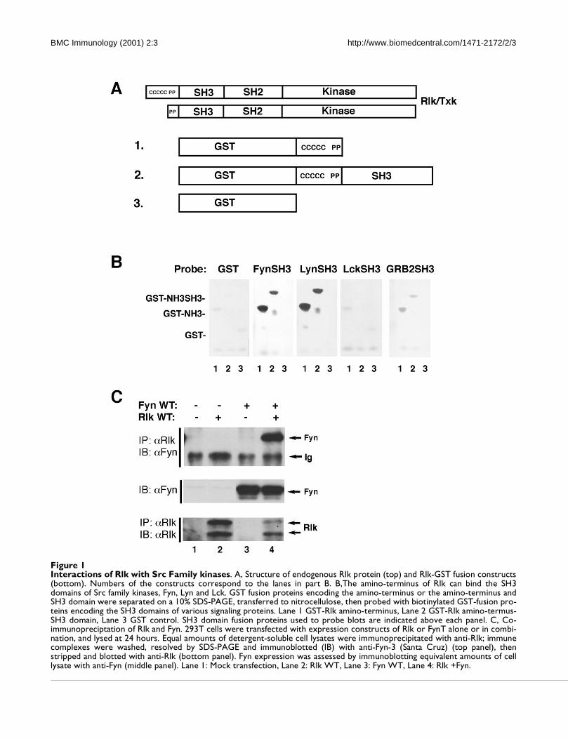
The proline-rich region of Btk has been found to bind to
SH3 domains of Src family kinses in vitro. To determine
whether the amino-terminus of Rlk/Txk interacts with
Src family SH3 domains in vitro, we performed in vitro
filter binding analyses. GST fusions encoding the Rlk
amino-terminal and SH3 domains or the amino-termi-
nus alone (containing the cysteine string and proline-
rich region) bound biotinylated GST fusions of the SH3
domains of Fyn, Hck, and Lyn, but not those of Src or
Lck, despite the ability of these constructs to bind other
proline rich sequences (Fig. 1B and data not shown)
[25,33]. The Rlk fusion proteins also failed to efficiently
bind SH3 domains from other kinases, including Abl,
Csk and Btk. A weaker but detectable binding to the SH3
domain of Grb2, however, was observed. These data
agree both with two-hybrid experiments and in vitro fil-
ter binding assays that have demonstrated specific bind-
ing of the proline-rich region near the amino-terminus of
BTK and Tec to the SH3 domains of the Src family kinas-
es Fyn, Lyn and Hck [22,25,26].
Because Fyn is expressed in T cells and Hck and Lyn are
not, we pursued the connection between Rlk and the T
cell-specific isoform p59FynT. Co-immunoprecipitation
demonstrated an interaction between these proteins in
extracts of cells in which both Rlk and Fyn were ex-
pressed (Fig. 1C). Anti-Rlk serum precipitated Fyn from
lysates only when Rlk was expressed (Fig. 1C, lane 4).
Likewise, anti-Fyn serum could precipitate Rlk onlywhen both Rlk and Fyn were produced (data not shown).
This interaction was not dependent on the activity of ei-
ther kinase, as co-immunoprecipitation was observed
with either wild-type (WT) or kinase-inactive versions of
the two proteins. Furthermore, both isoforms of Rlkwere also able to co-precipitate with Fyn (data not
shown). Finally, we have not observed co-precipitation
of full-length Rlk with several other proteins involved in
TCR signaling (unpublished observations).
Rlk co-localizes with a portion of FynTo further examine potential interactions between Rlk
and Fyn we compared the subcellular distribution of Rlk
to that of p59FynT. For these and subsequent analyses, we
utilized Green Fluorescent Protein (GFP)-tagged ver-
sions of Rlk. We have previously demonstrated that
these fusion proteins are kinase-active and exhibit local-
ization patterns similar to those observed by indirect im-
munofluorescence for wild type Rlk . Furthermore, we
have found that, like WT Rlk, they co-precipitate with
Fyn (data not shown). However, unlike WT Rlk, Rlk-GFP
does not co-migrate with immunoglobulin heavy chain.
Since these GFP fusions behaved similarly to wildtype
Rlk in all known respects except for electrophoretic mo-
bility, we used GFP fusions of mutant and wild type ver-
sions of Rlk to facilitate studies of Rlk localization and
activation.
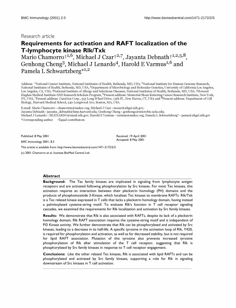
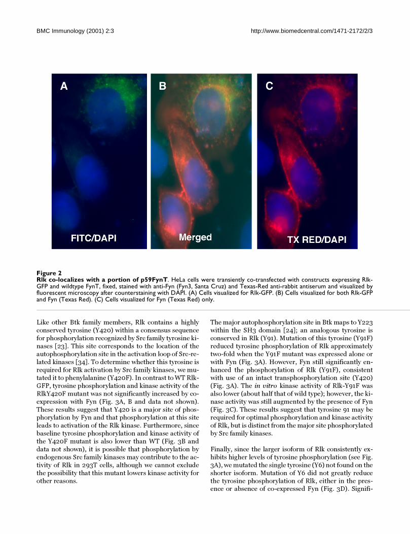
We co-transfected Fyn and Rlk-GFP expressing con-
structs into HeLa cells, and visualized Fyn with anti-Fynserum and Texas-red conjugated secondary antibody
(Fig. 2). Examination of cells expressing both kinases
demonstrated that a portion of Fyn co-localized with a
portion of the Rlk protein as indicated by the yellow
staining in overlapping images (center panel). Thus, Fyn
and Rlk can reside in similar compartments within the
cell, further supporting a potential interaction between
these two molecules.
Fyn phosphorylates and activates Rlk: A Conserved Tyro-sine (Y420) Provides Specificity For Phosphorylation of Rlk By Src KinasesTo further examine the consequences of the interaction
between Rlk and Src Family kinases, we examined tyro-
sine phosphorylation of Rlk by Fyn using co-expression
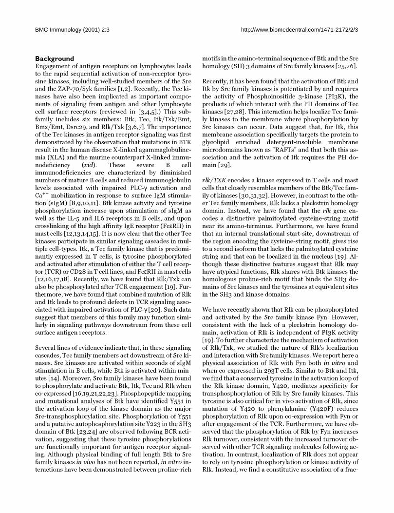
studies. As previously demonstrated, co-expression of
kinase-active Fyn and Rlk-GFP in 293T cells led to a
marked increase in the tyrosine phosphorylation and ki-
nase activity of wild type Rlk-GFP (Fig. 3A, lane 1 and 2;
3B, lane 1 and 2). This was not observed in cells trans-
fected with kinase-inactive Fyn, and the increase in ki-
nase activity on enolase appeared to be due to Rlk rather
than Fyn, since we barely detect phosphorylation of eno-
lase when WT Fyn is co-expressed with kinase-inactive
Rlk [19]. Thus, the phosphorylation of Rlk appears tocause an increase in Rlk kinase activity.
BMC Immunology (2001) 2:3 http://www.biomedcentral.com/1471-2172/2/3
Figure 1Interactions of Rlk with Src Family kinases. A, Structure of endogenous Rlk protein (top) and Rlk-GST fusion constructs(bottom). Numbers of the constructs correspond to the lanes in part B. B,The amino-terminus of Rlk can bind the SH3domains of Src family kinases, Fyn, Lyn and Lck. GST fusion proteins encoding the amino-terminus or the amino-terminus andSH3 domain were separated on a 10% SDS-PAGE, transferred to nitrocellulose, then probed with biotinylated GST-fusion pro-teins encoding the SH3 domains of various signaling proteins. Lane 1 GST-Rlk amino-terminus, Lane 2 GST-Rlk amino-termus-SH3 domain, Lane 3 GST control. SH3 domain fusion proteins used to probe blots are indicated above each panel. C, Co-immunopreciptation of Rlk and Fyn. 293T cells were transfected with expression constructs of Rlk or FynT alone or in combi-nation, and lysed at 24 hours. Equal amounts of detergent-soluble cell lysates were immunoprecipitated with anti-Rlk; immunecomplexes were washed, resolved by SDS-PAGE and immunoblotted (IB) with anti-Fyn-3 (Santa Cruz) (top panel), thenstripped and blotted with anti-Rlk (bottom panel). Fyn expression was assessed by immunoblotting equivalent amounts of celllysate with anti-Fyn (middle panel). Lane 1: Mock transfection, Lane 2: Rlk WT, Lane 3: Fyn WT, Lane 4: Rlk +Fyn.
BMC Immunology (2001) 2:3 http://www.biomedcentral.com/1471-2172/2/3
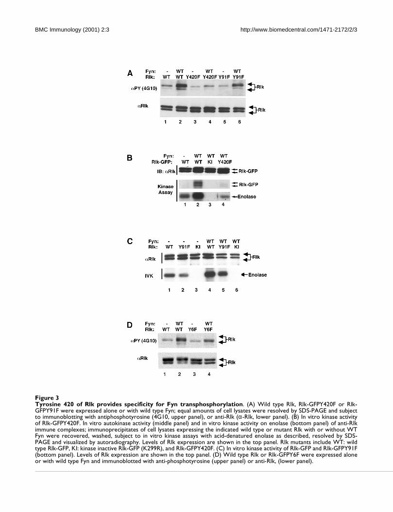
Like other Btk family members, Rlk contains a highly
conserved tyrosine (Y420) within a consensus sequence
for phosphorylation recognized by Src family tyrosine ki-
nases [23]. This site corresponds to the location of the
autophosphorylation site in the activation loop of Src-re-
lated kinases [34]. To determine whether this tyrosine is
required for Rlk activation by Src family kinases, we mu-
tated it to phenylalanine (Y420F). In contrast to WT Rlk-
GFP, tyrosine phosphorylation and kinase activity of the
RlkY420F mutant was not significantly increased by co-
expression with Fyn (Fig. 3A, B and data not shown).
These results suggest that Y420 is a major site of phos-
phorylation by Fyn and that phosphorylation at this site
leads to activation of the Rlk kinase. Furthermore, since
baseline tyrosine phosphorylation and kinase activity of
the Y420F mutant is also lower than WT (Fig. 3B and
data not shown), it is possible that phosphorylation by
endogenous Src family kinases may contribute to the ac-
tivity of Rlk in 293T cells, although we cannot exclude
the possibility that this mutant lowers kinase activity for
other reasons.
The major autophosphorylation site in Btk maps to Y223
within the SH3 domain [24]; an analogous tyrosine is
conserved in Rlk (Y91). Mutation of this tyrosine (Y91F)
reduced tyrosine phosphorylation of Rlk approximately
two-fold when the Y91F mutant was expressed alone or
with Fyn (Fig. 3A). However, Fyn still significantly en-
hanced the phosphorylation of Rlk (Y91F), consistent
with use of an intact transphosphorylation site (Y420)
(Fig. 3A). The in vitro kinase activity of Rlk-Y91F was
also lower (about half that of wild type); however, the ki-
nase activity was still augmented by the presence of Fyn
(Fig. 3C). These results suggest that tyrosine 91 may be
required for optimal phosphorylation and kinase activity
of Rlk, but is distinct from the major site phosphorylated
by Src family kinases.
Finally, since the larger isoform of Rlk consistently ex-
hibits higher levels of tyrosine phosphorylation (see Fig.
3A), we mutated the single tyrosine (Y6) not found on the
shorter isoform. Mutation of Y6 did not greatly reduce
the tyrosine phosphorylation of Rlk, either in the pres-ence or absence of co-expressed Fyn (Fig. 3D). Signifi-
Figure 2Rlk co-localizes with a portion of p59FynT. HeLa cells were transiently co-transfected with constructs expressing Rlk-GFP and wildtype FynT, fixed, stained with anti-Fyn (Fyn3, Santa Cruz) and Texas-Red anti-rabbit antiserum and visualized byfluorescent microscopy after counterstaining with DAPI. (A) Cells visualized for Rlk-GFP. (B) Cells visualized for both Rlk-GFPand Fyn (Texas Red). (C) Cells visualized for Fyn (Texas Red) only.
BMC Immunology (2001) 2:3 http://www.biomedcentral.com/1471-2172/2/3
Figure 3Tyrosine 420 of Rlk provides specificity for Fyn transphosphorylation. (A) Wild type Rlk, Rlk-GFPY420F or Rlk-GFPY91F were expressed alone or with wild type Fyn; equal amounts of cell lysates were resolved by SDS-PAGE and subjectto immunoblotting with antiphosphotyrosine (4G10, upper panel), or anti-Rlk (α-Rlk, lower panel). (B) In vitro kinase activityof Rlk-GFPY420F. In vitro autokinase activity (middle panel) and in vitro kinase activity on enolase (bottom panel) of anti-Rlkimmune complexes; immunoprecipitates of cell lysates expressing the indicated wild type or mutant Rlk with or without WTFyn were recovered, washed, subject to in vitro kinase assays with acid-denatured enolase as described, resolved by SDS-PAGE and visualized by autoradiography. Levels of Rlk expression are shown in the top panel. Rlk mutants include WT: wildtype Rlk-GFP, KI: kinase inactive Rlk-GFP (K299R), and Rlk-GFPY420F. (C) In vitro kinase activity of Rlk-GFP and Rlk-GFPY91F(bottom panel). Levels of Rlk expression are shown in the top panel. (D) Wild type Rlk or Rlk-GFPY6F were expressed aloneor with wild type Fyn and immunoblotted with anti-phosphotyrosine (upper panel) or anti-Rlk, (lower panel).
BMC Immunology (2001) 2:3 http://www.biomedcentral.com/1471-2172/2/3
cantly, the larger isoform still exhibited a higher level of
tyrosine phosphorylation than the shorter form of Rlk.
Thus, the increased tyrosine phosphorylation of the larg-
er isoform cannot be attributed solely to a phosphoryla-tion site present on the larger protein species.
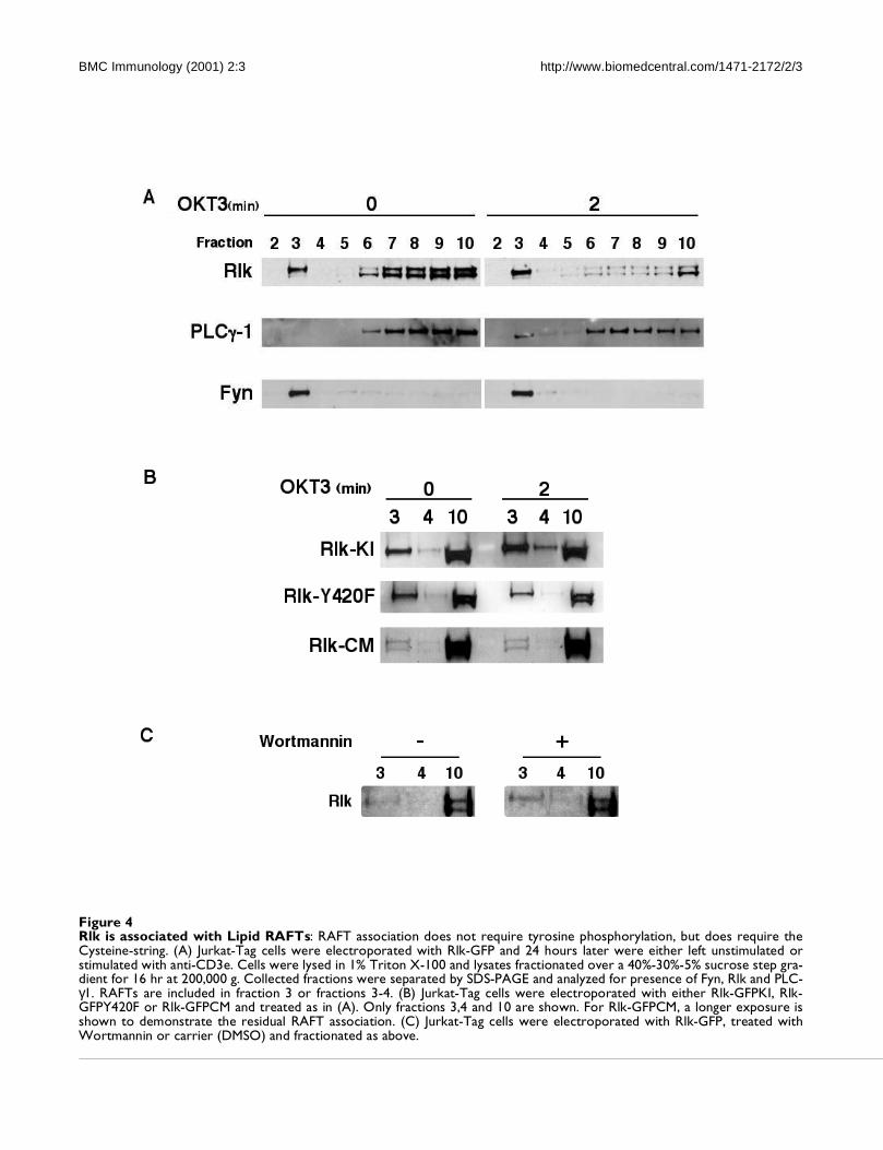
Rlk localizes in RAFTsSince the relative increase in tyrosine phosphorylation of
the larger form of Rlk did not result solely from an addi-
tional site for tyrosine phosphorylation, it was possible
that this increased phosphorylation of the longer isoform
of Rlk may result from preferential localization close to
Src family kinases. Like other proteins modified by
palmitoylation, the Src family kinase Fyn is found local-
ized in RAFTs, a membrane microdomain fraction en-
riched in signaling molecules that can be isolated by
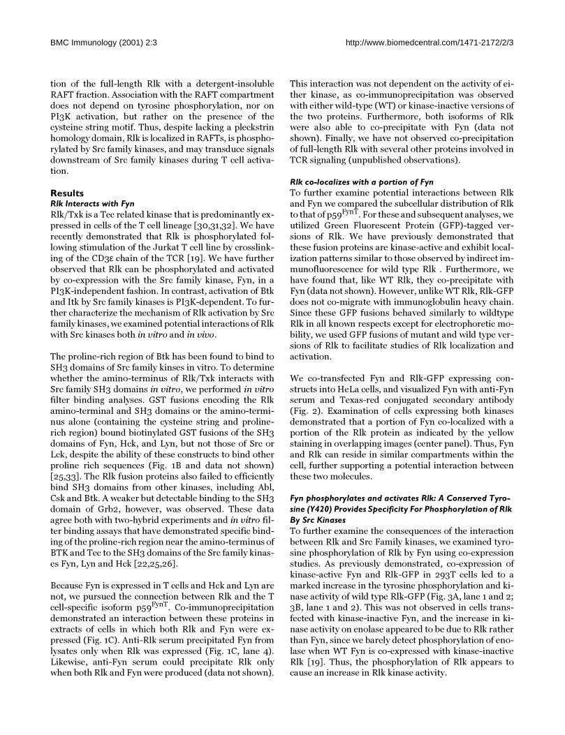
sucrose gradient fractionation. To examine whether Rlk
is also associated with RAFTs, we performed sucrose
gradiant fractionation of lysates from Jurkat lymphoma
cell lines transfected with Rlk-GFP. Upon such fraction-
ation, we observe the Src family kinase Fyn constitutively
associated with the RAFT fraction (Fig. 4A). In contrast,
PLC-γ was not found in the RAFT fraction constitutively,
but a portion became inducibly associated with RAFTs
upon activation. Similar to Fyn, a portion of Rlk was also
found constitutively associated with RAFTs. Notably,
this association consisted primarily of the larger form of
Rlk which contains the cysteine-string and is palmi-
toylated. Furthermore, the association of Rlk withRAFTs only marginally increased upon stimulation
through the TCR.
Upon TCR signaling, many molecules have been found to
show markedly increased association with RAFTs. Since
we have found that Rlk can be phosphorylated by Src
family kinases leading to activation of its kinase, we eval-
uated whether phosphorylation could influence RAFT
association. Neither mutation of a conserved lysine in
the kinase domain, which rendered Rlk kinase inactive,
nor mutation of the Src family kinase phosphorylation
site, tyrosine 420, prevented association with the RAFT
fraction. Thus, phosphorylation and activation of Rlk
does not appear to contribute to its association with
RAFTs (Fig. 4B).
A key mechanism of targeting proteins to RAFTs is lipid
modification. Since the long form of Rlk is palmitoylated
on its cysteine string motif and palmitoylated proteins
can be associated with RAFTs, we examined the localiza-
tion of a mutant version of RlkCM in which the cysteine
string is mutated, thereby preventing palmitoylation of
the long form of Rlk. The cysteine-string mutant demon-
strated marked reduction in RAFT association (Fig. 4B).
Indeed, while a small amount of the Rlk cysteine-stringmutant was still found associated with the RAFT frac-
tion, the amount of the long isoform of RlkCM in the
RAFT fraction was equivalent to that of the short iso-
form. This observation suggests that the cysteine-string
may be a major mechanism of targeting full-length Rlk tolipid RAFTs. However, the localization of a small portion
of both the short and long isoforms of the cysteine string
mutant to RAFTs suggests that other targeting mecha-
nisms may also contribute to the localization of this ki-
nase in RAFTs.
Recent data suggests that both Itk and Btk can also be as-
sociated with lipid RAFTs [29,35]. For Itk, this associa-
tion requires the PH domain and products of PI3K. For
example, in the Jurkat lymphoma cell line, which is defi-
cient in the inositol phosphatase PTEN, Itk is constitu-
tively associated with the membrane [36]. Since our
RAFT analyses of RLK were performed in Jurkat cells,
we also examined the effects of PI3K inhibition on Rlk lo-
calization. Treatment of Jurkat cells with Wortmannin
had no effect on Rlk association with RAFTs (Fig. 4C).
Thus, RAFT localization of Rlk, like its activation by Src
family kinases, appears to be independent of the activity
of PI3K.
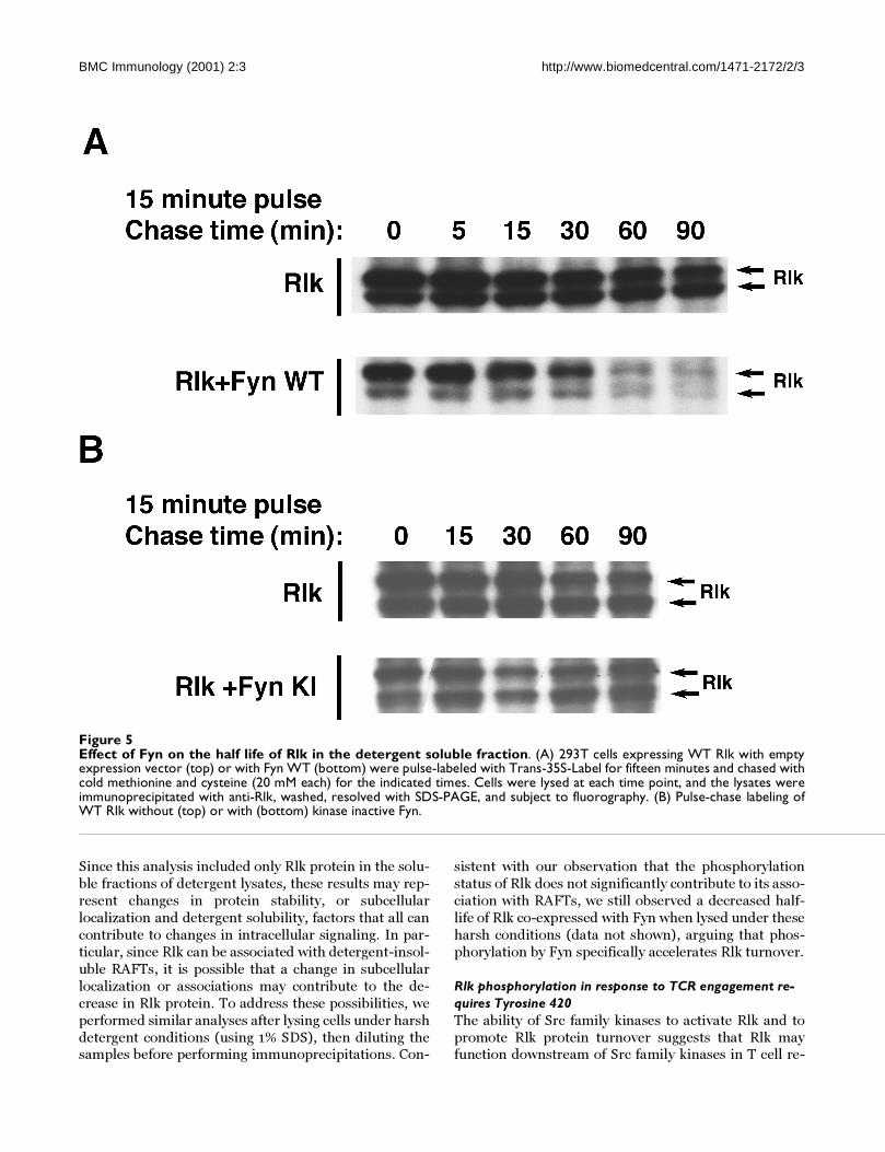
Fyn alters the half life of Rlk in the detergent soluble frac-tion of transfected cellsIn experiments with Rlk, we often observed a change in
the relative ratio of the two isoforms of Rlk when co-ex-
pressed with Fyn. Furthermore, activation of Jurkats ap-peared to be associated with a loss of Rlk from non-RAFT
fractions (see Fig. 4A). Since phosphorylation of Rlk did
not seem to affect association with RAFTs, we examined
whether Fyn altered other properties of the Rlk protein.
To start to address this issue, we measured the effect of
Fyn on the protein stability of Rlk. Rlk was expressed in
293T cells, either in the absence or the presence of Fyn;
the cells were labeled for 15 minutes with 35S methionine
and cysteine, then incubated for increasing times with
unlabelled methionine and cysteine. When expressed
alone, labeled Rlk appeared stable in the detergent-solu-
ble fraction for 90 minutes (Fig. 5A, top panel). When co-
expressed with Fyn, however, Rlk disappeared from the
soluble fraction at a significantly faster rate (Fig. 5A, low-
er panel, half-life between 30 and 60 minutes). The
change in half-life was particularly noticeable for the
larger form of Rlk. In the presence of kinase-inactive
Fyn, the half-life of Rlk was not altered, suggesting that
the phosphorylation of Rlk by Fyn is necessary for the de-
crease in half-life of Rlk (Fig. 5B). Similarly, co-transfec-
tion of wild type Fyn with RlkY420F did not alter the
half-life of RlkY420F in the soluble fraction (data not
shown), again arguing that phosphorylation of Rlk by
Fyn is required for this increased turnover in the soluble
fraction.
BMC Immunology (2001) 2:3 http://www.biomedcentral.com/1471-2172/2/3
Figure 4Rlk is associated with Lipid RAFTs: RAFT association does not require tyrosine phosphorylation, but does require theCysteine-string. (A) Jurkat-Tag cells were electroporated with Rlk-GFP and 24 hours later were either left unstimulated orstimulated with anti-CD3e. Cells were lysed in 1% Triton X-100 and lysates fractionated over a 40%-30%-5% sucrose step gra-dient for 16 hr at 200,000 g. Collected fractions were separated by SDS-PAGE and analyzed for presence of Fyn, Rlk and PLC-γ1. RAFTs are included in fraction 3 or fractions 3-4. (B) Jurkat-Tag cells were electroporated with either Rlk-GFPKI, Rlk-GFPY420F or Rlk-GFPCM and treated as in (A). Only fractions 3,4 and 10 are shown. For Rlk-GFPCM, a longer exposure isshown to demonstrate the residual RAFT association. (C) Jurkat-Tag cells were electroporated with Rlk-GFP, treated withWortmannin or carrier (DMSO) and fractionated as above.
BMC Immunology (2001) 2:3 http://www.biomedcentral.com/1471-2172/2/3
Since this analysis included only Rlk protein in the solu-
ble fractions of detergent lysates, these results may rep-
resent changes in protein stability, or subcellular
localization and detergent solubility, factors that all can
contribute to changes in intracellular signaling. In par-
ticular, since Rlk can be associated with detergent-insol-
uble RAFTs, it is possible that a change in subcellular
localization or associations may contribute to the de-
crease in Rlk protein. To address these possibilities, we
performed similar analyses after lysing cells under harsh
detergent conditions (using 1% SDS), then diluting thesamples before performing immunoprecipitations. Con-
sistent with our observation that the phosphorylation
status of Rlk does not significantly contribute to its asso-
ciation with RAFTs, we still observed a decreased half-
life of Rlk co-expressed with Fyn when lysed under these
harsh conditions (data not shown), arguing that phos-
phorylation by Fyn specifically accelerates Rlk turnover.
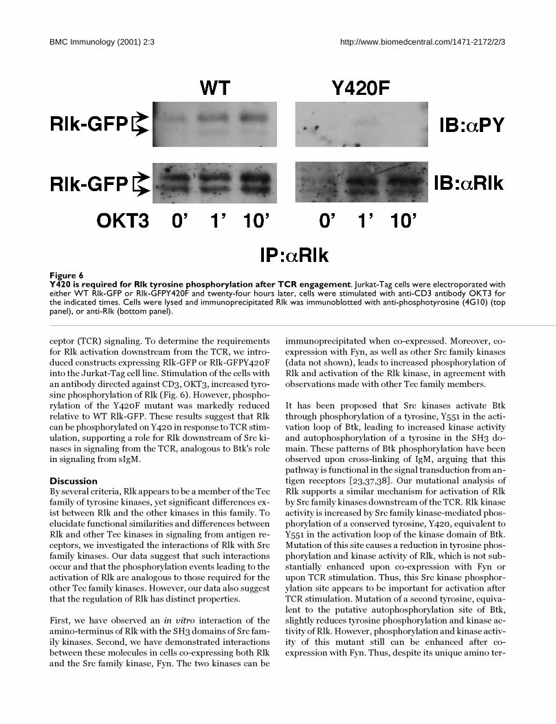
Rlk phosphorylation in response to TCR engagement re-quires Tyrosine 420The ability of Src family kinases to activate Rlk and to
promote Rlk protein turnover suggests that Rlk mayfunction downstream of Src family kinases in T cell re-
Figure 5Effect of Fyn on the half life of Rlk in the detergent soluble fraction. (A) 293T cells expressing WT Rlk with emptyexpression vector (top) or with Fyn WT (bottom) were pulse-labeled with Trans-35S-Label for fifteen minutes and chased withcold methionine and cysteine (20 mM each) for the indicated times. Cells were lysed at each time point, and the lysates wereimmunoprecipitated with anti-Rlk, washed, resolved with SDS-PAGE, and subject to fluorography. (B) Pulse-chase labeling ofWT Rlk without (top) or with (bottom) kinase inactive Fyn.
BMC Immunology (2001) 2:3 http://www.biomedcentral.com/1471-2172/2/3
ceptor (TCR) signaling. To determine the requirements
for Rlk activation downstream from the TCR, we intro-
duced constructs expressing Rlk-GFP or Rlk-GFPY420F
into the Jurkat-Tag cell line. Stimulation of the cells with
an antibody directed against CD3, OKT3, increased tyro-
sine phosphorylation of Rlk (Fig. 6). However, phospho-
rylation of the Y420F mutant was markedly reduced
relative to WT Rlk-GFP. These results suggest that Rlk
can be phosphorylated on Y420 in response to TCR stim-
ulation, supporting a role for Rlk downstream of Src ki-
nases in signaling from the TCR, analogous to Btk's rolein signaling from sIgM.
DiscussionBy several criteria, Rlk appears to be a member of the Tec
family of tyrosine kinases, yet significant differences ex-
ist between Rlk and the other kinases in this family. To
elucidate functional similarities and differences between
Rlk and other Tec kinases in signaling from antigen re-
ceptors, we investigated the interactions of Rlk with Src
family kinases. Our data suggest that such interactions
occur and that the phosphorylation events leading to the
activation of Rlk are analogous to those required for the
other Tec family kinases. However, our data also suggest
that the regulation of Rlk has distinct properties.
First, we have observed an in vitro interaction of the
amino-terminus of Rlk with the SH3 domains of Src fam-
ily kinases. Second, we have demonstrated interactions
between these molecules in cells co-expressing both Rlk
and the Src family kinase, Fyn. The two kinases can be
immunoprecipitated when co-expressed. Moreover, co-
expression with Fyn, as well as other Src family kinases
(data not shown), leads to increased phosphorylation of
Rlk and activation of the Rlk kinase, in agreement with
observations made with other Tec family members.
It has been proposed that Src kinases activate Btk
through phosphorylation of a tyrosine, Y551 in the acti-
vation loop of Btk, leading to increased kinase activity
and autophosphorylation of a tyrosine in the SH3 do-
main. These patterns of Btk phosphorylation have beenobserved upon cross-linking of IgM, arguing that this
pathway is functional in the signal transduction from an-
tigen receptors [23,37,38]. Our mutational analysis of
Rlk supports a similar mechanism for activation of Rlk
by Src family kinases downstream of the TCR. Rlk kinase
activity is increased by Src family kinase-mediated phos-
phorylation of a conserved tyrosine, Y420, equivalent to
Y551 in the activation loop of the kinase domain of Btk.
Mutation of this site causes a reduction in tyrosine phos-
phorylation and kinase activity of Rlk, which is not sub-
stantially enhanced upon co-expression with Fyn or
upon TCR stimulation. Thus, this Src kinase phosphor-
ylation site appears to be important for activation after
TCR stimulation. Mutation of a second tyrosine, equiva-
lent to the putative autophosphorylation site of Btk,
slightly reduces tyrosine phosphorylation and kinase ac-
tivity of Rlk. However, phosphorylation and kinase activ-
ity of this mutant still can be enhanced after co-
expression with Fyn. Thus, despite its unique amino ter-
Figure 6Y420 is required for Rlk tyrosine phosphorylation after TCR engagement. Jurkat-Tag cells were electroporated witheither WT Rlk-GFP or Rlk-GFPY420F and twenty-four hours later, cells were stimulated with anti-CD3 antibody OKT3 forthe indicated times. Cells were lysed and immunoprecipitated Rlk was immunoblotted with anti-phosphotyrosine (4G10) (toppanel), or anti-Rlk (bottom panel).

BMC Immunology (2001) 2:3 http://www.biomedcentral.com/1471-2172/2/3
minus, Rlk utilizes a transphosphorylation activation
mechanism similar to other Btk family members.
Although our original SH3 domain binding experimentssuggested a specific interaction between Tec kinases and
the Src kinases Fyn, Lyn and Hck, we have been able to
observe both activation by and co-precipitation with oth-
er Src family kinases including Lck and Src in 293T cells
(data not shown). Thus, in vivo, interactions with Rlk
may not be restricted to a particular subset of Src family
kinases. Indeed, we have found that mutation of the pro-
line-rich region did not abolish Rlk co-precipitation with
either Fyn or Lck (data not shown), suggesting that inter-
actions between these kinases may not occur solely
through an SH3 mediated interaction, at least in the
293T expression system. Nonetheless, whether there is a
specific connection between Rlk and the Src family ki-
nase Fyn in T cells remains an interesting question. Re-
cent data suggests that phosphorylation of the T cell
adaptor molecule SLP-76 occurs after stimulation of T
cells with altered peptide ligands in a Fyn-dependent
manner that is independent of the adaptor molecule LAT
[39,40]. Notably, Rlk has also been found to be capable
of phosphorylating SLP-76 [41]. It will be of interest to
determine whether Rlk participates in this novel activa-
tion pathway.
While Rlk is phosphorylated similarly to Btk and Itk,
there are marked differences between these two kinases.Perhaps the most obvious of these differences is the lack
of the pleckstrin homology domain in Rlk. Data suggest
that membrane association of Itk and Btk requires the
interaction of their PH domains with products of PI3K,
thereby facilitating phosphorylation by Src family kinas-
es [27,28]. In contrast, we have recently demonstrated
that PI3K inhibitors do not alter activation of Rlk by Fyn,
suggesting that the requirements for activation of Rlk by
Src family kinases are distinct from the other Tec kinas-
es. It is also of note that we can reproducibly co-immu-
noprecipitate Rlk and Fyn, perhaps due to their
association with RAFTs, whereas we have been unable to
co-precipitate Itk with Src family kinases when co-ex-
pressed in 293T cells (data not shown), again demon-
strating distinct properties of the Rlk protein compared
to certain other members of the Btk family. The combi-
nation of similar and distinct features is further support-
ed by the observation that TXK can rescue phospholipase
C-γ activation in BTK deficient DT40 cells, but fails to
rescue a defect in apoptosis [42].
Interestingly, we also find that the full-length form of Rlk
has a higher basal level of tyrosine phosphorylation and
kinase activity than Itk when expressed in heterologous
cells, consistent with potentially different localizationsand upstream interactions (unpublished observations).
The full-length isoform of Rlk also exhibits higher tyro-
sine phosphorylation and kinase activity than the short
isoform. This differential phosphorylation does not ap-
pear to result solely from a distinct phosphorylation siteon the full-length Rlk, since mutation of the only addi-
tional tyrosine found in the full-length Rlk (Y6) does not
greatly reduce the phosphorylation or activity of Rlk
when expressed either alone or in conjunction with Fyn.
Instead, the increased phosphorylation may result in
part from distinct properties of full-length Rlk. Indeed,
we report here that the palmitoylated cysteine-string is
responsible for the targeting of Rlk to RAFTs: detergent-
insoluble, glycolipid-enriched membrane microdomains
that contain many signaling molecules, including Src
family kinases. Our data may provide the first demon-
stration that a cysteine-string motif can lead to protein
association with RAFTs. Of note, Itk and Btk have been
found to be associated with lipid RAFTs upon activation
of antigen receptors [29,35]. However, the association of
Itk with RAFTs requires its PH domain and is dependent
on the products of PI3K. Indeed, in Jurkat cells, which
are deficient in the inositol phophatase PTEN, Itk is con-
stitutively associated with the membrane unless the cells
are treated with inhibitors of PI3K or transfected with
PTEN [36]. In contrast, we have found that Rlk associa-
tion with RAFTs, like its activation by Src family kinases,
is PI3K independent. Thus, although Rlk, like Itk and
Btk, is activated by phosphorylation by Src family kinas-
es and can be found in RAFTs, its requirements for thisactivation may be unique.
Co-expression of Rlk and Fyn also led to a rapid disap-
pearance of Rlk from the detergent soluble fraction as
measured by pulse-chase analysis (Fig. 5). This phenom-
enon could result from either a change in protein loca-
tion to a detergent-insoluble fraction or increased
turnover of Rlk. Activation of tyrosine kinases and other
signal transduction molecules is associated both with
changes in subcellular distribution and with changes in
protein stability. For example, crosslinking of the T-cell
receptor results in a transition of the receptor complex to
a detergent-insoluble fraction [43,44,45], as well as to
increased turnover of TCR components [46]. Nonethe-
less, the decreased half-life of Rlk in both detergent-sol-
uble and insoluble fractions argues that activation of Rlk
leads to increased degradation of the Rlk protein, similar
to that observed for other components of the TCR com-
plex, but not previously reported for Tec family kinases.
The dependence of this increased turnover on phospho-
rylation of Rlk suggests that a phosphotyrosine-depend-
ent association with another molecule may be required
for this process, reminiscent of the increased degrada-
tion of the PDGF receptor and Syk caused by association
with the ring-finger protein Cbl [47,48,49] or the Lck-de-pendent degradation of the TCR [50].
BMC Immunology (2001) 2:3 http://www.biomedcentral.com/1471-2172/2/3
Recent evidence from gene-targeted mice suggests that
Rlk may synergize with Itk in signaling from the T cell re-
ceptor to activate phospholipase C-γ [20]. The similarity
of the phosphorylation of Rlk by Src family kinases tothat which activates other Tec kinases suggests that Rlk
also functions downstream of Src kinases in TCR signal
transduction pathways. Rlk and Itk, may therefore func-
tion together in these TCR signaling events in a fashion
similar to Btk in BCR signaling. Recent data argue that
Itk interacts directly with Slp-76 and Grb2 to participate
in a LAT-nucleated T cell signaling complex involved in
PLC-γ activation [5,29]. It will be of interest to examine
the interactions of Rlk with other components of this
complex. Indeed, the potential in vitro binding of the
amino-terminus of Rlk to the SH3 domain of Grb2 hints
of similar protein interactions. Thus, although Rlk ap-
pears to be distinct among the Tec kinases in the lack of
a PH domain, the presence of a palmitoylated cysteine
string and the existence of a shorter isoform that can lo-
calize to the nucleus, Rlk may contribute to similar sign-
aling pathways. Indeed, our data suggest that the
cysteine string may serve as a surrogate to the PH do-
main for targeting Rlk to RAFTs. It is also notable that
recent data suggest that Btk can translocate to the nucle-
us upon B cell activation, similar to the nuclear translo-
cation we have reported for Rlk [19,51]. Nonetheless, the
PI3K independence of the activation of Rlk activation
and its association with RAFTs suggests that Rlk may
also have distinct contributions to these signaling path-ways.
Materials and MethodsCell Culture and Reagents293T and HeLa cells were maintained in Dulbecco's
modified Eagle's medium (DMEM) supplemented with
10% fetal calf serum, penicillin, streptomycin, and 10
mM glutamine. Jurkat cells expressing SV40 large T an-
tigen (Jurkat Tag) were a generous gift from Dr. Gerald
Crabtree (Stanford) and were maintained in RPMI 1640
supplemented with 10% fetal calf serum, penicillin,
streptomycin, glutamine, and 50 µM 2-mercaptoetha-
nol. Anti-murine Rlk was previously described. Anti-Fyn
(FYN3) was purchased from Santa Cruz Biotechnology
(Santa Cruz, CA); anti-phosphotyrosine (4G10) from
Upstate Biotechnologies (Lake Placid, New York).
TransfectionsConstructs were introduced into 293T cells by calcium
phosphate transfection in medium containing 25 µM
chloroquine (Sigma) [52]. 2-5 µg of each plasmid was
used for each transfection (2 × 10⁶ cells); the cells were
harvested 24 to 36 hours later. Transfections into HeLa
cells were performed identically, without the addition of
chloroquine. Jurkat Tag cell lines (107 cells) were trans-fected by electroporation using a Biorad electroporator
at 250 V and 960 µF or by square wave electroporation
on a BTX 830 using settings of 300 V and 10 ms. 24
hours later, cells were stimulated with OKT3, lysed and
Rlk analyzed as below.
Constructs and MutagenesisFull-length Rlk cDNA expression constructs pCIR,
pcDNA3Rlk and Rlk-GFP have been described previous-
ly [19]. Point mutations were introduced into Rlk-GFP by
a PCR based strategy [53] with oligonucleotides encod-
ing the required mutations (Mutated residues are in
bold): Y420F (GGAC GAT GAA TTC ATC AGT TCT TCT
G), Y91F (GTC AAG GCT CTT TTT GAC TTC CTG CC).
All mutations were confirmed by dideoxy sequencing.
pLNCX-FynT was a generous gift from Dr. Cliff Lowell
(USCF, San Francisco, CA). Glutathione S-Transferase
(GST) fusion constructs were previously described [19].
In vitro filter binding assaysGST-Rlk fusion proteins were grown and induced as pre-
viously described [19]. Bacteria were sonicated in phos-
phate-buffered saline (PBS), Ph7.4 with 0.1% Triton X-
100, insoluble material removed by centrifugation for 10
minutes at 14,000 rpm and soluble GST fusion proteins
purified with GST-Sepharose beads (Pharmacia). Ap-
proximately 2 µg of GST fusion proteins were separated
by SDS-PAGE, transferred to Nitrocellulose and probed
with biotinylated SH3-GST fusion proteins as previously
described [25].
ImmunofluorescenceHeLa cells were transfected with Rlk and Fyn expression
constructs, 20 hours later passed onto coverslips and 12-
24 hours later fixed with 1% paraformaldehyde for 10
minutes at room temperature. Cells were stained with
Fyn3 antiserum at 1: 500, washed with PBS and stained
with Texas Red conjugated anti-Rabbit Goat serum
(Cappel). Nuclei were counterstained with DAPI, cells
were visualized on a Zeiss Axioplan microscope with a
Charge Coupled Device (CCD) camera.
Western Blotting and Immunoprecipitation24 hours following transfection cells were washed once
with PBS and lysed in NP40 Lysis Buffer (0.5% Nonidet-
P 40, 50 mM HEPES pH 7.4, 5 mM EDTA, 50 mM NaCl,
10 mM NaPO4, 50 mM NaF, 1 mM sodium orthovanad-
ate, 1 mM AEBSF, 2 ug/ml aprotinin) for 10 minutes on
ice. Total protein extracts were directly mixed with 2X
SDS protein sample buffer (1X: 50 mM Tris pH 6.8, 2%
SDS, 10% glycerol, 0.1% Bromphenol Blue plus 5% B-
mercaptoethanol). Alternatively, lysates were clarified
by centrifugation and detergent-soluble protein was an-
alyzed. Equivalent amounts of protein were boiled in
SDS sample buffer and separated by 10% SDS-PAGE andtransferred onto nitrocellulose (Schleicher and Schuell).
BMC Immunology (2001) 2:3 http://www.biomedcentral.com/1471-2172/2/3
The membranes were blocked in TBST (10 mM Tris-HCl
pH 8.0, 150 mM NaCl, 0.1% Tween-20) with 5% nonfat
dry milk (w/v). The membranes were incubated with the
appropriate primary antibody at 1/2000 (anti-Rlk and4G10, anti-phosphotyrosine) or 1:1000 (anti-Fyn) dilu-
tion overnight at 4°C or 1-2 hours at room temperature.
Membranes were washed in TBST, incubated with the
appropriate secondary antibody at 1/10000 dilution
(Boehringer Mannheim) for 1 hr and protein was visual-
ized by enhanced chemiluminescence (Amersham).
For immunoprecipitation studies, equivalent amounts of
detergent soluble protein were incubated with affinity
purified anti-Rlk sera for 2-4 hours at 4°C. The complex-
es were recovered with Protein A-Sepharose (Sigma) and
washed two times with ice cold NP40 wash buffer (1%
NP40, PBS, 1 mM sodium orthovanadate) and analyzed
by Western blotting using either anti-Rlk serum or anti-
Fyn serum (1:1000).
In Vitro Kinase AssaysCell extracts were prepared in NP40 Lysis Buffer and im-
munoprecipitated as described above. Complexes were
recovered with Protein A Sepharose, washed twice with
ice cold NP40 wash buffer, washed twice with ice cold
Rlk kinase buffer (30 mM HEPES pH 7.4, 150 mM NaCl,
5 mM MgCl2, 5 mM MnCl2, 100 µM sodium orthovanad-
ate). The kinase reaction was carried out in kinase buffer
supplemented with 1 µg acid denatured enolase and 10µCi of [γ-32P] ATP (Redivue, Amersham) for 5 minutes at
room temperature. The reaction was quenched with SDS
sample buffer, boiled and separated by 10% SDS-PAGE.
Phosphorylated proteins were detected by autoradiogra-
phy or quantified on a PhosphoImager. The level of Rlk
protein was analyzed by immunoblotting.
Raft IsolationJurkat Tag cells were transfected by square wave electro-
poration using the parameters of 300 V and 10 ms. For
each condition 8 cuvettes containing 107 cells in 0.5 ml
were electroporated with 25 µg of the appropriate DNA.
Cells were grown over night in RPMI containing 20% fe-
tal calf serum. In some experiments, cells were pretreat-
ed with Wortmannin (100 nM) for 3 hrs prior to
fractionation. For TCR stimulation, cells for each condi-
tion were concentrated into 0.5 ml, warmed to 37°C for
10 minutes and then treated with 10 µg OKT3. Ice cold
PBS was added to the cells after 2 minutes OKT3 treat-
ment and the cells were quickly pelleted and lysed in 0.5
ml TENV (10 mM Tris 7.5, 150 mM NaCl, 5 mM EDTA, 1
mM Na3VO4) containing 1% Triton X-100. The cells were
homogenized with 12 strokes in 2 ml Dounce homogeniz-
ers after 30 minutes of incubation on ice. Homogenates
were adjusted to 40% sucrose by addition of TENV con-taining 80% sucrose. The lysate was transferred to a
TLS55 ultracentrifuge tube, over-layered sequentially
with 30% and 5% sucrose in TENV. Sucrose gradients
were centrifuged at 200,000 g for 16 hours and 200 µl
fractions were removed from the top of the gradient foranalysis by SDS-PAGE.
Metabolic Labeling and Pulse Chase5 × 106 293T cells were transfected with 10 µg of the in-
dicated Rlk construct and 10 µg of empty vector or the in-
dicated Fyn construct. Twenty four hours after
transfection, the cells were split equally among five to
seven plates and grown in complete DMEM for another
24 hours. The cells were placed in serum-free Met(-),
Cys(-) DMEM for 1 hour prior to pulse labeling. Cells
were pulse labeled with 0.5 mCi of Trans- [35S]-Label
(ICN) in 0.5 ml of Met(-)Cys(-) medium for 15 minutes at
37°C. After 15 minutes, the monolayers were chased with
complete DMEM supplemented with 20 mM unlabeled
methionine and cysteine (Sigma) at 37°C. At the indicat-
ed chase time, the cells were lysed with 1 ml cold NP40
Lysis Buffer and the lysates were clarified by centrifuga-
tion. Rlk was immunoprecipitated with affinity purified
anti-Rlk antibody for 3 hours at 4°C and immune com-
plexes were recovered with Protein A-Sepharose, washed
as described above, and boiled in SDS-sample buffer. La-
beled proteins were separated by 10% PAGE, and ana-
lyzed by fluorography.
AbbreviationsPH, pleckstrin homology; PI3K, phosphoinositide 3-Ki-
nase; SH2, Src homology-2, SH3, Src homology-3; XLA,
X-linked agammaglobulinemia; xid; x-linked immuno-
deficiency; TCR, T cell receptor; sIgM, surface immu-
noglobulin M; (GAP), GTPase-Activating Protein; GST,
Glutathione S-Transferase; SDS, Sodium Dodecyl Sul-
fate; PBS, phosphate buffered saline; NP40, Nonidet P-
40; PAGE, polyacrylamide gel electrophoresis.
AcknowledgementsMario Chamorro, Michael J Czar and Jayanta Debnath are equal contribu-tors to this article.
P.L.S. was a Special Fellow of the Leukemia Society of America and is pres-ently a Searle Scholar. M.C. is presently in the Program in Cell Biology, Cor-nell Weill-Sloan Kettering Institute Graduate Program. The authors would like to thank D. W. McVicar, R. Wange and members of the Schwartzberg, Varmus and Lenardo laboratories for critical feedback and L. Matis for the original Rlk cDNA clone.
References1. Wange RL, Samelson LE: Complex complexes: signaling at the
TCR. Immunity 1996, 5:197-2052. Weiss A, Littman DR: Signal transduction by lymphocyte anti-
gen receptors. Cell 1994, 76:263-2743. Rawlings DJ, Witte ON: The Btk subfamily of cytoplasmic tyro-
sine kinases: structure, regulation and function. Semin Immunol1995, 7:237-246
4. Satterthwaite AB, Li Z, Witte ON: Btk function in B cell develop-ment and response. Semin Immunol 1998, 10:309-316
5. Schaeffer EM, Schwartzberg PL: Tec family kinases in lymphocytesignaling and function. Curr Opin Immunol 2000, 12:282-288
BMC Immunology (2001) 2:3 http://www.biomedcentral.com/1471-2172/2/3
6. Mano H, Ishikawa F, Nishida J, Hirai H, Takaku F: A novel protein-tyrosine kinase, tec, is preferentially expressed in liver. Onco-gene 1990, 5:1781-1786
7. Desiderio S, Siliciano JD: The Itk/Btk/Tec family of protein-tyro-sine kinases. Chem Immunol 1994, 59:191-210
8. Rawlings DJ, Saffran DC, Tsukada S, Largaespada DA, Grimaldi JC,Cohen L, Mohr RN, Bazan JF, Howard M, Copeland NG, et al: Muta-tion of unique region of Bruton's tyrosine kinase in immuno-deficient XID mice. Science 1993, 261:358-361
9. Thomas JD, Sideras P, Smith CI, Vorechovsky I, Chapman V, Paul WE:Colocalization of X-linked agammaglobulinemia and X-linked immunodeficiency genes. Science 1993, 261:355-358
10. Tsukada S, Saffran DC, Rawlings DJ, Parolini O, Allen RC, Klisak I,Sparkes RS, Kubagawa H, Mohandas T, Quan S, et al: Deficient ex-pression of a B cell cytoplasmic tyrosine kinase in human X-linked agammaglobulinemia. Cell 1993, 72:279-290
11. Vetrie D, Vorechovsky I, Sideras P, Holland J, Davies A, Flinter F,Hammarstrom L, Kinnon C, Levinsky R, Bobrow M, et al: The geneinvolved in X-linked agammaglobulinaemia is a member ofthe src family of protein-tyrosine kinases. Nature 1993,361:226-233
12. Kawakami Y, Yao L, Miura T, Tsukada S, Witte ON, Kawakami T: Ty-rosine phosphorylation and activation of Bruton tyrosine ki-nase upon Fc epsilon RI cross-linking. Mol Cell Biol 1994,14:5108-5113
13. Matsuda T, Takahashi-Tezuka M, Fukada T, Okuyama Y, Fujitani Y,Tsukada S, Mano H, Hirai H, Witte ON, Hirano T: Association andactivation of Btk and Tec tyrosine kinases by gp130, a signaltransducer of the interleukin-6 family of cytokines. Blood 1995,85:627-633
14. Saouaf SJ, Mahajan S, Rowley RB, Kut SA, Fargnoli J, Burkhardt AL,Tsukada S, Witte ON, Bolen JB: Temporal differences in the ac-tivation of three classes of non-transmembrane protein tyro-sine kinases following B-cell antigen receptor surfaceengagement. Proc Natl Acad Sci U S A 1994, 91:9524-9528
15. Sato S, Katagiri T, Takaki S, Kikuchi Y, Hitoshi Y, Yonehara S, TsukadaS, Kitamura D, Watanabe T, Witte O, et al: IL-5 receptor-mediat-ed tyrosine phosphorylation of SH2/SH3-containing proteinsand activation of Bruton's tyrosine and Janus 2 kinases. J ExpMed 1994, 180:2101-2111
16. Heyeck SD, Wilcox HM, Bunnell SC, Berg LJ: Lck phosphorylatesthe activation loop tyrosine of the Itk kinase domain and ac-tivates Itk kinase activity. J Biol Chem 1997, 272:25401-25408
17. Gibson S, August A, Branch D, Dupont B, Mills GM: Functional LCKIs required for optimal CD28-mediated activation of theTEC family tyrosine kinase EMT/ITK. J Biol Chem 1996,271:7079-7083
18. August A, Gibson S, Kawakami Y, Kawakami T, Mills GB, Dupont B:CD28 is associated with and induces the immediate tyrosinephosphorylation and activation of the Tec family kinase ITK/EMT in the human Jurkat leukemic T-cell line. Proc Natl AcadSci U S A 1994, 91:9347-9351
19. Debnath J, Chamorro M, Czar MJ, Schaeffer EM, Leonardo MJ, Var-mus HE, Schwartzberg PL: Rlk/Txk encodes two forms of a novelcysteine-string tyrosine kinae activated by Src family kinas-es. Mol Cell Biol 1999, 19:1498-1507
20. Schaeffer EM, Debnath J, Yap G, McVicar D, Liao XC, Littman DR,Sher A, Varmus HE, Lenardo MJ, Schwartzberg PL: Requirementfor Tec kinases Rlk and Itk in T cell receptor signaling andimmunity. Science 1999, 284:638-641
21. Mahajan S, Fargnoli J, Burkhardt AL, Kut SA, Saouaf SJ, Bolen JB: Srcfamily protein tyrosine kinases induce autoactivation of Bru-ton's tyrosine kinase. Mol Cell Biol 1995, 15:5304-5311
22. Mano H, Yamashita Y, Miyazato A, Miura Y, Ozawa K: Tec protein-tyrosine kinase is an effector molecule of Lyn protein-tyro-sine kinase. Faseb J 1996, 10:637-642
23. Rawlings DJ, Scharenberg AM, Park H, Wahl MI, Lin S, Kato RM,Fluckiger AC, Witte ON, Kinet JP: Activation of BTK by a phos-phorylation mechanism initiated by SRC family kinases. Sci-ence 1996, 271:822-825
24. Park H, Wahl MI, Afar DE, Turck CW, Rawlings DJ, Tam C, Scharen-berg AM, Kinet JP, Witte ON: Regulation of Btk function by amajor autophosphorylation site within the SH3 domain. Im-munity 1996, 4:515-525
25. Cheng G, Ye ZS, Baltimore D: Binding of Bruton's tyrosine ki-nase to Fyn, Lyn, or Hck through a Src homology 3 domain-mediated interaction. Proc Natl Acad Sci U S A 1994, 91:8152-8155
26. Yang W, Malek SN, Desiderio S: An SH3-binding site conservedin Bruton's tyrosine kinase and related tyrosine kinases me-diates specific protein interactions in vitro and in vivo. J BiolChem 1995, 270:20832-20840
27. August A, Sadra A, Dupont B, Hanafusa H: Src-induced activationof inducible T cell kinase (ITK) requires phosphatidylinositol3-kinase activity and the Pleckstrin homology domain of in-ducible T cell kinase. Proc Natl Acad Sci U S A 1997, 94:11227-11232
28. Li Z, Wahl M, Eguino A, Stephens L, Hawkins P, Witte O: Phospho-tidylinositol 3-kinase-γ activates Bruton's tyrosine kinase inconcert with Src family kinases. Proc. Natl. Acad. Sci., USA 1997,94:13820-13825
29. Bunnell S, Diehn M, Yaffe M, Findell P, Cantley L, Berg L: Biochemi-cal Interactions Integrating Itk with the T cell Receptor-ini-tiated Signaling Cascade. J. Biol. Chem. 2000, 275:2219-2230
30. Haire RN, Ohta Y, Lewis JE, Fu SM, Kroisel P, Litman GW: TXK, anovel human tyrosine kinase expressed in T cells shares se-quence identity with Tec family kinases and maps to 4p12.Hum Mol Genet 1994, 3:897-901
31. Hu Q, Davidson D, Schwartzberg PL, Macchiarini F, Lenardo MJ, Blue-stone JA, Matis LA: Identification of Rlk, a novel protein tyro-sine kinase with predominant expression in the T celllineage. J Biol Chem 1995, 270:1928-1934
32. Sommers CL, Huang K, Shores EW, Grinberg A, Charlick DA, KozakCA, Love PE: Murine txk: a protein tyrosine kinase gene regu-lated by T cell activation. Oncogene 1995, 11:245-251
33. Alexandropoulos K, Cheng G, Baltimore D: Proline-rich sequenc-es that bind to Src homology 3 domains with individual spe-cificities. Proc Natl Acad Sci U S A 1995, 92:3110-3114
34. Hanks SK, Quinn AM, Hunter T: The protein kinase family: con-served features and deduced phylogeny of the catalytic do-mains. Science 1988, 241:42-52
35. Guo B, Kato RM, Garcia-Lloret M, Wahl MI, Rawlings DJ: Engage-ment of the human pre-B cell receptor generates a lipid raft-dependent calcium signaling complex. Immunity 2000, 13:243-253
36. Shan X, Czar MJ, Bunnell SC, Liu P, Liu Y, Schwartzberg PL, WangeRL: Deficiency of PTEN in Jurkat T cells causes constitutivelocalization of Itk to the plasma membrane and hyperre-sponsiveness to CD3 stimulation. Mol Cell Biol 2000, 20:6945-6957
37. Wahl MI, Fluckiger AC, Kato RM, Park H, Witte ON, Rawlings DJ:Phosphorylation of two regulatory tyrosine residues in theactivation of Bruton's tyrosine kinase via alternative recep-tors. Proc Natl Acad Sci U S A 1997, 94:11526-11533
38. Nisitani S, Kato RM, Rawlings DJ, Witte ON, Wahl MI: In situ detec-tion of activated Bruton's tyrosine kinase in the Ig signalingcomplex by phosphopeptide-specific monoclonal antibodies.Proc Natl Acad Sci U S A 1999, 96:2221-2226
39. Denny MF, Patai B, Straus DB: Differential T-cell antigen recep-tor signaling mediated by the Src family kinases Lck and Fyn.Mol Cell Biol 2000, 20:1426-1435
40. Huang J, Tilly D, Altman A, Sugie K, Grey HM: Inaugural article: T-cell receptor antagonists induce vav phosphorylation by se-lective activation of fyn kinase [In Process Citation]. Proc NatlAcad Sci U S A 2000, 97:10923-10929
41. Schneider H, Guerette B, Guntermann C, Rudd CE: Resting lym-phocyte kinase (Rlk/Txk) targets lymphoid adaptor SLP-76in the cooperative activation of interleukin-2 transcription inT-cells. J Biol Chem 2000, 275:3835-3840
42. Tomlinson MG, Kurosaki T, Berson AE, Fujii GH, Johnston JA, BolenJB: Reconstitution of Btk signaling by the atypical tec familytyrosine kinases Bmx and Txk. J Biol Chem 1999, 274:13577-13585
43. Rozdzial M, Malissen B, Finkel T: Tyrosine-phosphorylated T cellreceptor zeta chain associates with the actin cytoskeletonupon activation of mature T lymphocytes. Immunity 1995,3:623-633
44. Montixi C, Langlet C, Bernard AM, Thimonier J, Dubois C, WurbelMA, Chauvin JP, Pierres M, He HT: Engagement of T cell recep-tor triggers its recruitment to low-density detergent-insolu-ble membrane domains. Embo J 1998, 17:5334-5348
BMC Immunology (2001) 2:3 http://www.biomedcentral.com/1471-2172/2/3
45. Xavier R, Brennan T, Li Q, McCormack C, Seed B: Membrane com-partmentation is required for efficient T cell activation. Im-munity 1998, 8:723-732
46. Valitutti S, Muller S, Salio M, Lanzavecchia A: Degradation of T cellreceptor (TCR)-CD3-zeta complexes after antigenic stimu-lation [see comments]. J Exp Med 1997, 185:1859-1864
47. Ma YC, Huang XY: Identification of the binding site for Gqalphaon its effector Bruton's tyrosine kinase. Proc Natl Acad Sci U S A1998, 95:12197-12201
48. Miyake S, Lupher ML Jr, Druker B, Band H: The tyrosine kinaseregulator Cbl enhances the ubiquitination and degradationof the platelet-derived growth factor receptor alpha. Proc NatlAcad Sci U S A 1998, 95:7927-7932
49. Miyake S, Mullane-Robinson KP, Lill NL, Douillard P, Band H: Cbl-mediated negative regulation of platelet-derived growth fac-tor receptor-dependent cell proliferation. A critical role forCbl tyrosine kinase-binding domain. J Biol Chem 1999,274:16619-16628
50. D'Oro U, Vacchio MS, Weissman AM, Ashwell JD: Activation ofthe Lck tyrosine kinase targets cell surface T cell antigen re-ceptors for lysosomal degradation. Immunity 1997, 7:619-628
51. Webb CF, Yamashita Y, Ayers N, Evetts S, Paulin Y, Conley ME, SmithEA: The transcription factor bright associates with Bruton'styrosine kinase, the defective protein in immunodeficiencydisease. J Immunol 2000, 165:6956-6965
52. Pear WS, Nolan GP, Scott ML, Baltimore D: Production of high-titer helper-free retroviruses by transient transfection. ProcNatl Acad Sci U S A 1993, 90:8392-8396
53. Horton RM: In vitro recombination and mutagenesis of DNA.SOEing together tailor-made genes. Methods Mol Biol 1997,67:141-149
Publish with BioMedcentral and every scientist can read your work free of charge
"BioMedcentral will be the most significant development for disseminating the results of biomedical research in our lifetime."
Paul Nurse, Director-General, Imperial Cancer Research Fund
Publish with BMc and your research papers will be:
available free of charge to the entire biomedical community
peer reviewed and published immediately upon acceptance
cited in PubMed and archived on PubMed Central
yours - you keep the copyright
[email protected] your manuscript here:http://www.biomedcentral.com/manuscript/
BioMedcentral.comBioMedcentral.com