Embed Size (px)
Citation preview
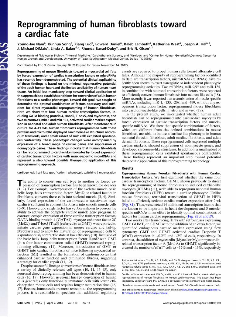
Reprogramming of human fibroblasts towarda cardiac fateYoung-Jae Nama, Kunhua Songa, Xiang Luob, Edward Daniela, Kaleb Lambetha, Katherine Westa, Joseph A. Hilla,b,J. Michael DiMaioc, Linda A. Bakerd,e, Rhonda Bassel-Dubya, and Eric N. Olsona,1
Departments of aMolecular Biology, bInternal Medicine, cCardiothoracic Surgery, and dUrology, and eCenter for Human Genetics/McDermott Center forHuman Growth and Development, University of Texas Southwestern Medical Center, Dallas, TX 75390
Contributed by Eric N. Olson, January 30, 2013 (sent for review November 14, 2012)
Reprogramming of mouse fibroblasts toward a myocardial cell fateby forced expression of cardiac transcription factors or microRNAshas recently been demonstrated. The potential clinical applicabilityof these findings is based on the minimal regenerative potentialof the adult human heart and the limited availability of human hearttissue. An initial but mandatory step toward clinical application ofthis approach is to establish conditions for conversionof adult humanfibroblasts to a cardiac phenotype. Toward this goal, we sought todetermine the optimal combination of factors necessary and suffi-cient for direct myocardial reprogramming of human fibroblasts.Here we show that four human cardiac transcription factors, in-cluding GATA binding protein 4, Hand2, T-box5, and myocardin, andtwomicroRNAs,miR-1 andmiR-133, activated cardiacmarker expres-sion in neonatal and adult human fibroblasts. After maintenance inculture for 4–11 wk, human fibroblasts reprogrammed with theseproteins and microRNAs displayed sarcomere-like structures and cal-cium transients, and a small subset of such cells exhibited spontane-ous contractility. These phenotypic changes were accompanied byexpression of a broad range of cardiac genes and suppression ofnonmyocyte genes. These findings indicate that human fibroblastscan be reprogrammed to cardiac-like myocytes by forced expressionof cardiac transcription factors with muscle-specific microRNAs andrepresent a step toward possible therapeutic application of thisreprogramming approach.
cardiogenesis | cell fate specification | phenotypic switching | regeneration
The ability to convert one cell type to another by forced ex-pression of transcription factors has been known for decades
(1, 2). For example, overexpression of the skeletal muscle basichelix–loop–helix transcription factor, MyoD or related factors infibroblasts, converts these cells into skeletal muscle (3, 4). Simi-larly, forced expression of the cardiovascular coactivator myo-cardin is sufficient to convert fibroblasts into smooth muscle cells(5–9). However, no single factor has yet been shown to possess theability to activate the complete cardiac muscle gene program. Incontrast, ectopic expression of three cardiac transcription factors,GATA binding protein 4 (GATA4), myocyte enhancer factor 2C(Mef2c), andT-box5 (Tbx5) (referred to asGMT), was reported toinitiate cardiac gene expression in mouse cardiac and tail-tipfibroblasts and to allow for maturation of reprogrammed cells toa spontaneously contractile state at low efficiency (10). Inclusion ofthe basic helix–loop–helix transcription factor Hand2 with GMT(in a four-factor combination called GHMT) increased reprog-ramming efficiency (11). Moreover, introduction of GMT orGHMT into cardiac fibroblasts of mice following myocardial in-farction (MI) resulted in the formation of cardiomyocytes thatenhanced cardiac function and diminished fibrosis, suggestinga strategy for cardiac repair (11, 12).Despite successful lineage conversions of mouse fibroblasts into
a variety of clinically relevant cell types (10, 11, 13–15), onlyneuronal direct reprogramming has been demonstrated in humancells (16, 17). However, neuronal lineage conversion of humancells generates only functionally immature cells with lower effi-ciency than mouse cells and requires longer maturation time (16,17). Because human cells aremore resistant to the reprogrammingprocess, it is reasonable to speculate that additional regulatory
events are required to propel human cells toward alternative cellfates. Although the majority of reprogramming factors identifiedto date are transcription factors, microRNAs (miRNAs) have re-cently been shown to exert synergistic or independent phenotypicreprogramming activities. Two miRNAs, miR-9/9* and miR-124,in combination with neuronal transcription factors, were reportedto efficiently convert human fibroblasts into neuron-like cells (18).More recently, it was reported that a combination of muscle-specificmiRNAs, including miR-1, -133, -208, and -499, without any ex-ogenous transcription factor, reprogrammed mouse fibroblastsinto cardiomyocyte-like cells in vitro and in vivo (19).In the present study, we investigated whether human adult
fibroblasts can be reprogrammed into cardiac-like myocytes byforced expression of cardiac transcription factors and muscle-specific miRNAs. We show that specific combinations of factors,which are different from the defined combinations in mousefibroblasts, are able to induce a cardiac-like phenotype in humanneonatal foreskin fibroblasts, adult cardiac fibroblasts, and adultdermal fibroblasts. These reprogrammed cells expressed multiplecardiac markers, showed suppression of nonmyocyte genes, anddeveloped sarcomere-like structures. In addition, a small subset ofthese reprogrammed cells exhibited spontaneous contractility.These findings represent an important step toward potentialtherapeutic application of this reprogramming technology.
ResultsReprogramming Human Foreskin Fibroblasts with Human CardiacTranscription Factors. We first examined whether the same fourcardiac transcription factors, GHMT, shown previously to directthe reprogramming of mouse fibroblasts to induced cardiac-likemyocytes (iCLMs) (11), were able to reprogram neonatal humanforeskin fibroblasts (HFFs) toward a cardiac phenotype. Unlikemouse fibroblasts, retroviral transduction of GHMT in HFFsfailed to efficiently activate cardiac marker expression after 2 wk(Fig. S1). Thus, we selected 14 additional transcription factors thatare known to be important in heart development and 3 muscle-specific miRNAs in an effort to identify optimal combinations offactors for human cardiac reprogramming (Fig. S2 A and B).Two weeks after transducing HFFs with retroviruses expressing
GMT, GHMT, or GHMT with the addition of an extra factor, wequantified endogenous cardiac marker expression using flowcytometry. GMT and GHMT activated cardiac Troponin T(cTnT) expression in ∼0.2% and ∼2% of cells, respectively. Incontrast, the addition of myocardin (Myocd or My) or myocardin-related transcription factor-A (Mrtf-A) to GHMT, significantly in-creased the number of cTnT+ cells to∼17% and∼13%, respectively
Author contributions: Y.-J.N., K.S., R.B.-D., and E.N.O. designed research; Y.-J.N., K.S., X.L.,E.D., K.L., and K.W. performed research; Y.-J.N., K.S., J.M.D., and L.A.B. contributed newreagents/analytic tools; Y.-J.N., K.S., X.L., J.A.H., R.B.-D., and E.N.O. analyzed data; andY.-J.N., K.S., R.B.-D., and E.N.O. wrote the paper.
Conflict of interest statement: E.N.O., Y.-J.N., and K.S. have all filed a patent relating toreprogramming of human fibroblasts to human cardiomyocytes. This patent has beenlicensed by LoneStar Heart, Inc. E.N.O. is a cofounder of this company and holds equity.1To whom correspondence should be addressed. E-mail: [email protected].
This article contains supporting information online at www.pnas.org/lookup/suppl/doi:10.1073/pnas.1301019110/-/DCSupplemental.
5588–5593 | PNAS | April 2, 2013 | vol. 110 | no. 14 www.pnas.org/cgi/doi/10.1073/pnas.1301019110
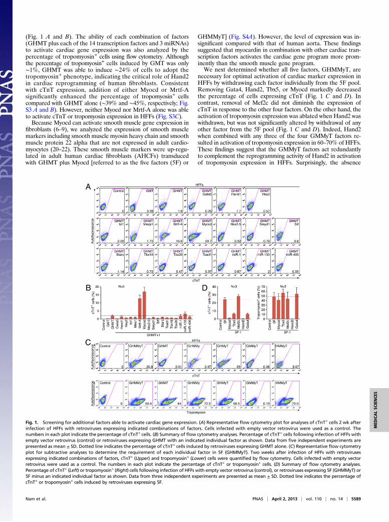
(Fig. 1 A and B). The ability of each combination of factors(GHMT plus each of the 14 transcription factors and 3 miRNAs)to activate cardiac gene expression was also analyzed by thepercentage of tropomyosin+ cells using flow cytometry. Althoughthe percentage of tropomyosin+ cells induced by GMT was only∼1%, GHMT was able to induce ∼24% of cells to adopt thetropomyosin+ phenotype, indicating the critical role of Hand2in cardiac reprogramming of human fibroblasts. Consistentwith cTnT expression, addition of either Myocd or Mrtf-Asignificantly enhanced the percentage of tropomyosin+ cellscompared with GHMT alone (∼39% and ∼45%, respectively; Fig.S3 A and B). However, neither Myocd nor Mrtf-A alone was ableto activate cTnT or tropomyosin expression in HFFs (Fig. S3C).Because Myocd can activate smooth muscle gene expression in
fibroblasts (6–9), we analyzed the expression of smooth musclemarkers including smooth muscle myosin heavy chain and smoothmuscle protein 22 alpha that are not expressed in adult cardio-myocytes (20–22). These smooth muscle markers were up-regu-lated in adult human cardiac fibroblasts (AHCFs) transducedwith GHMT plus Myocd [referred to as the five factors (5F) or
GHMMyT] (Fig. S4A). However, the level of expression was in-significant compared with that of human aorta. These findingssuggested that myocardin in combination with other cardiac tran-scription factors activates the cardiac gene program more prom-inently than the smooth muscle gene program.We next determined whether all five factors, GHMMyT, are
necessary for optimal activation of cardiac marker expression inHFFs by withdrawing each factor individually from the 5F pool.Removing Gata4, Hand2, Tbx5, or Myocd markedly decreasedthe percentage of cells expressing cTnT (Fig. 1 C and D). Incontrast, removal of Mef2c did not diminish the expression ofcTnT in response to the other four factors. On the other hand, theactivation of tropomyosin expression was ablated when Hand2 waswithdrawn, but was not significantly altered by withdrawal of anyother factor from the 5F pool (Fig. 1 C and D). Indeed, Hand2when combined with any three of the four GMMyT factors re-sulted in activation of tropomyosin expression in 60–70% of HFFs.These findings suggest that the GMMyT factors act redundantlyto complement the reprogramming activity of Hand2 in activationof tropomyosin expression in HFFs. Surprisingly, the absence
Fig. 1. Screening for additional factors able to activate cardiac gene expression. (A) Representative flow cytometry plot for analyses of cTnT+ cells 2 wk afterinfection of HFFs with retroviruses expressing indicated combinations of factors. Cells infected with empty vector retrovirus were used as a control. Thenumbers in each plot indicate the percentage of cTnT+ cells. (B) Summary of flow cytometry analyses. Percentage of cTnT+ cells following infection of HFFs withempty vector retrovirus (control) or retroviruses expressing GHMT with an indicated individual factor as shown. Data from five independent experiments arepresented as mean ± SD. Dotted line indicates the percentage of cTnT+ cells induced by retroviruses expressing GHMT alone. (C) Representative flow cytometryplot for subtractive analyses to determine the requirement of each individual factor in 5F (GHMMyT). Two weeks after infection of HFFs with retrovirusesexpressing indicated combinations of factors, cTnT+ (Upper) and tropomyosin+ (Lower) cells were quantified by flow cytometry. Cells infected with empty vectorretrovirus were used as a control. The numbers in each plot indicate the percentage of cTnT+ or tropomyosin+ cells. (D) Summary of flow cytometry analyses.Percentage of cTnT+ (Left) or tropomyosin+ (Right) cells following infection of HFFs with empty vector retrovirus (control), or retroviruses expressing 5F (GHMMyT) or5F minus an indicated individual factor as shown. Data from three independent experiments are presented as mean ± SD. Dotted line indicates the percentage ofcTnT+ or tropomyosin+ cells induced by retroviruses expressing 5F.
Nam et al. PNAS | April 2, 2013 | vol. 110 | no. 14 | 5589
MED
ICALSC
IENCE
S
of Hand2 almost eliminated both cTnT and tropomyosin expres-sion in HFFs, demonstrating the irreplaceable activity of thisfactor for activation of cardiac contractile gene programs in hu-man fibroblasts, unlike its synergistic role in mouse myocardialreprogramming (11).Because withdrawing any of the five factors (GHMMyT) failed
to further increase cardiac marker expression, we searched for anadditional sixth factor that could cooperatively enhance cardiac
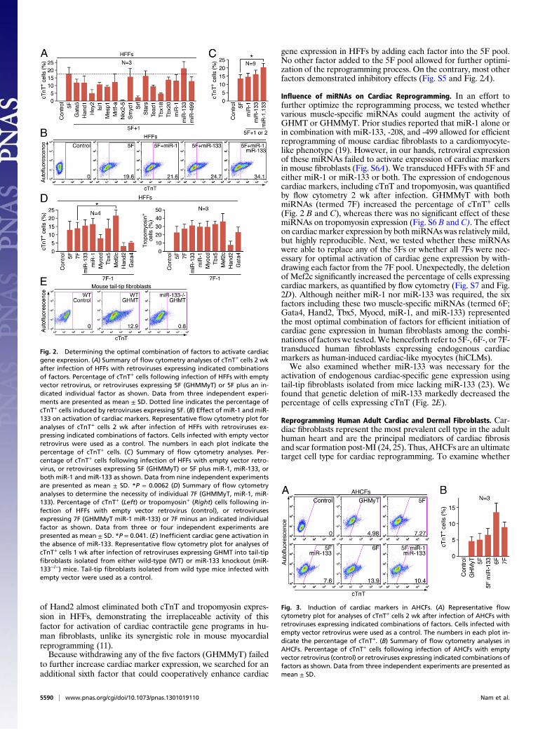
gene expression in HFFs by adding each factor into the 5F pool.No other factor added to the 5F pool allowed for further optimi-zation of the reprogramming process. On the contrary, most otherfactors demonstrated inhibitory effects (Fig. S5 and Fig. 2A).
Influence of miRNAs on Cardiac Reprogramming. In an effort tofurther optimize the reprogramming process, we tested whethervarious muscle-specific miRNAs could augment the activity ofGHMT or GHMMyT. Prior studies reported that miR-1 alone orin combination with miR-133, -208, and -499 allowed for efficientreprogramming of mouse cardiac fibroblasts to a cardiomyocyte-like phenotype (19). However, in our hands, retroviral expressionof these miRNAs failed to activate expression of cardiac markersin mouse fibroblasts (Fig. S6A). We transduced HFFs with 5F andeither miR-1 or miR-133 or both. The expression of endogenouscardiac markers, including cTnT and tropomyosin, was quantifiedby flow cytometry 2 wk after infection. GHMMyT with bothmiRNAs (termed 7F) increased the percentage of cTnT+ cells(Fig. 2 B and C), whereas there was no significant effect of thesemiRNAs on tropomyosin expression (Fig. S6 B and C). The effecton cardiacmarker expression by both miRNAs was relatively mild,but highly reproducible. Next, we tested whether these miRNAswere able to replace any of the 5Fs or whether all 7Fs were nec-essary for optimal activation of cardiac gene expression by with-drawing each factor from the 7F pool. Unexpectedly, the deletionof Mef2c significantly increased the percentage of cells expressingcardiac markers, as quantified by flow cytometry (Fig. S7 and Fig.2D). Although neither miR-1 nor miR-133 was required, the sixfactors including these two muscle-specific miRNAs (termed 6F;Gata4, Hand2, Tbx5, Myocd, miR-1, and miR-133) representedthe most optimal combination of factors for efficient initiation ofcardiac gene expression in human fibroblasts among the combi-nations of factors we tested.We henceforth refer to 5F-, 6F-, or 7F-transduced human fibroblasts expressing endogenous cardiacmarkers as human-induced cardiac-like myocytes (hiCLMs).We also examined whether miR-133 was necessary for the
activation of endogenous cardiac-specific gene expression usingtail-tip fibroblasts isolated from mice lacking miR-133 (23). Wefound that genetic deletion of miR-133 markedly decreased thepercentage of cells expressing cTnT (Fig. 2E).
Reprogramming Human Adult Cardiac and Dermal Fibroblasts. Car-diac fibroblasts represent the most prevalent cell type in the adulthuman heart and are the principal mediators of cardiac fibrosisand scar formation post-MI (24, 25). Thus, AHCFs are an ultimatetarget cell type for cardiac reprogramming. To examine whether
Fig. 2. Determining the optimal combination of factors to activate cardiacgene expression. (A) Summary of flow cytometry analyses of cTnT+ cells 2 wkafter infection of HFFs with retroviruses expressing indicated combinationsof factors. Percentage of cTnT+ cells following infection of HFFs with emptyvector retrovirus, or retroviruses expressing 5F (GHMMyT) or 5F plus an in-dicated individual factor as shown. Data from three independent experi-ments are presented as mean ± SD. Dotted line indicates the percentage ofcTnT+ cells induced by retroviruses expressing 5F. (B) Effect of miR-1 and miR-133 on activation of cardiac markers. Representative flow cytometry plot foranalyses of cTnT+ cells 2 wk after infection of HFFs with retroviruses ex-pressing indicated combinations of factors. Cells infected with empty vectorretrovirus were used as a control. The numbers in each plot indicate thepercentage of cTnT+ cells. (C) Summary of flow cytometry analyses. Per-centage of cTnT+ cells following infection of HFFs with empty vector retro-virus, or retroviruses expressing 5F (GHMMyT) or 5F plus miR-1, miR-133, orboth miR-1 and miR-133 as shown. Data from nine independent experimentsare presented as mean ± SD. *P = 0.0062 (D) Summary of flow cytometryanalyses to determine the necessity of individual 7F (GHMMyT, miR-1, miR-133). Percentage of cTnT+ (Left) or tropomyosin+ (Right) cells following in-fection of HFFs with empty vector retrovirus (control), or retrovirusesexpressing 7F (GHMMyT miR-1 miR-133) or 7F minus an indicated individualfactor as shown. Data from three or four independent experiments arepresented as mean ± SD. *P = 0.041. (E) Inefficient cardiac gene activation inthe absence of miR-133. Representative flow cytometry plot for analyses ofcTnT+ cells 1 wk after infection of retroviruses expressing GHMT into tail-tipfibroblasts isolated from either wild-type (WT) or miR-133 knockout (miR-133−/−) mice. Tail-tip fibroblasts isolated from wild type mice infected withempty vector were used as a control.
Fig. 3. Induction of cardiac markers in AHCFs. (A) Representative flowcytometry plot for analyses of cTnT+ cells 2 wk after infection of AHCFs withretroviruses expressing indicated combinations of factors. Cells infected withempty vector retrovirus were used as a control. The numbers in each plot in-dicate the percentage of cTnT+. (B) Summary of flow cytometry analyses inAHCFs. Percentage of cTnT+ cells following infection of AHCFs with emptyvector retrovirus (control) or retroviruses expressing indicated combinations offactors as shown. Data from three independent experiments are presented asmean ± SD.
5590 | www.pnas.org/cgi/doi/10.1073/pnas.1301019110 Nam et al.
combinations of factors could activate cardiac gene expression inthis population of cells, we isolated AHCFs from human heartsprovided by heart transplantation recipients or disqualified organdonors using an explant culture in which AHCFs migrated fromminced heart tissue in fibroblast growth medium. This methodavoids contamination of adult cardiomyocytes, which are unable tomigrate or survive in this medium. We transduced into AHCFsmultiple combinations of factors including the 6F combination andexpression of cardiac markers was analyzed 2 wk later. The 6Fcombination induced∼13% of AHCFs to become cTnT+, whereasother combinations we tested showed lower efficiency of gener-ating cTnT+ cells (Fig. 3 A and B). Overall, the reprogrammingefficiency of AHCFs was much lower than that of HFFs. Thisdifference results, at least in part, from relatively inefficient ret-roviral transduction of AHCFs compared with HFFs because ofthe slower proliferation rate of AHCFs. Moreover, AHCFs fromhumans of at least 20 y of age have likely established more stableepigenetic programs, which are more refractory to reprogrammingthan HFFs isolated from newborns.We also tested whether adult human dermal fibroblasts (AHDFs)
could be reprogrammed into hiCLMs. Twoweeks after transductionof AHDFs with retroviruses expressing 6F or 7F, cardiac markerexpression was quantified by flow cytometry. Consistent with thereprogramming of HFFs and AHCFs, the presence of 6F activateda higher percentage of AHDFs to express cTnT than 7F (9.5% vs.4.4%), whereas there was no significant difference in tropomyosinexpression with each combination (Fig. S8).Analysis of the time course of cardiac marker expression in
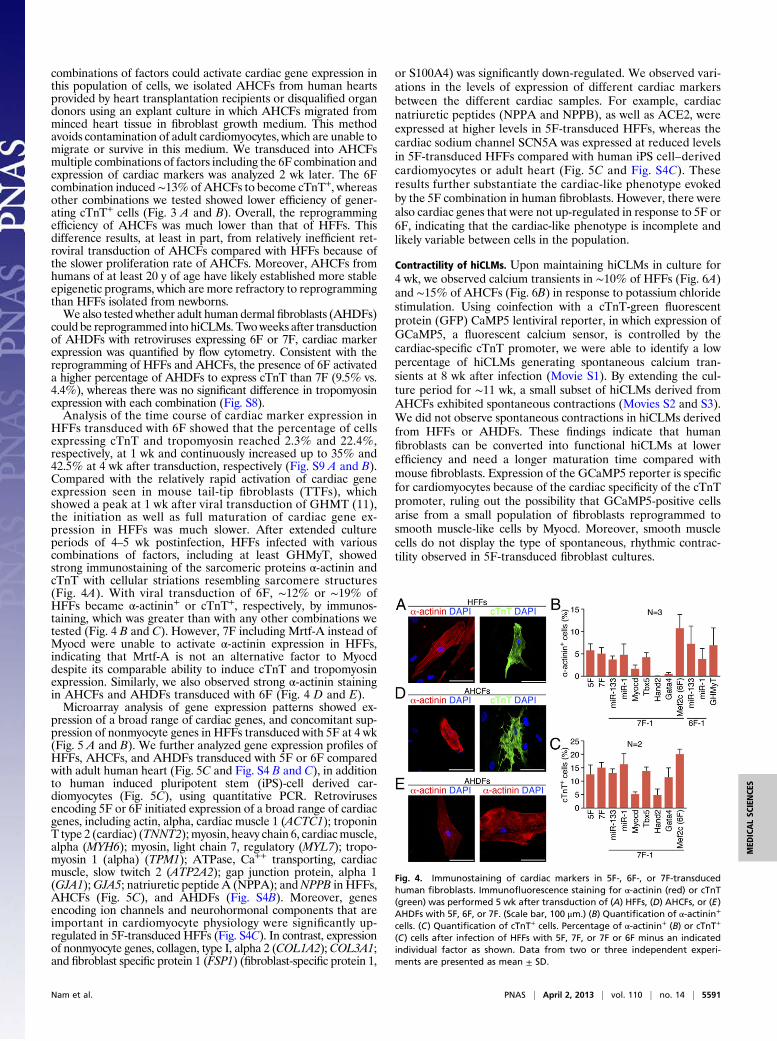
HFFs transduced with 6F showed that the percentage of cellsexpressing cTnT and tropomyosin reached 2.3% and 22.4%,respectively, at 1 wk and continuously increased up to 35% and42.5% at 4 wk after transduction, respectively (Fig. S9 A and B).Compared with the relatively rapid activation of cardiac geneexpression seen in mouse tail-tip fibroblasts (TTFs), whichshowed a peak at 1 wk after viral transduction of GHMT (11),the initiation as well as full maturation of cardiac gene ex-pression in HFFs was much slower. After extended cultureperiods of 4–5 wk postinfection, HFFs infected with variouscombinations of factors, including at least GHMyT, showedstrong immunostaining of the sarcomeric proteins α-actinin andcTnT with cellular striations resembling sarcomere structures(Fig. 4A). With viral transduction of 6F, ∼12% or ∼19% ofHFFs became α-actinin+ or cTnT+, respectively, by immunos-taining, which was greater than with any other combinations wetested (Fig. 4 B and C). However, 7F including Mrtf-A instead ofMyocd were unable to activate α-actinin expression in HFFs,indicating that Mrtf-A is not an alternative factor to Myocddespite its comparable ability to induce cTnT and tropomyosinexpression. Similarly, we also observed strong α-actinin stainingin AHCFs and AHDFs transduced with 6F (Fig. 4 D and E).Microarray analysis of gene expression patterns showed ex-
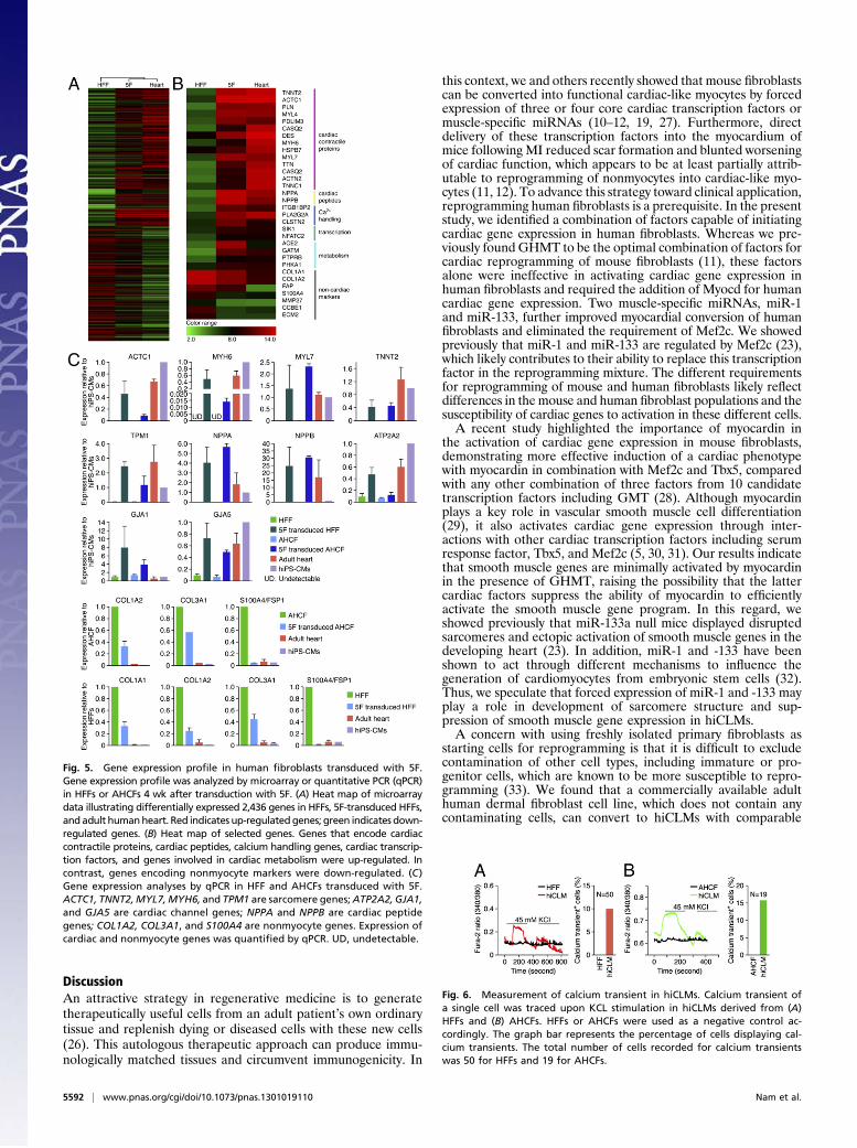
pression of a broad range of cardiac genes, and concomitant sup-pression of nonmyocyte genes in HFFs transduced with 5F at 4 wk(Fig. 5 A and B). We further analyzed gene expression profiles ofHFFs, AHCFs, and AHDFs transduced with 5F or 6F comparedwith adult human heart (Fig. 5C and Fig. S4 B and C), in additionto human induced pluripotent stem (iPS)-cell derived car-diomyocytes (Fig. 5C), using quantitative PCR. Retrovirusesencoding 5F or 6F initiated expression of a broad range of cardiacgenes, including actin, alpha, cardiac muscle 1 (ACTC1); troponinT type 2 (cardiac) (TNNT2); myosin, heavy chain 6, cardiacmuscle,alpha (MYH6); myosin, light chain 7, regulatory (MYL7); tropo-myosin 1 (alpha) (TPM1); ATPase, Ca++ transporting, cardiacmuscle, slow twitch 2 (ATP2A2); gap junction protein, alpha 1(GJA1);GJA5; natriuretic peptide A (NPPA); andNPPB in HFFs,AHCFs (Fig. 5C), and AHDFs (Fig. S4B). Moreover, genesencoding ion channels and neurohormonal components that areimportant in cardiomyocyte physiology were significantly up-regulated in 5F-transduced HFFs (Fig. S4C). In contrast, expressionof nonmyocyte genes, collagen, type I, alpha 2 (COL1A2);COL3A1;and fibroblast specific protein 1 (FSP1) (fibroblast-specific protein 1,
or S100A4) was significantly down-regulated. We observed vari-ations in the levels of expression of different cardiac markersbetween the different cardiac samples. For example, cardiacnatriuretic peptides (NPPA and NPPB), as well as ACE2, wereexpressed at higher levels in 5F-transduced HFFs, whereas thecardiac sodium channel SCN5A was expressed at reduced levelsin 5F-transduced HFFs compared with human iPS cell–derivedcardiomyocytes or adult heart (Fig. 5C and Fig. S4C). Theseresults further substantiate the cardiac-like phenotype evokedby the 5F combination in human fibroblasts. However, there werealso cardiac genes that were not up-regulated in response to 5F or6F, indicating that the cardiac-like phenotype is incomplete andlikely variable between cells in the population.
Contractility of hiCLMs. Upon maintaining hiCLMs in culture for4 wk, we observed calcium transients in ∼10% of HFFs (Fig. 6A)and ∼15% of AHCFs (Fig. 6B) in response to potassium chloridestimulation. Using coinfection with a cTnT-green fluorescentprotein (GFP) CaMP5 lentiviral reporter, in which expression ofGCaMP5, a fluorescent calcium sensor, is controlled by thecardiac-specific cTnT promoter, we were able to identify a lowpercentage of hiCLMs generating spontaneous calcium tran-sients at 8 wk after infection (Movie S1). By extending the cul-ture period for ∼11 wk, a small subset of hiCLMs derived fromAHCFs exhibited spontaneous contractions (Movies S2 and S3).We did not observe spontaneous contractions in hiCLMs derivedfrom HFFs or AHDFs. These findings indicate that humanfibroblasts can be converted into functional hiCLMs at lowerefficiency and need a longer maturation time compared withmouse fibroblasts. Expression of the GCaMP5 reporter is specificfor cardiomyocytes because of the cardiac specificity of the cTnTpromoter, ruling out the possibility that GCaMP5-positive cellsarise from a small population of fibroblasts reprogrammed tosmooth muscle-like cells by Myocd. Moreover, smooth musclecells do not display the type of spontaneous, rhythmic contrac-tility observed in 5F-transduced fibroblast cultures.
Fig. 4. Immunostaining of cardiac markers in 5F-, 6F-, or 7F-transducedhuman fibroblasts. Immunofluorescence staining for α-actinin (red) or cTnT(green) was performed 5 wk after transduction of (A) HFFs, (D) AHCFs, or (E)AHDFs with 5F, 6F, or 7F. (Scale bar, 100 μm.) (B) Quantification of α-actinin+
cells. (C) Quantification of cTnT+ cells. Percentage of α-actinin+ (B) or cTnT+
(C) cells after infection of HFFs with 5F, 7F, or 7F or 6F minus an indicatedindividual factor as shown. Data from two or three independent experi-ments are presented as mean ± SD.
Nam et al. PNAS | April 2, 2013 | vol. 110 | no. 14 | 5591
MED
ICALSC
IENCE
S
DiscussionAn attractive strategy in regenerative medicine is to generatetherapeutically useful cells from an adult patient’s own ordinarytissue and replenish dying or diseased cells with these new cells(26). This autologous therapeutic approach can produce immu-nologically matched tissues and circumvent immunogenicity. In
this context, we and others recently showed that mouse fibroblastscan be converted into functional cardiac-like myocytes by forcedexpression of three or four core cardiac transcription factors ormuscle-specific miRNAs (10–12, 19, 27). Furthermore, directdelivery of these transcription factors into the myocardium ofmice following MI reduced scar formation and blunted worseningof cardiac function, which appears to be at least partially attrib-utable to reprogramming of nonmyocytes into cardiac-like myo-cytes (11, 12). To advance this strategy toward clinical application,reprogramming human fibroblasts is a prerequisite. In the presentstudy, we identified a combination of factors capable of initiatingcardiac gene expression in human fibroblasts. Whereas we pre-viously found GHMT to be the optimal combination of factors forcardiac reprogramming of mouse fibroblasts (11), these factorsalone were ineffective in activating cardiac gene expression inhuman fibroblasts and required the addition of Myocd for humancardiac gene expression. Two muscle-specific miRNAs, miR-1and miR-133, further improved myocardial conversion of humanfibroblasts and eliminated the requirement of Mef2c. We showedpreviously that miR-1 and miR-133 are regulated by Mef2c (23),which likely contributes to their ability to replace this transcriptionfactor in the reprogramming mixture. The different requirementsfor reprogramming of mouse and human fibroblasts likely reflectdifferences in themouse and human fibroblast populations and thesusceptibility of cardiac genes to activation in these different cells.A recent study highlighted the importance of myocardin in
the activation of cardiac gene expression in mouse fibroblasts,demonstrating more effective induction of a cardiac phenotypewith myocardin in combination with Mef2c and Tbx5, comparedwith any other combination of three factors from 10 candidatetranscription factors including GMT (28). Although myocardinplays a key role in vascular smooth muscle cell differentiation(29), it also activates cardiac gene expression through inter-actions with other cardiac transcription factors including serumresponse factor, Tbx5, and Mef2c (5, 30, 31). Our results indicatethat smooth muscle genes are minimally activated by myocardinin the presence of GHMT, raising the possibility that the lattercardiac factors suppress the ability of myocardin to efficientlyactivate the smooth muscle gene program. In this regard, weshowed previously that miR-133a null mice displayed disruptedsarcomeres and ectopic activation of smooth muscle genes in thedeveloping heart (23). In addition, miR-1 and -133 have beenshown to act through different mechanisms to influence thegeneration of cardiomyocytes from embryonic stem cells (32).Thus, we speculate that forced expression of miR-1 and -133 mayplay a role in development of sarcomere structure and sup-pression of smooth muscle gene expression in hiCLMs.A concern with using freshly isolated primary fibroblasts as
starting cells for reprogramming is that it is difficult to excludecontamination of other cell types, including immature or pro-genitor cells, which are known to be more susceptible to repro-gramming (33). We found that a commercially available adulthuman dermal fibroblast cell line, which does not contain anycontaminating cells, can convert to hiCLMs with comparable
Fig. 5. Gene expression profile in human fibroblasts transduced with 5F.Gene expression profile was analyzed by microarray or quantitative PCR (qPCR)in HFFs or AHCFs 4 wk after transduction with 5F. (A) Heat map of microarraydata illustrating differentially expressed 2,436 genes in HFFs, 5F-transduced HFFs,and adult humanheart. Red indicates up-regulatedgenes; green indicates down-regulated genes. (B) Heat map of selected genes. Genes that encode cardiaccontractile proteins, cardiac peptides, calcium handling genes, cardiac transcrip-tion factors, and genes involved in cardiac metabolism were up-regulated. Incontrast, genes encoding nonmyocyte markers were down-regulated. (C)Gene expression analyses by qPCR in HFF and AHCFs transduced with 5F.ACTC1, TNNT2,MYL7,MYH6, and TPM1 are sarcomere genes;ATP2A2, GJA1,and GJA5 are cardiac channel genes; NPPA and NPPB are cardiac peptidegenes; COL1A2, COL3A1, and S100A4 are nonmyocyte genes. Expression ofcardiac and nonmyocyte genes was quantified by qPCR. UD, undetectable.
Fig. 6. Measurement of calcium transient in hiCLMs. Calcium transient ofa single cell was traced upon KCL stimulation in hiCLMs derived from (A)HFFs and (B) AHCFs. HFFs or AHCFs were used as a negative control ac-cordingly. The graph bar represents the percentage of cells displaying cal-cium transients. The total number of cells recorded for calcium transientswas 50 for HFFs and 19 for AHCFs.
5592 | www.pnas.org/cgi/doi/10.1073/pnas.1301019110 Nam et al.
efficiency to freshly isolated adult fibroblasts, excluding potentialcontributions of stem or progenitor cells in this process. Humancardiomyocytes have also been differentiated from human iPScells or cardiac progenitors (34, 35). The ability to reprogramhuman fibroblasts that were expanded multiple times after iso-lation could, with further optimization, eventually allow large-scale production of hiCLMs for possible transplantation. In thiscontext, a recent study demonstrated that human ES cell–derivedcardiomyocytes can integrate into an infarcted guinea pig heartand suppress development of post-MI arrhythmias (36). Giventhat human ES cells have limitations as a clinical source for celltransplantation, hiCLMs may be a viable alternative for thisapproach in the future.As with other reprogrammed cells generated from human
fibroblasts, including human iPSCs and human-induced neurons,human iCLMs are functionally immature, as indicated by theirmorphology, low-amplitude calcium transients in response toelectrical stimulation, and relatively rare spontaneous contractility.In addition, the human iCLMs generated in this study were het-erogeneous, containing cells with varying levels of expression ofcardiac and noncardiac genes. Phenotypic conversion to a cardiaccell fate probably requires a precise stoichiometry as well as certainlevels of expression of reprogramming factors, which are achievedonly in a small subset of fibroblasts. Heterogeneity of hiCLMs islikely to reflect variations in the stoichiometry and levels of ex-pression of reprogramming factors in individual cells. Moreover,variations in the percentage of cardiac marker–expressing cellsfrom experiment to experiment are likely also attributable to het-erogeneity of human fibroblasts that were isolated from humansubjects of various ages and genetic backgrounds. Human fibro-blasts whose epigenetic stabilities vary depending on their originsare likely to have a wide spectrum of susceptibility to reprogram-ming. In addition, the differences that exist in each viral prepara-tion also contribute to variability in reprogramming efficiency. For
similar reasons, we observed variable reprogramming efficiencyfrom experiment to experiment even in mouse fibroblasts (11).Nevertheless, the results of this study indicate that diverse types ofhuman fibroblasts can be reprogrammed toward a cardiac fate andestablish a foundation for further optimization of this process andthe eventual generation of more mature and homogeneous pop-ulations of hiCLMs. It will also be of particular interest to modifythis process using pharmacologic agents and to generate specializedcells involved in cardiac conduction as a strategy for modulatingcardiac contractility. Such studies are under way.
Materials and MethodsAll deidentified human heart and foreskin tissues were obtained and bankedwith proper informed consent under approval of the University of TexasSouthwestern Institutional Review Board (IRB CR00001649/STU 032011-174and IRB 092010-193). Normal human foreskin was obtained from neonates(weighing less than 10 lb) undergoing routine circumcision. Adult humanhearts were obtained from heart transplantation recipients, lung trans-plantation donors, or disqualified organ donors. Isolation of human fibro-blasts, production of retroviruses, induction of reprogramming, flowcytometry, immunocytochemistry, real-time PCR and DNA microarray, andcalcium transient measurements are described in SI Materials and Methods.
ACKNOWLEDGMENTS. We thank Dr. J. Gearhart (University of Pennsylvania)for the cTnT-GCaMP5 FUdeltaGW-rtTA construct; J. Cabrera for graphics andthe University of Texas Southwestern Microarray Core Facility for collectinggene expression data; Dr. Megan Kong for analyzing the microarray data;and Ankit Garg, Thomas Haden, Dr. Ning Liu, and Dr. Ji-Hoon Lee fortechnical advice. E.N.O. is supported by grants from the National Institutes ofHealth (NIH), the Donald W. Reynolds Center for Clinical CardiovascularResearch, the Robert A. Welch Foundation (Grant I-0025), the LeducqFoundation-Transatlantic Network of Excellence in Cardiovascular ResearchProgram, the American Heart Association–Jon Holden DeHaan Foundation,and the Cancer Prevention and Research Institute of Texas; L.A.B. is sup-ported by the Seay Endowment and a grant from the NIH; and Y.-J.N. wassupported by NIH Grant K08 HL111420-02.
1. Graf T, Enver T (2009) Forcing cells to change lineages. Nature 462(7273):587–594.2. Vierbuchen T, Wernig M (2011) Direct lineage conversions: Unnatural but useful? Nat
Biotechnol 29(10):892–907.3. Davis RL, Weintraub H, Lassar AB (1987) Expression of a single transfected cDNA
converts fibroblasts to myoblasts. Cell 51(6):987–1000.4. Olson EN (1990)MyoD family: A paradigm for development?Genes Dev 4(9):1454–1461.5. Wang D, et al. (2001) Activation of cardiac gene expression by myocardin, a tran-
scriptional cofactor for serum response factor. Cell 105(7):851–862.6. Wang Z, Wang DZ, Pipes GC, Olson EN (2003) Myocardin is a master regulator of
smooth muscle gene expression. Proc Natl Acad Sci USA 100(12):7129–7134.7. Chen J, Kitchen CM, Streb JW, Miano JM (2002) Myocardin: A component of a mo-
lecular switch for smooth muscle differentiation. J Mol Cell Cardiol 34(10):1345–1356.8. Du KL, et al. (2003) Myocardin is a critical serum response factor cofactor in the
transcriptional program regulating smooth muscle cell differentiation. Mol Cell Biol23(7):2425–2437.
9. Yoshida T, et al. (2003) Myocardin is a key regulator of CArG-dependent transcriptionof multiple smooth muscle marker genes. Circ Res 92(8):856–864.
10. Ieda M, et al. (2010) Direct reprogramming of fibroblasts into functional car-diomyocytes by defined factors. Cell 142(3):375–386.
11. Song K, et al. (2012) Heart repair by reprogramming non-myocytes with cardiactranscription factors. Nature 485(7400):599–604.
12. Qian L, et al. (2012) In vivo reprogramming of murine cardiac fibroblasts into inducedcardiomyocytes. Nature 485(7400):593–598.
13. Zhou Q, Brown J, Kanarek A, Rajagopal J, Melton DA (2008) In vivo reprogramming ofadult pancreatic exocrine cells to beta-cells. Nature 455(7213):627–632.
14. Huang P, et al. (2011) Induction of functional hepatocyte-like cells from mouse fi-broblasts by defined factors. Nature 475(7356):386–389.
15. Sekiya S, Suzuki A (2011) Direct conversion of mouse fibroblasts to hepatocyte-likecells by defined factors. Nature 475(7356):390–393.
16. Pang ZP, et al. (2011) Induction of human neuronal cells by defined transcriptionfactors. Nature 476(7359):220–223.
17. Qiang L, et al. (2011) Directed conversion of Alzheimer’s disease patient skin fibro-blasts into functional neurons. Cell 146(3):359–371.
18. Yoo AS, et al. (2011) MicroRNA-mediated conversion of human fibroblasts to neu-rons. Nature 476(7359):228–231.
19. Jayawardena TM, et al. (2012) MicroRNA-mediated in vitro and in vivo direct re-programming of cardiac fibroblasts to cardiomyocytes. Circ Res 110(11):1465–1473.
20. Kuro-o M, et al. (1989) Developmentally regulated expression of vascular smooth
muscle myosin heavy chain isoforms. J Biol Chem 264(31):18272–18275.21. Rovner AS, Thompson MM, Murphy RA (1986) Two different heavy chains are found
in smooth muscle myosin. Am J Physiol 250(6 Pt 1):C861–C870.22. Owens GK (1995) Regulation of differentiation of vascular smooth muscle cells.
Physiol Rev 75(3):487–517.23. Liu N, et al. (2008) microRNA-133a regulates cardiomyocyte proliferation and sup-
presses smooth muscle gene expression in the heart. Genes Dev 22(23):3242–3254.24. Brown RD, Ambler SK, Mitchell MD, Long CS (2005) The cardiac fibroblast: Thera-
peutic target in myocardial remodeling and failure. Annu Rev Pharmacol Toxicol 45:
657–687.25. Zeisberg EM, Kalluri R (2010) Origins of cardiac fibroblasts. Circ Res 107(11):1304–1312.26. Dimmeler S, Zeiher AM, Schneider MD (2005) Unchain my heart: The scientific
foundations of cardiac repair. J Clin Invest 115(3):572–583.27. SrivastavaD, IedaM (2012) Critical factors for cardiac reprogramming. Circ Res 111(1):5–8.28. Protze S, et al. (2012) A new approach to transcription factor screening for re-
programmingoffibroblasts to cardiomyocyte-like cells. JMol Cell Cardiol 53(3):323–332.29. Li S, Wang DZ, Wang Z, Richardson JA, Olson EN (2003) The serum response factor
coactivator myocardin is required for vascular smooth muscle development. Proc Natl
Acad Sci USA 100(16):9366–9370.30. Ghosh TK, et al. (2009) Physical interaction between TBX5 and MEF2C is required for
early heart development. Mol Cell Biol 29(8):2205–2218.31. Wang C, Cao D, Wang Q, Wang DZ (2011) Synergistic activation of cardiac genes by
myocardin and Tbx5. PLoS ONE 6(8):e24242.32. Ivey KN, et al. (2008) MicroRNA regulation of cell lineages in mouse and human
embryonic stem cells. Cell Stem Cell 2(3):219–229.33. Zhou Q, Melton DA (2008) Extreme makeover: Converting one cell into another. Cell
Stem Cell 3(4):382–388.34. Islas JF, et al. (2012) Transcription factors ETS2 and MESP1 transdifferentiate human
dermal fibroblasts into cardiac progenitors. Proc Natl Acad Sci USA 109(32):
13016–13021.35. Zwi L, et al. (2009) Cardiomyocyte differentiation of human induced pluripotent stem
cells. Circulation 120(15):1513–1523.36. Shiba Y, et al. (2012) Human ES-cell-derived cardiomyocytes electrically couple and
suppress arrhythmias in injured hearts. Nature 489(7415):322–325.
Nam et al. PNAS | April 2, 2013 | vol. 110 | no. 14 | 5593
MED
ICALSC
IENCE
S