Embed Size (px)
Citation preview

Review
Focus: Induced Pluripotency & Cellular Reprogramming
Reprogramming of human cancer cells topluripotency for models of cancer progressionJungsun Kim & Kenneth S Zaret*
Abstract
The ability to study live cells as they progress through the stages ofcancer provides the opportunity to discover dynamic networksunderlying pathology, markers of early stages, and ways to assesstherapeutics. Genetically engineered animal models of cancer,where it is possible to study the consequences of temporal-specificinduction of oncogenes or deletion of tumor suppressors, haveyielded major insights into cancer progression. Yet differences existbetween animal and human cancers, such as in markers of progres-sion and response to therapeutics. Thus, there is a need for humancell models of cancer progression. Most human cell models of cancerare based on tumor cell lines and xenografts of primary tumor cellsthat resemble the advanced tumor state, from which the cells werederived, and thus do not recapitulate disease progression. Yet asubset of cancer types have been reprogrammed to pluripotency ornear-pluripotency by blastocyst injection, by somatic cell nucleartransfer and by induced pluripotent stem cell (iPS) technology. Thereprogrammed cancer cells show that pluripotency can transientlydominate over the cancer phenotype. Diverse studies show thatreprogrammed cancer cells can, in some cases, exhibit early-stagephenotypes reflective of only partial expression of the cancergenome. In one case, reprogrammed human pancreatic cancer cellshave been shown to recapitulate stages of cancer progression, fromearly to late stages, thus providing a model for studying pancreaticcancer development in human cells where previously such couldonly be discerned from mouse models. We discuss these findings,the challenges in developing such models and their current limita-tions, and ways that iPS reprogramming may be enhanced todevelop human cell models of cancer progression.
Keywords cancer; iPS; pluripotency; progression
DOI 10.15252/embj.201490736 | Received 5 December 2014 | Revised 24
December 2014 | Accepted 2 January 2015 | Published online 20 February
2015
The EMBO Journal (2015) 34: 739–747
Introduction
Over the past two decades, diverse cellular models of cancer, in
combination with biochemical and imaging tools, have greatly
improved the early diagnosis and treatment of various cancers.
Tests exist for the early detection of cervical, colon, and breast
cancers and mortality has been steadily declining (Arteaga et al,
2014). Despite these advances, cancers such as pancreatic ductal
adenocarcinoma (PDAC) retain a dismal prognosis due to a paucity
of clinical symptoms and biomarkers for early-stage disease
(Cantley et al, 2012). The failure to detect early disease prevents the
application and testing of therapeutics that could be used to inhibit
disease progression. Genetically engineered animal models in mouse
and zebrafish have been developed for diverse cancers (Tuveson &
Jacks, 2002; Liu & Leach, 2011; Ablain & Zon, 2013). Such models
can provide insights into the basis of cancer development that have
helped generate treatments, such as for acute promyelocytic
leukemia (Wang & Chen, 2008; Nardella et al, 2011). Yet there are
cross-species differences between animal and human cancers with
regard to the size of tumors, cancer susceptibility, spectrum of age-
related cancers, and telomere lengths (DePinho, 2000; Rangarajan &
Weinberg, 2003). Cancer therapeutics that work in mouse models
can fail in clinical human trials (Ledford, 2011; Begley & Ellis,
2012). To complement these limitations, human solid tumors, orga-
noid cultures thereof (Gao et al, 2014; Li et al, 2014), and cell lines
(Sharma et al, 2010) have been engrafted into immunocompromised
mice either as tumor fragments (Tentler et al, 2012), dispersed cells,
or cells sorted for tumor initiating cells/cancer stem cells (CSC)
(Ishizawa et al, 2010; Nguyen et al, 2012). In these contexts, tumors
inevitably arise that resemble the parental tumor state from which
the cells were derived and do not undergo early-stage progression
(Tentler et al, 2012). Primary human mammary epithelial cells can
undergo cancer progression in mouse xenografts, but require prior
transformation with oncogenes rather than employing endogenous
genetic changes found in tumors (Wu et al, 2009). Currently, there
are few human cell models of cancer progression that are dependent
upon naturally occurring genetic mutations.
Here, we describe the use of somatic cell nuclear transfer (SCNT)
on mouse cancer cells and induced pluripotent stem (iPS) cell tech-
nology to model human cancer progression. Cancer progression by
the SCNT or iPS reprogrammed cells can help reveal new networks
for early-stage disease, potential early-stage biomarkers, and human
cell models in which therapeutics can be assessed. We first discuss
the history of reprogramming of cancer cells to pluripotency, as
initiated with blastocyst injection and somatic nuclear transfer.
We also describe mechanisms that may underlie a temporary
Department of Cell and Developmental Biology, Institute for Regenerative Medicine, Abramson Cancer Center, Tumor Biology Program, Perelman School of Medicine,University of Pennsylvania, Philadelphia, PA, USA
*Corresponding author. Tel: +1 215 573 5813; E-mail: [email protected]
ª 2015 The Authors The EMBO Journal Vol 34 | No 6 | 2015 739
Published online: February 20, 2015
dominance of pluripotency over the cancer genome and the use of
iPS technology to elicit such dominance. We then review the
advances and challenges associated with iPS-based approaches,
along with the future prospects for developing new human cell
models of cancer progression.
Early examples of cancer cell reprogrammingto pluripotency
Several lines of evidence show that cancer progression can be elic-
ited by reversible epigenetic changes as well as by irreversible muta-
tions in oncogenic and tumor suppressor genes (Esteller, 2007). The
first experimental evidence of tumor reversibility was described in
crown-gall tumors in plants over a half century ago (Braun, 1959).
Braun had hypothesized that the individual pluripotent teratoma
cells of crown-gall tumors might be recovered from the cancer in
grafting experiments. To test his idea, he grafted the shoots from
crown-gall teratoma cells serially to the cut stem ends of the healthy
tobacco plants. The grafted teratoma tissue gradually developed
more normal appearing shoots, which eventually flowered and
developed seeds. He concluded that ‘the cellular alteration in crown
gall did not involve a somatic mutation at the nuclear gene level
and rather some yet uncharacterized cytoplasmic entity is responsi-
ble for the cellular changes that underlie the tumorous state in the
crown-gall disease.’
In mammalian development, the fertilized egg, or zygote, under-
goes cell divisions to reach the blastocyst stage. The blastocyst
contains the first differentiated structures, consisting of an outer
layer of trophectoderm and an inner cell mass (ICM) that develops
into the embryo and yolk sac. Many cells within the inner cell mass
are considered pluripotent, because they give rise to all three germ
layers (endoderm, ectoderm, and mesoderm) and derived tissues in
the embryo. The most stringent test of whether exogenous mamma-
lian cells are pluripotent is to inject them into a blastocyst and deter-
mine whether they contribute to the embryo and, ultimately, the
animal proper (Lin, 1966). Individual malignant stem cells of
murine embryonal teratomas (embryonic carcinoma, EC) injected
into 280 blastocysts yielded 45 apparently normal embryos or
fetuses, and such cells injected into 183 blastocysts yielded 48
apparently normal live mice (Mintz & Illmensee, 1975). All of three
fetuses analyzed were mosaic, with substantial tissue contributions
from teratoma cells, indicating the cells’ pluripotency. Notably,
many genes that had been silent or undetectable in the tumor, such
as immunoglobulin, hemoglobin, MUP, agouti genes, were
expressed in the appropriate tissues. The authors concluded that
‘conversion to neoplasia did not involve structural changes in the
genome, but rather a change in gene expression.’
The developmental pluripotency of somatic cell nucleus can be
tested by implanting them into enucleated oocytes, that is, somatic
cell nuclear transfer (SCNT) (Gurdon et al, 1958). By SCNT, a
subset of cancer cells such as certain renal tumor cells, medulloblas-
toma cells, RAS-induced melanoma cells, and EC cells were able to
be reprogrammed to pluripotency (McKinnell et al, 1969; Blelloch
et al, 2004; Hochedlinger et al, 2004). Donor nuclei from triploid
frog renal tumors were reprogrammed by injection into enucleated
frog eggs, developed into blastulas with a higher efficiency than
diploid nuclei (23% versus 7.7%). Yet only 21% of the triploid,
tumor-derived blastulas developed into swimming embryos
compared to 100% of the blastulas from normal diploid nuclei
(McKinnell et al, 1969). The living triploid tadpoles differentiated
into functional embryonic tissues of many types and the descendant
tail fins regenerated appropriately, demonstrating pluripotency of
the reprogrammed tumor genome (McKinnell et al, 1969).
Hochedlinger et al (2004) attempted the reprogramming by
SCNT of diverse mouse cancer cells, including a p53�/� lymphoma,
moloney murine leukemia virus-induced leukemia, PML-RAR trans-
gene-induced leukemia, hypomethylated Chip/c lymphoma, p53�/�
breast cancer cell line, and an ink4a/Arf�/�, RAS-inducible mela-
noma cell line. All SCNT-reprogrammed cancer cell lines, but no
primary tumor cells, were able to develop normal appearing blast-
ocysts, with much greater efficiency in cancer cell lines harboring
mutant tumor suppressors. SCNT-derived blastocysts whose zona
pellucida was removed were placed onto irradiated murine embry-
onic fibroblast to derive embryonic stem (ES) cells. However, such
SCNT-ES cell lines were only made from an Ink4a/Arf�/�, RAS-
inducible melanoma cell line, suggesting that only certain cancer
genomes or cell types are amenable to the manipulation.
To assess their autonomous developmental potential, melanoma
SCNT-ES cells were injected into tetraploid blastocysts, where trans-
planted wild-type ES cells can exclusively give rise to the embryo
and tetraploid cells become the placenta (Wang et al, 1997). The
resulting embryos developed up to day 9.5 of mouse gestation
(E9.5) with a beating heart, closed neural tube, and developing limb
and tail buds. Yet at later stages, embryos were not recovered,
presumably because of the reactivation of irreversible genetic altera-
tions of the melanoma.
However, in a chimeric blastocyst assay, where normal and
donor cells can contribute to the embryo, the SCNT-ES cells, despite
having major severe chromosomal changes from the original cancer,
showed remarkable differentiation by contributing to skin, intestine,
heart, kidney, lungs, thymus, and liver in newborn chimeric mice
and to adult lineages such as the lymphoid compartment in Rag2-
deficient chimeric mice (Hochedlinger et al, 2004). These findings
show that the oocyte cytoplasm can reprogram the epigenetic state
of the donor cancer cell nucleus into a pluripotent state that
supports differentiation into multiple somatic cell types.
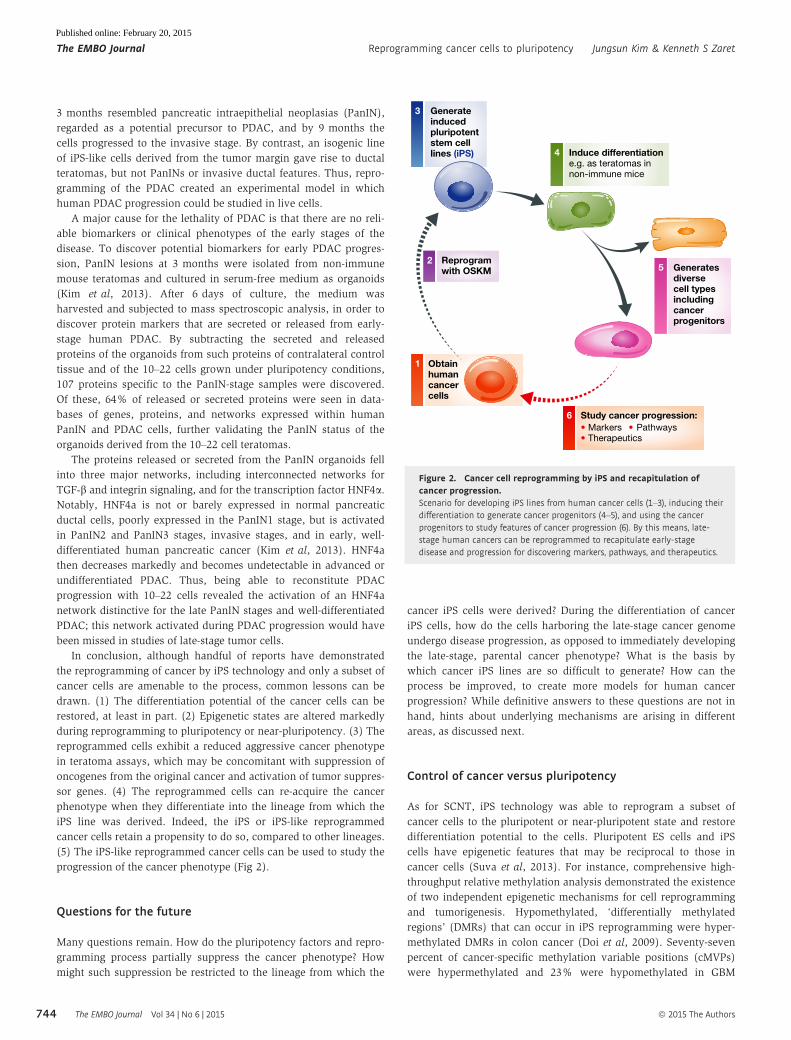
The SCNT-ES-derived adult chimeric mice developed multiple
primary melanoma lesions with an average latency of 19 days,
comparable to the latency required for the development of recurrent
tumors or the emergence of tumors derived from transplanted mela-
noma cells (Chin et al, 1999). Interestingly, 33% of the chimeras
from melanoma SCNT-ES cells developed rhabdomyosarcoma,
which has an overlapping pathway with melanoma, showing the
consequence of the ‘melanoma genome’ expressed in a different
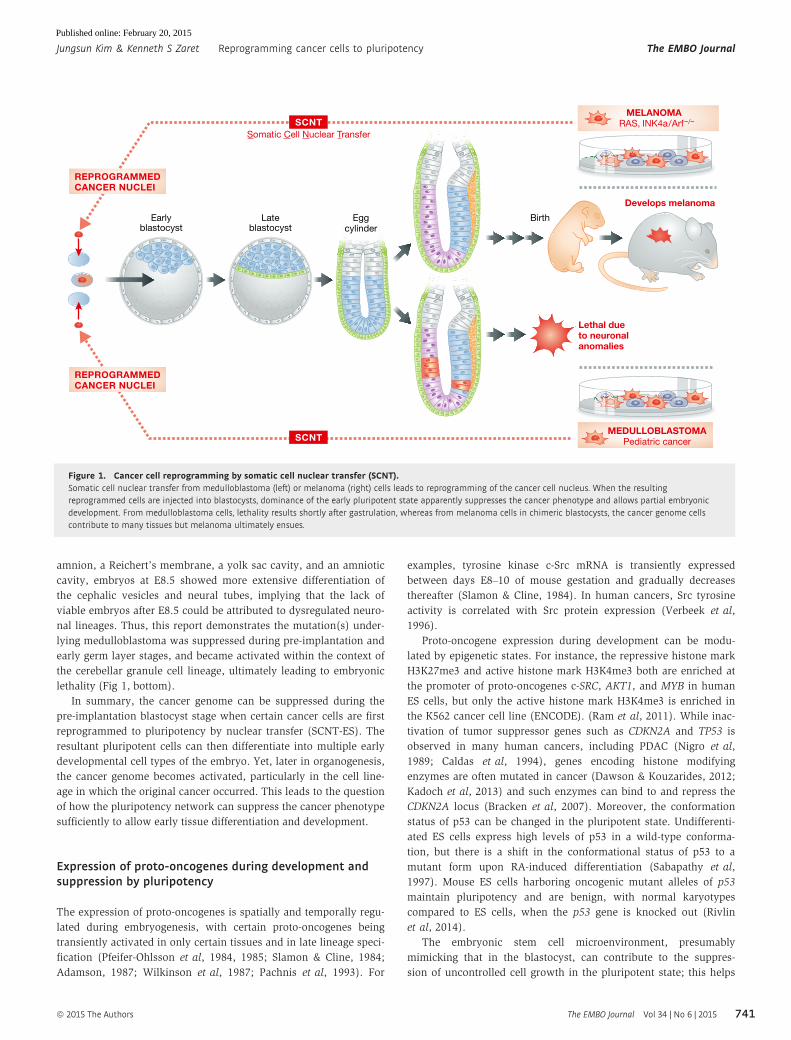
tissue (Hochedlinger et al, 2004) (Fig 1, top).
Li et al (2003) tested the epigenetic reprogramming of medullo-
blastoma, a pediatric brain tumor, originating from the granule
neuron precursors of the developing cerebellum. The medullo-
blastoma cells were isolated from Ptc+/� mice and used for SCNT.
Although transferred SCNT cells developed into blastocysts that
were morphologically indistinguishable from those derived nuclei of
spleen control cells, no viable embryos were identified after E8.5 in
the transplanted pseudo-pregnant mice. Intriguingly, while the
embryos at E7.5 days appeared grossly normal and contained all
three germ layers as well as an ectoplacental cone, a chorion, an
The EMBO Journal Vol 34 | No 6 | 2015 ª 2015 The Authors
The EMBO Journal Reprogramming cancer cells to pluripotency Jungsun Kim & Kenneth S Zaret
740
Published online: February 20, 2015
amnion, a Reichert’s membrane, a yolk sac cavity, and an amniotic
cavity, embryos at E8.5 showed more extensive differentiation of
the cephalic vesicles and neural tubes, implying that the lack of
viable embryos after E8.5 could be attributed to dysregulated neuro-
nal lineages. Thus, this report demonstrates the mutation(s) under-
lying medulloblastoma was suppressed during pre-implantation and
early germ layer stages, and became activated within the context of
the cerebellar granule cell lineage, ultimately leading to embryonic
lethality (Fig 1, bottom).
In summary, the cancer genome can be suppressed during the
pre-implantation blastocyst stage when certain cancer cells are first
reprogrammed to pluripotency by nuclear transfer (SCNT-ES). The
resultant pluripotent cells can then differentiate into multiple early
developmental cell types of the embryo. Yet, later in organogenesis,
the cancer genome becomes activated, particularly in the cell line-
age in which the original cancer occurred. This leads to the question
of how the pluripotency network can suppress the cancer phenotype
sufficiently to allow early tissue differentiation and development.
Expression of proto-oncogenes during development andsuppression by pluripotency
The expression of proto-oncogenes is spatially and temporally regu-
lated during embryogenesis, with certain proto-oncogenes being
transiently activated in only certain tissues and in late lineage speci-
fication (Pfeifer-Ohlsson et al, 1984, 1985; Slamon & Cline, 1984;
Adamson, 1987; Wilkinson et al, 1987; Pachnis et al, 1993). For
examples, tyrosine kinase c-Src mRNA is transiently expressed
between days E8–10 of mouse gestation and gradually decreases
thereafter (Slamon & Cline, 1984). In human cancers, Src tyrosine
activity is correlated with Src protein expression (Verbeek et al,
1996).
Proto-oncogene expression during development can be modu-
lated by epigenetic states. For instance, the repressive histone mark
H3K27me3 and active histone mark H3K4me3 both are enriched at
the promoter of proto-oncogenes c-SRC, AKT1, and MYB in human
ES cells, but only the active histone mark H3K4me3 is enriched in
the K562 cancer cell line (ENCODE). (Ram et al, 2011). While inac-
tivation of tumor suppressor genes such as CDKN2A and TP53 is
observed in many human cancers, including PDAC (Nigro et al,
1989; Caldas et al, 1994), genes encoding histone modifying
enzymes are often mutated in cancer (Dawson & Kouzarides, 2012;
Kadoch et al, 2013) and such enzymes can bind to and repress the
CDKN2A locus (Bracken et al, 2007). Moreover, the conformation
status of p53 can be changed in the pluripotent state. Undifferenti-
ated ES cells express high levels of p53 in a wild-type conforma-
tion, but there is a shift in the conformational status of p53 to a
mutant form upon RA-induced differentiation (Sabapathy et al,
1997). Mouse ES cells harboring oncogenic mutant alleles of p53
maintain pluripotency and are benign, with normal karyotypes
compared to ES cells, when the p53 gene is knocked out (Rivlin
et al, 2014).
The embryonic stem cell microenvironment, presumably
mimicking that in the blastocyst, can contribute to the suppres-
sion of uncontrolled cell growth in the pluripotent state; this helps
Birth
Lethal due to neuronalanomalies
MEDULLOBLASTOMAPediatric cancer
Develops melanoma
Earlyblastocyst
Lateblastocyst
Eggcylinder
MELANOMA RAS, INK4a/Arf–/–
REPROGRAMMEDCANCER NUCLEI
REPROGRAMMEDCANCER NUCLEI
SCNT
SCNTSomatic Cell Nuclear Transfer
Figure 1. Cancer cell reprogramming by somatic cell nuclear transfer (SCNT).Somatic cell nuclear transfer from medulloblastoma (left) or melanoma (right) cells leads to reprogramming of the cancer cell nucleus. When the resultingreprogrammed cells are injected into blastocysts, dominance of the early pluripotent state apparently suppresses the cancer phenotype and allows partial embryonicdevelopment. From medulloblastoma cells, lethality results shortly after gastrulation, whereas from melanoma cells in chimeric blastocysts, the cancer genome cellscontribute to many tissues but melanoma ultimately ensues.
ª 2015 The Authors The EMBO Journal Vol 34 | No 6 | 2015
Jungsun Kim & Kenneth S Zaret Reprogramming cancer cells to pluripotency The EMBO Journal
741
Published online: February 20, 2015
to keep the balance between self-renewal and differentiation.
Aggressive melanoma cells were reprogrammed into melanocyte-
like cells and invasiveness was reduced, at least in part, by
culturing the cells on Matrigel that was conditioned by human ES
cells, suggesting suppressive, anti-invasive cues associated with
the huES microenvironment (Postovit et al, 2006). The aggressive
melanoma and breast carcinoma cells express Nodal, which is
essential for human ES cell pluripotency, yet these cancers did
not express Lefty, an inhibitor of Nodal, which is expressed
in human ES cells. Exposure of the cancer cell lines to
ES-conditioned Matrigel resulted in a decrease in tumorigenesis
accompanied by a reduction in clonogenicity and an increase in
apoptosis, directly associated with secretion of Lefty from huES
cells (Postovit et al, 2008).
In conclusion, early developmental signals naturally regulate
proto-oncogenes so that their expression can be suppressed until an
appropriate developmental stage where the genes function. Concor-
dantly, the changing early embryonic environment and the mimic of
such environments during ES cell culture can suppress oncogenic
phenotypes of cancer-derived cells.
Reprogramming of somatic cells to pluripotency by OSKM
Human and mouse somatic cells (such as fibroblasts, blood cells,
etc.) can be reprogrammed into a pluripotent ES-like cells, called
induced pluripotent stem (iPS) cells, by the ectopic expression of
transcription factors such as Oct4, Sox2, Klf4, and c-Myc (OSKM)
(Takahashi & Yamanaka, 2006). As this method does not need
oocytes or blastocysts that are used for SCNT, it is a much more
accessible technique and it side-steps ethical issues associated with
using early human embryos. The pluripotency of human iPS cells
can be validated by cell markers, genomic RNA expression profiles,
epigenetic profiles, and teratoma assays; the latter being when (e.g.,
human) iPS cells are injected subcutaneously into mice that are
genetically deficient in innate and acquired immunity (e.g., NOD-
SCID (Shultz et al, 2005)). Details of iPS cell generation and use are
discussed elsewhere in this issue.
Reprogramming of cancer cells to pluripotency ornear-pluripotency by OSKM
In the first experiments on cancer reprogramming by SCNT
described above, only a subset of cancer cell types could be repro-
grammed to pluripotency. Despite the remarkable demonstration of
the potential dominance of the pluripotent state over cancer, major
questions remained. Are there particular cancer mutations that
allow or block the ability of a cancer cells to be reprogrammed to
pluripotency? Does such ability to be reprogrammed relate to the
tissue type of the cancer? At what stage of using re-differentiation
of the reprogrammed cancer cells does the cancer genome regain
dominance over the cell phenotype? Can understanding the transi-
tion between pluripotency and cancer provide new insight into
how to control the growth of cancer cells? Now that the relatively
simple iPS technology can be applied to reprogram cancer cells,
independent of oocytes and blastocysts, these questions have been
revisited.
Reprogramming of chronic myeloid leukemia (CML)
The initial chronic phase of CML, which originates from hematopoi-
etic stem cells of the bone marrow, is caused by a BCR-ABL fusion
mutation that drives cell expansion, while the CML clones retain
differentiation potential (Melo & Barnes, 2007). The chronic phase
of CML progresses into an accelerated phase, followed by the blast
crisis, terminal phase of CML upon acquisition of a second lesion.
Once CML reaches the blast crisis stage, the cells lose the ability to
differentiate and immature leukemia cells overgrow. Based upon the
dependency of CML on BCR-ABL activated tyrosine kinase, tyrosine
kinase inhibitors such as imatinib improved the long-term survival
rate of CML patients. However, the inhibitor cannot completely
eradicate CML cells and often lead to the recurrence of CML clones
after its discontinuation (Melo & Barnes, 2007). Can CML in the
terminal blast crisis stage be reprogrammed into iPS cells? If so, can
CML-iPS cells recapitulate the initial chronic phase of CML, which
has differentiation potential? Can be the dependency of CML on
BCR-ABL signaling be altered?
Carette et al (2010) reprogrammed a cell line derived from blast
crisis stage CML by infecting with a retrovirus expressing OSKM.
Subcutaneous injection of the resulting CML-iPS cells into NOD-
SCID mice revealed teratomas which contained cells of three germ
layers, indicating pluripotency. During in vitro differentiation, the
CML-iPS cells were able to differentiate into cells expressing the
pan-T cell marker CD43+ and the hematopoietic lineage marker
CD45+, as well as the stem cell marker CD34+, demonstrating a
restoration of differentiation potential into hematopoietic lineages.
The loss of the CML phenotype in CML-iPS cells and the recovery of
differentiation can be viewed as a recapitulation of the chronic
phase of CML, despite starting with blast crisis stage CML-iPS cells.
Interestingly, whereas parental CML cell lines were dependent on
the BCR-ABL pathway, the CML-iPS cell lines were independent of
BCR-ABL signaling and showed resistance to imatinib, an inhibitor
of BCR-ABL signaling (Carette et al, 2010). The loss of BCR-ABL
dependency was also observed in cells differentiated in vitro into
neuronal or fibroblast-like cells. Yet when the cells were differenti-
ated in vitro to hematopoietic lineage cells, they became sensitive to
imatinib, suggesting that the recovery of oncogenic dependency as
the CML-iPS cells underwent hematopoietic differentiation. Thus,
oncogenic mutations can be dynamically expressed when cancer
cells are converted to pluripotency and then re-differentiated.
Similar observations were seen by another group that generated
iPS cells from primary CD34+ cells which were isolated from bone
marrow mononuclear cells of a CML chronic phase patient, by retro-
viral infection with OSKM (Kumano et al, 2012). The CML-iPS cells
underwent normal hematopoiesis during in vitro differentiation. The
differentiated hematopoietic progenitors (CD34+CD45+) from CML-
iPS cells produced colonies of mature erythroid cells, granulocyte–
macrophage cells, or mix lineages with a distribution of colony size,
morphologies, and kinetics of growth and maturation that was simi-
lar to non-cancer iPS cells. The CML-iPS-derived hematopoietic
progenitors did not show a CML phenotype in vivo, when intrave-
nously engrafted into NSG mice receiving minimal irradiation.
Whereas the parental CD34+ cells responded to imatinib, the
CML-iPS derivatives and immature CD34+CD38�CD45+CD90+ cells
derived from the CML-iPS cells were not sensitive to imatinib.
Yet mature hematopoietic cells (CD34�CD45+) derived from the
CML-iPS cells restored their sensitivity to imatinib. Given that the
The EMBO Journal Vol 34 | No 6 | 2015 ª 2015 The Authors
The EMBO Journal Reprogramming cancer cells to pluripotency Jungsun Kim & Kenneth S Zaret
742
Published online: February 20, 2015
expression of the proto-oncogene C-ABL is high at mouse embryonic
day 10 when the first definitive hematopoietic stem cells are gener-
ated (Muller et al, 1982; Sanchez et al, 1996), the CML-iPS cells,
corresponding to mouse blastocyst cells at embryonic day 3–5, may
express regulatory factors that suppress C-ABL signaling. Thus,
understanding the underlying mechanisms that counteract BCR-ABL
signaling in CML-iPS cells and their immature, newly differentiated
progeny could provide insight into the resistant to imatinib. Further-
more, it may be possible to screen new drugs that can inhibit CML
clones at this stage and then treat CML patients with such drugs in
combination with classical inhibitors. Taken together, these studies
show how reprogramming to pluripotency can modulate oncogene
expression and recapitulate the initial chronic phase of CML.
Reprogramming of gastrointestinal cancer cell lines
Miyoshi et al (2010) hypothesized that reprogramming of gastroin-
testinal cancer cell lines into iPS would allow the cells to undergo
differentiation and enhanced sensitivity to therapeutics. The iPS
cells arising in their experiments were capable of differentiation into
cells of the three germ layers in vitro. Interestingly, while the paren-
tal gastrointestinal cancer cell lines generated tumors within four
months of injection into NOD/SCID mice, such tumorigenesis was
not seen with the differentiated cells arising from the iPS cells
derived from the cell lines. In accord with these observations, the
gastrointestinal cancer-derived iPS cells, upon differentiation,
expressed higher levels of the tumor suppressor genes p16 Ink4a and
p53, slower proliferation and were sensitive to differentiation-
inducing treatment (Miyoshi et al, 2010). These findings show that
the pluripotency state imposed by the OSKM factors can partially
suppress the cancer phenotype in the gastrointestinal cell lines.
Reprogramming of glioblastoma (GBM) neural stem cells and
human sarcoma
To determine whether cancer-specific epigenetic changes can be
altered or erased by reprogramming and how such might correlate
with transcriptional changes and suppression of malignancy,
primary GBM-derived neural stem cells (GNS) were reprogrammed
using piggyBac transposon vectors expressing OCT4 and KLF4
(Stricker et al, 2013). Widespread resetting of epigenetic methyla-
tion occurred in the GNS-iPS cells in cancer-specific methylation
variable positions (cMVPs) and also at the GBM tumor suppressor
genes CDKN1C (cyclin-dependent kinase inhibitor 1C) and TES
and was associated with the genes’ derepression. Interestingly,
teratomas from the GNS-iPS cells generated compact and non-
infiltrative cells of all three germ layers. The majority of cells in
the teratomas developed into highly proliferating neural progeni-
tors, showing epigenetic memory of the cell type used to generate
iPS cells.
Primary GBM develops rapidly from neural precursors, appar-
ently without clinical or histopathological evidence of less malig-
nant precursor lesions (Ohgaki & Kleihues, 2013). In accordance
with the human cancer, neural progenitor cells that were differenti-
ated from iPS-GNS recapitulated aggressive glioblastoma when
transplanted into the adult mouse brain (Stricker et al, 2013). In
contrast, non-neural mesodermal progenitors, differentiated from
two independent GNS-iPS clones in vitro, sustained the expression
of TES and CDKN1C formed benign tumors and failed to infiltrate
the surrounding brain (Stricker et al, 2013).
To determine whether human cancer cells can be reprogrammed
to pluripotency and then terminally differentiated with concomitant
loss of tumorigenicity, human sarcoma cell lines were repro-
grammed by infecting with human OSKM along with NANOG and
LIN28 (Zhang et al, 2013). In xenograft assays in non-immune mice,
the reprogrammed sarcoma cells formed tumors at slower rates than
their parental cell lines. The sarcoma-iPS-derived tumors were of
lower grade, exhibited more necrosis, reduced staining for a marker
of proliferation, and reduced expression of the vimentin mesenchy-
mal marker than tumors from the sarcoma parental cell lines. Thus,
reprogramming decreased the aggressiveness of the cancer
compared to the cells’ parental counterparts. All 32 oncogenes and
82 tumor suppressor genes whose promoter DNA was initially
methylated, were demethylated as a result of reprogramming, indi-
cating that the reprogramming process was accompanied by major
epigenetic changes in growth- and cancer-related genes. By ANOVA
of gene expression profiles and principal component analysis, the
reprogrammed sarcoma cells, though only partially reprogrammed,
were more like embryonic stem cells compared to mesenchymal
stem cells (MSCs) and partially reprogrammed fibroblasts, demon-
strating that the sarcoma cell line was de-differentiated into a pre-
MSC state (Zhang et al, 2013).
The above studies with cancer cell lines showed that the pluripo-
tency factors and the pluripotency state can suppress features of the
cancer phenotype, restore differentiation potential, perturb epigenet-
ics via DNA methylation, and alter cancer-related gene expression.
Reprogramming of human primary pancreatic ductal adenocarcinoma
Taking together the principles learned from SCNT and iPS studies of
cancer cell lines, it seemed possible that generating iPS cells from
primary human cancer cells would allow the cells to be propagated
indefinitely in the pluripotent state and that, upon differentiation, a
subset of the cells would undergo early developmental stages of the
human cancer, thereby providing a live cell human model to study
cancer progression (Kim et al, 2013). However, generating iPS had
not been achieved with cancer epithelial cells from primary human
adenocarcinomas. To test this idea, primary pancreatic epithelial
cells isolated from pancreatic ductal adenocarcinoma (PDAC) cells
and normal cells at the margin of the tumors were reprogrammed
by introducing OSKM. Colonies came up frequently from the margin
epithelial cultures and very rarely from the cancer epithelial
cultures. Once OSKM factors were suppressed, all ES-like colonies
differentiated or died; therefore, low level of OSKM expression was
retained and the ES-like colonies that arose were called ‘iPS-like’, as
they were apparently unable to completely sustain a pluripotency
program.
One cancer iPS-like line, designated 10–22 cells, harbored classi-
cal PDAC mutations, including an activated KRAS allele, a CDKN2A
heterozygous deletion, and decreased SMAD4 gene copy levels, as
well as retained the gross chromosomal alterations seen in the
parental, primary cancer epithelial cells, demonstrating that the
PDAC-iPS line was derived from advanced PDAC cells (Kim et al,
2013). The 10-22, PDAC-iPS-like line differentiated into all three
germ layer descendants during in vitro embryoid body differentia-
tion, though neuronal lineages were under-represented. In vivo tera-
toma assays in non-immune mice showed that the 10–22 cells
generated multiple germ layer tissues, but preferred to generate
endodermal ductal structures. Notably, the ductal structures at
ª 2015 The Authors The EMBO Journal Vol 34 | No 6 | 2015
Jungsun Kim & Kenneth S Zaret Reprogramming cancer cells to pluripotency The EMBO Journal
743
Published online: February 20, 2015
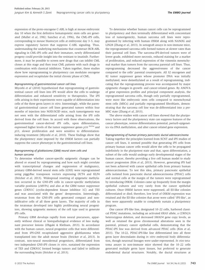
3 months resembled pancreatic intraepithelial neoplasias (PanIN),
regarded as a potential precursor to PDAC, and by 9 months the
cells progressed to the invasive stage. By contrast, an isogenic line
of iPS-like cells derived from the tumor margin gave rise to ductal
teratomas, but not PanINs or invasive ductal features. Thus, repro-
gramming of the PDAC created an experimental model in which
human PDAC progression could be studied in live cells.
A major cause for the lethality of PDAC is that there are no reli-
able biomarkers or clinical phenotypes of the early stages of the
disease. To discover potential biomarkers for early PDAC progres-
sion, PanIN lesions at 3 months were isolated from non-immune
mouse teratomas and cultured in serum-free medium as organoids
(Kim et al, 2013). After 6 days of culture, the medium was
harvested and subjected to mass spectroscopic analysis, in order to
discover protein markers that are secreted or released from early-
stage human PDAC. By subtracting the secreted and released
proteins of the organoids from such proteins of contralateral control
tissue and of the 10–22 cells grown under pluripotency conditions,
107 proteins specific to the PanIN-stage samples were discovered.
Of these, 64% of released or secreted proteins were seen in data-
bases of genes, proteins, and networks expressed within human
PanIN and PDAC cells, further validating the PanIN status of the
organoids derived from the 10–22 cell teratomas.
The proteins released or secreted from the PanIN organoids fell
into three major networks, including interconnected networks for
TGF-b and integrin signaling, and for the transcription factor HNF4a.Notably, HNF4a is not or barely expressed in normal pancreatic
ductal cells, poorly expressed in the PanIN1 stage, but is activated
in PanIN2 and PanIN3 stages, invasive stages, and in early, well-
differentiated human pancreatic cancer (Kim et al, 2013). HNF4a
then decreases markedly and becomes undetectable in advanced or
undifferentiated PDAC. Thus, being able to reconstitute PDAC
progression with 10–22 cells revealed the activation of an HNF4a
network distinctive for the late PanIN stages and well-differentiated
PDAC; this network activated during PDAC progression would have
been missed in studies of late-stage tumor cells.
In conclusion, although handful of reports have demonstrated
the reprogramming of cancer by iPS technology and only a subset of
cancer cells are amenable to the process, common lessons can be
drawn. (1) The differentiation potential of the cancer cells can be
restored, at least in part. (2) Epigenetic states are altered markedly
during reprogramming to pluripotency or near-pluripotency. (3) The
reprogrammed cells exhibit a reduced aggressive cancer phenotype
in teratoma assays, which may be concomitant with suppression of
oncogenes from the original cancer and activation of tumor suppres-
sor genes. (4) The reprogrammed cells can re-acquire the cancer
phenotype when they differentiate into the lineage from which the
iPS line was derived. Indeed, the iPS or iPS-like reprogrammed
cancer cells retain a propensity to do so, compared to other lineages.
(5) The iPS-like reprogrammed cancer cells can be used to study the
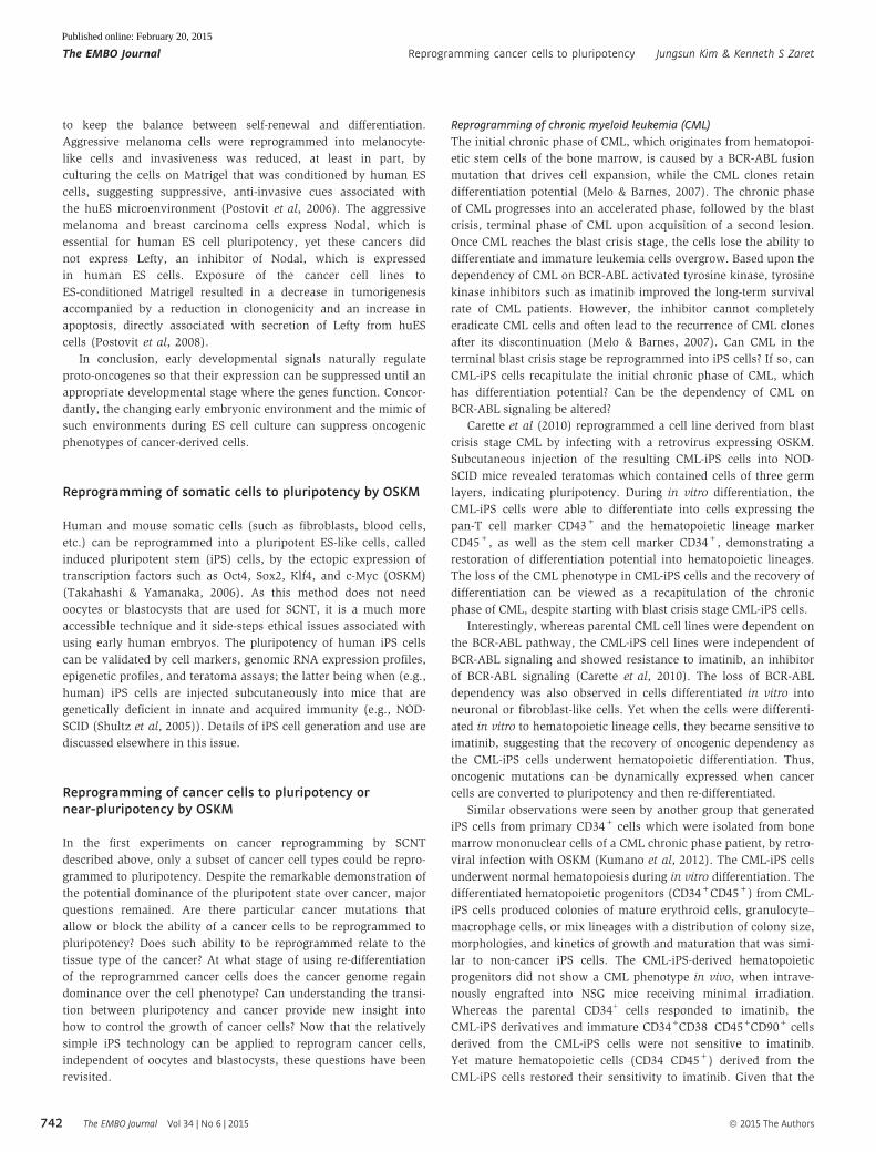
progression of the cancer phenotype (Fig 2).
Questions for the future
Many questions remain. How do the pluripotency factors and repro-
gramming process partially suppress the cancer phenotype? How
might such suppression be restricted to the lineage from which the
cancer iPS cells were derived? During the differentiation of cancer
iPS cells, how do the cells harboring the late-stage cancer genome
undergo disease progression, as opposed to immediately developing
the late-stage, parental cancer phenotype? What is the basis by
which cancer iPS lines are so difficult to generate? How can the
process be improved, to create more models for human cancer
progression? While definitive answers to these questions are not in
hand, hints about underlying mechanisms are arising in different
areas, as discussed next.
Control of cancer versus pluripotency
As for SCNT, iPS technology was able to reprogram a subset of
cancer cells to the pluripotent or near-pluripotent state and restore
differentiation potential to the cells. Pluripotent ES cells and iPS
cells have epigenetic features that may be reciprocal to those in
cancer cells (Suva et al, 2013). For instance, comprehensive high-
throughput relative methylation analysis demonstrated the existence
of two independent epigenetic mechanisms for cell reprogramming
and tumorigenesis. Hypomethylated, ‘differentially methylated
regions’ (DMRs) that can occur in iPS reprogramming were hyper-
methylated DMRs in colon cancer (Doi et al, 2009). Seventy-seven
percent of cancer-specific methylation variable positions (cMVPs)
were hypermethylated and 23% were hypomethylated in GBM
1 Obtainhumancancer cells
2 Reprogramwith OSKM
3 Generate induced pluripotent stem cell lines (iPS) 4 Induce differentiation
e.g. as teratomas in non-immune mice
5 Generates diverse cell types including cancerprogenitors
6 Study cancer progression:• Markers • Pathways • Therapeutics
Figure 2. Cancer cell reprogramming by iPS and recapitulation ofcancer progression.Scenario for developing iPS lines from human cancer cells (1–3), inducing theirdifferentiation to generate cancer progenitors (4–5), and using the cancerprogenitors to study features of cancer progression (6). By this means, late-stage human cancers can be reprogrammed to recapitulate early-stagedisease and progression for discovering markers, pathways, and therapeutics.
The EMBO Journal Vol 34 | No 6 | 2015 ª 2015 The Authors
The EMBO Journal Reprogramming cancer cells to pluripotency Jungsun Kim & Kenneth S Zaret
744
Published online: February 20, 2015
neural stem cell (GNS) (Stricker et al, 2013). More than 44% of
these cMVPs were reset during reprogramming of GNS to GNS-iPS
along with a majority of targets of the Polycomb complex (Stricker
et al, 2013). Furthermore, oncogenes and tumor suppressor genes
can become demethylated as a result of iPS reprogramming, which
is the opposite of the methylation of tumor suppressor genes seen
during cancer progression (Zhang et al, 2013). ATP-dependent SWI/
SNF complexes are considered to be tumor suppressors, since recur-
rent mutations of Brg1 subunits of the complex have been observed
in various human cancers (Wilson & Roberts, 2011), including
pancreatic cancer (Jones et al, 2008; Shain et al, 2012). The loss of
the Brg1 BAF complex promoted the formation of intraductal papil-
lary mucinous neoplasms and PDAC (von Figura et al, 2014). By
contrast, overexpression of Brg1 and Baf155 components of the
BAF complex, along with OSKM, enhances reprogramming to iPS
(Singhal et al, 2010). Together, these findings show that common
factors and epigenetic features can differentially elicit tumor forma-
tion or reprogramming to pluripotency.
While the coordinate, ectopic expression of OSKM induces repro-
gramming of somatic cells pluripotency (Takahashi & Yamanaka,
2006), activation of individual pluripotency factors can contribute to
tumorigenesis. For instance, the inducible expression of OCT4 in
mice initiated dysplasia by preventing the differentiation of multipo-
tent lineages (Hochedlinger et al, 2005). The ectopic expression of
OCT4 in human melanoma cells produced a more aggressive, cancer
stem cell-like melanoma (Kumar et al, 2012). Furthermore, whereas
reprogramming to pluripotency in the mouse can produce well-
differentiated teratomas (Abad et al, 2013), partial reprogramming
in mice yields tumors (Ohnishi et al, 2014). These findings indicate
that the pluripotency transcription factors are integrated into
networks that govern cancer phenotypes.
An unexplained feature of the use of cancer-derived iPS cells is
that the cells harbor the mutations of the late-stage cancer from
which the iPS cells were derived, but, as described above, the
cancer iPS cells can exhibit disease progression; that is, not simply
return to the late-stage phenotype from which they were derived. It
follows that the disease progression exhibited by the models may
not reflect that seen during the sequential accumulation of cancer
driver mutations as might occur in natural tumor development. Yet
in human PDAC progression, PanIN2 cells can harbor somatic muta-
tions required for PDAC development, but it can take years for the
cells to acquire metastatic activity (Yachida et al, 2010; Murphy
et al, 2013). Thus, even during natural PDAC tumor development,
epigenetic or extrinsic factors may be crucial for progression. The
attenuation of such factors may occur transiently during reprogram-
ming to pluripotency, and the release from pluripotency can thus
mirror the progression to the cancer phenotype seen in vivo.
Low efficiency of reprogramming cancer cellsto pluripotency
Cancer cells are reprogrammed very inefficiently and only subset of
cancers are amenable to reprogramming. Certain mutations might
make cancer cells refractory to reprogramming; that is, ‘non-
suppressible’ by pluripotency. One iPS line out of 8 derived from
PDAC contained the primary PDAC driver allele KRASG12D, even
though 78% of starting primary cancer epithelial cells contained
KRASG12D (Kim et al, 2013). Conceivably, secondary mutations
arising during iPS formation allowed the KRASG12D cells to be repro-
grammed. Furthermore, aneuploidy in cancer could cause the cellu-
lar stress responses such as activation of p53 through the stress
kinase p38 (Thompson & Compton, 2010), thus possibly impeding
reprogramming. PDAC epithelial cells from patients pre-treated with
radiation did not produce any iPS colonies, perhaps due to senes-
cence induced by irradiation and DNA damage (J.K. and K.S.Z,
unpublished observations). Given that many cancer patients are
treated with chemotherapy and irradiation prior to surgical resec-
tion, such treatments may prevent the creation of iPS lines.
The apparent preference for pluripotent cells to regenerate the
cancer type from which they were derived reflects the tendency of
iPS cell lines to preferentially differentiate into their lineages of
origin (Bar-Nur et al, 2011; Kim et al, 2011). This ‘deficiency’ in
exhibiting equal pluripotency for all cell lineages can be an advan-
tage in developing human cell models of cancer progression,
whereby the cancer iPS lines preferentially recapitulate stages of the
cancer type of interest. Thus, despite the difficulties and caveats in
generating human cancer iPS models, the examples covered in this
review provide new insights into disease progression. It is hoped
that a better understanding of how to create iPS cells from human
cancers, and epithelial cancers in particular, will provide more
opportunities to model and understand other types of solid tumors.
AcknowledgementsResearch on the project is supported by NIH grant R37GM36477 and funding
from the Abramson Cancer Center with Penn Medicine to K.S.Z.
Conflict of interestThe authors declare that they have no conflict of interest.
References
Abad M, Mosteiro L, Pantoja C, Canamero M, Rayon T, Ors I, Grana O, Megias
D, Dominguez O, Martinez D, Manzanares M, Ortega S, Serrano M (2013)
Reprogramming in vivo produces teratomas and iPS cells with totipotency
features. Nature 502: 340 – 345
Ablain J, Zon LI (2013) Of fish and men: using zebrafish to fight human
diseases. Trends Cell Biol 23: 584 – 586
Adamson ED (1987) Oncogenes in development. Development 99: 449 – 471
Arteaga CL, Adamson PC, Engelman JA, Foti M, Gaynor RB, Hilsenbeck SG,
Limburg PJ, Lowe SW, Mardis ER, Ramsey S, Rebbeck TR, Richardson AL,
Rubin EH, Weiner GJ, Sweeney SM, Honey K, Bachen J, Driscoll P, Hobin J,
Ingram J et al (2014) AACR cancer progress report 2014. Clin Cancer Res
20: S1 – S112
Bar-Nur O, Russ HA, Efrat S, Benvenisty N (2011) Epigenetic memory and
preferential lineage-specific differentiation in induced pluripotent stem
cells derived from human pancreatic islet Beta cells. Cell Stem Cell 9:
17 – 23
Begley CG, Ellis LM (2012) Drug development: raise standards for preclinical
cancer research. Nature 483: 531 – 533
Blelloch RH, Hochedlinger K, Yamada Y, Brennan C, Kim M, Mintz B, Chin L,
Jaenisch R (2004) Nuclear cloning of embryonal carcinoma cells. Proc Natl
Acad Sci USA 101: 13985 – 13990
Bracken AP, Kleine-Kohlbrecher D, Dietrich N, Pasini D, Gargiulo G, Beekman
C, Theilgaard-Monch K, Minucci S, Porse BT, Marine JC, Hansen KH, Helin
ª 2015 The Authors The EMBO Journal Vol 34 | No 6 | 2015
Jungsun Kim & Kenneth S Zaret Reprogramming cancer cells to pluripotency The EMBO Journal
745
Published online: February 20, 2015

K (2007) The Polycomb group proteins bind throughout the INK4A-ARF
locus and are disassociated in senescent cells. Genes Dev 21: 525 – 530
Braun AC (1959) A demonstration of the recovery of the crown-gall tumor
cell with the use of complex tumors of single-cell origin. Proc Natl Acad
Sci USA 45: 932 – 938
Caldas C, Hahn SA, da Costa LT, Redston MS, Schutte M, Seymour AB,
Weinstein CL, Hruban RH, Yeo CJ, Kern SE (1994) Frequent somatic
mutations and homozygous deletions of the p16 (MTS1) gene in
pancreatic adenocarcinoma. Nat Genet 8: 27 – 32
Cantley LC, Dalton WS, DuBois RN, Finn OJ, Futreal PA, Golub TR, Hait WN,
Lozano G, Maris JM, Nelson WG, Sawyers CL, Schreiber SL, Spitz MR, Steeg
PS (2012) AACR cancer progress report 2012. Clin Cancer Res 18: S1 – S100
Carette JE, Pruszak J, Varadarajan M, Blomen VA, Gokhale S, Camargo FD,
Wernig M, Jaenisch R, Brummelkamp TR (2010) Generation of iPSCs from
cultured human malignant cells. Blood 115: 4039 – 4042
Chin L, Tam A, Pomerantz J, Wong M, Holash J, Bardeesy N, Shen Q, O’Hagan
R, Pantginis J, Zhou H, Horner JW 2nd, Cordon-Cardo C, Yancopoulos GD,
DePinho RA (1999) Essential role for oncogenic RAS in tumour
maintenance. Nature 400: 468 – 472
Dawson MA, Kouzarides T (2012) Cancer epigenetics: from mechanism to
therapy. Cell 150: 12 – 27
DePinho RA (2000) The age of cancer. Nature 408: 248 – 254
Doi A, Park IH, Wen B, Murakami P, Aryee MJ, Irizarry R, Herb B, Ladd-Acosta
C, Rho J, Loewer S, Miller J, Schlaeger T, Daley GQ, Feinberg AP (2009)
Differential methylation of tissue- and cancer-specific CpG island shores
distinguishes human induced pluripotent stem cells, embryonic stem cells
and fibroblasts. Nat Genet 41: 1350 – 1353
Esteller M (2007) Cancer epigenomics: DNA methylomes and histone-
modification maps. Nat Rev Genet 8: 286 – 298
von Figura G, Fukuda A, Roy N, Liku ME, Morris Iv JP, Kim GE, Russ HA, Firpo
MA, Mulvihill SJ, Dawson DW, Ferrer J, Mueller WF, Busch A, Hertel KJ,
Hebrok M (2014) The chromatin regulator Brg1 suppresses formation of
intraductal papillary mucinous neoplasm and pancreatic ductal
adenocarcinoma. Nat Cell Biol 16: 255 – 267
Gao D, Vela I, Sboner A, Iaquinta PJ, Karthaus WR, Gopalan A, Dowling C,
Wanjala JN, Undvall EA, Arora VK, Wongvipat J, Kossai M, Ramazanoglu S,
Barboza LP, Di W, Cao Z, Zhang QF, Sirota I, Ran L, MacDonald TY et al
(2014) Organoid cultures derived from patients with advanced prostate
cancer. Cell 159: 176 – 187
Gurdon JB, Elsdale TR, Fischberg M (1958) Sexually mature individuals of
Xenopus laevis from the transplantation of single somatic nuclei. Nature
182: 64 – 65
Hochedlinger K, Blelloch R, Brennan C, Yamada Y, Kim M, Chin L, Jaenisch R
(2004) Reprogramming of a melanoma genome by nuclear
transplantation. Genes Dev 18: 1875 – 1885
Hochedlinger K, Yamada Y, Beard C, Jaenisch R (2005) Ectopic expression of
Oct-4 blocks progenitor-cell differentiation and causes dysplasia in
epithelial tissues. Cell 121: 465 – 477
Ishizawa K, Rasheed ZA, Karisch R, Wang Q, Kowalski J, Susky E, Pereira K,
Karamboulas C, Moghal N, Rajeshkumar NV, Hidalgo M, Tsao M, Ailles L,
Waddell TK, Maitra A, Neel BG, Matsui W (2010) Tumor-initiating cells are
rare in many human tumors. Cell Stem Cell 7: 279 – 282
Jones S, Zhang X, Parsons DW, Lin JC, Leary RJ, Angenendt P, Mankoo P,
Carter H, Kamiyama H, Jimeno A, Hong SM, Fu B, Lin MT,
Calhoun ES, Kamiyama M, Walter K, Nikolskaya T, Nikolsky Y,
Hartigan J, Smith DR et al (2008) Core signaling pathways in human
pancreatic cancers revealed by global genomic analyses. Science 321:
1801 – 1806
Kadoch C, Hargreaves DC, Hodges C, Elias L, Ho L, Ranish J, Crabtree GR
(2013) Proteomic and bioinformatic analysis of mammalian SWI/SNF
complexes identifies extensive roles in human malignancy. Nat Genet 45:
592 – 601
Kim J, Hoffman JP, Alpaugh RK, Rhim AD, Reichert M, Stanger BZ, Furth EE,
Sepulveda AR, Yuan CX, Won KJ, Donahue G, Sands J, Gumbs AA, Zaret KS
(2013) An iPSC line from human pancreatic ductal adenocarcinoma
undergoes early to invasive stages of pancreatic cancer progression. Cell
Rep 3: 2088 – 2099
Kim K, Zhao R, Doi A, Ng K, Unternaehrer J, Cahan P, Huo H, Loh YH, Aryee
MJ, Lensch MW, Li H, Collins JJ, Feinberg AP, Daley GQ (2011) Donor cell
type can influence the epigenome and differentiation potential of human
induced pluripotent stem cells. Nat Biotechnol 29: 1117 – 1119
Kumano K, Arai S, Hosoi M, Taoka K, Takayama N, Otsu M, Nagae G, Ueda K,
Nakazaki K, Kamikubo Y, Eto K, Aburatani H, Nakauchi H, Kurokawa M
(2012) Generation of induced pluripotent stem cells from primary chronic
myelogenous leukemia patient samples. Blood 119: 6234 – 6242
Kumar SM, Liu S, Lu H, Zhang H, Zhang PJ, Gimotty PA, Guerra M, Guo W, Xu
X (2012) Acquired cancer stem cell phenotypes through Oct4-mediated
dedifferentiation. Oncogene 31: 4898 – 4911
Ledford H (2011) Translational research: 4 ways to fix the clinical trial.
Nature 477: 526 – 528
Li L, Connelly MC, Wetmore C, Curran T, Morgan JI (2003) Mouse embryos
cloned from brain tumors. Cancer Res 63: 2733 – 2736
Li X, Nadauld L, Ootani A, Corney DC, Pai RK, Gevaert O, Cantrell MA, Rack
PG, Neal JT, Chan CW, Yeung T, Gong X, Yuan J, Wilhelmy J, Robine S,
Attardi LD, Plevritis SK, Hung KE, Chen CZ, Ji HP et al (2014) Oncogenic
transformation of diverse gastrointestinal tissues in primary organoid
culture. Nat Med 20: 769 – 777
Lin TP (1966) Microinjection of mouse eggs. Science 151: 333 – 337
Liu S, Leach SD (2011) Zebrafish models for cancer. Annu Rev Pathol 6: 71 – 93
McKinnell RG, Deggins BA, Labat DD (1969) Transplantation of pluripotential
nuclei from triploid frog tumors. Science 165: 394 – 396
Melo JV, Barnes DJ (2007) Chronic myeloid leukaemia as a model of disease
evolution in human cancer. Nat Rev Cancer 7: 441 – 453
Mintz B, Illmensee K (1975) Normal genetically mosaic mice produced from
malignant teratocarcinoma cells. Proc Natl Acad Sci USA 72: 3585 – 3589
Miyoshi N, Ishii H, Nagai K, Hoshino H, Mimori K, Tanaka F, Nagano H,
Sekimoto M, Doki Y, Mori M (2010) Defined factors induce reprogramming
of gastrointestinal cancer cells. Proc Natl Acad Sci USA 107: 40 – 45
Muller R, Slamon DJ, Tremblay JM, Cline MJ, Verma IM (1982) Differential
expression of cellular oncogenes during pre- and postnatal development
of the mouse. Nature 299: 640 – 644
Murphy SJ, Hart SN, Lima JF, Kipp BR, Klebig M, Winters JL, Szabo C, Zhang L,
Eckloff BW, Petersen GM, Scherer SE, Gibbs RA, McWilliams RR, Vasmatzis
G, Couch FJ (2013) Genetic alterations associated with progression from
pancreatic intraepithelial neoplasia to invasive pancreatic tumor.
Gastroenterology 145: 1098 – 1109 e1091
Nardella C, Lunardi A, Patnaik A, Cantley LC, Pandolfi PP (2011) The APL
paradigm and the “co-clinical trial” project. Cancer Discov 1: 108 – 116
Nguyen LV, Vanner R, Dirks P, Eaves CJ (2012) Cancer stem cells: an evolving
concept. Nat Rev Cancer 12: 133 – 143
Nigro JM, Baker SJ, Preisinger AC, Jessup JM, Hostetter R, Cleary K, Bigner SH,
Davidson N, Baylin S, Devilee P, Glover T, Collins FS, Weslon A, Modali R,
Harris CC, Vogelstein B (1989) Mutations in the p53 gene occur in diverse
human tumour types. Nature 342: 705 – 708
Ohgaki H, Kleihues P (2013) The definition of primary and secondary
glioblastoma. Clin Cancer Res 19: 764 – 772
The EMBO Journal Vol 34 | No 6 | 2015 ª 2015 The Authors
The EMBO Journal Reprogramming cancer cells to pluripotency Jungsun Kim & Kenneth S Zaret
746
Published online: February 20, 2015

Ohnishi K, Semi K, Yamamoto T, Shimizu M, Tanaka A, Mitsunaga K, Okita K,
Osafune K, Arioka Y, Maeda T, Soejima H, Moriwaki H, Yamanaka S,
Woltjen K, Yamada Y (2014) Premature termination of reprogramming in
vivo leads to cancer development through altered epigenetic regulation.
Cell 156: 663 – 677
Pachnis V, Mankoo B, Costantini F (1993) Expression of the c-ret proto-
oncogene during mouse embryogenesis. Development 119: 1005 – 1017
Pfeifer-Ohlsson S, Goustin AS, Rydnert J, Wahlstrom T, Bjersing L, Stehelin D,
Ohlsson R (1984) Spatial and temporal pattern of cellular myc oncogene
expression in developing human placenta: implications for embryonic cell
proliferation. Cell 38: 585 – 596
Pfeifer-Ohlsson S, Rydnert J, Goustin AS, Larsson E, Betsholtz C, Ohlsson R
(1985) Cell-type-specific pattern of myc protooncogene expression in
developing human embryos. Proc Natl Acad Sci USA 82: 5050 – 5054
Postovit LM, Seftor EA, Seftor RE, Hendrix MJ (2006) A three-dimensional
model to study the epigenetic effects induced by the microenvironment of
human embryonic stem cells. Stem Cells 24: 501 – 505
Postovit LM, Margaryan NV, Seftor EA, Kirschmann DA, Lipavsky A, Wheaton
WW, Abbott DE, Seftor RE, Hendrix MJ (2008) Human embryonic stem cell
microenvironment suppresses the tumorigenic phenotype of aggressive
cancer cells. Proc Natl Acad Sci USA 105: 4329 – 4334
Ram O, Goren A, Amit I, Shoresh N, Yosef N, Ernst J, Kellis M, Gymrek M,
Issner R, Coyne M, Durham T, Zhang X, Donaghey J, Epstein CB, Regev A,
Bernstein BE (2011) Combinatorial patterning of chromatin regulators
uncovered by genome-wide location analysis in human cells. Cell 147:
1628 – 1639
Rangarajan A, Weinberg RA (2003) Opinion: comparative biology of mouse
versus human cells: modelling human cancer in mice. Nat Rev Cancer 3:
952 – 959
Rivlin N, Katz S, Doody M, Sheffer M, Horesh S, Molchadsky A, Koifman G,
Shetzer Y, Goldfinger N, Rotter V, Geiger T (2014) Rescue of embryonic
stem cells from cellular transformation by proteomic stabilization of
mutant p53 and conversion into WT conformation. Proc Natl Acad Sci USA
111: 7006 – 7011
Sabapathy K, Klemm M, Jaenisch R, Wagner EF (1997) Regulation of ES cell
differentiation by functional and conformational modulation of p53.
EMBO J 16: 6217 – 6229
Sanchez MJ, Holmes A, Miles C, Dzierzak E (1996) Characterization of the first
definitive hematopoietic stem cells in the AGM and liver of the mouse
embryo. Immunity 5: 513 – 525
Shain AH, Giacomini CP, Matsukuma K, Karikari CA, Bashyam MD, Hidalgo M,
Maitra A, Pollack JR (2012) Convergent structural alterations define
SWItch/Sucrose NonFermentable (SWI/SNF) chromatin remodeler as a
central tumor suppressive complex in pancreatic cancer. Proc Natl Acad
Sci USA 109: E252 – E259
Sharma SV, Haber DA, Settleman J (2010) Cell line-based platforms to
evaluate the therapeutic efficacy of candidate anticancer agents. Nat Rev
Cancer 10: 241 – 253
Shultz LD, Lyons BL, Burzenski LM, Gott B, Chen X, Chaleff S, Kotb M, Gillies
SD, King M, Mangada J, Greiner DL, Handgretinger R (2005) Human
lymphoid and myeloid cell development in NOD/LtSz-scid IL2R gamma
null mice engrafted with mobilized human hemopoietic stem cells.
J Immunol 174: 6477 – 6489
Singhal N, Graumann J, Wu G, Arauzo-Bravo MJ, Han DW, Greber B, Gentile L,
Mann M, Scholer HR (2010) Chromatin-remodeling components of the
BAF complex facilitate reprogramming. Cell 141: 943 – 955
Slamon DJ, Cline MJ (1984) Expression of cellular oncogenes during
embryonic and fetal development of the mouse. Proc Natl Acad Sci USA
81: 7141 – 7145
Stricker SH, Feber A, Engstrom PG, Caren H, Kurian KM, Takashima Y, Watts
C, Way M, Dirks P, Bertone P, Smith A, Beck S, Pollard SM (2013)
Widespread resetting of DNA methylation in glioblastoma-initiating cells
suppresses malignant cellular behavior in a lineage-dependent manner.
Genes Dev 27: 654 – 669
Suva ML, Riggi N, Bernstein BE (2013) Epigenetic reprogramming in cancer.
Science 339: 1567 – 1570
Takahashi K, Yamanaka S (2006) Induction of pluripotent stem cells from
mouse embryonic and adult fibroblast cultures by defined factors. Cell
126: 663 – 676
Tentler JJ, Tan AC, Weekes CD, Jimeno A, Leong S, Pitts TM, Arcaroli JJ,
Messersmith WA, Eckhardt SG (2012) Patient-derived tumour xenografts
as models for oncology drug development. Nat Rev Clin Oncol 9:
338 – 350
Thompson SL, Compton DA (2010) Proliferation of aneuploid human cells is
limited by a p53-dependent mechanism. J Cell Biol 188: 369 – 381
Tuveson DA, Jacks T (2002) Technologically advanced cancer modeling in
mice. Curr Opin Genet Dev 12: 105 – 110
Verbeek BS, Vroom TM, Adriaansen-Slot SS, Ottenhoff-Kalff AE, Geertzema JG,
Hennipman A, Rijksen G (1996) c-Src protein expression is increased in
human breast cancer. An immunohistochemical and biochemical analysis.
J Pathol 180: 383 – 388
Wang ZQ, Kiefer F, Urbanek P, Wagner EF (1997) Generation of completely
embryonic stem cell-derived mutant mice using tetraploid blastocyst
injection. Mech Dev 62: 137 – 145
Wang ZY, Chen Z (2008) Acute promyelocytic leukemia: from highly fatal to
highly curable. Blood 111: 2505 – 2515
Wilkinson DG, Bailes JA, McMahon AP (1987) Expression of the proto-
oncogene int-1 is restricted to specific neural cells in the developing
mouse embryo. Cell 50: 79 – 88
Wilson BG, Roberts CW (2011) SWI/SNF nucleosome remodellers and cancer.
Nat Rev Cancer 11: 481 – 492
Wu M, Jung L, Cooper AB, Fleet C, Chen L, Breault L, Clark K, Cai Z,
Vincent S, Bottega S, Shen Q, Richardson A, Bosenburg M, Naber SP,
DePinho RA, Kuperwasser C, Robinson MO (2009) Dissecting genetic
requirements of human breast tumorigenesis in a tissue transgenic
model of human breast cancer in mice. Proc Natl Acad Sci USA 106:
7022 – 7027
Yachida S, Jones S, Bozic I, Antal T, Leary R, Fu B, Kamiyama M,
Hruban RH, Eshleman JR, Nowak MA, Velculescu VE, Kinzler KW,
Vogelstein B, Iacobuzio-Donahue CA (2010) Distant metastasis occurs
late during the genetic evolution of pancreatic cancer. Nature 467:
1114 – 1117
Zhang X, Cruz FD, Terry M, Remotti F, Matushansky I (2013) Terminal
differentiation and loss of tumorigenicity of human cancers via
pluripotency-based reprogramming. Oncogene 32: 2249 – 2260, 2260
e2241–2221
ª 2015 The Authors The EMBO Journal Vol 34 | No 6 | 2015
Jungsun Kim & Kenneth S Zaret Reprogramming cancer cells to pluripotency The EMBO Journal
747
Published online: February 20, 2015