Embed Size (px)
Citation preview
REPRODUCTIVE BIOLOGY OFTHE SPOTTED SEATROUT, CYNOSCION NEBULOSUS, IN SOUTH TEXASl
NANCY BROWN-PETERSON,2 PETER THOMAS,3 AND CONNIE R. ARNOLOS
ABSTRACT
The spotted seatrout, CY1lO8Cion nebulosus, spawns from April through the end of September inshallow bays in South Texas. All females were sexually mature at 300 mm SL and males at 200 mmSL. Histological examination of the testes showed that spermatogenesis began in January and continued until September, although spermatozoa remained in the testes until November. Spermatogenesiswas more active in the peripheral lobules of the testes than in the central lobules during the latterhalf of the spawning season. The sequence and timing of final oocyte maturation (FOMJ was investigated for the first time in this species. Lipid coalescence began at dawn and germinal vesicle migration started by midmorning. By early afternoon, oocytes were hydrated and spawning occurred atdusk.
Evidence of multiple spawning was examined. Morphological and histological data showed thatoocytes were continually recruited from March through the end ofSeptember, and the percentages ofvitellogenic and fully yolked oocytes did not decline during the spawning season. An average of only15.5% ofthe vitellogenic oocytes underwent FOM and hydration during a single spawn. Postovulatoryfollicles were found in many fish with mature ovaries throughout the reproductive season. Laboratorystudies showed that this species is capable of repeated spawns. Batch fecundity was best predicted byovary-free body weight of the fish and averaged 451 ± 43 eggslg ovary-free body weight. Estimatesof spawning frequency ranged from every other day to once every three weeks.
The spotted seatrout, Cynoscion nebulosus, supports important commercial and recreationalfisheries throughout its range (Chesapeake Bay,Virginia to Tampico, Mexico; Tabb 1966).Whereas aspects of the reproductive biology ofthis sciaenid species have been documentedthroughout its range (i.e., Chesapeake Bay:Brown 1981; Georgia: Mahood 1975; Florida:Moody 1950, Klima and Tabb 1959, Tabb 1961;Mississippi: Overstreet 1983; Louisiana: Heinand Shepard 1979; Texas: Pearson 1929, Miles1950, 1951), a comprehensive study of reproduction in spotted seatrout does not exist for anyarea. An extensive knowledge of the reproductivelife history of a species is necessary to understandcertain aspects of its reproductive physiology andendocrinology. Most previous studies have concentrated on the size at sexual maturity and theextended spawning season of this species (Pearson 1929; Moody 1950; Tabb 1961; Mahood 1975).A protracted spawning season is generally char-
1Contribution No. 696, University ofTexas at Austin, MarineScience Institute, Port Aransas, TX 78373.
2Marine Science Institute, University of 1'exas at Austin,Port Aransas, TX; present address: Florida Department ofNatural Resources, Indian River Lagoon Aquatic Preserves, 4842 S.U.S. Highway I, Fort Pierce, FL 34982.
3Marine Science Institute, University of Texas at Austin,Port Aransas, TX 78373.
Manuscript accepted February 1988.FISHERY BULLETIN: VOL. 86, NO.2, 1988.
acteristic of multiple spawners (Nikolskii 1969).In addition, Overstreet's (1983) histological datasuggest that C. nebulosus may be a multiplespawner. However, the possibility that spottedseatrout are multiple spawners has not been thoroughly discussed in the literature, and previousestimates of fecundity (Sundararaj and Suttkus1962; Overstreet 1983> have not considered themultiple spawning nature ofC. nebulosus or haveincluded estimates of batch fecundity.
In the present study, the reproductive biologyof C. nebulosus in South Texas was investigated,and particular attention was given to evidence formultiple spawning and estimates of batch fecundity and spawning frequency. The spawning season, spawning sites, time of spawning, and percentage of running ripe fish were documented, Inaddition, the temporal pattern of final oocytematuration was determined. The histological appearance ofthe gonads was examined, and particular attention was given to the presence of postovulatory follicles in ovarian tissue, an indicatorof multiple spawning in the northern anchovy,Engraulis mordax (Hunter and Goldberg 1980).The size-frequency distribution of vitellogenicoocytes was examined, since this can indicatemultiple spawning (deVlaming 1983). Batch fecundity was determined and spawning frequency
373

was estimated from both field-caught and laboratory fish.
MATERIALS AND MEmODS
Collection of Samples
Spotted seatrout were collected in Redfish Bayor Lydia Ann Channel near Port Aransas, TX,U.S.A. at depths of 1-3 m (Fig. 1). Fish were captured using a 300 m gill net (82 mm stretch) or a300 m trammel net (outer panels, 178 mmstretch; inner panel, 89 mm stretch) over shallowbeds of turtle grass, Thalassia testudinum orshoal grass, Halodule wrightii, bordered by a 1-2m drop-off into a channel. Fish <300 mm SL werecaptured with hook and Hne in 1-2 m of water.
Samples were collected weekly or twicemonthly from March 1982 through early May1985. No samples were taken in November andDecember 1983, October and December 1984, andFebruary 1985. During 1982, samples were collected at dusk only; in 1983, 1984, and 1985, samples from dawn, midday, and midnight were alsotaken. During each sampling period, salinity and
FISHERY BULLETIN: VOL. 86, NO.2
temperature were recorded as well as the time ofcapture.
Analysis of Fish and Gonads
Total length (TL) and standard length (SLlwere measured to the nearest mm for each specimen and total body weight (WT) was determinedto the nearest 10 g. Gonads were removed andweighed to the nearest 0.1 g (gonad weight, GW)and the gonadosomatic index (GSIJ was calculated, using the formula: GSI = (GW/WT) x 100.
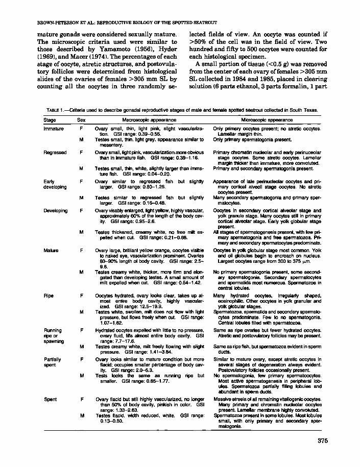
Reproductive stage of the gonads was assessedmacroscopically using the criteria in Table 1. Themacroscopic criteria used were similar to thoseused by Overstreet (1983) and Macer (1974). Asmall portion of tissue was removed from theanterior or midsection ofone gonad from each fishand preserved in Davidson's fixative for histological analysis (Jones 1966). Tissues were dehydrated and embedded in paraffin. Seven micronsections were cut and stained with Harris' hematoxylin and counterstained with eosin. Reproductive stage of each sample was assessed microscopically using the criteria in Table 1. Fish with
Gulf of Mexico1--1
1 km
FIGURE I.-Location of spotted seatrout sampling sites in South Texas. Asterisks denote sampling sites.
374
BROWN-PETERSON ET AL.: REPRODUCTIVE BIOLOGY OF THE SPOTTED SEATROUT
mature gonads were considered sexually mature.The microscopic criteria used were similar tothose described by Yamamoto (1956), Hyder(1969), and Macer (1974). The percentages ofeachstage of oocyte, atretic structures, and postovulatory follicles were determined from histologicalslides of the ovaries of females >305 mm SL bycounting all the oocytes in three randomly se-
lected fields of view. An oocyte was counted if>50% of the cell was in the field of view. Twohundred and fifty to 500 oocytes were counted foreach histological specimen.
A small portion of tissue «0.5 g) was removedfrom the center ofeach ovary offemales >305 mmSL collected in 1984 and 1985, placed in clearingsolution (6 parts ethanol, 3 parts formalin, 1 part
TABLE 1.-Criteria used to describe gonadal reproductive stages of male and female spotted seatrout collected in South Texas.
Stage
Immature
Regressed
Earlydeveloping
Developing
Mature
Ripe
Runningripe orspawning
Partiallyspent
Spent
Sex
F
M
F
M
F
M
F
M
F
M
F
M
F
M
F
M
F
M
Macroscopic appearance
Ovary small, thin, light pink, slight vascularization. GSI range: 0.39-0.55.
Testes small, thin, light grey, appearance similar tomesentery.
Ovary small, light pink, vascularization more obviousthan in immature fish. GSI range: 0.39-1.16.
Testes small, thin, white, slightly larger than immature fish. GSI range: 0.04-0.20.
Ovary similar to regressed fish but slighdylarger. GSI range: 0.80-1.26.
Testes similar to regressed fish but slightlylarger. GSI range: 0.16-0.48.
Ovary visably enlarged, light yellow, highly vascular,approximately 60% of the length of the body cavity. GSI range: 0.95-2.6.
Testes thickened, creamy white, no free milt expelled when cut. GSI range: 0.21-0.68.
Ovary large, brilliant yellow orange, oocytes visibleto naked eye, vascularization prominent. Ovaries80-90% length of body cavity. GSI range: 2.59.6.
Testes creamy white, thicker, more firm and elongated than developing testes. A small amount ofmilt expelled when cut. GSI range: 0.54-1.42.
Oocytes hydrated, ovary looks clear, takes up almost entire body cavity, highly vascularized. GSI range: 12.5-19.9.
Testes white, swollen, milt does not flow with lightpressure, but flows freely when cut. GSI range:1.07-1.62.
Hydrated oocytes expelled with little to no pressure,ovary fluid, fills almost entire body cavity. GSIrange: 7.7-17.6.
Testes creamy white, milt freely flowing with slightpressure. GSI range: 1.41-3.84.
Ovary looks similar to mature condition but moref1acid; occupies smaller percentage of body cavity. GSI range: 2.0-5.3.
Tests looks the same as running ripe butsmaller. GSI range: 0.85-1.n.
Ovary f1acid but still highly vascularized, no longerthan 50% of body cavity, pinkish in color. GSIrange: 1.33-2.63.
Testes flacid, width reduced, White. GSI range:0.13-0.80.
Microscopic appearance
Only primary oocytes present; no atretic oocytes.Lamellar margin thin.
Only primary spermatogonia present.
Primary chromatin nucleolar and early perinuceolarstage oocytes. Some atretic oocytes. Lamellarmargin thicker than immature, more convolutad.
Primary and secondary spermatogonia present.
Appearance of late perinucleolar oocytes and primary cortical alveoli stage oocytes. No atreticoocytes present.
Many secondary spermatogonia and primary spermatocytes.
Oocytes in secondary cortical alveolar stage andyolk granule stage. Many oocytes still in primarycortical alveolar stage. Early yolk globular stagepresent.
All stages of spermatogenesis present, with few primary spermatogonia and free spermatozoa. Primary and secondary spermatocytes predominata.
Oocytes in yolk globular stage most common. Yolkand oil globules begin to encroach on nucleus.Largest oocytes range from 300 to 375 ....m.
No primary spermatogonia present, some secondary spermatogonia. Secondary spermatocytesand spermatids most numerous. Spermatozoa incentral lobules.
Many hydrated oocytes, irregularly shaped,eosinophilic. Other oocytes in yolk granular andyolk globular stages.
Spermatozoa, spermatids and secondary spermatocytes predominate. Few to no spermatogonia.Central lobules filled with spermatozoa.
Same as ripe ovaries but fewer hydrated oocytes.Atretic and postovulatory follicles may be present.
Same as ripe fish, but spermatozoa evident in spermducts.
Similar to mature ovary, except atretic oocytes inseveral stages of degeneration always evident.Postovulatory follicles occasionally present.
No spermatogonia, few primary spermatoocytes.Most active spermatogenesis in peripheral lobules. Spermatozoa parlially filling lobules andabundant in sperm ducts.
Massive atresia of all remaining vitellogenic oocytes.Many primary and chromatin nucleolar oocytespresent. Lamellar membrane highly convoluted.
Spermatozoa present in some lobules. Most lobulessmall, with only primary and secondary spermatogonia.
375
glacial acetic acid), and vigorously shaken for 30seconds. Within a few minutes the cytoplasmcleared and the germinal vesicle could be easilyobserved microscopically. Ovarian fragmentswere taken from females collected at the spawning site over a 24-h period, placed in clearing solution and then examined under low-power magnification to determine the stage of final oocytematuration.
Oocyte Size-Frequency Distributionsand Estimates of Batch Fecundity
To determine fecundity and the frequency distribution of oocyte diameters, a 2-15 g piece oftissue was removed from the midsection of theovaries of 57 fish and weighed to the nearest0.01 g. The tissues were placed in a modifiedGilson's solution lBagenal1966) for 3-12 monthsand periodically shaken to separate the oocytesfrom connective tissues. Ovaries containing hydrated oocytes were examined after three monthssince hydrated oocytes of spotted seatrout beganto disintegrate when left in Gilson's solution for alonger period of time.
The volumetric method was used to estimatefecundity <Bagenal and Braum 1971). The oocytesamples were suspended in 500-1,500 mL ofwater and three replicate 0.5 or 1 mL subsampleswere taken. All the oocytes >30 IJom werecounted, and those >80 IJom in diameter (thegrowing oocytes) in each sample were measuredto the nearest 15 IJom using an ocular micrometer.A total of 556-1,110 growing oocytes were measured in each sample. The number of restingoocytes (oocyte diameter 30-80 IJom) was determined by diluting the original oocyte suspension1:10, and counting three replicate subsamples.Altogether, the frequency distributions of oocytesfrom 48 fish were analyzed l3 in developing stage;9 in mature, spawning not imminent stage; 14 inmature, just prior to spawning stage; and 22 inrunning ripe stage). Fecundity was calculated following Macer's (1974) formula and expressed asrelative fecundity of number of eggs per gramovary-free body weight. Batch fecundity (BF) isdefined as all oocytes >350 IJom which wereundergoing final oocyte maturation that formed adistinct batch, and all hydrated oocytes. This definition of batch fecundity is in agreement withHunter and Macewicz's (1985) statement thatoocytes undergoing final oocyte maturation maybe included as hydrated oocytes when hydrationoccurs very rapidly.
376
FISHERY BULLETIN: VOL. 86, NO.2
Spawning of Fish in the Laboratory
Four female and two male spotted seatroutwere maintained in a 30,000 L recirculating system. The tank, filtration system and feedingregime of the fish has been described previously(Arnold et at 1976). The salinity ranged from 25to 30%0. Spawning was induced by increasing thetemperature and photoperiod from wintertimesettings of 13°C, 9L:15D to 26°C and 15L:9D(Arnold et at 1976). The filter boxes were checkeddaily for the presence of buoyant, newly fertilizedeggs.
Statistical Analysis
Simple linear regression, oneway analysis ofvariance, and analysis of covariance were computed for the data using SPSS packaged programs(SPSS 1981).
RESULTS
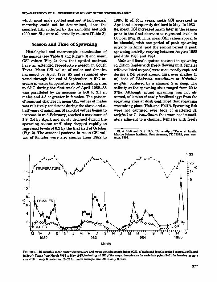
Size at Maturity
Some female spotted seatrout were sexuallymature after they reached 231 mm SL and >90%ofthe females had reached sexual maturity at 271mm SL (Table 2). By 300 mm SL, all female spotted seatrout were sexually mature. Fish 300 romSL or larger made up 85.4%, and immature fishcomprised 6.5%, of all the females sampled.
Male spotted seatrout reached sexual maturityat a much smaller size than females. The size at
TABLE 2.-Number and percentage of mature female and malespoiled seatrout by 10 mm size categories collected in SouthTexas, April 1982-May 1985. Maturity was judged by histologicaland macroscopic inspection.
Standard Female Malelength(mm) N % mature N % mature
201-210 5 0 5 100211-220 4 0 4 100221-230 4 0 1 100231-240 4 50 3 100241-250 6 83 3 67251-260 6 100 5 100261-270 5 80 7 100271-280 11 91 10 100281-290 14 100 11 100291-300 24 96 20 100301-310 60 100 40 100311-320 94 100 68 100321-330 115 100 80 100
>330 945 100 507 100
Total 1,297 764
BROWN-PETERSON ET AL.: REPRODUCTIVE BIOLOGY OF THE SPOTTED SEATROUT
which most male spotted seatrout attain sexualmaturity could not be determined, since thesmallest fish collected by the sampling methods(200 mm SL) were all sexually mature (Table 2).
Season and Time of Spawning
Histological and macroscopic examination ofthe gonads (see Table 3 and Figure 3) and meanGSI values (Fig. 2) show that spotted seatrouthave an extended reproductive season in SouthTexas. Mean OSI values of males and femalesincreased by April 1982-85 and remained elevated through the end of September. A 5°C increase in water temperature at the sampling sitesto 23°C during the first week of April 1982-85was paralleled by an increase in OSI to 2.1 inmales and 4.5 or greater in females. The patternof seasonal changes in mean OSI values of maleswas relatively consistent during the three-and-ahalfyears of sampling. Mean OSI values began toincrease in mid-February, reached a maximum of1.9-2.4 by April, and slowly declined during thespawning season until they dropped rapidly toregressed levels of 0.2 by the first half of October(Fig. 2). The seasonal patterns in mean OSI values of females were also similar from 1982 to
10
1981). In all four years, mean GSI increased inApril and subsequently declined in May. In 198284, mean OSI increased again later in the seasonprior to the final decrease to regressed levels inOctober (Fig. 2). Thus, mean OSI values appear tobe bimodal, with one period of peak spawningactivity in April, and the second period of peakspawning activity varying between August 1982and July 1983 and 1984.
Male and female spotted seatrout in spawningcondition (males with freely flowing milt, femaleswith ovulated oocytes) were consistently capturedduring a 2-h period around dusk over shallow (1m) beds of Thalassia testudinum or Halodulewrightii bordered by a channel 2 m deep. Thesalinity at the spawning sites ranged from 20 to37%0. Although actual spawning was not observed, collection ofnewly fertilized eggs from thespawning area at dusk confirmed that spawningwas taking place (Holt and Holt4). Spawning fishwere not captured over beds of scattered H.wrightii or T. testudinum that were not immediately adjacent to a channel. Females with freely
48. A. Holt and G. J. Holt, University of Texas at Austin,Marine Science Institute, Port Aransas, TX 78373, pers. com·mun.1983.
33
..~.. 25.. .. ·C.' ....
17
9
8en"6
4
2
Month
M J S1984
M M1985
FIGURE 2.-Bi.monthly mean water temperature and mean gonadosomatic index (GSII ofmale and female spotted seatrout collectedin South Texas from March 1982 to May 1985. including ±1 8E of the mean. Sample size for each data point: 3-61 for females (samplesize <15 in only 9 cases) and 2-52 for males (sample size <9 in only 9 cases).
377
flowing oocytes were only captured at dusk andmilt appeared to flow more freely in males at duskthan at other times of the day.
Gonadal Development in Males
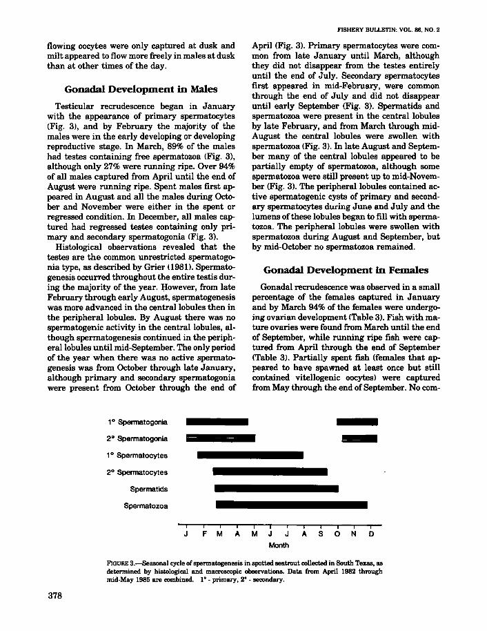
Testicular recrudescence began in Januarywith the appearance of primary spermatocytes(Fig. 3), and by February the majority of themales were in the early developing or developingreproductive stage. In March, 89% of the maleshad testes containing free spermatozoa (Fig. 3),although only 27% were running ripe. Over 94%of all males captured from April until the end ofAugust were running ripe. Spent males first appeared in August and all the males during October and November were either in the spent orregressed condition. In December, all males captured had regressed testes containing only primary and secondary spermatogonia (Fig. 3).
Histological observations revealed that thetestes are the common unrestricted spermatogonia type, as described by Grier (1981). Spermatogenesis occurred throughout the entire testis during the majority of the year. I:!owever, from lateFebruary through early August, spermatogenesiswas more advanced in the central lobules then inthe peripheral lobules. By August there was nospermatogenic activity in the central lobules, although spermatogenesis continued in the peripherallobules until mid-September. The only periodof the year when there was no active spermatogenesis was from October through late January,although primary and secondary spermatogoniawere present from October through the end of
10 Spermatogonia
2 0 Spermatogonia
10 Spermatocytes
20 Spermatocytes
Spermatids
Spermatozoa
FISHERY BULLETIN: VOL. 86. NO.2
April (Fig. 3). Primary spermatocytes were common from late January until March, althoughthey did not disappear from the testes entirelyuntil the end of July. Secondary spermatocytesfirst appeared in mid-February, were commonthrough the end of July and did not disappearuntil early September (Fig. 3). Spermatids andspermatozoa were present in the central lobulesby late February, and from March through midAugust the central lobules were swollen withspermatozoa (Fig. 3). In late August and September many of the central lobules appeared to bepartially empty of spermatozoa, although somespermatozoa were still present up to mid-November (Fig. 3). The peripheral lobules contained active spermatogenic cysts of primary and secondary spermatocytes during June and July and thelumens of these lobules began to fill with spermatozoa. The peripheral lobules were swollen withspermatozoa during August and September, butby mid-October no spermatozoa remained.
Gonadal Development in Females
Gonadal recrudescence was observed in a smallpercentage of the females captured in Januaryand by March 94% of the females were undergoing ovarian development (Table 3). Fish with mature ovaries were found from March until the endof September, while running ripe fish were captured from April through the end of September(Table 3). Partially spent fish (females that appeared to have spawned at least once but stillcontained vitellogenic oocytesJ were capturedfrom May through the end of September. No com-
iii IJ FM AM J J AS 0 NO
Month
FIGURE 3.-Seasonal cycle ofspermatogenesis in spotted seatrout collected in South Texas, asdetermined by histological and macroscopic observations. Data from April 1982 throughmid.May 1985 are combined. 1° - primary, 2° - secondary.
378
BROWN·PETERSON ET AL.: REPRODUCTIVE BIOLOGY OF THE SPOTTED SEATROUT
TABLE 3.-Percentage of female spotted seatrout in seven reproductive stages by month, as assessed by histological and macroscopic examination of the ovaries. Data from April 1982 to May1985 are combined. REG = Regressed, E DEV = Early Developing, DEV = Developing. MAT = Mature, RR = Ripe and RunningRipe, P SP = Partially Spent, SP = Spent.
Percent in each reproductive stageMonth N REG E DEV DEV MAT RR P SP SP
January 70 98 2February 51 55 43 2March 124 4 56 31 9April 372 4 6 78 12May 220 4 84 5 7June 104 1 56 19 24July 114 48 30 20 1August 100 58 32 7 3September 37 14 37 30 11 8October 8 87 13November 27 100December 51 98 2
Total 1,278
pletely spent fish were captured before July andfew were captured during the remainder of thereproductive season.
Histological observations offish with regressedovaries collected from late September to midFebruary showed only primary chromatin nucleolar and early perinucleolar oocytes. Atreticoocytes were present from September until midDecember; no atretic oocytes were observed frommid-December through the end of February. Theappearance oflate perinucleolar and primary cortical alveoli stage oocytes in late January, February, and early March represented the initialstages of ovarian recrudescence. Ovarian development was proceeding rapidly by early Marchand oocytes in the secondary cortical alveoli andyolk granule stages were common.
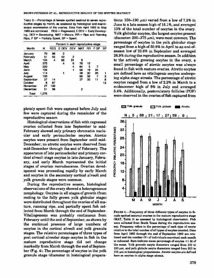
During the reproductive season, histologicalobservations of the ovary showed a heterogeneousmorphology. Oocytes in all stages of growth (fromresting to the fully grown yolk globular stage)were distributed throughout the ovaries ofall mature, running ripe, and partially spent fish collected from March through the end of September.Vitellogenesis was probably continuous fromFebruary until the end ofSeptember, as shown bythe continual presence of large numbers ofoocytes in the cortical alveoli and yolk granulestages. The relative percentages of three types ofpost cortical alveolar stage oocytes in fish in themature reproductive stage did not changemarkedly from March through the end ofSeptember (Fig. 41. The percentage ofoocytes in the yolkgranule stage (diameter in histological prepara-
tions: 150-190 f.l.m) varied from a low of 7.3% inJune to a late season high of 16.1%, and averaged13% of the total number of oocytes in the ovary.Yolk globular oocytes, the largest oocytes present(diameter: 200-375 f.l.m), were most common. Thepercentage of oocytes in the yolk globular stageranged from a high of 30.8% in April to an end-ofseason low of 22.6% in September and averaged26.9% during the reproductive season. In additionto the actively growing oocytes in the ovary, asmall percentage of atretic oocytes was alwaysfound in fish with mature ovaries. Atretic oocytesare defined here as vitellogenic oocytes undergoing alpha stage atresia. The percentage of atreticoocytes ranged from a low of 2.2% in March to amidsummer high of 8% in July and averaged5.4%. Additionally, postovulatory follicles (PDF)were observed in the ovaries offish captured from
rzzJ Yolk granule C Yolk globule • Atretic
N I 5 I 58 21 I 17 I 271 29 I 9 I
36
24
20
16 :
12
8
4 IiI I•M A M . ~ SMONTH
FIGURE 4.-Frequency of three different types of oocytes in female spotted seatrout ovaries in the mature reproductive stage(MAT, Table 3) as assessed by histological observation. Fishwere collected from South Texas during the reproductive season. Frequency refers to the percentage of each type of oocyterelative to the total number ofall types ofoocytes counted. Datafrom April 1982 through the end of September 1983 are combined and the number CN) of individuals examined each monthis indicated. Bars indicate mean percentage of oocytes ± 1 BE ofthe mean. Yolk granule oocyte diameters ranged from 150 to190 m, and yolk globule oocyte diameters ranged from 200 to375 m in histological preparations. Atretic oocytes are definedhere as oocytes in alpha stage atresia.
379
FISHERY BULLETIN: VOL. 86. NO.2
2
80 200 320 440 560 680
OOCYTE DIAMETER <Jim)
A
2
4
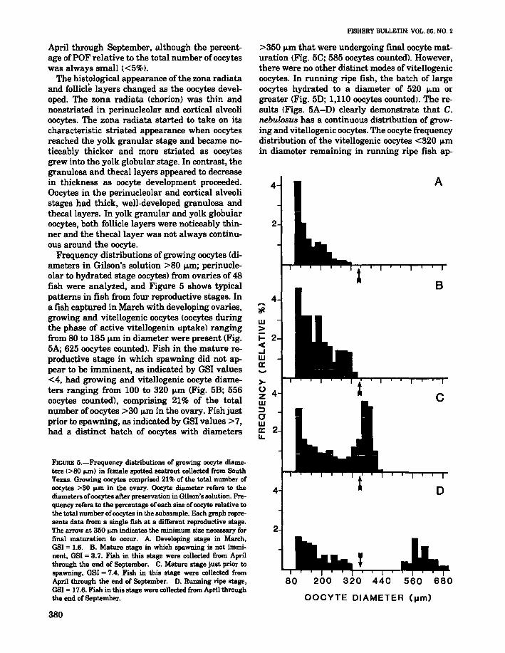
>350 jJ.m that were undergoing final oocyte maturation (Fig. 5C; 585 oocytes counted). However,there were no other distinct modes of vitellogenicoocytes. In running ripe fish, the batch of largeoocytes hydrated to a diameter of 520 jJ.m orgreater (Fig. 5D; 1,110 oocytes counted>. The results (Figs. 5A-D) clearly demonstrate that C.nebulosus has a continuous distribution of growing and vitellogenic oocytes. The oocyte frequencydistribution of the vitellogenic oocytes <320 jJ.min diameter remaining in running ripe fish ap-
4,..."#.w>I- 2<..JWc:....>-0 4zW:Jawc: 2u.
FIGURE 5.-Frequency distributions of growing oocyte diameters (>80 ...m) in female spotted seatrout collected from SouthTexas. Growing oocytes comprised 21% of the total number ofoocytes >30 ....m in the ovary. Oocyte diameter refers to thediameters ofoocytes after preservation in Gilson's solution. Frequency refers to the percentage of each size ofoocyte relative tothe total number ofoocytes in the subsample. Each graph represents data from a single fish at a different reproductive stage.The arrow at 350 ...m indicates the minimum size necessary forfinal maturation to occur. A. Developing stage in March.OSI = 1.6. B. Mature stage in which spawning is not imminent, OSI = 3.7. Fish in this stage were collected from Aprilthrough the end of September. C. Mature stage just prior tospawning. GSI = 7.4. Fish in this stage were collected fromApril through the end of September. D. Running ripe stage,OSI = 17.6. Fish in this stage were collected from April throughthe end of September.
April through September, although the percentage ofPOF relative to the total number ofoocyteswas always small «5%1.
The histological appearance of the zona radiataand follicle layers changed as the oocytes developed. The zona radiata (chorion) was thin andnonstriated in perinucleolar and cortical alveolioocytes. The zona radiata started to take on itscharacteristic striated appearance when oocytesreached the yolk granular stage and became noticeably thicker and more striated as oocytesgrew into the yolk globular stage. In contrast, thegranulosa and thecal layers appeared to decreasein thickness as oocyte development proceeded.Oocytes in the perinucleolar and cortical alveolistages had thick, well-developed granulosa andthecal layers. In yolk granular and yolk globularoocytes, both follicle layers were noticeably thinner and the thecal layer was not always continuous around the oocyte.
Frequency distributions of growing oocytes (diameters in Gilson's solution >80 jJ.m; perinucleolar to hydrated stage oocytes) from ovaries of 48fish were analyzed, and Figure 5 shows typicalpatterns in fish from four reproductive stages. Ina fish captured in March with developing ovaries,growing and vitellogenic oocytes (oocytes duringthe phase of active vitellogenin uptake) rangingfrom 80 to 185 jJ.m in diameter were present (Fig.5A; 625 oocytes counted). Fish in the mature reproductive stage in which spawning did not appear to be imminent, as indicated by GSI values<4, had growing and vitellogenic oocyte diameters ranging from 100 to 320 jJ.m (Fig. 5B; 556oocytes counted), comprising 21% of the totalnumber ofoocytes >30 jJ.m in the ovary. Fish justprior to spawning, as indicated by GSI values >7,had a distinct batch of oocytes with diameters
380
BROWN-PETERSON ET AL.: REPRODUCTIVE BIOLOGY OF THE SPOTTED SEATROUT
peared similar to the oocyte distribution in nonspawning fish (Fig. 5B, 0). The percentage (21%)of growing and vitellogenic oocytes in the ovaryremained constant throughout the reproductiveseason in fish in the mature and running ripestages.
first observed at dawn (0545), and GVM startedat 0900. By 1430, all fish undergoing final maturation had hydrated oocytes and ovulation andspawning commenced at dusk (1830) and continued until 2100. None of the fish collected from2100 to 0500 were undergoing FOM.
Batch Fecundity
TABLE 4.-Monthly mean batch fecundityexpressed as number eggslg ovary-freebody weight 01 spotted seatrout in SouthTexas. All means were not statistically different.
Spawning Frequency
To estimate the spawning frequency of spottedseatrout in South Texas, the percentage of running ripe females captured monthly from Aprilthrough the end of September in 1982 through1985 was examined. Only fish captured at duskand >305 mm SL were included in this analysis.The percentage of spawning females ranged from
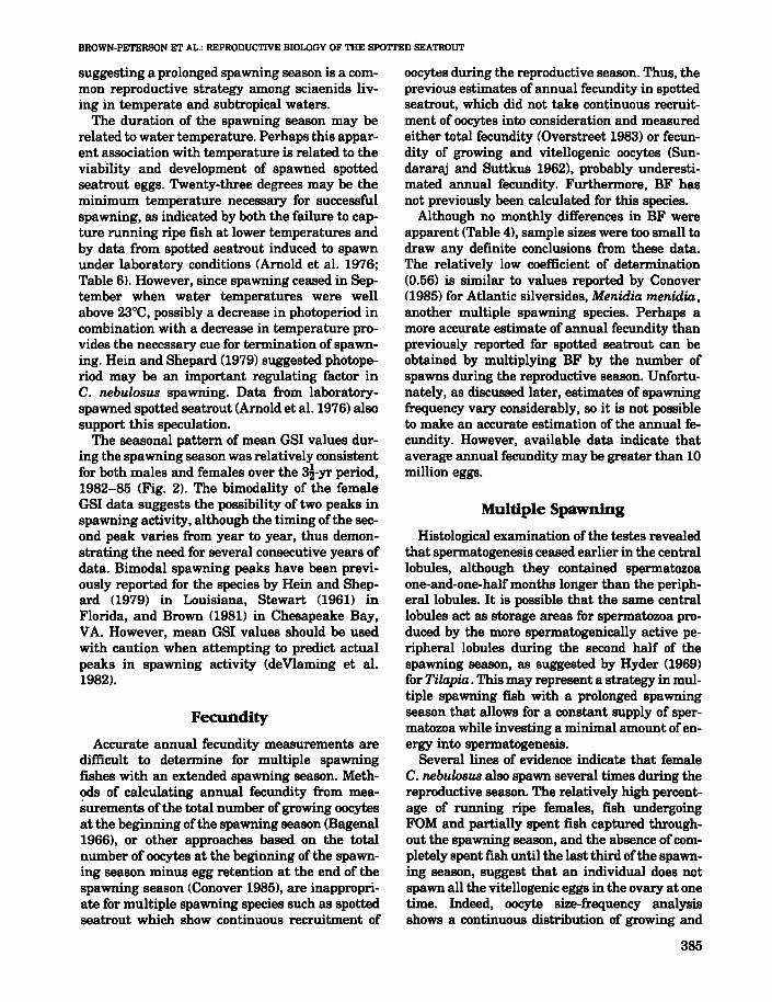
The significant positive relationship (J' <0.001) between BF and ovary-free body weightcan be best described by the following equation:BF = 459WT - 56,066, r2 = 0.56 (Fig. 8), whilecurvilinear equations best described the relationship between BF and SL and TL. The coefficientsof determination in all cases were <0.56.
A one-way analysis of variance showed thatmean BF (number of eggs per gram ovary-freeweight) did not vary significantly during theApril through September spawning season. Meanrelative batch fecundity was highest in September, lowest in May, and varied little during April,June, and July (Table 4).
A prominent batch of oocytes was present onlyin females that were in all stages of final oocytematuration or were running ripe (Figs. 5C, D; 7).The average batch size calculated from 14 fishcontaining hydrated oocytes and no postovulatoryfollicles was 451 ± 43 eggslg ovary-free bodyweight. This number averaged 15.5 ± 2.5% of thenumber of growing and vitellogenic oocytes in theovary.
N Mean fecundity ± 1 SE
19 477 ± 422 320± 723 435± 1095 409± 763 361 ± 603 560± 79
Month
AprilMayJuneJulyAugustSeptamber
5A 400 !Lm live oocyte equals a 350 !Lm oocyte preserved inGilson's solution. Both measurements represent oocytes beginningFOM.
Final Oocyte Maturation
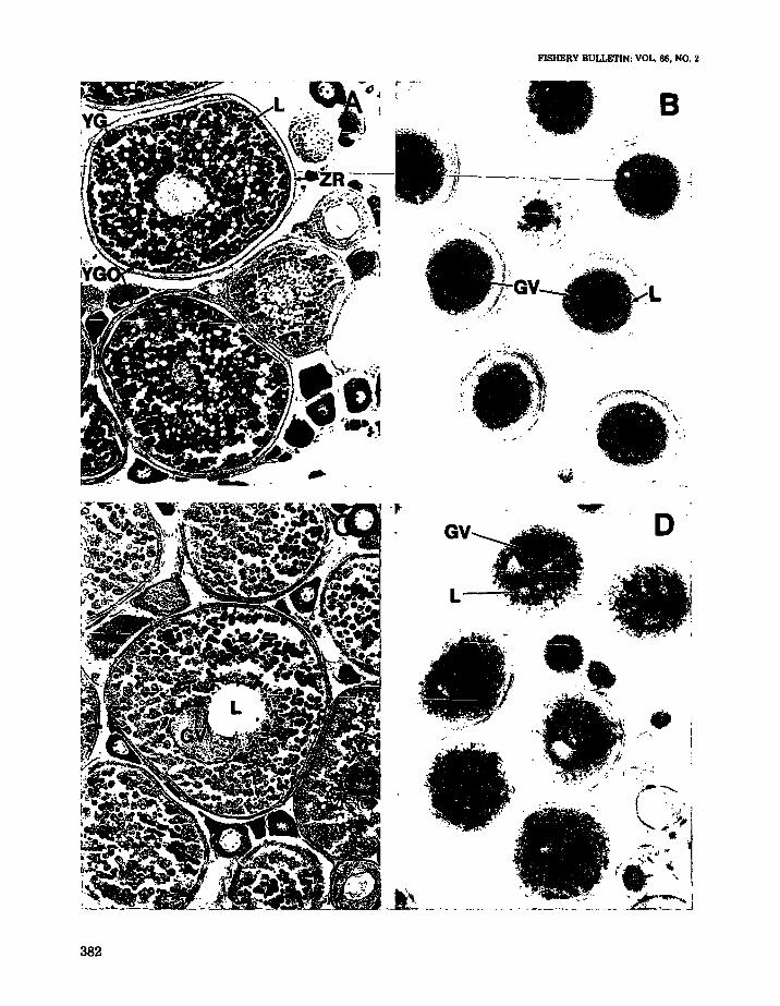
Final oocyte maturation (FOM) was highly synchronized in spotted seatrout and occurred only inoocytes >400 J.l.m 5. Figure 6A shows a photomicrograph of a histological section of a spottedseatrout ovary in the mature reproductive stagethat was not undergoing final oocyte maturation.Many oocytes were in the yolk globular stage andappeared to be fully grown. The first readily observable stage of FOM in "cleared" oocytes waslipid coalescence (Fig. 6B). The oil droplets in theoocytes began to coalesce around the germinalvesicle (nucleus) and subsequently formed one tothree large oil droplets. This stage was not alwaysobserved in histological preparations since manyof the oocytes were not sectioned through theircenters. The yolk globules remained discrete during lipid coalescence. After the lipids had coalesced, the germinal vesicle (GV) began to migrate to the periphery of the oocyte (germinalvesicle migration, or GVM). GVM could be seenin both histological sections (Fig. 6C) and in"cleared" oocytes (Fig. 6D). The oil droplet occupies the center of the oocytes shown in Figure 6C,D. Histological observation of this stage (Fig. 6C)showed that the yolk globules were not coalesced,the oil droplets had coalesced to form one or twolarge droplets and the GV had begun to lose itsintegrity and often appeared semicircular. At thecompletion of GVM, the nuclear (germinal vesicle) membrane broke down (GVBD) and the nuclear material intermingled with the cytoplasm ofthe oocyte. Hydration occurred shortly thereafter,followed by ovulation and spawning of the fullymature oocyte.
Final oocyte maturation occurred within 10hours in spotted seatrout in the natural environment (Fig. 7). A total of 209 fish were collectedover eight 24-h periods from April through August in 1984 and 1985. Forty-three percent of thefish collected between the hours of 0500 and 1500were undergoing FOM. Lipid coalescence was
381
FISHERY BULLETIN: VOL. 86, NO.2
,."•.~,,:.;.~"".~-';\.~..
. '
. ". ' ..
*i .
, _.
':0. .. '
'\)cGV-,:.' "":""'" 'L/". . .
. ." :'.... ."
".-" . -"
- . .--.' :"
D
•.........
;.-. -:----.( . : " I
\, .-;',\.;..... 1·
.~' ",
.'.--;.,' '"I .. '. I. \• ......_ I
... ......__.... __ ..~._. _....J
L
. .. ) ....
•....
... ' .
. ?
t~. ,)
"'. ..'.. .'. .' ....
.."
. --~~ -- . - - -.. -.
382
BROWN-PETERSON ET AL.: REPRODUCTIVE BIOLOGY OF THE SPO'ITED SEATROUT
FIGURE 6.-Photomicrographs of oocytes from spotted seatroutin the mature reproductive stage collected from SouthTexas. A. Histological section of an ovary in the mature stagethat is not undergoing final oocyte maturation IFOMI. Oocytedevelopment is continuous, with fully grown yolk globularoocytes co-occurring with oocytes in earlier stages of development (Magnification 120x). B. "Cleared" oocytes in the lipidcoalescence stage, the first stage of FOM (magnification 40X).C. Histological section ofan oocyte undergoing germinal vesiclemigration (GVM). (Magnification 160><). D. "Cleared" oocytesin the GVM stage (magnification 40x). Key: GV =germinalvesicle or nucleus. L = Lipid droplets, YG = yolk globules,YGO =yolk globular oocyte, ZR =Zona radiata.
Lipid Coalescence
GVM
Hydration
Ovulation and Spawning14L:l0D
0060 O~OO 08bo 12bo
Time
2000
FIGURE 7.-Time-course of final oocyte maturation in spotted seatrout collected fromSouth Texas. Lipid coalescence is the initial stage in final oocyte maturation. Dataobtained from 46 fish undergoing final oocyte maturation in April and May1985. GVM = germinal vesicle migration. 14L: 10D = hours of light and dark.
1750150012501000
Weight (g)
750
SF = 469 WT - 56,066
r 2 = .56
n = 32
••••• •
••
•• • •••
500
750
.......0 625...)(.....>.
500...=cc:::J0
~ 375..c:0a;al 250
125FIGURE 8.-Relation between batch fecundity (BF)and ovary-free body weight (WT) of spotted seatroutfrom April 1984 to May 1985. Thirty·three fish withoocytes >350 IJom that were undergoing final oocytematuration and formed a distinct batch of oocytes orhad hydrated oocytes are included.
383
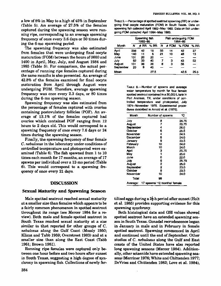
TABLE 6.-Number of spawns and averagewater temperature by month for four femalespotted seatrout contained in a 30,000 L tank inPort Aransas, TX, under conditions of controlled temperature and photoperiod, July1974-November 1975. Experimental procedures described in Amold at al. (1976).
26.7526.7526.025.524.524.023.524.024.025.7525.022.025.7525.023.023.524.0
Number of spawnsMonth
July 8August 7September 4~ober 6November 4December 8January 9February 10March 10April 3May 1June 0July 6August 2Se~ember 2~ober 9November 10
Total 99
Average: 17 spawns! 12 months! female
FISHERY BULLETIN: VOL. 86. NO.2
TABlE 5.-Percentage of spotted seatrout spawning (RR) or under-going final oocyte maturation (FOM) in South Texas. Data onspawning fish collected April 1982-May 1985. Data on fish under-going FOM collected April 1984-May 1985.
Spawning fish Fish undergoing FOM1982-85 1984-85
Month N # RR "IoRR N #FOM "IoFOM "IoRR
April 256 42 16 33 14 42 37May 140 11 8 21 10 48 13June 101 20 20July 82 33 40 7 3 43 52August 101 36 36 8 3 38September 20 9 45
Mean 27.5 42.8 25.5
tilized eggs during a 21-h period after sunset (Holtet al. 1985) provides supporting evidence for thisspawning synchrony.
Both histological data and GSI values showedspotted seatrout have an extended spawning season in South Texas. Gonadal recrudescence beganin January in male and in February in femalespotted seatrout. Spawning commenced in Apriland continued until the end of September. Otherstudies of C. nebulosus along the Gulf and Eastcoasts of the United States have also reportedlong spawning seasons (Mercer 1984). Additionally, other sciaenids have extended spawning seasons (Merriner 1976; White and Chittenden 1977;DeVries and Chittenden 1982; Love et al. 1984),
DISCUSSION
Sexual Maturity and Spawning Season
Male spotted seatrout reached sexual maturityat a smaller size than females which appears to bea fairly common phenomenon in spotted seatroutthroughout its range (see Mercer 1984 for a review). Both male and female spotted seatrout inSouth Texas reached sexual maturity at a sizesimilar to that reported for other groups of C.nebulosus along the Gulf Coast (Moody 1950;Klima and Tabb 1959; Overstreet 1983) and at asmaller size than along the East Coast (Tabb1961; Brown 1981).
Running ripe females were captured only between one hour before and two hours after sunsetin South Texas, suggesting a high degree of synchrony in spawning fish. Collections of newly fer-
a low of8% in May to a high of 45% in September(Table 5). An average of 27.5% of the femalescaptured during the spawning season were running ripe, corresponding to an average spawningfrequency of once every 3.6 days or 50 times during the 6-mo spawning period.
The spawning frequency was also estimatedfrom females that were undergoing final oocytematuration (FOM) between the hours of0600 and1400 in April, May, July, and August 1984 and1985 (Table 5). For comparison, the actual percentage of running ripe females captured duringthe same months is also presented. An average of42.8% of the females examined for final oocytematuration from April through August wereundergoing FOM. Therefore, average spawningfrequency was once every 2.3 days, or 80 timesduring the 6-mo spawning season.
Spawning frequency was also estimated fromthe percentage of females captured with ovariescontaining postovulatory follicles (POF). An average of 13.1% of the females captured hadovaries which contained POF ranging from 12hours to 2 days old. This would correspond to aspawning frequency of once every 7.6 days or 24times during the spawning season.
Finally, the spawning frequency of four femaleC. nebulosus in the laboratory under conditions ofcontrolled temperature and photoperiod were examined (Table 6). The fish spawned from 1 to 10times each month for 17 months, an average of 17spawns per individual over a 12-mo period (Table6). This would correspond to a spawning frequency of once every 21 days.
384
BROWN-PETERSON ET AL.: REPRODUCTIVE BIOLOGY OF THE SPOTTED SEATROUT
suggesting a prolonged spawning season is a common reproductive strategy among sciaenids living in temperate and subtropical waters.
The duration of the spawning season may berelated to water temperature. Perhaps this apparent association with temperature is related to theviability and development of spawned spottedseatrout eggs. Twenty-three degrees may be theminimum temperature necessary for successfulspawning, as indicated by both the failure to capture running ripe fish at lower temperatures andby data from spotted seatrout induced to spawnunder laboratory conditions (Arnold et al. 1976;Table 61. However, since spawning ceased in September when water temperatures were wellabove 23°C, possibly a decrease in photoperiod incombination with a decrease in temperature provides the necessary cue for termination of spawning. Hein and Shepard (1979) suggested photoperiod may be an important regulating factor inC. nebulosus spawning. Data from laboratoryspawned spotted seatrout (Arnold et al. 1976) alsosupport this speculation.
The seasonal pattern of mean aSI values during the spawning season was relatively consistentfor both males and females over the 3i-yr period,1982-85 (Fig. 2). The bimodality of the femaleGSI data suggests the possibility of two peaks inspawning activity, although the timing ofthe second peak varies from year to year, thus demonstrating the need for several consecutive years ofdata. Bimodal spawning peaks have been previously reported for the species by Hein and Shepard (1979) in Louisiana, Stewart (1961) inFlorida, and Brown (1981) in Chesapeake Bay,VA. However, mean aSI values should be usedwith caution when attempting to predict actualpeaks in spawning activity (deVlaming et al.1982).
Fecundity
Accurate annual fecundity measurements aredifficult to determine for multiple spawningfishes with an extended spawning season. Methods of calculating annual fecundity from measurements of the total number of growing oocytesat the beginning of the spawning season (Bagenal1966), or other approaches based on the totalnumber of oocytes at the beginning of the spawning season minus egg retention at the end of thespawning season (Conover 1985), are inappropriate for multiple spawning species such as spottedseatrout which show continuous recruitment of
oocy~sduring the reproductive season. Thus, theprevious estimates of annual fecundity in spottedseatrout, which did not take continuous recruitment of oocytes into consideration and measuredeither total fecundity (Overstreet 1983) or fecundity of growing and vitellogenic oocytes (Sundararaj and Suttkus 1962), probably underestimated annual fecundity. Furthermore, BF hasnot previously been calculated for this species.
Although no monthly differences in BF wereapparent (Table 4), sample sizes were too small todraw any definite conclusions from these data.The relatively low coefficient of determination(0.56) is similar to values reported by Conover(1985) for Atlantic silversides, Menidia menidia,another multiple spawning species. Perhaps amore accurate estimate of annual fecundity thanpreviously reported for spotted seatrout can beobtained by multiplying BF by the number ofspawns during the reproductive season. Unfortunately, as discussed later, estimates of spawningfrequency vary considerably, so it is not possibleto make an accurate estimation of the annual fecundity. However, available data indicate thataverage annual fecundity may be greater than 10million eggs.
Multiple Spawning
Histological examination of the testes revealedthat spermatogenesis ceased earlier in the centrallobules, although they contained spermatozoaone-and-one-half months longer than the peripheral lobules. It is possible that the same centrallobules act as storage areas for spermatozoa produced by the more spermatogenically active peripheral lobules during the second half of thespawning season, as suggested by Hyder (1969)for Tilapia. This may represent a strategy in multiple spawning fish with a prolonged spawningseason that allows for a constant supply of spermatozoa while investing a minimal amount of energy into spermatogenesis.
Several lines of evidence indicate that femaleC. nebulosus also spawn several times during thereproductive season. The relatively high percentage of running ripe females, fish undergoingFOM and partially spent fish captured throughout the spawning season, and the absence ofcompletely spent fish until the last third ofthe spawning season, suggest that an individual does notspawn all the vitellogenic eggs in the ovary at onetime. Indeed, oocyte size-frequency analysisshows a continuous distribution of growing and
385
vitellogenic oocytes (Fig. 5) and fecundity estimates show that only about 15% of the growingoocytes undergo FOM prior to a spawn. Histological data shows that the percentage ofvitellogenicoocytes in the ovary remains constant throughoutthe spawning season (Fig. 4), which suggests thatnew oocytes may be recruited into the vitellogenicphase as rapidly as mature oocytes are released.Convincing histological evidence of multiplespawning is the presence ofpostovulatory follicles(POF) from May through the end of September inovaries containing many vitellogenic oocytes.Hunter and Goldberg (1980) characterized postovulatory follicles in laboratory-spawned Engraulis mordax, a multiple spawning fish, andfound POF in all females that had spawned in thelaboratory one or two days previously. F'inally,laboratory studies also show that spotted seatroutare capable ofmultiple spawning under relativelyconstant environmental conditions (Table 6).Tucker and Faulkner (1987) also found that sixfemale fish kept in raceways outdoors at the ambient summer temperature and photoperiodspawned repeatedly.
Spawning Frequency
It is especially difficult to determine thespawning frequency of wide-ranging, multiplespawning marine fishes such as C. nebulosus thatare not group-synchronous spawners. Onemethod to estimate spawning frequency is tocount the number of distinct batches of vitellogenic oocytes in the ovary (Shackley and King19771. However, only one distinct batch of vitellogenic or hydrated oocytes can be distinguishedin spotted seatrout ovaries at anyone time (Fig.5) and the reliability of this method has beenquestioned (deVlaming 1983). Therefore, threetechniques were used to estimate spawning frequency in spotted seatrout.
Spawning frequency was estimated to be onceevery 3.6 days from the percentage of runningripe fish caught on the spawning grounds. Although this is probably an overestimate owing tosampling bias, the error may not be susbtantial,since the spawning grounds are also the feedinggrounds for this species (Moody 1950) and manynonspawning individuals were captured. Thetime of sample collection did not significantly influence the estimate of spawning frequency. Highspawning frequencies were also obtained (every2.3 days) when fish were captured 6-12 hoursprior to spawning and examined for signs of final
386
FISHERY BULLETIN: VOL. 86. NO.2
oocyte maturation (Table 5). The spawning frequency of other sciaenids fishes has been estimated by this technique (DeMartini and Fountain 1981; Love et aI. 1984). Additionally, Hunterand Macewicz (1985) suggested that this methodproduces a useful first approximation ofspawningfrequency.
The proportion of fish having POF in the ovaryhas also been used to determine spawning frequency (Hunter and Goldberg 1980; Alheit et aI.1984; Hunter and Macewicz 19851. Spottedseatrout were found to have POF throughout thespawning season, although the age of the POFwas often difficult to determine. Furthermore, detailed laboratory studies have not been undertaken to accurately age POF in spotted seatrout.However, the unce a week estimate of spawningfrequency obtained using thi8 method is similarto spawning frequencies reported for two othersciaenids, the queenfish, Seriphus politus, (DeMartini and Fountain 1981) and the whitecroaker, Genyonemus lineatus, (Love et aI. 1984).Spawing frequency estimates from POF are probably more reliable than estimates based on thenumber of spawning fish since sampling biasis less likely to occur when capturing fish withPOF.
Another method used to quantify spawning frequencies in various species is direct observationof spawning in the laboratory or in "controlled"field situations, such as impoundments (Gale andDeutsch 1985; Hubbs 1985; Heins and Rabito1986). Spotted seatrout spawned an average ofonce every three weeks per individual under controlled temperature and photoperiod in the laboratory (Table 6). Tucker and Faulkner (1987)found that six female spotted seatrout kept outdoors at ambient temperature and photoperiodaveraged one spawn per individual every 2.3weeks. The same spawning frequency was notedfor an individual female, although that same individual later spawned three times in four days(Tucker and Faulkner 1987). Thus, spottedseatrout appear to be capable of the high spawning frequencies estimated from field-caught fishwith hydrated ovaries, although this frequency isprobably not sustained throughout the entirespawning season. In general, the spawning frequencies in both laboratory studies are lowerthan those estimated from actual spawning fishin the field. However, it is unclear whether this isdue to an overestimation of spawning frequencyin the field or to a decline in spawning frequencyowing to confinement in the laboratory.
BROWN-PETERSON ET AL.: REPRODUCTIVE BIOLOGY OF THE SPOTTED SEATROUT
ACKNOWLEDGMENTS
We thank John Trant, John Gourley, JohnSmith, and Wayne Wofford for their help with thefield work and other aspects of the research.Funding was provided by grants from the SidRichardson Foundation and the Caesar KlebergFoundation.
LITERATURE CITED
ALHEIT. J., V. H. ALARON. AND B. J. MACEWICZ.1984. Spawning frequency and sex ratio in the peruvian
anchovy, Engraulis ringens. CaICOFI. Rep. 25:43-52.ARNOLD, C. R.. T. D. WILLIAMS, W. A. FABLE,JR.,J. L. LASSWELL.
AND W. H. BAILEY.
1976. Methods and techniques for spawning and rearingspotted seatrout IC.vnoscion nebul08us) in the laboratory.Proc. 30th Conf. Southeast. Assoc. Game Fish Comm.30:167-178.
BAGENAL. T. B.1966. The ecological and geographical aspects of the fe
cundity of the plaice. J. Mar. BioI. Assoc. U.K. 46:161186.
BAGENAL, T. 8., AND E. BRAUM.1971. Eggs and early life history. In W. E. Ricker (edi
tor), Methods for assessment of fish production in freshwaters. p 159-181. IBP lint. BioI. Prgrammel. Handb.3, 2d ed., Blackwell Sci. Publ. Oxford, England.
BROWN,N.J.1981. Reproductive biology and recreational fishery for
spotted seatrout, Cynoscion nebul08us, in the Chesapeake Bay area. MA Thesis, College of William andMary, Gloucester Point, VA, 120 p.
CONOVER, D. O.1985. Field and laboratory assessment of patterns in fe
cundity of a multiple spawning fish: The Atlantic silverside. Menidia menidia. Fish. BulL, US. 83:331-341.
DEMARTINI, E. E., AND R. K. FOUNTAIN.1981. Ovarian cycling frequency and batch fecundity in
the queenfish, Seriphus palitus: attributes representative of serial spawning fishes. Fish. Bull., U.S. 79:547560.
DEVLAMlNG, V.1983. Oocyte development patterns and hormonal in
volvements among teleosts. In J. C. Rankin. T. J.Pitcher, and R. T. Duggan (editors), Control processes infish physiology, p. 176-199. Wiley & Sons, N.Y.
DEVLAMING, V., G. GROSSMAN, AND F. CHAPMAN.1982. On the use of the gonosomatic index. Compo
Biochem. Physiol. 73A:31-39.DEVRIES, D. A., AND M. E. CHITTENDEN, JR.
1982. Spawning, age determination, longevity and mortality ofthe silver seatrout, Cyn08cion nothus, in the Gulfof Mexico. Fish. BulL, U.S. 80:487-500.
GALE, W. F., AND W. G. DEUTSCH.1985. Fecundity and spawning frequency ofcaptive tesse
lated darters - fractional spawners. Trans. Am. Fish.Soc. 114:220-229.
GRIER,H.J.
1981. Cellular organization of the testis and spermatogenesis in fishes. Am. Zool. 21:345-357.
HEIN. S .. ANDJ. SHEPARD.1979. Spawning ofspotted seatrout in a Louisiana estuar-
ine ecosystem. Proc. Annu. Conf. Southeast. Assoc.Fish. Wildl. Agencies 33:451-465.
HEINS, D. L .. AND F. G. RABITO, JR.
1986. Spawning performance in North American minnows: Direct evidence of the occurrence of multipleclutches in the genus Notropis. J. Fish BioI. 28:343357.
HOLT, G. J .. S. A. HOLT, AND C. R. ARNOLD.
1985. Diel periodicity of spawning in sciaenids. Mar.Ecol. Prog. Ser. 27:1-7.
HUBBS,C.
1985. Darter reproductive season. Copeia 1985:56-68.HUNTER, J. R.. AND S. R. GoLDBERG.
1980. Spawning incidence and batch fecundity in northern anchovy, Engraulis morda:r. Fish. Bull., U.S.77:641-652.
HUNTER J. R., AND B. J. MACEWICZ.
1985. Measurement of spawning frequency in multiplespawning fishes. In R. Lasker (editor), An egg production method for estimation spawning biomass of pelagicfish: application to the northern anchovy, Engraulis mordax, p. 79-94. U.S. Dep. Commer., NOAA Tech. Rep.NMFS36.
HYDER.M.
1969. Histological studies on the testis of Tilapia leucosticta and other species of the genus Tilapia (Pisces:TeleosteiJ. Trans. Am. Microsc. Soc. 88:211-231.
JONES, R. McC.
1966. Basic microscopic techniques. 5th ed. ChicagoUniv. Press. (See p. 249).
KLIMA. E. F .. AND D. C. TABB.
1959. A contribution to the biology of the spotted weakfish Cyn08cion nebulosus from northwest Florida, with adescription of the fishery. Fla. State Board Conserv.Tech. Ser. 30, 24 p.
LoVE, M. S., G. E. MCGoWEN, W. WESTPHAL. R. J. LAVENBERG, ANDL.MARTIN.
1984. Aspects of the life history and fishery of the whitecroaker, Genyonemus lineatus (Sciaenidae), 01T California. Fish. Bull., U.S. 82:179-198.
MACER.C. T.1974. The reproductive biology of the horse mackerel.
Trachurus trachurus IL.) in the North Sea and EnglishChannel. J. Fish BioI. 6:415-438.
MAHOOD, R. K.1975. Spotted seatrout in coastal waters of Geor
gia. Proc. Annu. Conf. Southeast. Assoc. Game FishComm.29:195-207.
MERCER, L. P.1984. A biological and fisheries profile ofspotted seatrout,
Cynoscion nebulosus. N.C. Dep. Nat. Res. Comm. Dev.Div. Mar. Fish., Spec. Sci. Rep. No. 40, 87 p.
MERRINER. J. V.1976. Aspects of the reproductive biology of the weakfish,
Cynoscion regalis ISciaenidael, in North Carolina. Fish.BulL, U.S. 74:18-26.
MILES, D. W.1950. The life histories of the spotted seatrout. Cynoscion
nebulosus and the redfish, Sciaenops acellatus. Tex.Game Fish Comm. Mar. Lab. Annu. Rep. 1949-1950, 38p.
1951. The life histories of the seatrout, Cynoscion nebulosus, and the redfish, Sciaenops ocellatus: Sexual development. Tex. Game Fish Comm. Mar. Lab. Annu. Rep.1950-1951, 11 p.
387

MooDY.W.D.1950. A study of the natural history of the spotted
seatrout, Cynoscion nebulosus, in the Cedar Key, Floridaarea. Q. J. Fla. Acad. Sci. 12:147-172.
NIKOLSKU, G. V.1969. Theory offish population dynamics as the biological
backgtound for rational exploitation and management offishery resources. Oliver & Boyd, Edinb.• 323 p.
OVERSTREET, R. M.
1983. Aspects of the biology of the spotted seatrout.Cynoscion nebulosus, in Mississippi. Gulf Res. Rep.Supp\. 1:1-43.
PEARSON. J. C.1929. Natural history and conservation of redfish and
other commercial Sciaenids on the Texas coast. Bull.U.S. Bur. Fish. 44:129-214.
SHACKLEY, S. E.. AND P. E. KING.1977. The reproductive cycle and its control; frequency of
spawning and fecundity in Blennius pholis L. J. Exp.Mar. Bio\. Ecol. 30:73-83.
SPSSINC.1981. SPSS user's guide. 2d ed. SPSS. Inc., 988 p.
STEWART, K. W.1961. Contributions to the biology of the spotted seatrout
(Cynoscion nebulosusJ in the Everglades National Park,
388
FISHERY BULLETIN: VOL. 86, NO.2
Florida. M.S. Thesis. Univ. Miami, Coral Gables, FL,103 p.
SUNDARARAJ. B. I.. AND R. D. SUTTKUS.1962. Fecundity of the spotted seatrout Cynoscion nebula
sus (Cuvierl from Lake Borgne area, LA. Trans. Am.Fish. Soc. 91:84-88.
TABB.D.C.1961. A contribution to the biology ofthe spotted seatrout,
C.vnoscion nebulosus of East Central Florida. Fla. StateBoard Conserv. Tech. Ser. 35, 21 p.
1966. The estuary as a habitat for spotted seatrout.Cynoscion nebulosus. Am. Fish. Soc. Spec. Pub\. 3:5967.
TuCKER. J. W .. JR.. AND B. E. FAULKNER.1987. Voluntary spawning patterns of captive spotted
seatrout. Northeast Gulf Sci. 9:59-63.WHITE. M. L.. AND M. E. CHITTENDEN, JR.
1977. Age determination, reproduction and populationdynamics of the Atlantic croaker, Micropogonias undulatus. Fish. Bull., U.S. 75:109-123.
YAMAMOTO. N.1956. Studies on the formation of fish eggs. I. Annual
cycle in the development of ovarian eggs in the flounder,Liopsetta obscura. J. Fac. Sci. Hokkaido Univ. Ser. VI,Zoo\. 12:362--373.