Embed Size (px)
Citation preview

REPRODUCTIVE AND LARVAL BIOLOGY OF THE NORTHEASTERN PACIFIC
POLYCHAETE OWENIA COLLARIS (FAMILY OWENIIDAE) IN COOS BAY, OR
by
TRACEY IRENE SMART
A DISSERTATION
Presented to the Department of Biology
and the Graduate School of the University of Oregon
in partial fulfillment of the requirements
for the degree of
Doctor of Philosophy
December 2008
ii
“Reproductive and Larval Biology of the Northeastern Pacific Polychaete Owenia
collaris (Family Oweniidae) in Coos Bay, OR,” a dissertation prepared by Tracey Irene
Smart in partial fulfillment of the requirements for the Doctor of Philosophy degree in the
Department of Biology. This dissertation has been approved and accepted by:
____________________________________________________________
Barbara A. Roy, Chair of the Examining Committee
________________________________________
Date
Committee in Charge: Barbara Roy, Chair
Richard Emlet
Craig Young
Charles Kimmel
William Orr
Accepted by:
____________________________________________________________
Dean of the Graduate School

iii
© 2008 Tracey Irene Smart

iv
An Abstract of the Dissertation of
Tracey Irene Smart for the degree of Doctor of Philosophy
in the Department of Biology to be taken December 2008
Title: REPRODUCTIVE AND LARVAL BIOLOGY OF THE NORTHEASTERN
PACIFIC POLYCHAETE OWENIA COLLARIS (OWENIIDAE) IN COOS BAY,
OR
Approved: _______________________________________________
Richard B. Emlet
Approved: _______________________________________________
Craig M. Young
The polychaete worm Owenia collaris (Family Oweniidae) is found in soft
sediment habitats along the northeastern Pacific coast, particularly within bays and
estuaries. Seasonally, these small tubeworms spawn gametes freely into the water
column where they develop into planktotrophic mitraria larvae. After three to four weeks
at ambient temperatures, they undergo a dramatic metamorphosis and return to the
bottom. The reproductive and larval biology of a population of O. collaris in Coos Bay,
OR was investigated over several years. The development of this polychaete has several
unusual features, including a stomodeum not derived from the blastopore and continued
proliferation of trochoblast descendents, producing simple cilia on monociliated cells.

v
The description of larval and juvenile structures provided morphological characteristics
useful for distinguishing this species from the congener O. fusiformis. The consequences
of the unusual larval morphology of O. collaris (i.e. simple cilia, convoluted ciliated
band) were investigated by comparing the feeding performance and growth of this
species with those of invertebrate larvae representative of the more typical tornaria-type
larval forms found in deuterostomes and trochophore-type larval forms found in the
lophotrochozoa. Feeding and growth patterns were similar in the convergent mitraria and
deuterostome larval forms. In an experiment designed to test the relationship between
abiotic factors and the seasonal reproduction, the onset of breeding was cued by
photoperiod, but seasonal trends in temperature, alkalinity, food availability for larvae,
and salinity may drive reproductive patterns as well. Within the Coos Bay estuary, the
intertidal distribution of O. collaris is related to adult salinity tolerances. Low salinity
limits horizontal distribution and also reduces potential for reproduction. Most life-
history stages are tolerant of a wide range of temperatures, both higher and lower than
those typically seen in Coos Bay. There is little evidence to support the hypothesis that
sediment characteristics limit distribution within the bay, although settling juveniles show
some preference for small grain sizes and may not be able to recruit to mudflats that
completely lack these size fractions.

vi
CURRICULUM VITAE
NAME OF AUTHOR: Tracey Irene Smart
PLACE OF BIRTH: Marion, OH
DATE OF BIRTH: November 18, 1978
GRADUATE AND UNDERGRADUATE SCHOOLS ATTENDED:
University of Oregon, Oregon Institute of Marine Biology
University of Arizona
DEGREES AWARDED:
Doctor of Philosophy, December 2008, University of Oregon
Master of Science, December, 2003, University of Oregon
Bachelor of Science, May 2001, University of Arizona
AREAS OF SPECIAL INTEREST:
Larval Ecology
Reproductive Ecology
PROFESSIONAL EXPERIENCE:
Research Assistant, 2001-2008, University of Oregon
Supervisors: Richard Emlet, Craig Young
Teaching Assistant, 2003-2008, University of Oregon
Supervisors: Richard Emlet, Nora Terwilliger, Craig Young
NSF GK-12 Fellow, 2004-2007, Oregon Institute of Marine Biology
Supervisors: Janet Hodder, Patricia Mace, Alan Shanks

vii
PUBLICATIONS:

viii
ACKNOWLEDGMENTS
I wish to express sincere appreciation to Drs. Richard Emlet and Craig Young for
their guidance and support throughout the work embodied by this dissertation. In
addition, special thanks are due to Dr. George von Dassow for his technical guidance,
advice, and encouragement in choosing to work with a poorly understood species. I
would also like to thank Dr. Barbara Roy for her assistance with statistics, composition,
and for keeping everyone on track. I also thank Drs. Charles Kimmel and William Orr
for their advice and support. This work would not have been possible without the help of
many faculty and graduate and undergraduate students at the Oregon Institute of Marine
Biology. In particular, I greatly appreciate the help of Shawn Arellano, Katie Bennet,
Sandra Brooke, Timothy Davidson, Christina Geierman, Ben Grupe, AnnMarie Jones,
Holly Keammerer, Alix Laferriere, Jose Marin Jarrin, Michelle Schuiteman, Ahna Van
Gaest, and Maya Wolf. This investigation was partially funded by a National Science
Foundation GK-12 grant to Drs. Jan Hodder and Alan Shanks at OIMB and National
Science Foundation Biological Oceanography grants to Drs. Richard Emlet and Craig
Young.

ix
TABLE OF CONTENTS
Chapter Page
I. GENERAL INTRODUCTION ............................................................................ 1
Development of Polychaetes ......................................................................... 1
Larval Performance ....................................................................................... 2
Seasonal Reproduction in Polychaetes ......................................................... 4
Control of Distribution Within Estuaries ...................................................... 4
Scope and Objectives .................................................................................... 5
II. PROPERTIES OF PROTOSTOMY AND DEUTEROSTOMY IN THE
EMBRYOGENESIS AND LARVAL DEVELOPMENT OF
OWENIA COLLARIS (ANNELIDA: POLYCHAETA) ............................... 8
Introduction ................................................................................................... 8
Materials and Methods .................................................................................. 10
Results ........................................................................................................... 12
General Embryology ................................................................................ 12
Fate of the Blastopore ............................................................................... 15
Prototroch Formation ............................................................................... 17
Larval Development ................................................................................. 20
Metamorphosis ......................................................................................... 23
Juvenile Worm Morphology .................................................................... 26
Discussion ..................................................................................................... 29
Bridge I ......................................................................................................... 33
III. FUNCTIONAL MORPHOLOGY OF CILIATED BANDS: DOES
PHYLOGENY OR FORM GOVERN THE PERFORMANCE OF
THE LARVAL CILIATED BANDS OF THE POLYCHAETE
OWENIA COLLARIS (FAMILY OWENIIDAE)? ........................................ 34
Introduction ................................................................................................... 34
Materials and Methods .................................................................................. 38
Results ........................................................................................................... 43

x
Chapter Page
Discussion ..................................................................................................... 52
Bridge II ........................................................................................................ 60
IV. ENVIRONMENTAL CUES AND THE REPRODUCTIVE CYCLE OF
A TEMPERATE POPULATION OF OWENIA COLLARIS
(ANNELIDA: POLYCHAETA) .................................................................. 61
1. Introduction ............................................................................................... 61
2. Materials and Methods .............................................................................. 66
2.1 Natural Reproductive Cycle ............................................................... 66
2.2 Gonad Development Experiment ....................................................... 70
3. Results ....................................................................................................... 74
3.1 Natural Reproductive Cycle ............................................................... 74
3.2 Gonad Development Experiment ....................................................... 80
4. Discussion ................................................................................................. 81
Bridge III ....................................................................................................... 88
V. PHYSICAL FACTORS AND THE ESTUARINE DISTRIBUTION OF
[PG] THE TEMPERATE POLYCHAETE OWENIA COLLARIS (FAMILY
OWENIIDAE) IN COOS BAY, OR ............................................................ 89
Introduction ................................................................................................... 89
Methods ......................................................................................................... 95
Field Survey ............................................................................................. 95
Adult Transplant ....................................................................................... 100
Physiological Tolerances ......................................................................... 104
Settlement and Sediment Choice ............................................................. 106
Results ........................................................................................................... 108
Field Survey ............................................................................................. 108
Adult Transplant ....................................................................................... 109
Physiological Tolerances ......................................................................... 113
Settlement and Sediment Choice ............................................................. 118
Discussion ..................................................................................................... 120

xi
Chapter Page
VI. GENERAL CONCLUSION .............................................................................. 130
BIBLIOGRAPHY ..................................................................................................... 134

xii
LIST OF FIGURES
Figure Page
1.1. Adult and Gametes of O. collaris .............................................................. 13
1.2. Early Embryonic Development of O. collaris ........................................... 14
1.3. Late Embryonic Development and Gastrulation of O. collaris ................. 15
1.4. Gastrulation in O. collaris .......................................................................... 17
1.5. Trochoblast Division in O. collaris ........................................................... 19
1.6. Formation of Prototroch in O. collaris ...................................................... 20
1.7. Early Larval Morphology of O. collaris .................................................... 22
1.8. Juvenile Rudiment Development of O. collaris ........................................ 23
1.9. Metamorphosis of O. collaris .................................................................... 25
1.10. Early Juvenile Morphology of O. collaris ................................................. 27
1.11. Juvenile Morphology of O. collaris ........................................................... 28
1.12. Advanced Juvenile Morphology ................................................................ 29
2.1. Clearance Rates of Tornaria, Mitraria, and Trochophore Larvae .............. 44
2.2. Weight-Specific Clearance Rates of Larvae .............................................. 45
2.3. Weight-Specific Ciliated Band Lengths of Larvae .................................... 46
2.4. Ciliated Band Lengths of Larvae ............................................................... 48
2.5. Ciliated-Band-Length-Specific Clearance Rates of Larvae ....................... 49
2.6. Weight-Specific Protein Content of Larvae ............................................... 50
2.7. Protein Content of Larvae .......................................................................... 51
2.8. Protein-Content-Specific Clearance Rates of Larvae ................................ 52
3.1. Map of Coos Bay Estuary, Coos Bay, Oregon .......................................... 67
3.2. Stages of Gonad Development in Owenia collaris .................................... 68
3.3. Diagram for Gonad Development Experiment ........................................... 71
3.4. Natural Reproductive Cycle ....................................................................... 77
3.5. Environmental Variables and Reproduction in Owenia collaris ............... 79
3.6. Gonad Development Experiment ............................................................... 82
4.1. Salinity and Temperature for Coos Bay, OR ............................................. 97
4.2. Images of Adult Transplants ...................................................................... 102

xiii
Figure Page
4.5. Adult O. collaris Transplant Experiment Results ......................................
Page
4.3. Schematic of Settlement/ Sediment Choice Experiments .......................... 108
4.4. Sediment Grain Size Profiles for Mudflats in Coos Bay, OR ................... 110
4.5. Results of Adult Transplant Experiment..................................................... 112
4.6. Results of Exposure for Early Life-History Stages of O. collaris ............. 115
4.7. Results of Exposure for Adult O. collaris ................................................. 117
4.8. Percent of Competent Larvae Recruiting ................................................... 119
4.9. Tubes of O. collaris ................................................................................... 120

xiv
LIST OF TABLES
Table Page
1.1. Juvenile Worms of O. collaris and O. fusiformis ...................................... 32
2.1. Larval Characteristics of Species Cultured ................................................ 39
2.2. Two-way ANOVA for Weight-Specific Clearance Rates ......................... 45
2.3. Two-way ANOVA for Ciliated Band Length ............................................ 47
2.4. Two-way ANOVA for Protein Content ..................................................... 50
2.5. Summary for Tornaria-type, Mitraria, and Trochophore-type Larvae ...... 54
3.1. Summary of Cues for Seasonally Reproducing Polychaete Worms .......... 62
3.2. Natural Reproductive Cycle of Owenia collaris ANOVA ......................... 76
3.3. Natural Reproductive Cycle of O. collaris Multiple Regression ................ 78
3.4. Gonad Development Experiment ANCOVA ............................................. 81
4.1. Details of Sites Surveyed in Coos Bay, OR ............................................... 98
4.2.Two-way ANOVA for Fine-grained Sediment ........................................... 111
4.3. Statistical Analysis of Transplantion of Adult O. collaris ......................... 113
4.4. Statistical Analysis for Early Life History Stages ...................................... 116
4.5. Statistical Analysis of td50 for Adult O. collaris ...................................... 116
4.6. Statistical Analysis of Recruitment into Sediment .................................... 120

xv
LIST OF GRAPHS
Graph Page
2.1. Body Mass and Clearance Rates of Three Larval Types ........................... 44
2.2. Weight-Specific Clearance Rates of Three Larval Types .......................... 45
2.3. Weight-Specific Ciliated Band Lengths of Three Larval Types ............... 46
2.4. Ciliated Band Lengths and Clearance Rates of Three Larval Types ......... 48
2.5. Ciliated-Band-Length-Specific Clearance Rates of Three Larval Types .. 49
2.6. Weight-Specific Protein Content of Three Larval Types .......................... 50
2.7. Protein Content and Clearance Rates of Three Larval Types .................... 51
2.8. Protein-Content-Specific Clearance Rates of Three Larval Types ............ 52
3.4. Natural Reproductive Cycle ....................................................................... 77
3.5. Environmental Variable and Reproduction in Owenia collaris ................. 79
3.6. Gonad Development Experiment ............................................................... 82
4.4. Sediment Grain Size Profiles for Mudflats in Coos Bay, OR ................... 111
4.5. Adult O. collaris Transplant Experiment Results ...................................... 113
4.6. Tolerances of Early Life History Stages of O. collaris ............................. 116
4.7. Tolerances of Adult O. collaris ................................................................. 118
4.8. Settlement and Sediment Choice Experiment Results ............................... 120

xvi

1
CHAPTER I
GENERAL INTRODUCTION
Polychaete worms are a morphologically and ecologically diverse group.
Polychaetes display a variety of life history strategies, and the factors that control
reproduction, larval biology, and adult distributions are as varied as these strategies.
Reproduction ranges from direct development, to complex life cycles with planktonic
larvae and benthic adults, to even more complex life cycles involving benthic adults that
metamorphose into pelagic adults that disperse prior to spawning (epitoky) and produce
either lecithotrophic or pelagic larvae. Adults can be free-living or epibiotic, parasitic or
mutualistic, wandering, swimming, sedentary, or sessile. Within species that produce
larvae, larvae can be short-lived, recruiting into the adult habitat after only a few hours or
days, or long-lived, capable of broad dispersals. For larvae with relatively longer
planktonic periods, they may be modified in morphology, behavior, or metabolism to
assist in dispersal. In sedentary species, populations are connected primarily by larval
dispersal and distributions and ranges often reflect larval ecology and reproductive
strategies. Despite the overwhelming diversity within this group, some generalizations
can be made about their development, reproduction and ecology.
Development of Polychaetes
Embryogenesis in polychaetes occurs via total, often unequal, spiral cleavage and
organogenesis via determinate cleavage and protostomy (Okada, 1970). Gastrulation
commonly occurs via epiboly (animal cells overgrow yolky vegetal cells), ingression

2
(vegetal cells divide inward, detaching and continuing to divide filling the blastocoel), or
invagination (vegetal cells grow inward as a sac) (Shankland and Savage, 1997). Beyond
embryogenesis, direct and indirect development are both common. Within those species
that undergo indirect development, the resulting larvae are referred to as trochophores
followed by a setiger larva (Nielson, 2001). The hypothetical ancestral trochophore larva
is characterized by the presence of an apical tuft, a pre-oral prototroch derived from
trochoblasts, an adoral ciliary zone, and post-oral meta-, gastro- and telotrochs, all of
which are formed from multiciliate cells (Nielson, 2004). The setiger larva develops by
addition of segments through teloblastic growth anterior to the anus (Schroeder and
Hermans, 1975).
In a few cases, the ancestral trochophore has been lost or modified and the
resulting larval forms lack some aspect of the trochophore’s defining features. For
example, larvae of Marphysa sp. have no distinct ciliary bands; instead the whole surface
of the larva is ciliated (Aiyar, 1931). The endolarva of Polygordius appendiculatus and
the mitraria larva of Owenia fusiformis develop the segmented body within the larval
hyposphere (Wolterek, 1902; Okada, 1970; Wilson, 1932). Modifications in larval forms
suggest modifications to cells fates, cleavage patterns, or organogenesis during
embryogenesis.
Larval Performance
High feeding rates may result in shortened planktonic periods and increased size
at metamorphosis, which may ultimately reduce mortality of larvae and juveniles of
species with planktotrophic larvae (Havenhand, 1995; Hart and Strathmann, 1995;
3
Sogard, 1997). Larvae that feed using ciliated bands (e.g., the polychaete prototroch) can
maximize the rate at which they clear particles from water by increasing the length or
velocity of their cilia, the length of the ciliated band, or the number of cilia creating a
current (Strathmann et al., 1972; Strathmann and Leise, 1979; Hart, 1996; Miner et al.,
1999). These are effective ways of increasing the volume of water that passes within the
capture zone of the feeding apparatus.
The changes to the cilia or ciliated band that can be made are constrained by the
type of cilia present, which is generally constrained by phylogeny. These physical
restrictions produce two strategies for increased feeding rates. Trochozoan larvae (found
in the phyla Annelida, Mollusca, Nemertea, etc.) possess multiciliated cells and
compound cilia (Nielson, 1985). Polychaete larvae with multiciliated cells can increase
the length of cilia and the velocity of the effective stroke by fusing cilia together into
compound cilia, which provide stiffness during the effective stroke that propels water and
food (Sleigh, 1962). These larvae generally retain the circular band of the trochophore
throughout development. Several polychaete larvae develop ciliary bands that increase in
length over the course of larval development (i.e. mitraria larvae of the Family
Oweniidae, rostraria larvae of the Family Amphinomidae). The growth patterns in these
bands are similar to those of tornaria larvae of deuterostomes, despite being
phylogenetically dissimilar. These growth patterns may confer a performance advantage
for the modified polychaete larvae, allowing them to capture food at much higher rates
than polychaetes that retain the ancestral trochophore and setiger larvae.

4
Seasonal Reproduction in Polychaetes
Seasonal reproduction (i.e., spawning or gametogenesis restricted to certain parts
of the year) is common in temperate polychaetes. Nutrient availability, photoperiod,
salinity and temperature are common environmental controllers of gametogenesis in
polychaetes (Orton, 1920; Schroeder and Hermans 1975; Tenore, 1977; Olive, 1980;
Garwood and Olive, 1982; Clark, 1988). Within habitats such as estuaries, a host of
environmental variables vary seasonally and are likely to affect phenology and success of
reproduction in invertebrates. Generally, seasonal species have developed reproductive
systems in which either gametogenesis or spawning is strongly synchronized within a
population or individuals are able to store and maintain gametes until the appropriate
time. Synchrony provides the best chances for successful reproduction, ensuring
availability of other gametes sufficient for fertilization, adequate environment for
embryonic and larval development and survival, or excess energy for adults to apply to
the production of gametes or brood care. Seasonal reproduction appears to have adaptive
advantages in temperate waters, but the cues that entrain this seasonality vary
substantially among taxa (Giese and Kanatani, 1987; Olive et al., 2000).
Control of Distribution Within Estuaries
Polychaetes are common members of estuarine ecosystems. For polychaete
worms, community composition and reproductive ability can be highly dependent on
salinity and temperature gradients (Pardal et al., 1993). Within temperate estuaries,
salinity and temperature tend to covary along the estuarine gradient from river to mouth
and interactions between these two physical factors may strongly influence species

5
distributions through their impacts on benthic and pelagic stages. Salinity alone,
however, has been investigated in more detail than temperature in estuarine polychaetes.
Low salinities cause mortality, lower fecundity, and prevent reproduction to varying
degrees (Gasiunas, 1956; Bogucki, 1963; Kube and Powilleit, 1997; Daunys et al., 2000;
Pechenik et al., 2000). Salinity tolerances in polychaete larvae often reflect adult
distribution, with the most tolerant larvae having the broadest adult estuarine distribution
(Lyster, 1965; Kube and Powilleit, 1997; Qui and Qian, 1997).
Polychaete species richness and diversity (Gambi and Giangrade, 1986), density
(Scaps et al., 1998; Gutierrez et al., 2000), and community composition (Bilyard and
Carey, 1979; Pardal et al., 1993; Scaps et al., 1998; Bromberg et al., 2000; Elias et al.,
2001; Maggiore and Keppel, 2007) are related closely to sediment characteristics in
intertidal and subtidal soft sediment habitats. These observed patterns may be related to
preferences or tolerances of adults or the result of differential recruitment based on
sediment type (Wilson, 1952; Hardredge et al, 1998; Duchene, 2004). However,
polychaete larvae may not always differentially respond to sediment type, as exemplified
by the lack of selectivity in some estuarine species (Grassle et al., 1992; Rohri, 1997).
Scope and Objectives
My primary objective in developing this dissertation project was to examine the
connections between the biology of the polychaete Owenia collaris and its environment.
Owenia collaris (Family Oweniidae) is found in soft sediment habitats along the
northeastern Pacific coast, particularly within bays and estuaries (Blake, 2000; Smart,
personal observation). This species is dieocious and ovoviviparous (Blake, 2000; Smart,

6
personal observation). Embryos develop in the water column into planktotrophic larvae
(the “mitraria”), which later undergo a dramatic metamorphosis to the juvenile stage.
The link between biology and the environment was examined through the use of
manipulative laboratory and field experiments. This required developing techniques for
culturing and maintaining embryos, larvae, juveniles, and adults in the laboratory and
provided the opportunity to observe other aspects of the biology of this species in detail.
Chapters II and III of this dissertation focus on describing the development of O.
collaris from zygote to juvenile worm and closely examining the consequences of
unusual developmental aspects on larval performance. During the development of the
congener O. fusiformis, Wilson (1932) noted that the ciliated band of the mitraria larva
was unusual for a polychaete in that the band was much longer and more convoluted than
that seen in typical polychaete larvae, although they retained the ancestral opposed band
system (Nielson, 1995). Emlet and Strathmann (1994) further concluded that short,
simple cilia on monociliated cells comprised the ciliated band of the mitraria larva,
reminiscent of larvae of echinoderms and hemichordates (very distant relatives to the
polychaetes). They proposed that the long, convoluted ciliated band of the mitraria (of
unknown origin in the embryo) was an adaptation to enhance feeding rates and growth in
the unusual mitraria. This hypothesis is tested in Chapter III by comparing feeding
performance of mitraria of O. collaris to other species of larvae that represent the typical
larval form of the lophotrochozoa (i.e., Annelids, Molluscs) and the typical larval form of
the deuterostomia (i.e., Echinoderms, Hemichordates). In Chapter II, I identify the origin

7
of the unusual ciliated band in the mitraria larva and describe the particulars of
development for O. collaris.
In Chapter IV, I focus on reproductive cycles in O. collaris. Embryos and larval
cultures could only be obtained during spring and summer months, indicating that this
species reproduced seasonally. I examined the effects of environmental factors on gonad
development in O. collaris in two ways. Field samples of adult worms were collected on
monthly intervals for two years in Coos Bay, OR. The stage of gonad development was
determined for these samples and compared to environmental factors that vary on
monthly and seasonal time-scales in temperate estuaries (e.g., photoperiod, salinity,
temperature, primary production). A controlled laboratory experiment was used to
determine this species’ ability to respond to variations in photoperiod and food
availability by regulating gametogenic cycles.
In Chapter V, I examine the relationship between the biology of the different life
history stages of O. collaris and the distribution of this species within the Coos Bay
estuary. I tested the effects of salinity and temperature on survival of embryos, larvae
and juveniles in the laboratory and on survival and reproduction of adults in the
laboratory and the field. The effects of sediment of different sizes on survival and
reproduction of adults and recruitment of juveniles were examined in the field and the
laboratory.

8
CHAPTER II
PROPERTIES OF PROTOSTOMY AND DEUTEROSTOMY IN THE
EMBRYOGENESIS AND LARVAL DEVELOPMENT OF OWENIA COLLARIS
(ANNELIDA: POLYCHAETA)
Introduction
Embryogenesis in annelids is defined by total, often unequal, spiral cleavage and
protostomy (Okada 1970). Within these two constraints, however, embryogenesis and
development are as diverse as the habitats and morphologies of the adults. Eggs can
either be very yolky and produce stereoblastulae or have little to no yolk and produce
coeloblastulae. The mechanism by which gastrulation occurs is generally determined by
the amount of yolk with epiboly occurring in yolky embryos and invagination occurring
in non-yolky embryos (Okada 1970). Direct and indirect development are both common.
Within those species that undergo indirect development, yolky eggs frequently produce
non-feeding larvae, whereas non-yolky eggs produce feeding larvae, with the ancestral
larval form being that of the trochophore (Nielson 2001). The hypothetical ancestral
trochophore larva is characterized by the presence of an apical tuft, a pre-oral prototroch
derived from trochoblasts, an adoral ciliary zone, and post-oral meta-, gastro- and
telotrochs, all of which are formed from multiciliate cells (Nielson 2004). Within
polychaetes, this typical form also undergoes post-embryonic growth to form segments
through teloblastic growth (Schroeder and Hermans 1975). Segments develop
9
sequentially from pockets of mesoderm which grow posterior to the progenitor segment
and form new coelomic compartments through schizocoely, along with continued
elongation of the epiderm.
In a few cases, the ancestral trochophore has been lost or modified and the
resulting larval forms lack some aspect of the trochophore’s defining features. For
example, the endolarva of Polygordius appendiculatus develops the segmented body
within the larval hyposphere (Wolterek 1902, Okada 1970). Larvae of Marphysa sp.
have no distinct ciliary bands; instead the whole surface of the larva is ciliated (Aiyar
1931). One of the most modified forms is the mitraria larva of the family Oweniidae.
The hyposphere of mitraria larvae is greatly reduced and they lack both gastro- and
telotrochs. The segmental body in this family also forms within the larval body as a
juvenile rudiment. Within the genus Owenia, the prototroch and metatroch are not
formed from multiciliate cells, but rather monociliate cells (Emlet and Strathmann 1994),
making this a highly unusual larval form.
D.P. Wilson (1932) first documented larval and juvenile development of Owenia
fusiformis demonstrating both the unusual morphology of the mitraria and the unusual
metamorphosis of this larva. However, there are no published studies on the early
development of O. fusiformis or any species within the family Oweniidae, linking cell
lineage and embryogenesis to larval morphology. The current study is an account of
embryogenesis and development through the juvenile stage of Owenia collaris (Hartman
1969). My primary focus was to document the development of features that unite the
Polychaeta: cleavage, gastrulation, ciliary bands. Secondarily, the development through

10
metamorphosis of this species was used to determine whether there are commonalities
that can be used to define developmental patterns for the genus Owenia or to differentiate
species within this genus.
Materials and Methods
Adult Owenia collaris were collected from mudflats in the Coos Bay estuary,
Oregon, USA during spring and summer in the years 2004-2007 and were taken to the
Oregon Institute of Marine Biology where individuals were held separately in 0.45 μm
filtered seawater (FSW) at ambient seawater temperatures (10-13ºC). Worms were held
until they spawned naturally. Both sexes released gametes from a pair of posterior pores.
If worms failed to spawn, removing worms from tubes with fine forceps and piercing
holes in the epidermis through which gametes could escape from the coelom could
produce viable gametes.
Concentrated sperm were diluted in filtered seawater to a concentration of about 1
x 105/ mL and added to culture dishes containing a monolayer of eggs and 0.45 m FSW.
Cultures were covered and kept in an incubator set at 12C. Occasionally polyspermy
occurred, but at very low frequencies. Developing embryos and larvae were
photographed in vivo using light microscopy (LM) or fixed for either confocal
microscopy (CM) or scanning electron microscopy (SEM). To improve images, jelly
coats were often removed after fertilization by placing eggs into a 1:1 mixture of 0.25M
NaCitrate: 1M Sucrose and then passing fertilized embryos through a 73 μm sieve
(Render 1982; Winesdorfer 1967). Time-lapse videos were made with a digital camera
mounted on a Zeiss compound scope in a temperature-controlled box (Styrofoam with a

11
recirculating water bath). Images were captured and converted to video using bTV Pro
for Mac OS 9 (Ben’s Software, Inc.).
To observe organogenesis, specimens were fixed for CM in 4% paraformaldehyde
for 45 minutes and then washed two to three times in 1X phosphate buffer solution (von
Dassow, pers. comm.). Specimens were stored for no more than one week at 4ºC before
they were stained with propidium iodide (PI) and either Alexafluor phalloidin or
BODIPY-phalloidin (PH) (Molecular Probes, Inc.) and viewed on either a Zeiss 310
LSM or a BioRad Radiance 2100 CM.
Specimens for SEM were fixed two ways. 1) They were fixed with 2.5%
glutaraldehyde for 30 minutes, followed by 2 washes in Millonig’s phosphate buffer
wash and then post-fixed with 4% osmium tetroxide in SW for 30 minutes, followed by 2
washes in Millonig’s phosphate buffer wash. 2) Specimens were directly fixed in 4%
osmium tetroxide for 30 minutes then washed twice with FSW and freshwater over a 24-
hr period.
To examine the formation of cilia in embryos, some embryos at 20 and 22 hours
post fertilization were fixed in 4% paraformaldehyde as described above for CM.
However, these embryos were then stained with fluorescent-labeled tubulin antibodies
(Molecular Probes, Inc.), which bind to microtubules found in cleavage spindles and
cilia.
Larvae were reared through metamorphosis using standard techniques for
polychaetes (Strathmann 1987). Larvae were kept in 4 L glass culture jars at a density of
1 per mL and gently stirred with plexiglass paddles. Cultures were cleaned every other

12
day with FSW and fed a mixture of Rhodomonas lens and Isochrysis galbana (5,000 and
10,000 cells/mL, respectively).
Results
General Embryology
Gametes are loose in the coelom in mature males and females and can flow
between segments, although gametes are much more tightly packed in the mid-abdominal
segments than in the posterior segments (Fig. 1.1A, B). Primary oocytes and
spermatozoa either singly or in bundles (i.e., late stage rosettes with heads packed
together and tails radiating outward) are spawned through paired pores at the posterior
end of the animal (Fig. 1.1C, D). Upon contact with seawater, rosettes break apart into
individual spermatozoa. Sperm have a small head (4 μm) and long flagellum (30 μm).
Newly released eggs, whether spawned naturally or removed from ripe females, had an
irregular shape, a pronounced germinal vesicle, a spawning membrane, and jelly coat
upon release (Figs. 1.1C, 1.2B,C). After an hour or two, eggs became spherical and
underwent germinal vesicle breakdown (GVBD). Eggs are clear to white and 70 μm in
diameter. Removal of the jelly coat does not prevent fertilization or development,
however, removal of the spawning membrane does. Eggs could be fertilized only after
GVBD was completed.
Cleavage follows the typical spiral pattern through the 128-cell stage (Figs. 1.2-
1.3). The first polar body is given off by the embryo approximately one-hour post
fertilization at 12ºC, followed by a second after another hour (Fig. 1.2A). The first two
divisions are meridional and occur at 3 and 4 hours post fertilization, respectively (Fig.

13
1.2B, C). Cleavage is dexiotropic and unequal at the 8-cell stage with the animal cells
(micromeres) being slightly larger than the vegetal cells (macromeres) (Fig. 1.2D). The
blastocoel first becomes apparent in the 16-cell stage (Fig. 1.2E, F) and is very
pronounced beginning with the 32-cell stage and remains obvious throughout
embryogenesis (Fig. 1.2G-I). At the 64-cell stage (Fig. 1.3A, B), the vegetal cells of the
embryo thicken into the blastocoel, forming a flat plate (Fig. 1.3C).
Figure 1.1. Light micrograph of adults and gametes of O. collaris: (A) adult female with
free-floating oocytes concentrated along the ventral side of the coelom in an abdominal
segment; (B) adult male with densely packed sperm bundles along the ventral side of the
coelom in an abdominal segment. Scale bars: 1 mm. (C) Newly spawned primary oocyte
with spawning membrane (sm) and jelly coat not visible in this photograph; (D) newly
spawned sperm bundle (sperm in late stage rosette). Scale bars: 20 μm.
Gastrulation via invagination begins approximately 8-9 hours post fertilization,
after the eighth cleavage, the 128-cell stage (Fig. 1.3D). Gastrulation is initiated when

14
the vegetal cells constrict at their apical ends and their basal regions extend into the
blastocoel (Fig. 1.3F). After this initial rearrangement, the vegetal cells resume dividing
and the archenteron takes up the majority of the blastocoel within four hours (Fig. 1.3F).
Figure 1.2. Light micrograph sequence of early embryonic development of O. collaris:
(A) fertilized zygote with 2 polar bodies (pb); (B) 2-cell with spawning membrane (sm);
(C) 4-cell with sperm (sp) in surrounding jelly coat (jc); (D) 8-cell viewed from vegetal
pole showing small size of vegetal cross furrow and relative sizes of macromeres (ma)
and micromeres (mi); (E) 16-cell viewed from animal pole (polar bodies out of focal
plane); (F) 16-cell viewed from vegetal pole with cross furrow; (G) 32-cell viewed from
animal pole; (H) 32-cell viewed from vegetal pole; (I) 32-cell blastula with blastocoel
(bc). All photographs were taken at the same magnification. Scale bar: 20 μm.

15
Figure 1.3. Light and confocal micrograph sequence of late embryonic development and
gastrulation of O. collaris: (A) 64-cell viewed from animal pole, CM; (B) 64-cell viewed
from vegetal pole, CM; (C) 64-cell viewed from side with flattened vegetal cells in
blastocoel (bc), LM; (D) 128-cell viewed from animal pole, CM; (E) 9-h embryo with
vegetal cells extending into the blastocoel, LM; (F) 13-h early gastrula with open
blastopore (bp), LM. Scale bar: 20 μm.
Fate of the Blastopore
The blastopore initially opens 12-13 hours post-fertilization, after the invagination
forming the archenteron takes place (Fig. 1.3F). The blastopore remains open throughout
gastrulation and is situated in the center of the vegetal pole of the embryo (Fig. 1.4A-D).
Once the archenteron reaches the apical end of the embryo, it folds over in the opposite
direction from which mesodermal cells are proliferating in the blastocoel at the vegetal
pole (Fig. 1.4A). Two hours after the archenteron makes contact with the apical pole, a
group of cells grows out of the base of the archenteron and grows outward and upward

16
joining the top portion of the archenteron (Fig. 1.4B, C). On the opposite side of the
archenteron, the epidermis of the embryo has extended outward just below the
mesodermal pockets (Fig. 1.4C). This area forms the chaetal sacs and chaetae of the
mitraria larva. At 21 hours post-fertilization, the gut has almost completely formed and
one of two pairs of dorsal levator muscles connecting the chaetal sac to the apical
epidermis have formed, confirming the location of the chaetal sac (Fig. 1.4D). At 22-
hours the mouth and anus of the larva are both present and open (Fig. 1.4E). The location
of the chaetal sac and the folding of the archenteron suggests that the blastopore is the
opening immediately adjacent to the chaetal sac in the larva and thus is destined to
become the anus. In contrast the mouth may form as a secondary opening of the
archenteron with the anus between it and the chaetal sac or may form through the
expansion of the blastopore to encompass both ends of the archenteron. At this point, the
U-shaped digestive track is completely formed and 2 pockets of mesodermal cells have
appeared around the presumptive intestine (Fig. 1.4F). Further growth of the epidermis
and gut separate the mouth and the anus, but the digestive system remains U-shaped.
Because of the strong curve of the digestive track, the ventral surface at this point
consists of only the tissue between the mouth and anus at the vegetal end of the embryo.
The ventral surface grows minimally by comparison to the dorsal surface of the larva, but
cells in this region do proliferate over time.
17
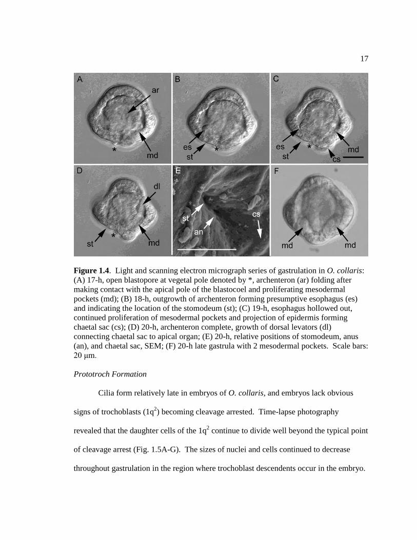
Figure 1.4. Light and scanning electron micrograph series of gastrulation in O. collaris:
(A) 17-h, open blastopore at vegetal pole denoted by *, archenteron (ar) folding after
making contact with the apical pole of the blastocoel and proliferating mesodermal
pockets (md); (B) 18-h, outgrowth of archenteron forming presumptive esophagus (es)
and indicating the location of the stomodeum (st); (C) 19-h, esophagus hollowed out,
continued proliferation of mesodermal pockets and projection of epidermis forming
chaetal sac (cs); (D) 20-h, archenteron complete, growth of dorsal levators (dl)
connecting chaetal sac to apical organ; (E) 20-h, relative positions of stomodeum, anus
(an), and chaetal sac, SEM; (F) 20-h late gastrula with 2 mesodermal pockets. Scale bars:
20 μm.
Prototroch Formation
Cilia form relatively late in embryos of O. collaris, and embryos lack obvious
signs of trochoblasts (1q2) becoming cleavage arrested. Time-lapse photography
revealed that the daughter cells of the 1q2 continue to divide well beyond the typical point
of cleavage arrest (Fig. 1.5A-G). The sizes of nuclei and cells continued to decrease
throughout gastrulation in the region where trochoblast descendents occur in the embryo.

18
Nucleus diameter decreased from 5 μm to 3 μm to the point that they cannot easily be
distinguished (Figs. 1.5A, E, H, respectively). The diameter along the animal-vegetal
axis of these cells decreased from 11 to 7 to 5 μm at the same timepoints. Typical
polychaetes become ciliated at the 64- to128-cell stage, which occurs in O. collaris at 8
hours post-fertilization, but cilia did not appear in embryos of O. collaris until 19 hours
after fertilization at 12ºC (Fig. 1.5H, 6A). By this point, gastrulation has taken place and
the archenteron is well developed. Tubulin antibody staining also demonstrated that the
cells in the ciliated band are actively forming cleavage spindles, well beyond the point
that there should have been a distinct band of very large, cleavage arrested cells in the
band (Fig. 1.6B-D). When cilia first form, each is isolated from the others and although
they appear to form in four regions around the embryo, at no point are compound cilia
from a few large cells observed in embryos (Figs. 1.6B-D).
At 24 hours post-fertilization, larvae begin to swim off the bottom of culture
dishes. Larvae do not hatch out of the spawning membrane, but rather the cilia of the
apical tuft and prototroch grow through it (Fig. 1.6C). The epidermal outpocketing
formed at the vegetal end of the embryo, just posterior to the anus, becomes the chaetal
sac and soon the first pair of larval chaetae appears (Fig. 1.6E). The ciliary band is
formed by a double row of simple cilia uniformly arranged around the embryo (Fig. 1.6E,
F). Confocal imaging also revealed the vast number of cells in the ciliary band and the
continued proliferation of these cells in larvae within the first few days post-fertilization
(Fig. 1.7C-F).

19
Figure 1.5. Light and scanning electron micrograph series of trochoblast division in O.
collaris. Arrows indicate location of descendent of trochoblasts (1q2) and area of cilia
development in image H. Embryos are 8, 10, 11, 12, 14, 15, 17, 19, and 18 h after
fertilization, respectively. Reduction of nucleus and cell diameters to the point that
individual cells are indistinguishable indicates continued division of trochoblast
descendants. Scale bars: 20 μm.

20
Figure 1.6. Formation of prototroch in O. collaris: (A) 22-h, weakly beating cilia (cl)
viewed from side, LM; (B) tubulin-stained 20-h, oblique apical view, short cilia around
equator and actively forming cleavage spindles (cv), CM; (C) tubulin-stained 22-h, apical
view, equatorial cilia making up the prototroch (pt) and cilia at animal pole forming the
apical tuft in the apical organ (ao), CM; (D) tubulin-stained 22-h, oblique vegetal view,
with stomodeum (st), anus (an), equatorial cilia forming the prototroch, and cleavage
spindles; (E) 22-h, oblique vegetal view, with first pair of larval chaetae (ch), SEM; (F)
22-h, with double row of prototroch cilia (arrows), SEM. Scale bars: 20 μm.
Larval Development
Larvae develop in the plankton for approximately 4 weeks at 12ºC and 3 weeks at
16ºC. The episphere of a new larva is relatively opaque with many cells closely packed
together (Fig. 1.7A), but soon the episphere expands, the cells of the epidermis become
less dense, and the episphere becomes nearly transparent (Fig. 1.7B). Within two to three
days the mitraria has formed its first two to three pairs of chaetae and both the chaetal sac
and esophagus have become highly muscular (Fig. 1.7C, D). The chaetal sac contains

21
two chaetal glands connected by a system of interwoven muscles that surround the glands
in a figure-eight pattern. The dorsal levators increase in size in the next few days,
becoming more distinct (Fig. 1.7C, E). These muscles connect the chaetal sac to the
apical organ, which now contains a pair of red larval eyes. The chaetal sac is also
connected to the esophagus by muscles running along the ventral surface of the larva
(Fig. 1.7D). By this time, the ciliary band contains thousands of cells and is
predominantly bright orange (Fig. 1.7D, F). The exact origin of the metatroch and food
groove that complete the ciliated band system have not been identified, but are fully
formed by this time (Fig. 1.7E, F). The ciliary band begins as a ring, but soon folds in the
dorsal direction at the anterior and posterior ends of the larva (Fig. 1.8A). As the mitraria
grows, folds also appear on either side of the larva forming lappets (Fig. 1.8B, C). Algal
cells are often entrained into the broad food groove between the proto- and metatrochs
from the posterior end of the larva and traverse along these folds to the anteriorly located
mouth.
The juvenile rudiment in O. collaris first appears as an epidermal invagination on
the larval ventral surface between the mouth and anus two weeks post fertilization at
12ºC (Fig. 1.8A). This invagination grows to occupy most of the hyposphere below the
curve formed by the esophagus, stomach, and intestine. Within days, this fold grows up
along the anterior portion of the intestine and forms a pocket which fills the space ventral
to the gut (Fig. 1.8B, C). Approximately 18-21 days post fertilization four pairs of
outpocketings form on the anterior and dorsal side of the rudiment (Fig. 1.8D-F). These
pockets (thread glands in Wilson 1932) will become the first nephridia in the adult worm,

22
derived from the ventral epidermis of the larva. Several smaller pairs of nephridia can be
found posterior to these between the fold on the rudiment invagination and the intestine.
The basal portions of the juvenile rudiment are still connected to the ventral epidermis of
the larva and the pocket remains open to the outside. Ventral to the nephridia, the trunk
of the juvenile worm begins to surround the intestine (Fig. 1.8F).
Figure 1.7. Early larval morphology of O. collaris: (A) 27-h, oblique apical view of
episphere (epi) with spawning membrane/larval cuticle (lc), apical organ (ao),
hyposphere (hyp) is out of focal plane but edge of chaetal sac (cs) can be made out; (B)
48-h, oblique apical view, with first two pairs of larval chaetae (ch) and fully formed
digestive system with esophagus (es), stomach (sto), and intestine; (C) 3-day larva
viewed equatorially, cell nuclei of all tissues, densest in prototroch (pt) and apical organ,
PI-stained CM; (D) 3-day larva viewed equatorially, musculature of esophagus, chaetal
sac, prototroch and connection between esophagus and chaetal sac (em), PH-stained CM;
(E) 4-day larva, anterior view, cell nuclei of all tissues showing second ciliated band or
metatroch (mt), PI-stained CM; (F) 4-day larva, anterior view, musculature connecting
apical organ to digestive system, dorsal levators (dl), PH-stained CM. Scale bars: 50
μm.

23
Figure 1.8. Juvenile rudiment development of O. collaris: (A) 14-day, side view
showing folding of the ciliated band (cb) and invagination (in) of ventral epiderm, LM;
(B) 17-day, expansion of rudiment invagination to fill the hyposphere below the digestive
system, fully formed ciliated band system with prototroch (pt), metatroch (mt) and
lappets (la), well developed apical organ (ao), PI-stained CM; (C) 17-day, fully
developed larval musculature with dorsal levators (dl) and chaetal sac (cs), rudiment is
now sac-like, PH-stained CM; (D) 19-day, outpocketing at apical end of rudiment
forming first nephridium (np), PI-stained CM; (E) 24-day, first 4 pairs of nephridia, PI-
stained CM; (F) 27-day, fully developed rudiment and apical organ with pair of eyespots
(ey), PI-stained CM. Scale bars: 100 μm.
Metamorphosis
Prior to the onset of metamorphosis, larvae typically sink to the bottom of culture
jars and the trunk segments begin to protrude from the larval hyposphere (Fig. 1.9A).
The process by which tissues rearrange during metamorphosis mostly conforms to that
described by Wilson (1932) for O. fusiformis, with a few exceptions.

24
Once the trunk segments protrude, the larval episphere begins to shrink and
retract toward the apical surface of the gut (Fig. 1.9B). The cilia of the ciliary band still
beat and propel the animal, but the band folds in on itself and slowly disintegrates (Fig.
1.9C). At the same time, the trunk begins to straighten out and this pulls the digestive
system and the nephridia down into the trunk segments (Fig. 1.9D). In contrast to
Wilson’s description of the juvenile worm consuming the larval episphere, there is no
compelling evidence of this occurring in O. collaris. The ciliary band remains distinctly
bright orange, but this color is absent from the gut after the ciliary band has disappeared,
and in most cases, the gut is empty after metamorphosis except for small clumps of
consumed algae (Fig. 1.9D, H). It appears that the larval episphere simply collapses and
is resorbed into the anterior end of the young worm. In Figure 1.9E-G, one can see the
remainder of the larval episphere shrinking onto the dorsal side of the worm while the
mouth of the juvenile remains unencumbered by this tissue on the ventral surface. The
episphere continues to recede and the dorsal levators contract and pull the apical organ
toward the esophagus until it becomes attached to the juvenile body (Fig. 1.9H). One of
the final larval features lost is the chaetal sac (Fig 1.9D, E, H). The chaetae are shed once
the episphere has collapsed and the sacs themselves are resorbed into the anterior collar
of the worm.

25
Figure 1.9. Metamorphosis of O. collaris: (A-I) 30-day: (A) initiation of metamorphosis
by extension of rudiment just below stomodeum (st) and above anus (an) and breakdown
of ciliated bands (cb), LM; (B) eversion of juvenile trunk (jt) including larval digestive
system and nephridia (np) past the chaetal sac (cs), retraction of episphere, PI-stained
CM; (C) folding and breakdown of ciliated bands, PI-stained CM; (D) retraction of larval
episphere onto dorsal surface, trunk extended beyond chaetal sac and larval chaetae (ch),
LM; (E) resorption of chaetal sac into dorsal surface, PI-stained CM; (F) elongation of
trunk with ventral groove (vg) and nephridiopores (npo) visible, PI-stained CM; (G)
musculature of elongated trunk with juvenile chaetae (jch) and uncini (un) demarcating
segments, PH-stained CM; (H) retraction of dorsal levators (dl) pulling the apical organ
(ao) with eyespots (ey) down to fuse with the trunk, stomodeum remains unencumbered
by the degenerating larval episphere, LM; (I) juvenile worm with apical organ and
eyespots fused to trunk, rows of uncini and juvenile chaetae can also be seen, PI-stained
CM. Scale bars: 100 μm.

26
Juvenile Worm Morphology
The young worm is equipped with a head (prostomium and peristomium), one
thoracic segment bearing two to three sets of chaetae, six or seven segments each bearing
a single pair of chaetae and one pair of tori (rows of uncini located at the anterior of each
segment), and a pygidium (Figs. 1.9I, 1.10A, B). The chaetae and uncini, which were
present before metamorphosis, now can be seen clearly along the trunk segments (Figs.
1.9I, 1.10A, B). The juvenile worm retains the larval eyes as part of the fused pro- and
peristomium, although they are generally lost in the adult (Fig. 1.10A). Internally, the
most anterior two pairs of nephridia come to reside in the thoracic segment (np1 and 2),
the first chaetigerous segment inherits the next nephridial pocket and the longest pair of
nephridia (np3 and 4), which fuse as the juvenile develops. One pair of nephridia resides
in each of the next three chaetigerous segments (np5-7) (Fig. 1.10C). Within the larva,
the juvenile trunk is folded over itself, but at metamorphosis the juvenile epiderm unfolds
and straightens to engulf the larval digestive system into the already segmented trunk.
After metamorphosis, juveniles are approximately 815 μm in length and 125 μm wide.
Soon after completing metamorphosis, the juvenile begins to gather materials for
its initial tube and often sticks to surfaces with its anterior end (Fig. 1.11A, B). The
initial tube is generally made of clear mucus and small particles such as algal cells and
bacterial film, but can also contain much larger items such as threads, larval chaetae, and
the tubes of other worms (Fig. 1.11C). Within the first week, they also develop a
pronounced sphincter at the opening to the stomach (Fig. 1.11D). When offered
sediment, young worms commonly use the smallest grains to add to their tubes or will

27
shed the initial tube and replace it with one anchored into the sediment (Fig. 1.12A). At
first, juveniles of O. collaris lack tentacles, but these soon form in pairs of buds (Fig.
1.12B). Within two weeks, juveniles grow three pairs of unbranched tentacles. Like the
adults, juveniles are surface deposit feeders, consuming small grains of sediments as well
as whatever is attached to them (Fig. 1.12B). When kept in the laboratory, juveniles
grow to a length of about 1300 μm by 24 days after metamorphosis.
Figure 1.10. Confocal micrographs of early juvenile morphology of O. collaris: (A)
juvenile 3 h after completion of metamorphosis, larval eye (ey) is retained and thoracic
segment has two sets of chaetae (tch) whereas posterior segments have single pairs of
chaetae (jch) and rows of uncini (un), PH-stained; (B) ventral view of fully extended 2-
day old juvenile, open stomodeum (st), pygidium (py), tori (to), and juveniles chaetae,
PH-stained; (C) ventral view of 2-day old juvenile,4 pairs of nephridia (np), PI-stained.
Scale bars: 100 μm.

28
Figure 1.11. Light micrographs of juvenile morphology of O. collaris: (A) dorsal view
of 3-day old juvenile beginning to gather particles together for initial tube with mucus at
anterior end, gut has elongated through the worm, terminating in the pygidium (py),
juvenile chaetae (jch) present on each segment and nephridia can still be seen through the
translucent body wall (np); (B) ventral view of 3-day juvenile with tori (to); (C) 5-day old
juvenile with complete juvenile mucus tube (jt), larval eye (ey) still present; (D) 1-wk old
juvenile showing growth of trunk, coelomic compartments (cc), sphincter (sp) at the
entrance to the stomach, and uncini (un) on head. Scale bars: 200 μm.

29
Figure 1.12. Light micrograph of advanced juvenile morphology: (A) 2-wk old juvenile
with complete sediment tube anchored to a fragment of an urchin spine (sf) at the
posterior end (ta) and unattached anterior end (ant); (B) 3-4 wk old juvenile removed
from tube with feeding tentacle buds (tb) and partially digested food (fo) in the digestive
system. Scale bars: 200 μm.
Discussion
Embryonic development in polychaetes does not follow one prescribed pattern.
There is a range between those species that exemplify every aspect of accepted
protostome development to those that possess modifications of cleavage patterns,
gastrulation, and larval morphology. Owenia collaris exhibits shared features of
development for annelids and protostomes, but also extreme modifications to the
accepted patterns. Sperm are of the typical form for polychaetes with a small head and
long flagellum (Okada 1970). At the 8-cell stage, animal cells (micromeres) that are

30
larger than the vegetal macromeres is a pattern similar to nemertean embryos but unlike
most polychaete embryos (Shankland and Savage 1997). Embryos undergo holoblastic
spiral cleavage to form a coeloblastula, which reflects the ancestral pattern for annelids.
Past this point, several key features of typical protostome development cease to occur.
The stomodaeum is not necessarily derived from the area in which the blastopore forms.
It may form as either a secondary opening or by the lateral compression of the blastopore,
followed by differentiation of the blastopore into a stomodeum and anus. However, the
blastopore of O. collaris appears to remain a small, circular opening throughout
gastrulation, suggesting that the anus alone develops from the blastopore (Nielson 2001).
Although rare, this is not unheard of in polychaetes. In Eunice kobiensis the anus also
forms in the region in which the archenteron opens and the stomodaeum forms as a
separate, secondary invagination (Akesson 1967). However, in the case of E. kobiensis,
the blastopore closes after gastrulation and reopens later in development as the anus.
Another modification found in O. collaris is the fate of the trochoblasts (1q2).
The derivatives of this lineage, those cells that generally form the prototroch in
trochophores, do not become cleavage arrested and do not become multiciliated. The
prototroch in O. collaris develops through continued proliferation of cells that may be
derivatives of the original trochoblasts and form cilia much later in development than
embryos of similar size (Rattenbury and Berg 1954, Okada 1970, Smith 1981,
Strathmann 1987). The cells that make up the region of the prototroch are also much
smaller than those of other protostomes, further supporting continued division. Estimates
of prototroch cell diameters for O. collaris after ciliation are in the range of 4-5 μm,
31
whereas those for the nemertean Carinoma tremaphoros, the polychaetes Nereis limbata
and Polygordius sp., and the limpet Patella vulgata are 25-50, 11-22, 15, and 30 μm,
respectively (Maslakova et al. 2004, Costello 1945, Woltereck 1902, Damen and Dictus
1994). Although comprised of smaller cells, the prototroch of early trochophores of O.
collaris covers approximately the same surface area relative to the total surface area of
the larva when compared with early trochophores of other polychaetes (14% as compared
to 11-38% for other polychaetes) (Costello 1945, Anderson 1959, Akeeson 1967). Adult
Owenia sp. also possess deuterostome-like epidermis and nephridia, based on the fact that
monociliated cells dominate both tissues (Gardiner 1978, Smith et al. 1987).
Monociliated cells are also found in the larvae and adult Magelona mirabilis
(Bartolomaeus 1995). The closest relatives of Owenia spp. and M. mirabilis within the
polychaetes, however, possess multiciliated cells (Gardiner 1979, Pettibone 1982, Rouse
and Fauchald 1997). Bartolomaeus (1995) suggested that in both O. fusiformis and M.
mirabilis, the formation of monociliated cells occurred through a suppression of
muliticiliarity in an early stage of ciliogenesis and thus monociliarity in these two
lineages is secondarily derived. Gardiner’s (1978) finding that some epidermal cells of
adult O. fusiformis are biciliated further supports this idea.
The deuterostome-like characteristics found in Owenia may be the result of
reversion to the plesiomorphic condition of the Urbilateria or they may be secondarily
derived characters that resemble deuterostomes by conversion (De Robertis and Sasai
1996). Since the stomodaeum forms secondary to the blastopore in members of both the
deuterostome and protostome lineages, this may indicate that the Urbilateria was a

32
deuterostome. If this is the case, protostomy in animals with complete digestive tracts
developed in the ancestor to modern day protostomes, with loss of this character several
times in individual genera (Aguinaldo and Lake 1997, Rouse and Fauchald 1997).
Comparisons between O. collaris and O. fusiformis reveal similarities in
development, but also differences that can be used to distinguish the two species as larvae
and juveniles (Wilson 1932). Owenia collaris and O. fusiformis vary in size throughout
development, with the former species smaller at each stage (Table 1). Coloration patterns
also vary between species, with the prototroch rim orange in the former and pale yellow
in the latter and the photoreceptors red in the former and brown in the latter. Coloration
in O. collaris is independent of the type of food consumed in culture. During
metamorphosis the overall series of events is the same between species, although some
differences occur. Owenia collaris resorbs its larval tissues rather than casting them off
or consuming them. Owenia collaris has fewer nephridia and chaetigerous segments at
metamorphosis than O. fusiformis. The difference in number of segments continues once
tentacles form, probably due to the smaller size of O. collaris. This is also reflected in
the relative size and number of segments in adults of these two species (Blake 2000, Koh
and Bhaud 2003).
Table 1.1. Characteristics of juvenile worms of O. collaris and O. fusiformis. Owenia
fusiformis values from Wilson (1932).
O. collaris O. fusiformis
# Nephridia 4 7
# Fused Thoracic Segs. 2 2
# Chaetigerous Segs. 6-7 11
Length (@ metamorphosis) 815 μm 870 μm
Length (3-4 weeks) 1300 μm 2560 μm
33
The study of embryogenesis and larval development in Owenia collaris supports
both conservation and high levels of modification of characters, depending on the
taxonomic scale examined. Within the genus Owenia there is variation in size and
segmentation, but larval and juvenile development appears to be relatively conserved.
Although typically thought of as determinant, spiralian cell fates have some plasticity,
with different sets of cells becoming the stomodaeum in at least a few lineages and
developmental arrest of trochoblasts is not universal. Ultimately, protostome
development is much more plastic than was once thought, and occasionally the ancestral
features may emerge or deuterostome characteristics can be secondarily derived
(convergent evolution).
BRIDGE I
Embryogenesis in Owenia collaris produces a larva (mitraria) with a ciliated band
composed of simple cilia on monociliated cells not derived from the typical polychaete
trochoblasts cells. The cellular arrangement of this ciliated band prevents the formation
of compound cilia, which can produce fast feeding and swimming currents by growing to
longer lengths and moving faster through the water column. The ciliated band of mitraria
larvae may be an adaptation to allow high feeding rates similar to the ciliated bands of
other larval forms such as echinoderm plutei or bipinnaria, as suggested by Emlet and
Strathmann (1994). This hypothesis will be tested in Chapter III by comparing growth
and feeding rates of mitraria larvae with those of canonical larval types.

34
CHAPTER III
FUNCTIONAL MORPHOLOGY OF CILIATED BANDS: DOES PHYLOGENY OR
FORM GOVERN THE PERFORMANCE OF THE LARVAL CILIATED BANDS OF
THE POLYCHAETE OWENIA COLLARIS (FAMILY OWENIIDAE)?
Introduction
The role of a feeding larva can be considered from both evolutionary and
ecological perspectives (Hart and Strathmann, 1995). The evolutionary view focuses on
variation in larval form among taxa and over time, and explores the consequences of that
variation on performance. The ecological view focuses within taxa on the population and
community consequences of having planktotrophic larvae in the life cycle. Feeding
larvae are sometimes viewed as machines for turning small eggs into larger (and
therefore more fit) juveniles (Hart and Strathmann, 1995). Both approaches consider
how feeding larvae have evolved to survive the planktonic period and recruit successfully
into the adult population.
Feeding mechanisms of planktotrophic larvae have received much attention
because they determine the rates at which different larval forms can remove food
particles from suspension. High feeding rates may result in shortened planktonic periods
and increased size at metamorphosis, which may ultimately reduce mortality of larvae
and juveniles (Havenhand, 1995; Hart and Strathmann, 1995; Sogard, 1997). Larvae that
feed using ciliated bands can maximize the rate at which they clear particles from water

35
(clearance rate) by increasing the length or velocity of their cilia, the length of the ciliated
band, or the number of cilia creating a current (Strathmann et al., 1972; Strathmann and
Leise, 1979; Hart, 1996; Miner et al., 1999). Larvae may also increase clearance rates by
placing ciliated bands on ridges or other projections of the body (Emlet, 1991). Each of
these is an effective way of increasing the volume of water that passes within the capture
zone of the feeding apparatus. However, the potential changes to the cilia or ciliated
band are constrained by the type of cilia present, which is generally constrained by
phylogeny.
Physical restrictions imposed on cilia produce two major strategies for increased
feeding rates. Trochozoan larvae (found in the phyla Annelida, Mollusca, Nemertea, et
al.) possess multiciliated cells and compound cilia (Nielson, 1985). Larvae with
multiciliated cells can increase the length of cilia and the velocity of the effective stroke
by fusing cilia together into compound cilia, which provide stiffness during the effective
stroke that propels water and food (Sleigh, 1962). These larvae can also increase the
length of the ciliated band by incorporating accessory ciliated cells into the band (Damen
and Dictus, 1994). Tornaria-type larvae (found in the phyla Hemichordata and
Echinodermata), by contrast, possess monociliated cells and simple cilia and can only
increase feeding rate by increasing the length of the ciliated band or the number of cells
in the band (Nielson, 1985). Larvae with simple cilia on monociliated cells cannot
increase the length or velocity of their cilia because the resulting increase in resistance
would ultimately cause the cilia to cease functioning without the added support of a
neighbor.

36
The morphology and function of ciliated band systems in both lineages is derived
from programmed events that occur during embryonic development and from continued
growth during larval development. The morphology of trochophore-type ciliated bands is
initially set by the arrest of a limited number of trochoblast-derived cells in the embryo,
which form the large, multiciliated cells found in the primary ciliated band or prototroch
of hatched larvae (Nielson, 1985). The development of a second ciliated band, the
metatroch, completes the morphology of the opposed-band system. Particles are captured
by the interaction of currents between these two bands. Further growth of the ciliated
bands in derived larvae (e.g. veliger larvae) by the addition of secondary trochoblast cells
or accessory trochoblast cells and compound cilia increases the length of the ciliated band
and the magnitude of the feeding current. The morphology of tornaria-type ciliated bands
is initially set by the concentration of monociliated epidermal cells into a distinct band
following a uniformly-ciliated blastula stage (Wray, 1997). With continued addition of
epidermal cells, the ciliated band forms a convoluted loop of monociliated cells with
simple cilia. This single band may split during larval development to form two sections
of the loop, but particles are captured by a single current created by the band system and
the reversal of ciliary beat to drive particles to the mouth. In both larval types, continued
growth of ciliated bands by the increase in the number of cells increases the feeding and
swimming performances of these bands.
The mitraria larva of the polychaete family Oweniidae is morphologically and
developmentally very different from the trochophore larva typical of polychaetes.
Mitraria larvae possess a sinuous ciliated band that is set out from the body on a

37
pronounced ridge, rather than the simple circular band characteristic of most
trochophores. This band is comprised of simple cilia on monociliated cells that remain
the same length throughout the larval period (Emlet and Strathmann, 1994). Although
the cellular arrangement of this ciliated band is quite different from other trochophore
larvae, mitraria still capture particles with an opposed band system consisting of the
preoral prototroch and postoral metatroch. The unusual curvature of the ciliated band
(and thus the increase in band length) in the mitraria has been proposed as an adaptation
for increasing feeding rates since simple cilia cannot function if they increase in length or
velocity (Emlet and Strathmann, 1994). However, this hypothesis has yet to be tested,
and the feeding performance of this larval form has not been compared with those of
other larval forms.
The purpose of this study is to compare the feeding performance (i.e. clearance
rates) of mitraria larvae of Owenia collaris over the course of its development to larvae
that represent the two typical larval forms, trochophore-type larvae and tornaria-type
larvae, to assess if phylogeny (i.e. trochozoa) or form (e.g. opposed-band, simple cilia)
determines function in ciliated band systems. Trochophore and setiger larvae of the
polychaete Serpula vermicularis and trochophore and veliger larvae of the mussel
Mytilus californianus are used to represent trochophore-type larvae. Larvae of S.
vermicularis retain the typical circular ciliated band composed of multiciliated cells with
compound cilia throughout development, while M. californianus moves through the
trochophore stage to a veliger larva with a larger, but still circular, ciliated band.
Echinoplutei of the urchin Strongylocentrotus purperatus and bipinnaria of the seastar

38
Dermasterias imbricata are used to represent typical tornaria-type larvae with their single
bands of monociliated cells. The length of the ciliated band increases greatly during
development in both of these species. A summary of larval characteristics can be found
in Table 2.1.
Materials and Methods
The clearance rates and sizes of all species of larvae were measured in laboratory-
reared cultures. Because of differences in breeding seasons, each species was cultured
and tested at different times. Dermasterias imbricata were reared in July, 2007; Mytilus
californianus in October, 2007; Owenia collaris in July, 2006 and August, 2007; Serpula
vermicularis in September, 2006 and August, 2007; Strongylocentrotus purpuratus in
March, 2007. Gametes were obtained from many parents of each species using standard
techniques (Strathmann, 1987; see Ch. 2), and embryos were pooled after fertilization in
order to minimize parental effects. Standard larval rearing procedures were used, with
larvae of each species maintained in 4-L glass jars at a starting concentration of 1
embryo/mL of 0.45 μm filtered seawater (FSW) (Strathmann, 1987; Hart, 1996). A
minimum of 15 culture jars were maintained for each species. Cultures were held in
running seawater to maintain temperatures between 12 and 16ºC and they were stirred
gently with plexiglas paddles. Once the gut formed, larvae were fed a mixture of
cultured unicellular phytoplankton, Rhodomonas lens and Dunaliella tertiolecta for
echinoderms and R. lens and Isochrysis galbana for polychaetes and mussels, at
concentrations of 5,000 cells/mL. Cultures were cleaned every other day and given fresh
food following each cleaning.

39
Table 2.1. Larval characteristics of species cultured to test for differences in function of ciliated band systems.
Species Larval Type Band System Cell Type Cilia Type Band Shape
Dermasterias Tornaria Single Monociliated Simple Convoluted
imbricata
Strongylocentrotus Tornaria Single Monociliated Simple Convoluted
purpuratus
Mytilus Trochophore Opposed Multiciliated Compound Circular
californianus
Serpula Trochophore Opposed Multiciliated Compound Circular
vermicularis
Owenia Mitraria Opposed Monociliated Simple Convoluted
collaris

40
At various times throughout larval life, individuals of each species from each of
five randomly chosen replicate jars were removed for measurements. Maximum
clearance rates have been obtained for both bivalve veligers and echinoderm larvae at
concentrations of 1,000 to 5,000 cells/ mL and can be dependent on preconditioning
(Strathmann, 1971; Sprung, 1984; Hart, 1996), so clearance rates were measured using
cultured R. lens at 5,000 cells/mL, a food that all larvae consumed in culture. Twenty
larvae were removed from each of 5 culture jars and placed in 5 separate 20-mL
scintillation vials with 20 mL of FSW and R. lens. Rhodomonas lens was added from a
stock algal culture grown in f/2 media, which had been centrifuged, concentrated and the
concentration determined by hemacytometer counts. Vials were held for 48 hours in a
16ºC incubator equipped with fluorescent lights set to mimic a 16:8 Day:Night cycle.
Two control vials with R. lens but no larvae were also incubated alongside the five
replicate larval vials.
At the end of the 48 hour experimental period, 10 mL samples were removed
from each vial and transferred to a 15-mL centrifuge tube using a volumetric pipet.
Immediately after this, all samples were killed with a drop of concentrated formalin and
centrifuged at 3,000 rpm for 10 minutes in a Marathon 6K swinging bucket centrifuge
(Fisher Scientific, Inc.). Algal cells in a pellet at the bottom of each tube were
concentrated for counting by removing all but 1 mL of FSW and resuspending the pellet
with a Pasteur pipet. A minimum of three subsamples for each vial was then counted

41
with a hemacytometer on an Olympus BH-2 compound microscope and the final cell
concentration calculated.
Clearance rate (the volume of water cleared of particles) per animal over the 48-
hour time interval was calculated from equations in Frost (1972). Analysis of control
vials yielded an average algal growth constant, k, from:
C1 = C0e k(t
1-t
0)
(1)
where C0 and C1 were cell concentrations in the control vials at times t0 and t1. A grazing
coefficient g for each vial with larvae was calculated from:
C1* = C0*e (k-g)(t
1-t
0)
(2)
where C0* and C1* were the cell concentrations in the larval vials at times t0 and t1.
The clearance rate per animal over the 24-hour interval, F, was then calculated by:
F= Vg/N (3)
where V was the volume of the vial and N was the number of larvae in the vial.
The larval protein content was used as a proxy for the amount of metabolically
active tissue in all species. Protein was determined by the Bradford method, which
compares the absorption of samples stained with Coomassie blue reagent on a
spectrophotometer with bovine serum albumin (BSA) standards of known concentrations
(Bradford, 1976; Alexander and Griffiths, 1993). This method can be used with
microgram quantities of protein, thus limiting the need for high numbers of larvae. One
hundred larvae from each replicate culture jar were removed at the same time that
clearance rates were measured, frozen in FSW with liquid nitrogen and held at –80ºC
until analysis.

42
Ash free dry mass and ciliated band length also were measured as scaling factors
for these experiments since there are no distinct stages within the larval period that are
comparable for all species tested and the time from hatching to metamorphosis varies
among species (Wilson, 1932; Young and Chia, 1982; Strathmann, 1987). For ash free
dry mass, several hundred larvae were removed from each replicate culture jar at the
same time that clearance rate and protein content were measured. Larvae were frozen at
–20ºC in FSW and held until analysis. Samples were concentrated with a 53-μm mesh
and rinsed with fresh water several times, transferred to pre-weighed foil pans and dried
in a 60C oven to a constant mass (Jespersen and Olsen, 1982). Samples were then ashed
for 6 hours at 550ºC in a muffle furnace to remove all organic material and reweighed.
Ciliated band length was measured from fixed samples. Twenty larvae were held
for a minimum of 5 minutes in a 1:1 mix of FSW: 7.5% MgCl in order to relax them and
allow the ciliated bands to be viewed at their full size. Larvae were then fixed with 10%
formalin buffered with sodium borate. Photographs of 10 larvae per replicate were taken
from several different angles with an Optronics Microfire digital camera (Optronics, Inc.)
mounted on an Olympus BX50 compound microscope. Segments of convoluted ciliated
bands were traced and measured from series of images with Image J (National Institutes
of Health). Circular ciliated bands were measured by calculating circumference from the
measured diameter of the larva or velum.
Differences among performances of the various larval types were examined in
several ways. Clearance rates, ciliated band lengths, and protein content were divided by
the measured ash-free dry masses for each sample, thus standardizing these

43
measurements across all species by weight. Weight-specific measurements were then
compared among the tornaria-type larvae (D. imbricata and S. purpuratus), the
trochophore-type larvae (M. californianus and S. vermicularis), and the mitraria larva of
O. collaris by analysis of variance with larval type as a fixed factor, species as a random
factor, and with species nested within larval type. Analyses of variance were followed by
Tukey’s HSD post-hoc tests for larval type when differences were significant (Zar, 1999;
SPSS 14.0). Band-length- specific and protein-content-specific clearance rates were also
tested in this way.
Results
For the three larval types tested, clearance rates generally increased with growth
in mass, band length, and protein content. The highest clearance rates were observed in
the tornaria-type bipinnaria larvae of Dermasterias imbricata, followed by plutei of
Strongylocentrotus purpuratus and mitrariae of Owenia collaris (Fig. 2.1). The
trochophore-type larvae of Mytilus californianus and Serpula vermicularis cleared
particles at much lower rates. However, tornaria-type and mitraria larvae grew to much
larger sizes and differences in weight-specific clearance rates among larval types were
not significant (Fig. 2.2; Table 2.2).

44
Figure 2.1. Clearance rates of
(A) tornaria-type
(Dermasterias imbricata and
Strongylocentrotus
purpuratus), (B) mitraria
(Owenia collaris), and (C)
trochophore-type (Mytilus
californianus and Serpula
vermicularis) larvae.
Clearance rates and ash free
dry masses were measured for
each species over the course
development for larvae
randomly selected from 5
replicate culture jars. Data
points represent means with
standard deviations. Note the
differences in y-axis scale
between graphs.
A. Tornaria-type
0 1 2 3 4
Cle
ara
nce
Ra
te(m
L/la
rva
/da
y)
0
1
2
3
4
5
6
D. imbricata
S. purpuratus
B. Mitraria
0 1 2 3 4
Cle
ara
nce
Ra
te(m
L/la
rva
/da
y)
0
1
2
3
O. collaris
C. Trochophore-type
Ash-Free Dry Mass (g/larva)
-1 0 1 2 3 4
Cle
ara
nce
Ra
te(m
L/la
rva
/da
y)
0.0
0.5
1.0
1.5
M. californianus
S. vermicularis

45
Figure 2.2. Weight-specific clearance rates of tornaria-type (Dermasterias imbricata and
Strongylocentrotus purpuratus), mitraria (Owenia collaris), and trochophore-type
(Mytilus californianus and Serpula vermicularis) larvae. Clearance rates and ash-free dry
masses were measured for each species over the course of development for larvae
randomly selected from 5 replicate culture jars. Bars represent means with standard
deviations. Sample sizes range from 15 to 25.
Table 2.2. Results of 2-way nested ANOVA with larval type as a fixed factor and
species as a random factor to test for differences in weight-specific clearance rates for
tornaria-type (Dermasterias imbricata and Strongylocentrotus purpuratus), mitraria
(Owenia collaris), and trochophore-type (Mytilus californianus and Serpula
vermicularis) larvae.
Source df SS MS F p
Larval Type 2 7.357 3.679 4.588 0.201
Species (Larval Type) 2 1.562 0.781 1.379 0.257
Error 100 56.639 0.564
Tornaria-type and mitraria larvae developed much longer and more convoluted
ciliated bands than the trochophore-type larvae during development. Weight-specific
band lengths were not significantly different among larval types (Fig. 2.3, Table 2.3).
Ciliated bands in all three larval types developed proportionally to the body size of the
D. imbric
ata
S. purpuratus
O. colla
ris
M. califo
rnianus
S. verm
icularis
Weig
ht-
Specific
Cle
ara
nce R
ate
(mL/
g/larv
a/d
ay)
0.0
0.5
1.0
1.5
2.0
Tornaria Mitraria Trochophore

46
larva. Clearance rates increased proportionally to the band length in the tornaria-type and
mitraria larvae, but the relationship between these two factors was much less obvious in
the trochophore-type larvae, probably due to their relatively small size (Fig. 2.4). Band-
length-specific clearance rates did not differ significantly among larval types (Fig. 2.5,
Table 2.3).
Figure 2.3. Weight-specific ciliated band lengths of tornaria-type (Dermasterias
imbricata and Strongylocentrotus purpuratus), mitraria (Owenia collaris), and
trochophore-type (Mytilus californianus and Serpula vermicularis) larvae. Ciliated band
lengths and ash-free dry masses were measured for each species over the course of
development for larvae randomly selected from 5 replicate culture jars. Bars represent
means with standard deviations. Sample sizes range from 15 to 25.
D. imbric
ata
S. purpuratus
O. colla
ris
M. califo
rnianus
S. verm
icularis
Weig
ht-
Specific
Cili
ate
d B
and L
ength
(m
/g/larv
a)
0
2000
4000
6000
8000
10000Tornaria Mitraria Trochophore

47
Table 3. Results of 2-way nested ANOVAs with larval type as a fixed factor and species
as a random factor and to test for differences in (A) weight-specific band length (B)
ciliated-band-length-specific clearance rates for tornaria-type (Dermasterias imbricata
and Strongylocentrotus purpuratus), mitraria (Owenia collaris), and trochophore-type
(Mytilus californianus and Serpula vermicularis) larvae.
A. Weight-specific band length ANOVA
Source df SS MS F p
Larval Type 2 9.33E07 4.67E07 3.693 0.230
Species (Larval Type) 2 2.42E07 1.21E07 1.751 0.179
Error 100 6.93E08 6.93E06
B. Band-length-specific clearance rate ANOVA
Source df SS MS F p
Larval Type 2 5.44E-07 2.72E-07 8.857 0.177
Species (Larval Type) 2 6.99E-08 3.50E-08 0.443 0.643
Error 100 7.89E-06 7.89E-04

48
Figure 2.4. Ciliated band
lengths of (A) tornaria-type
(Dermasterias imbricata and
Strongylocentrotus
purpuratus), (B) mitraria
(Owenia collaris), and (C)
trochophore-type (Mytilus
californianus and Serpula
vermicularis) larvae. Ciliated
band lengths and clearance
rates were measured for each
species over the course of
development for larvae
randomly selected from 5
replicate culture jars. Data
points represent means with
standard deviations. Note the
differences in scale between
graphs.
A. Tornaria-type
0 2000 4000 6000 8000 10000
Cle
ara
nce R
ate
(mL/larv
a/d
ay)
0
1
2
3
4
5
6
D. imbricata
S. purpuratus
B. Mitraria
0 1000 2000 3000 4000 5000
Cle
ara
nce R
ate
(mL/larv
a/d
ay)
0
1
2
3
O. collaris
C. Trochophore-type
Ciliated Band Length (m)
0 200 400 600 800 1000
Cle
ara
nce R
ate
(mL/larv
a/d
ay)
0.0
0.5
1.0
1.5
M. californianus
S. vermicularis

49
Figure 2.5. Ciliated-band-length-specific clearance rates of tornaria-type (Dermasterias
imbricata and Strongylocentrotus purpuratus), mitraria (Owenia collaris), and
trochophore-type (Mytilus californianus and Serpula vermicularis) larvae. Ciliated band
lengths and clearance rates were measured for each species over the course of
development for larvae randomly selected from 5 replicate culture jars. Bars represent
means with standard deviations. Sample sizes range from 15 to 25.
Larval protein content increased as all larval types developed, but, again, tornaria-
type and mitraria larvae possessed much higher amounts of protein than trochophore-type
larvae later in development. Weight-specific protein content was not significantly
different among larval types (Fig. 2.6, Table 2.4). For tornaria-type and mitraria larvae,
the relationship between protein content and clearance rate was similar in shape to the
relationship between protein content and ash-free dry mass (Fig. 2.7). Higher protein
content did not coincide necessarily with high clearance rates in the trochophore-type
larvae. Protein-specific clearance rates were similar among larval types, although rates of
tornaria-type larvae tended to be higher than those of either mitraria or trochophore-type
larvae (Fig. 2.8, Table 2.4). A summary of all results can be found in Table 2.5.
D. imbric
ata
S. purpuratus
O. colla
ris
M. califo
rnianus
S. verm
icularis
Cili
ate
d-B
and-L
ength
-Specific
Cle
ara
nce R
ate
s
(mL/
m/d
ay)
0.0000
0.0002
0.0004
0.0006
0.0008
Tornaria Mitraria Trochophore

50
Figure 2.6. Weight-specific protein content of tornaria-type (Dermasterias imbricata
and Strongylocentrotus purpuratus), mitraria (Owenia collaris), and trochophore-type
(Mytilus californianus and Serpula vermicularis) larvae. Protein content and ash-free dry
masses were measured for each species over the course of development for larvae
randomly selected from 5 replicate culture jars. Bars represent means with standard
deviations. Sample sizes range from 15 to 25.
Table 2.4. Results of 2-way nested ANOVAs with larval type as a fixed factor and
species as a random factor to test for differences in (A) weight-specific protein content
and (B) protein-content-specific clearance rates for tornaria-type (Dermasterias imbricata
and Strongylocentrotus purpuratus), mitraria (Owenia collaris), and trochophore-type
(Mytilus californianus and Serpula vermicularis) larvae.
A. Weight-specific protein content ANOVA
Source df SS MS F p
Larval Type 2 6.18E06 3.09E06 6.579 0.169
Species (Larval Type) 2 9.58E05 4.79E05 0.839 0.435
Error 100 5.71E07 5.71E05
B. Protein-content-specific clearance rate ANOVA
Source df SS MS F p
Larval Type 2 2.66E-05 1.33E-05 31.173 0.094
Species (Larval Type) 2 1.01E-06 5.04E-07 0.385 0.681
Error 100 0.000 2.00E-07
D. imbric
ata
S. purpuratus
O. colla
ris
M. califo
rnianus
S. verm
icularis
We
igh
t-S
pe
cific
Pro
tein
Co
nte
nt
(ng
/g
/la
rva
)
0
500
1000
1500
2000
2500Tornaria Mitraria Trochophore

51
Figure 2.7. Protein content
of (A) tornaria-type
(Dermasterias imbricata and
Strongylocentrotus
purpuratus), (B) mitraria
(Owenia collaris), and (C)
trochophore-type (Mytilus
californianus and Serpula
vermicularis) larvae. Protein
content and clearance rates
were measured for each
species over the course of
development for larvae
randomly selected from 5
replicate culture jars. Data
points represent means with
standard deviations. Note the
differences in scale between
graphs.
A. Tornaria-type
0 500 1000 1500 2000 2500
Cle
ara
nce
Ra
te(m
L/la
rva
/da
y)
0
1
2
3
4
5
6
D. imbricata
S. purpuratus
B. Mitraria
0 500 1000 1500 2000 2500
Cle
ara
nce
Ra
te(m
L/la
rva
/da
y)
0
1
2
3
O. collaris
C. Trochophore-type
Protein Content (ng/larva)
0 100 200 300 400 500
Cle
ara
nce
Ra
te(m
L/la
rva
/da
y)
0.0
0.5
1.0
1.5
M. californianus
S. vermicularis

52
Figure 2.8. Protein-content-specific clearance rates of tornaria-type (Dermasterias
imbricata and Strongylocentrotus purpuratus), mitraria (Owenia collaris), and
trochophore-type (Mytilus californianus and Serpula vermicularis) larvae. Protein
content and clearance rates were measured for each species over the course of
development for larvae randomly selected from 5 replicate culture jars. Bars represent
means with standard deviations. Sample sizes range from 15 to 25.
Discussion
The search for differences and similarities in the feeding abilities of different
larval forms allows us to test general hypotheses about the connection between larval
form, functional morphology and components of fitness in species with complex life
cycles that include feeding larvae (Hart, 1996). Selection for efficient feeding in
planktotrophic larvae is driven by the need to reduce mortality by either shortening
planktonic periods or increasing size in the larval stage and at metamorphosis
(Havenhand, 1995; Hart and Strathmann, 1995; Sogard, 1997). Among the species
investigated, tornaria-type and mitraria larvae achieve higher clearance rates, masses,
ciliated band lengths, and protein contents than trochophore-type larvae. Clearance rates
D. imbric
ata
S. purpuratus
O. colla
ris
M. califo
rnianus
S. verm
icularis
Pro
tein
Sp
ecific
Cle
ara
nce
Ra
te(m
L/n
g/d
ay)
0.000
0.001
0.002
0.003
0.004Tornaria Mitraria Trochophore

53
of fully developed tornaria-type and mitraria larvae are 6-18 times higher than those of
fully developed trochophore-type larvae and ash free dry masses 2.5-3.5 times higher.
Ciliated bands of fully developed tornaria-type and mitraria larvae were 3-20 times
longer than those of trochophore-type larvae. Long, convoluted bands, despite being
composed of short, simple cilia, provide greater particle capture ability than the circular,
multiciliated bands tested here and are strongly associated with the larger body sizes
found in tornaria-type and mitraria larvae.
The data collected for clearance rates and ash-free dry masses in this study (Table
2.5) are comparable to those obtained by other workers with the same larval types.
Bipinnaria larvae of Dermasterias imbricata cleared particles at rates ranging from 0.1 to
4.8 mL/larva/day at sizes ranging from 0.9 to 2.6 μg/larva (Fig. 1), compared to
bipinnaria larvae of Acanthaster planci, which cleared particles at a rate of 1.9
mL/larva/day at a size of 0.9 μg (Lucas, 1982). Echinopluteus larvae of
Strongylocentrotus purpuratus cleared particles at rates ranging from 0.1 to 1.6
mL/larva/day at sizes of 0.5 to 2.8 μg/larva and (Fig. 2.1), compared to Paracentrotus
lividus, which cleared particles at a rate of 0.9 mL/larva/day at 0.2 μg/larva (Strathmann,
1975; Fenaux et al., 1985). Veligers of Mytilus californianus cleared particles at 0.1 to
0.2 mL/larva/day at 0.6 to 1.0 μg/larva (Fig. 2.1). Other bivalve veligers investigated to
date achieved clearance rates ranging from 0.05 to 4.1 mL/larva/day at sizes from 0.04 to
0.86 μg/larva (Walne, 1965; Gallager and Mann, 1980; Riisgard et al., 1981; Jespersen
and Olsen, 1982; Gerdes, 1983; Sprung, 1984; Mann and Gallager, 1985). By
comparison, gastropod veligers, which were generally larger, cleared particles at rates
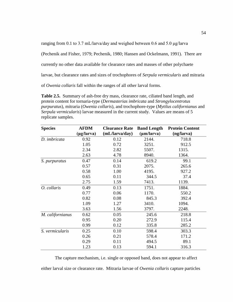
54
ranging from 0.1 to 3.7 mL/larva/day and weighed between 0.6 and 5.0 μg/larva
(Pechenik and Fisher, 1979; Pechenik, 1980; Hansen and Ockelmann, 1991). There are
currently no other data available for clearance rates and masses of other polychaete
larvae, but clearance rates and sizes of trochophores of Serpula vermicularis and mitraria
of Owenia collaris fall within the ranges of all other larval forms.
Table 2.5. Summary of ash-free dry mass, clearance rate, ciliated band length, and
protein content for tornaria-type (Dermasterias imbricata and Strongylocentrotus
purpuratus), mitraria (Owenia collaris), and trochophore-type (Mytilus californianus and
Serpula vermicularis) larvae measured in the current study. Values are means of 5
replicate samples.
Species AFDM Clearance Rate Band Length Protein Content
(μg/larva) (mL/larva/day) (μm/larva) (ng/larva)
D. imbricata 0.92 0.12 2144. 718.8
1.05 0.72 3251. 912.5
2.34 2.82 5507. 1315.
2.63 4.78 8940. 1364.
S. purpuratus 0.47 0.14 619.2 99.1
0.57 0.31 2075. 265.6
0.58 1.00 4195. 927.2
0.65 0.11 344.5 37.4
2.75 1.59 7413. 1139.
O. collaris 0.49 0.13 1751. 1884.
0.77 0.06 1170. 550.2
0.82 0.08 845.3 392.4
1.09 1.27 3410. 1094.
3.63 1.56 3797. 2248.
M. californianus 0.62 0.05 245.6 218.8
0.95 0.20 272.9 115.4
0.99 0.12 335.8 285.2
S. vermicularis 0.25 0.10 598.4 303.3
0.26 0.21 578.4 171.2
0.29 0.11 494.5 89.1
1.23 0.13 594.1 316.3
The capture mechanism, i.e. single or opposed band, does not appear to affect
either larval size or clearance rate. Mitraria larvae of Owenia collaris capture particles

55
with the opposed-band system using short, simple cilia, yet they clear particles at rates
comparable to those of echinoplutei of Strongylocentrotus purperatus or bipinnaria of
Dermasterias imbricata, both of which use a single-band system with short, simple cilia
(Fig. 1). The trochophore-type larvae of both Mytilus californianus and Serpula
vermicularis, on the other hand, appear to be limited in their clearance rates by their
small size, not by their use of the opposed band system. Particle velocities measured in
other opposed-bands of larvae and rotifers are generally higher than those of either
mitraria or of single-band systems (Strathmann et al., 1972; Strathmann and Leise, 1979;
Emlet 1983; McEdward and Strathmann, 1987; Gallager, 1988; Emlet, 1990; Emlet and
Strathmann, 1994). Based on these velocities, opposed bands should produce higher
clearance rates per unit length of ciliated band (Strathmann and Leise, 1979; Gallager,
1988). There is little evidence of this in the species tested in this study, although band-
length-specific clearance rates tended to be higher in M. californianus than in the other
species tested (Fig. 2.5). It is possible that the trochophore-type species tested here
simply do not increase cilium length as much as other, larger species of veliger or setiger.
These larvae also retain the circular ciliated band throughout development, rather than
producing convoluted bands similar to those developed by larger veligers or rostraria
larvae. Gastropod veligers with more convoluted bands clear particles at higher rates
than M. californianus or S. vermicularis (Pechenik, and Fisher, 1979; Pechenik, 1980),
further supporting the notion that opposed band systems are capable of creating feeding
currents comparable to those of tornaria-type larvae. The cumulative effects of longer
ciliated bands in tornaria-type and mitraria larvae (and larger veligers) overcome slower

56
particle velocities associated with short, simple cilia. High clearance rates are produced
by both systems.
Other workers have found high variation in clearance rates within a given particle
capture mechanism. Hart (1996) found that within the different echinoderm larval forms
that possess the single band system, bipinnariae (asteroids) and auriculariae
(holothuroids) clear particles at higher rates than either ophioplutei (ophiuroids) or
echinoplutei (echinoids), although these rates were not corrected for larval size (Hart and
Strathmann, 1995). Differences in the density of cilia within the ciliated bands in these
groups accounted for differences in clearance rates. Hart (1996) proposed that ciliated
bands of bipinnariae and auriculariae produced high clearance rates because they had
more cilia per unit band length and could thus create faster currents. Of larvae with the
opposed-band system, clearance rates of gastropod veligers vary with the length of
prototroch cilia, not just with body size or band length (Strathmann and Leise, 1979;
Gallager, 1988). It appears that across all larval forms investigated (bipinnaria, pluteus,
veliger, trochophore, mitraria, etc.) the strongest relationship to clearance rate is larval
body size and that variation in clearance rate within groups can be explained by the
adjustments allowed within the different capture mechanisms (i.e. cilium length, cilium
density, ciliated band length). There is currently no evidence to suggest inherent
differences in the potential clearance rates or larval sizes that can be achieved by the two
different capture mechanisms or lineages, since larvae with both capture mechanisms
achieve large size and feed at comparable levels for their size.

57
Although Hart (1996) demonstrated that the clearance rates of echinoderm larvae,
which possess simple cilia, increase proportionally to the lengths of the ciliated band, he
also presented preliminary data suggesting that long ciliated bands are not the only way
that different larval forms can compensate for low feeding rates. He hypothesized that
different larval forms may take several different routes to increase their size at
metamorphosis and to decrease the length of the larval period. Larvae may adjust their
metabolic rates or the relative amount of metabolically active tissues as a way to
compensate for low clearance rates. This may allow larvae with low clearance rates to
develop and metamorphose faster than if they relied solely on food assimilation. Hart
(1996) further suggested that larvae that employ morphological and physiological
methods to maximize feeding efficiency could metamorphose sooner and at larger sizes
than other larvae even if they start with the same egg size.
Protein content was used in this study as a proxy for the amount of metabolically
active tissue in each species examined. Protein content, and therefore metabolic demand,
increased dramatically along with clearance rate over the course of development for
tornaria-type and mitraria larvae (Fig. 2.7). The trochophore-type larvae tested here do
not reach such large sizes, and so metabolic demand and clearance rates remained low.
The larger tornaria-type and mitraria larvae appear to maximize feeding rates for their
high metabolic demand through the use of larger and more convoluted ciliated bands,
meeting the demands of large larval bodies and juvenile tissues. However, clearance
rates achieved by mitraria larvae do not increase as quickly as one would assume should
be necessary to meet their increasing metabolic demands.

58
Mitraria larvae tended to contain more protein for their mass relative to the other
two larval types investigated. Mitraria larvae develop two structures that probably
account for their relatively higher protein content compared to other larval forms: the
larval chaetal sac and the juvenile rudiment. The larval chaetal sac consists of muscle
fibers arranged in a figure-eight pattern around two chaetal glands, which are used to
erect chaetae when larvae are disturbed. The advanced juvenile rudiment possesses all of
the segmental musculature of the juvenile worm within the larval hyposphere. Ciliated
band growth and clearance rates of mitraria larvae may not be able to meet the metabolic
demand from these highly muscular structures. Since the juvenile structures are not
active in the larval stage, however, metabolic demand may not be as high as would be
predicted by protein content alone.
Clearance rates do not scale isometrically with body volume, or presumably with
metabolic demand, for other large polychaete larvae (Miner et al., 1999). Most
polychaete larvae grow by posterior elongation, producing long-bodied larvae with
relatively small heads and small, circular ciliated bands (Bhaud and Cazaux, 1987). This
allometric relationship between volume and clearance rate may be unfavorable in these
forms, either limiting further growth or causing larvae to develop alternative food-capture
mechanisms. Mitraria larvae may be constrained by phylogeny in that the allometry
between clearance rate and body size, or metabolic demand, becomes unfavorable to
continued growth at large sizes. This, in turn, may have driven the evolution of a
convoluted ciliated band to help offset this relationship. Perhaps this also drove the
evolution of the internalized juvenile rudiment found in mitraria, which would reduce

59
drag as larvae move through the water column and minimize interference with flow
around the ciliated band. The amphinomid rostraria larva, which extends its ciliated
bands along anterior palps (Järgersten, 1972), and the endo-larva of Polygordius
appendiculata, in which the juvenile trunk develops within the larval episphere (Cowles,
1903), may be other examples of larval forms that have evolved specialized morphology
in order to cope with the unfavorable feeding allometry within the polychaetes.
The current research supports the hypothesis that the long, convoluted ciliated
band of the mitraria larva is an adaptation to achieve high feeding rates, although the
order in which this adaptation has come about is still unknown. The likeliest explanation
is that mitraria larvae were constrained to simple cilia during embryonic development
(see Ch. 2), which required that this larval form develop some mechanism other than a
prototroch with long, compound cilia to allow for sufficient larval feeding rates and
development to competent size (Emlet and Strathmann, 1994). Mitraria larvae of Owenia
collaris develop a juvenile similar in size to that of Serpula vermicularis under similar
laboratory conditions, but in almost half the number of days required by S. vermicularis
(Young and Chia, 1982). It is plausible that the convoluted ciliated band either allowed
for relatively fast growth rates or was produced by evolutionary pressure to decrease
development time in the plankton. Together, evolutionary pressure (or pre-adaptation)
along with the development of primarily simple cilia produced a larval form highly
reminiscent of, and convergent upon, the tornaria-type larvae found among
hemichordates and echinoderms in morphology. In many respects, though, mitraria
larvae are a combination of morphology and performance of both ancestral larval types.

60
For all larval types, patterns in feeding performance are the result of continued larval and
band growth not the result of phylogenetic constraints. Continued growth of both ciliated
band types (opposed and single) increased feeding performance of larvae tested in this
study and others. This increase overcame restrictions in cilia morphology imposed by
phylogeny and development.
BRIDGE II
During the investigations of embryogenesis and larval feeding, embryonic and
larval material for Owenia collaris could only be obtained during the spring and summer
months. Wilson (1932) also noted that Owenia fusiformis produced gametes only during
the spring and summer months in Plymouth, U.K. The seasonal reproduction and the
environmental factors that control this seasonality were investigated in Chapter IV using
field and laboratory techniques.

61
CHAPTER IV
ENVIRONMENTAL CUES AND THE REPRODUCTIVE CYCLE OF A
TEMPERATE POPULATION OF OWENIA COLLARIS (ANNELIDA:
POLYCHAETA)
1. Introduction
Reproduction of most marine organisms is highly dependent on environmental
factors. Beginning with Orton (1920), temperature has often been invoked as the primary
influence determining onset of breeding, length of breeding period, and breeding success.
Over time, more and more authors have recognized the importance of other
environmental factors as well, either due to their interactions with temperature or their
influence on reproduction independent of temperature (Giese and Kanatani, 1987). In
reviewing reproductive cycles of polychaetes and other marine invertebrates, Olive
(1995) delineated several exceptions to Orton’s Rule of temperature dominance. Nutrient
availability, photoperiod, endogenous long-term interval timers, and gamete growth rates
can override or modulate reproductive responses to temperature (Tenore, 1977; Olive,
1980; Garwood and Olive, 1982; Clark, 1988; see Table 3.1 for details). Schroeder and
Hermans (1975) also documented the effect of salinity on gametogenesis for several
polychaete species. Within habitats such as estuaries, a host of environmental variables
vary seasonally and are likely to affect phenology and success of reproduction in
invertebrates. However, the effects of these variables are mostly untested.

62
Table 3.1. Summary of cues for onset of gametogenesis and or spawning in seasonally reproducing temperate polychaete worms.
Documentation of patterns involved the use of correlation studies, isochronic experiments (experimentally examining the effects of
environmental variables during the species reproductive season) or heterochronic experiments (experimentally examining the effects
of environmental variables out of the species’ reproductive season) experiments (Olive, 1984).
Species Family Cues Tested Evidence Source
Arenicola marina Arenicolidae air temperature isochronic Duncan, 1960
Bispira volutacornis Sabellidae water temperature correlation Nash and Keegan, 2003
Ditrupa arietina Serpulidae water temperature correlation Charles et al., 2003
chlorophyll a
Capitella sp. Capitellidae water temperature correlation Tenore, 1977
Eulalia viridis Phyllodocidae water temperature isochronic Garwood and Olive, 1982
Harmothoe imbricata Polynoidae photoperiod isochronic Garwood and Olive, 1982;
Clark, 1988
Hydroides dianthus Spirorbidae water temperature heterochronic Turner and Hanks, 1960
water temperature heterochronic Olive, 1984
Kefersteinia cirrata Hesionidae photoperiod isochronic Olive and Pillai, 1983
water temperature
Marenzelleria viridis Spionidae salinity correlation Bochert et al., 1996
water temperature correlation Daunys et al., 2000
Nereis diversicolor Nereidae water temperature isochronic; heterochronic Olive, 1984
Salinity correlation Gasiunas, 1956; Bogucki, 1963
Platynereis dumerillii Nereidae lunar cycle isochronic Hauenschild, 1955, 1956, 1960
Phragmatopoma lapidosa Sabellariidae wave disturbance isochronic McCarthy et al., 2003
photoperiod correlation
Typosyllis prolifera Syllidae photoperiod heterochronic Franke, 1980
water temperature
lunar cycle

63
In polychaetes, phenology can be divided into two broad categories, non-seasonal
and seasonal. Non-seasonal species are those that release gametes throughout the year or
do not respond to seasonally varying cues. Within seasonally reproducing species, there
is a range of reproductive strategies (Olive and Clark, 1978). Generally, seasonal species
have developed reproductive systems in which either gametogenesis or spawning is
strongly synchronized within a population or individuals are able to store and maintain
gametes until the appropriate time. Olive et al. (2000) noted that two types of factors
could maintain synchronicity, ultimate and proximate. Ultimate factors are evolutionary
determinants that give a fitness advantage to animals with a particular spawning
periodicity. Crisp’s Rule is a commonly cited ultimate cause in which larval success
(higher food availability, maximum opportunity for growth, favorable oceanography for
recruitment) favors spring reproduction in the Northern Hemisphere (Crisp, 1954; Qasim,
1956; Crisp, 1959). Addition of adult food does not necessarily lead to out-of-season
breeding in temperate invertebrates governed by Crisp’s Rule because breeding is not
controlled by immediate conditions but by those operating over a long period beforehand
(Crisp and Klegg, 1960; Barnes, 1963; Crisp, 1966; Crisp and Patel, 1969). Selective
pressures associated with fertilization and fecundity may favor seasonality (synchronicity
provides better chances of fertilization and predator swamping; reviewed by Olive et al.,
2000). Proximate factors are environmental inputs that are transduced by an organism
allowing physiological responses to occur that organize a temporal sequence of cellular
and physiological events that culminate in spawning. Within the polychaetes, proximate

64
factors vary considerably among and within taxonomic groupings, but the most common
are temperature, photoperiod, and lunar cycle.
Seasonal reproduction appears to have adaptive advantages in temperate waters,
but the cues that entrain this seasonality vary substantially among taxa (Giese and
Kanatani, 1987; Olive et al., 2000). In the sabellid polychaete Bispira volutacornis,
vitellogenesis occurs in response to the spring phytoplankton bloom whereas spawning is
associated with the autumn bloom (Nash and Keegan, 2003). In this species, adults and
larvae take advantage of seasonal peaks in food availability to complete the life cycle.
Peak numbers of recruits of Marenzelleria viridis are linked to the autumn phytoplankton
bloom in coastal waters of the southern Baltic (Bochert et al., 1996). In the North
Atlantic, Harmothoe imbricata undergoes gametogenesis in the winter when low
temperatures and lengthening days induce oocyte growth. Summer temperatures above
15ºC prevent vitellogenesis (Olive, 1984). Vitellogenesis is suppressed or prevented in
Eulalia viridis below 5ºC but occurs independently of day length (Olive, 1984).
Gametogenesis and spawning in the hesionid Kefersteinia cirrata and the nereid Nereis
virens are controlled synergistically by temperature and day length (Olive and Pillai,
1983; Olive, 1995), whereas vitellogenesis in Nereis diversicolor is not affected by
temperature or day length (Olive, 1984). In each of these species, synchronicity probably
allows animals to take advantage of food availability, amenable water conditions, and
availability of mates.
Relatively little is known about the phenology of breeding in the polychaete
family Oweniidae. Seasonal reproduction has been documented for populations of

65
Owenia fusiformis in Plymouth, U.K. (Wilson, 1932) and the Bay of Seine, France
(Menard et al., 1989) where reproductive adults and larvae are found in the spring and
summer months. In contrast, a population of O. fusiformis from Biscayne Bay, Florida,
was thought to have continuous or non-seasonal reproduction since adults with mature
gametes were found in all months except June, September, and December (McNulty and
Lopez, 1969). Seasonal cycles in environmental factors are not as dramatic in subtropical
Florida as they are in Europe. It may be that these distinct populations vary in
reproductive seasonality because the environmental cues that drive seasonality in Europe
are not present at sufficient levels in Florida. However, the causes underlying these
latitudinal differences remain a matter of speculation, since the relationship between
breeding and environmental conditions has not been specifically addressed with
experimental techniques. Moreover, reproductive traits have not been documented in any
other species or populations within the family Oweniidae.
In this study, I chronicled the breeding season of a temperate population of
Owenia collaris (Hartman, 1955) and identified the environmental factors that most
influence this timing. Adults feed on surface sediments and the associated bacterial and
algal communities. Gametes are free-spawned into the water column and feeding larvae
develop for three to four weeks in the plankton, depending on food and temperature
(Chapter 2). By reproducing seasonally in the spring and summer months, this species
may be able to take advantage of the spring phytoplankton bloom either as adults or as
larvae, as well as the presence of marine-dominated waters in the estuary and calmer
conditions for establishment of new recruits. I report evidence for seasonal reproduction

66
associated with environmental fluctuations and give the results of a controlled laboratory
experiment examining potential proximate factors organizing the seasonal breeding.
2. Materials and Methods
2.1. Natural Reproductive Cycle
Each month during one of the spring tide series, adult Owenia collaris were
collected from Coos Bay (N 43º20, W 124º19) from March 2005 through February 2007
(Figure 3.1). Twenty to thirty animals were collected each month and preserved with
10% formalin buffered with seawater for 24 hours, then transferred to 70% EtOH. After
preservation, worms were removed from their tubes and examined under a dissecting
microscope for the presence of identifiable gametes (oocytes or spermatozoa) (Figure
3.2). Males can be distinguished from females by loops of gonadal tissue in the third and
fourth segments, as well as sperm bundles free in the coelom. Female gonads are not so
organized. Eggs are present all along the worm from the third segment to the pygidium.
Gender and the presence or absence of gametes was scored for each individual collected
each month. At the same time as the preserved collections were made, several live
animals were examined for gametes. When gametes were present, eggs and sperm were
dissected out of the worm and used to check for viability and fertilization success. In all
samples in which gamete type could be identified, at least some gametes from each worm
proved to be mature and viable. All animals with identifiable gametes are therefore
referred to as “reproductive”. A one-way ANOVA was used to examine differences in
percent reproduction among months with month as a fixed factor. A Chi2 Goodness of

67
Fit Test was used to test whether the observed sex ratio found in the field was different
from the expected 50:50 ratio.
Figure 3.1. Map of Coos Bay Estuary, Coos Bay, Oregon. Worms and benthic
chlorophyll samples used in natural reproductive cycle analysis were collected from the
Fisherman’s Grotto (FG) mudflat. Water quality and meteorological sampling stations
maintained by the South Slough National Estuarine Research Reserve are located on the
OIMB campus and adjacent to the Fisherman’s Grotto mudflat, respectively. Worms
used in gametogenesis experiment were collected from the Fossil Point (FP) and the
Cribs (CRB) mudflats as well as Fisherman’s Grotto.

68
Figure 3.2. Stages of gonad development in Owenia collaris. A. Stage 0, where gonad is
completely absent from the ventral portion of the abdominal segments. B. Stage 1, gonad
appears as an opaque, cream-colored mass along the ventral blood vessel (vbv). C. Stage
2, male gonad fully formed as loops of tightly packed sperm bundles (sbl) in mid region
of worm. D. Stage 2, female gonad fully formed with oocytes (oc) tightly packed in
abdominal segments or free floating in posterior segments. Scale bars represent 1 mm.
Each month from March 2006 through February 2007, along with worm samples,
three replicate sediment cores (0.5 cm depth) were collected using a 5 mL syringe with
the end removed. Benthic chlorophyll was extracted with 90% acetone for 24 hrs in the
dark and measured with a Turner TD700 Fluorometer (Turner Designs, CA). The
percentage of the population with gametes (reproductive individuals) was correlated with
benthic chlorophyll estimates over this 12-month period with a Pearson correlation
(Quinn and Keough, 2002).

69
Environmental data were obtained from several online sources. Water quality and
meteorological data for the South Slough National Estuarine Research Reserve
Charleston sampling station were obtained from the Centralized Data Management
Office, NOAA (http://cdmo.baruch.sc.edu/). Day length for the sampling period was
calculated from sunrise and sunset data for Coos Bay obtained from the U.S. Naval
Observatory (http://aa.usno.navy.mil/). Linear multiple regression was used to test which
environmental variables had the greatest impact on reproduction in O. collaris (Quinn
and Keough, 2002). Variables were chosen based on their lack of colinearity, the
completeness of the data set, and Akaike’s Information Criterion (AIC; Akaike, 1978;
Quinn and Keough, 2002). Environmental variables considered were water temperature,
salinity, pH, day length, and water-column chlorophyll. To adjust for colinearity between
day length and both water temperature and chlorophyll, the unstandardized residuals of
day length were used in this analysis. Data analyzed were mean monthly values for the
two years for which worm samples were collected.
Spring transition dates for the Northwest Pacific were obtained from the
Columbia River DART program at the University of Washington
(http://www.cbr.washington.edu) but were not included in the statistical analysis because
the data points are annual, not monthly. Benthic chlorophyll was not used in the linear
regression analysis because these data were only collected during the second year of
sampling.

70
2.2. Gonad Development Experiment
The influence of two environmental factors (day length and benthic chlorophyll)
on reproductive condition was examined in depth using a controlled laboratory study. In
June 2006, I collected 1000 adult O. collaris from mudflats within Coos Bay, OR (Figure
3.1). Worms with intact tubes were haphazardly assigned to one of six indoor aquaria at
the Oregon Institute of Marine Biology (OIMB), Charleston, OR. These tanks were part
of a temperature controlled, recirculating seawater system that was maintained at 12ºC
throughout the course of the experiment. This system consisted of six tanks (150L each)
pumped through a common titanium-coil seawater chiller set at 12ºC (Figure 3.3). Prior
to the addition of worms, native sediment was collected from the same sites as the
worms, sieved, rinsed with freshwater, and added to the bottom of the tanks to a depth of
1 cm. Surface sediment samples were also collected prior to the beginning of the
experiment and placed in f/2 media (Guillard, 1975) in a 16ºC incubator with 16:8
Light:Dark cycle. The community of phytoplankton and bacteria grown in this manner
was added to the sediment in all of the tanks to provide food for the collected worms,
which can alternate between surface deposit and suspension feeding (Gambi, 1969).

71
Figure 3.3. Diagram of setup for Gonad Development Experiment. Black suns indicate
lights set to the reverse-photoperiod treatment six months out of phase with natural light
cycles. Lights above these tanks were wired to a photocell placed in a window adjacent
to the tank system. White suns indicate lights set to the in-phase photoperiod treatment
that mimics natural light cycles. Lights above these tanks were wired to a photocell
placed in view of one of the lights in the reverse treatment. Tanks are divided as
described in the methods section into high (dark gray) and low (white) food treatments.
Each tank was darkened on all sides with black plastic and equipped with a light
fixture and 15 watt fluorescent bulb. Three tanks were set in phase with the ambient light
cycle (in-phase) and the other three tanks were set out of phase with the ambient light
cycle (reverse) (Figure 3.3). Light treatment was alternated with every other tank
receiving the same treatment. Each set of tanks was wired in series and controlled by a
common system of photocells (Bingham, 1997). The first photocell was placed in a
window and would cue the lights in the reverse series to turn off at dawn and on at dusk,
producing light during nighttime hours. The second photocell was placed beneath one of
the lights in the reverse series, and would thus cue the in-phase series to turn on at dawn
when the reverse lights went off and to turn off at dusk. This produced an overall light
system in which the in-phase treatment mirrored the natural seasonal light regime and the
Photocells
Window

72
reverse treatment was 6 months out of phase with the natural seasonal light regime and
12 hours out of phase with the normal daylight hour. For example, in the summer months
if there were 15 hours between sunrise and sunset, the in-phase tanks received 15 hours
of light and 9 hours of darkness and the reverse tanks received 15 hours of darkness and 9
hours of light.
Due to limitations of space, food or benthic chlorophyll treatments were created
by dividing tanks into two identical halves, using a barrier to separate these two halves.
Barriers consisted of plastic mesh sheets covered with cotton batting and 250-µm mesh to
allow water flow between sides ensuring common water quality throughout all treatments
while minimizing the exchange of particulates between sides of the tanks. Every two
weeks over the course of the experiment, one of the two sides of each tank was spiked
with the phytoplankton and bacterial mixture previously mentioned. The water was
turned off in each tank, and the slurry allowed to settle for 24 hours. Food in the other
half consisted only of that which continued to grow under the established light cycles
after the initial addition. In this way, each tank within the two photoperiod treatments
also received high and low food treatments. Food treatments were systematically
assigned so that the high food side was not always on the same side of the tanks.
Once a week, the system was inspected for photocell and light function,
temperature, and barrier integrity. When barriers became clogged, they were carefully
cleaned using a Tygon aquarium vacuum.
Monthly, from June 2006 to February 2007 and again in May 2007, six
individuals from each half of each tank were removed for gonad analysis. Animals were

73
held overnight in separate containers of seawater to allow for gut evacuation and then
preserved in their tubes with 10% formalin buffered with seawater for 24 hrs followed by
storage in 70% EtOH. Concurrently, a small sediment core was removed from each half
of each tank using a 1-mL syringe with the end removed. I removed excess water from
these samples, then extracted chlorophyll using 90% acetone as mentioned above. After
extraction, sediment cores were dried and weighed, and these weights were used to
normalize chlorophyll measurements for the volume of sediment sampled.
All worms were removed from their tubes after preservation, and each worm was
scored for gonad stage. Individuals were examined under a dissecting microscope and
scored for reproductive condition by the appearance of the gonad through the body wall
and from coelomic fluid samples. The stages of gonad maturity were based on those used
in Gentil et al. (1990) but simplified into three categories: 0 if no gonad was identifiable
on the ventral surface of the worm, 1 if cream-colored gonad was present along the
ventral surface of the worm, and 2 if the gonad extended dorsally beyond the midline of
the worm (Figure 3.2). In this last stage, gametes could be identified as either sperm or
eggs and gametes were also present in the coelomic fluid. The length of the fourth
abdominal segment of each worm was measured, as Gentil and Dauvin (1989) found that
this segment had the strongest correlation to the whole worm length in O. fusiformis and
could be used in place of whole worm length when worms were damaged or had
regenerated. This relationship was also valid in O. collaris.
Gonad stage was analyzed using an analysis of covariance (ANCOVA) with
photoperiod and food treatments as fixed factors and worm length and chlorophyll as

74
covariates (Quinn and Keough 2002). Levene’s Test of Homogeneity revealed
significant heteroscedasticity (F=2.5, p<0.001) when month was included in the analysis.
I was unable to transform the data in such a way as to equalize variances across
treatments and the relationship between worm length and month also violated the
assumption of equality of slopes for an ANCOVA. However, sample sizes were equal
across all treatments, making this analysis relatively robust to heterogeneity of variances
and the assumption of equality of slopes (Underwood 1995).
3. Results
3.1. Natural Reproductive Cycle
Gonadal tissue first appeared in Owenia collaris along the ventral blood vessel in
the anterior abdominal segments at the end of February each year. This tissue thickened
outwardly into the coelomic compartment, growing up along the sides of the worm. As
more gametes reached their full size, the gonad extended into the posterior segments as
loose aggregations of gametes, which was the typical state of worms from March through
August. In samples collected in June and July of both years, the posterior segments
usually were devoid of gametes, but the central segments remained full, indicating that
spawning had occurred in these animals but that worms did not release all gametes in a
single spawning event. Worms held in the laboratory in light and temperature conditions
approximating natural conditions occasionally spawned without provocation. When this
occurred, not all gametes were released and worms survived beyond this spawning event
and into the next breeding season. In unprovoked spawning events, fecundity averaged

75
in the low thousands, whereas dissecting gametes could produce more than 40,000
mature oocytes per female.
There was a strong seasonal influence on the presence of gametes in O. collaris.
Gametes were found in a majority of animals from March through August, followed by a
precipitous decline in reproductive adults after September. By November, no
reproductive adults were found in either year. There was a significant effect of month on
the presence of reproductive products (Table 3.2). Post-hoc Tukey’s HSD pairwise
comparisons revealed specific differences between months. There were no significant
differences between most of the spring and summer months (March through August), nor
between the fall and winter months (October through January) (Figure 3.4). September
was statistically similar to both spring/summer and fall/winter months, because of
relatively higher variation between years in this month. It appears that these animals
invest considerable energy in gametes starting in March and continuing through the
summer. A drop in the presence of gametes characterized June and July, which probably
indicates a high frequency of spawnings during the weeks preceding these samples rather
than marking the end of the reproductive season, since there was a high frequency of
gametes in August. In both years, September was a transitional month, with reproduction
tailing off rather than coming to an abrupt halt. In all months when full-sized oocytes
were present, dissected gametes were viable and capable of fertilization, although
fertilization rates in March and September were depressed relative to April through
August.

76
Table 3.2. Natural Reproductive Cycle of Owenia collaris. Results of 1-way ANOVA
with month as a fixed factor and 2 replicated sampling years and post-hoc Tukey’s HSD
to test for differences between months.
Factor dfN, dfD Mean Square F p
Month 11, 12 2703.569 11.470 <0.001
Error 12 235.712
Total 23
More males than females were identified from the above samples from March
through August during the first year of sampling (70:57 male:female ratio). This ratio,
however, is not significantly different from the expected sex ratio of 50:50 (X2
= 1.13,
F0.05 = 3.84, p=0.287). It is possible that many of the worms for which sex could not be
determined were females whose gonads were not developed enough to be distinguished
as eggs through the body wall. Many of these worms had a characteristic amorphous
white mass in the anterior segments where gonads begin developing. When this material
was examined under the microscope, it could not be identified positively as either
developing eggs or sperm. The material examined, however, was that portion of the mass
that was exuded when the animal was pierced with fine forceps. Any developing eggs or
sperm that remained attached to the body wall might have been missed.
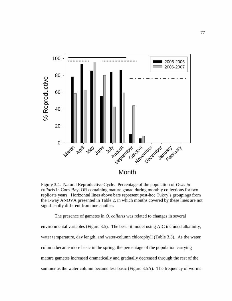
77
Figure 3.4. Natural Reproductive Cycle. Percentage of the population of Owenia
collaris in Coos Bay, OR containing mature gonad during monthly collections for two
replicate years. Horizontal lines above bars represent post-hoc Tukey’s groupings from
the 1-way ANOVA presented in Table 2, in which months covered by these lines are not
significantly different from one another.
The presence of gametes in O. collaris was related to changes in several
environmental variables (Figure 3.5). The best-fit model using AIC included alkalinity,
water temperature, day length, and water-column chlorophyll (Table 3.3). As the water
column became more basic in the spring, the percentage of the population carrying
mature gametes increased dramatically and gradually decreased through the rest of the
summer as the water column became less basic (Figure 3.5A). The frequency of worms
Month
Mar
chApr
ilM
ay
June Ju
ly
Augus
t
Septe
mbe
r
Octob
er
Nov
embe
r
Dec
embe
r
Janu
ary
Febru
ary
% R
epro
ductive
0
20
40
60
80
1002005-2006
2006-2007

78
possessing gametes increased dramatically when water temperatures rose above 11ºC
(Figure 3.5B). There was a similar jump between day lengths of 11 and 12 hours, from
<10% to >40%, respectively (Figure 3.5C). Water-column chlorophyll did not account
for a significant portion of the variation in reproduction between years, but the two peaks
in reproduction (May and August) are associated with the highest concentration of
chlorophyll in the water column (Figure 3.5D). Salinity was not included in the best-fit
model due to its weak relationship with reproduction (Figure 3.5E). Overall, alkalinity,
day length, and water temperature explained 74% of the variation in gamete presence
between months and years. In the second year of sampling, there was no correlation
between the benthic chlorophyll values and presence of gametes (r2=0.031, Figure 3.5F).
Table 3.3. Natural Reproductive Cycle of Owenia collaris. Results of linear multiple
regression using AIC between environmental variables and reproduction of O. collaris.
Alkalinity, water temperature, day length, and water column chlorophyll were included in
the model. Adjusted r2=0.741.
A) Model ANOVA
Factor dfN, dfD Mean Square F p
Regression 4, 19 6398.517 17.433 <0.001
Residual 19 367.039
Total 23
B) Individual Variables
Variable Standardized β t p
Alkalinity 0.470 4.329 <0.001
Water Temperature 0.431 3.293 0.004
Day length 0.123 2.204 0.040
Water Chlorophyll 0.264 2.013 0.059

79
Figure 3.5. Relationship between environmental variables and percent reproduction in
Owenia collaris. A-E) Data points represent mean monthly values for environmental
variables averaged over two years and bars represent mean monthly values for percentage
of population with mature gonad, averaged over two years. Variables are presented in
order from highest to lowest p-value (Table 3.3). F) Data points represent mean monthly
values of benthic chlorophyll from 3 replicate samples collected each month and bars
represent monthly values for percentage of population with mature gonad in the second
year of worm sampling.
E. Salinity
Month
J F M A M J J A S O N D
Sa
linity
24
26
28
30
32
34
% R
ep
rodu
ctive
0
20
40
60
80
100
% R
ep
rodu
ctive
0
20
40
60
80
100
% R
ep
rodu
ctive
0
20
40
60
80
100
D. Chlorophyll (Water Column)
Ch
loro
ph
yll
(ug
/L)
0
1
2
3
4
5
6
7
8
B. Water Temperature
Te
mpe
ratu
re (
oC
)
9
10
11
12
13
14
15
A. AlkalinitypH
7.8
7.9
8.0
8.1
8.2
8.3
F. Benthic Chlorophyll
Month
J F M A M J J A S O N D
Ch
loro
ph
yll
(ug
/L)
0
20
40
60
80
100
120
C. Daylength
Da
yle
ng
th (
hrs
)
8
9
10
11
12
13
14
15
16

80
3.2. Gonad Development Experiment
A 3-way ANCOVA with photoperiod, food, and month as fixed factors and
chlorophyll and worm length as covariates was used to examine the results of the
gametogenesis experiment (Table 3.4). Tank was used as the unit of replication (n=3).
Results from the 3-way ANCOVA analysis indicate a strong interaction between
photoperiod and food, which is best explained by the fact that those worms receiving
high food with a reversed photoperiod possessed gonads at more advanced stage of
development than those in any other treatment during the winter months (Figure 3.6).
There was also a significant effect of both month and photoperiod on gonad stage. The
effect of month is primarily due to the cessation of reproduction in the fall, which
occurred in both photoperiod and food treatments. However, the samples collected in the
winter months in the reverse photoperiod treatment possessed moderately developed
gonad, indicating that increasing day lengths out of season slowed the cessation of gonad
development or allowed it to continue through the winter months. Food availability had
no effect on gonad development by itself. The lengths of worms decreased markedly
over the course of the experiment, and length significantly covaried across all treatments.

81
Table 3.4. Gonad Development Experiment. Results of 3-way ANCOVA with
photoperiod, food, and month as fixed factors and chlorophyll and worm length as
covariates.
Factor dfN, dfD Mean Square F p
Worm Length 1, 78 0.406 6.192 0.015
Chlorophyll 1, 78 0.012 0.178 0.674
Photoperiod 1, 78 0.421 6.411 0.013
Food 1, 78 0.036 0.554 0.459
Month 9, 78 1.128 17.184 <0.001
Photoperiod*Month 9, 78 0.086 1.311 0.245
Photoperiod*Food 1, 78 0.399 6.076 0.016
Food*Month 9, 78 0.059 0.903 0.527
Photoperiod*Food*Month 9, 78 0.082 1.250 0.278
Error 78 0.066
Corrected Total 119
4. Discussion
Owenia collaris is a dioecious iteroparous species with a well-defined breeding
season (March through September in Coos Bay, OR). Onset of gonad development
occurs in early spring and worms advance rapidly to maturity in March. Two spawning
peaks characterize the breeding season, the first occurring between April and May and a
second shorter period in August. During the breeding season, a majority of the animals
in the population contain mature gametes and individuals can spawn multiple times
throughout the season.

82
Figure 3.6. Gonad score for O. collaris exposed to either in-phase or reverse light cycles and either high or low food concentrations.
Bars represent means of 3 replicate tanks with standard deviations. Zeroes represent treatments with gonad scores with mean values
of 0.
Month
June
July
August
Septem
ber
October
Novem
ber
Decem
ber
January
February
May
Go
nad S
core
0.0
0.5
1.0
1.5
2.0In-Phase / High Food
In-Phase / Low Food
Reverse / High Food
Reverse / Low Food
0 0 0 0

83
The major influences on seasonality of reproduction in temperate invertebrates are
food availability, temperature, and photoperiod (Giese and Kanatani, 1987). Often the
increase in day length corresponds with an increase in water temperature and primary
productivity, which in turn may allow invertebrates to begin devoting excess energy to
reproduction. Owenia collaris adults are primarily direct deposit feeders, consuming
surface sediments along with the associated algae, bacteria, and meiofauna (personal
observation). They may therefore be able to take advantage of increasing day length and
primary productivity in the surface sediments as winter progresses into spring.
However, the lack of a strong correlation between benthic chlorophyll and the presence
of gametes in O. collaris prior to the spring phytoplankton bloom indicates that food
availability in the sediment is not necessarily a factor limiting the production of gametes
for this species. By directly consuming the sediment, worms ingest not only benthic
phytoplankton but also small infaunal organisms and bacteria, which may be readily
available at all times of the year. Instead, the timing of reproduction in these animals
most likely is determined by cues that are consistent between years, such as longer days
and warmer water temperatures during spring and summer months.
The first mature individuals collected in 2005 were found in the second half of
March. One female and one male of this group spawned a small number of gametes
when removed from their tubes, but few of these eggs matured and developed. After this
point, spawning events in the laboratory each month until September produced many
more eggs and sperm and viable embryos. Presumably, spawning events in the field
become increasingly common from March through June and continue throughout the

84
summer months. Owenia collaris produce planktotrophic larvae that remain in the
plankton for three to four weeks, depending on temperature, before undergoing a
dramatic metamorphosis into a juvenile (Chapter 2). With the onset of spawning in
March or April, this should provide the larvae with high levels of phytoplankton and
favorable water conditions and also provide juveniles with enhanced levels of surface
primary productivity upon their return to the adult sediments. By spawning before
September and October, the pelagic stage is completed typically before winter storms
return, decreasing primary productivity and potentially washing new settlers away with
increased wave action. Adults in Coos Bay often are found in looser, sandy sediment
within mudflats and in protected embayments on the outer coast, which are prone to
shifting and thus burying the smaller juveniles at the surface. Adults and juveniles of O.
collaris are surface deposit feeders and suspension feeders and cannot tolerate extended
burial below the surface. Moreover, they have limited ability to reconstruct damaged
tubes.
Each year along the Oregon coast, winds from the north push surface waters
offshore via Eckman Transport, thereby pulling cold, nutrient-rich water up from depth
and promoting a spring phytoplankton bloom. This shift from winds from either the west
or south to predominantly northerly winds is referred to as the spring transition (Huyer et
al., 1979; Strub et al., 1987). Variations in the date of the spring transition strongly
influence the production of commercially important species, including Dungeness crab
and Coho salmon (Shanks and Roegner, 2007; Logerwell et al., 2003). Spring transition
dates from 2005-2007 occurred in mid March or late April, after mature gametes had

85
developed in the Coos Bay population of O. collaris. This also indicates that the adults
of this species do not convert increased primary production during the spring bloom to
the production of gametes. However, the larvae that are produced at this time of year
could take advantage of this bloom. This is further supported by the fact that there are
two peaks in reproduction, one in May when chlorophyll concentrations within the water
column are at their highest, and one in August when the chlorophyll concentrations again
peak (Figure 3.5D). This observation supports Crisp’s Rule (Crisp, 1954; Qasim, 1956;
Crisp, 1959); if a high proportion of the population spawns in concert with these peaks in
chlorophyll, larvae would have the advantage of increased phytoplankton productivity.
Changes in alkalinity can indicate changes in carbon dioxide concentration or,
ultimately, phytoplankton concentration (Sverdrup et al. 2006). Reproduction in O.
collaris is associated with the transition to more alkaline conditions in the spring,
indicating an association with low levels of carbon dioxide or high phytoplankton. By
itself, water alkalinity probably is not a factor driving reproduction in O. collaris, but
rather an indication of the progress of the spring phytoplankton bloom followed by the
increase in salinity as run-off diminishes in the summer.
Another advantage to reproducing in the spring and summer is that water
temperature is higher. Laboratory cultures of larvae of O. collaris can be sensitive to
temperature changes, and these affect both growth rates and the percentage of cultures
that successfully undergo metamorphosis (personal observation). At 12ºC, larvae require
up to 4 weeks to develop and metamorphosis occurs successfully in less than half of these
larvae, whereas larvae grown at 15ºC require only 3 weeks to develop and nearly three-

86
quarters of these undergo metamorphosis successfully (Smart, unpublished data). In the
field, spawning during times of warmer water temperatures would minimize loss through
advection, predation, and incomplete development (see reviews by Thorson, 1950;
Rumrill, 1990; Morgan, 1995).
Overall, reversing the photoperiod or adding food in the laboratory is not enough
to induce out-of-season development of mature gametes in O. collaris, at least not over
the span of a single breeding season. Polychaetes often have endogenous cycles that are
maintained for months or years in the absence of external cues, so long as conditions are
adequate for survival and reproduction (e.g. Franke, 1980; Olive, 1995). The temperature
in the field during the months that most worms possess mature gametes is 11ºC or above
and tanks were held at 12ºC. Sediment chlorophyll levels were comparable across the
range of both high and low food treatments and under field conditions, suggesting that
food may not have limited gonad development even in the low food treatments. In
addition, water quality was maintained throughout the experiment. Water clarity was
monitored and salinity was maintained by occasional additions of fresh seawater so
salinity did not drop below a level (salinity of 28) at which worms do not normally
produce gametes in the field (Figure 3.5E). Although worms collected at the end of the
experiment were smaller than those collected at the beginning, they were still above the
presumptive minimum size for reproduction since they were still similar in size to
reproductive adults collected from the field. If gametogenesis is cued by or maintained
by an endogenous rhythm in O. collaris, these animals may have continued the set cycle
under these adequate laboratory conditions. It is possible, if not probable, that onset of

87
gametogenesis is cued by a change in one of the environmental variables not addressed in
this experiment, and that adult O. collaris possess an internal clock that cannot be fully
overridden under these laboratory conditions.
Reproductive cycles in O. collaris are strongly seasonal and associated with both
proximate and ultimate environmental factors. Day length, water temperature, alkalinity
and chlorophyll concentration in the water column all correlate with the time at which
viable gametes can be produced and with the percentage of the population involved in
reproduction. Gametes are produced rapidly in the springtime once day length increases
above 11 hours and the mean water temperature reaches 11ºC. Gamete production trails
off once days shorten back to 11 hours and water temperatures decrease in the fall. The
effect of shorter days is somewhat reversible in the lab, and the reproductive season may
be extended if light cycles are disrupted. Peaks in reproduction (and therefore in the
production of larvae) mirror patterns of phytoplankton availability. Benthic food
availability, however, has no effect on adult condition in the laboratory and adults in the
field develop mature gonad before the onset of the spring phytoplankton bloom,
suggesting that phenology in this species has evolved to allow larvae (rather than adults)
to take advantage of enhanced phytoplankton production. One might postulate that the
lack of a response to food may put this species at a greater risk of mismatching when
larval food is available (Cushing, 1990). However, this species also appears to have an
endogenous clock that reduces the production of gametes out of season and a relatively
long breeding period encompassing the interannual variation in primary production
cycles. These factors minimize the chances of larvae encountering unfavorable

88
conditions. Together, these environmental cues serve to synchronize reproduction in this
species to ensure the best possible conditions for larval growth and recruitment.
BRIDGE III
Environmental factors play a major role in determining the reproductive biology
of Owenia collaris. This species can be found in bays and estuaries along the
northeastern Pacific coast, where the environment can change drastically on seasonal,
monthly, and daily timescales. Within the Coos Bay estuary, O. collaris are abundant at
several mudflats, but completely absent at others. The physical factors controlling the
distribution of O. collaris in the Coos Bay estuary were investigated in Chapter V. In the
following chapter, I concentrated on factors that vary along the estuarine gradient,
salinity, temperature, and sediment, using laboratory and field technique


90
CHAPTER V
PHYSICAL FACTORS AND THE ESTUARINE DISTRIBUTION OF THE
TEMPERATE POLYCHAETE OWENIA COLLARIS (FAMILY OWENIIDAE) IN
COOS BAY, OR
INTRODUCTION
Estuaries represent marginal ecosystems for two communities, those with species
adapted to brackish conditions and those with species adapted to marine conditions. The
range limits of a principally marine species in an estuary may be set either by the
extremes of its physiological tolerances or by its ability to tolerate the wide fluctuations
in environmental variables that occur seasonal, daily, and or even hourly. The primary
physical factors known to control distributions of benthic populations in estuaries include
salinity, temperature, wave action, and sediment grain size (Lyster, 1965; Carriker, 1967;
Tenore, 1972; Vernberg and Vernberg, 1974; Bulger et al., 1993; Dethier and Schoch,
2005). Variations in these factors among different portions of the estuary may lead to
reduced growth, fecundity, or survival of benthic stages, thus setting limits to populations
and producing observed patterns in species distributions.
Observed distributions of benthic organisms in marine soft-sediment habitats have
been attributed largely to post-settlement mortality, whereas the larval stages may be
distributed widely (Olafsson et al., 1994). Larvae dispersing within estuarine systems
must either be tolerant of environmental fluctuations or exhibit behaviors that prevent

91
them from exposure to detrimental regions of the estuary. It is commonly assumed that
the early life history stages are the most vulnerable to environmental variation (Orton,
1920; Hutchins, 1947; Passano, 1960; Anger, 2001; Sanford et al., 2006). However, a
handful of published studies have demonstrated that larval tolerance of the range of a
given physical variable does not always directly match patterns of occurrence of benthic
adults (Lyster, 1965; Grassle et al., 1992; Bhaud, 1993; Bhaud, 2000). For example,
Lyster (1965) found that the larval tolerances of estuarine polychaetes were much wider
than would be expected based on benthic adult distribution. This pattern could be
explained by narrower physiological tolerances or resource needs in earlier (e.g. embryo)
or later (e.g. juveniles) life-history stages. Unfortunately, Lyster (1965) was not able to
resolve the various alternative explanations.
Within temperate estuaries, salinity and temperature tend to covary along the
estuarine gradient from river to mouth and interactions between these two physical
factors may strongly influence species distributions through their impacts on benthic and
pelagic stages. Often lower salinities increase lethal and sublethal impacts of other
stressors such as pollution or high temperatures (see Carriker, 1967 for review). For
polychaete worms, species’ ranges can be highly dependent on salinity and temperature
gradients (Pardal et al., 1993). Salinity alone can greatly influence survival and condition
in multiple life-history stages. In the spionid Marenzelleria cf. viridis, low salinity
restricted larvae from entering and colonizing lagoonal areas in the SE Baltic Sea and
greatly reduced adult fecundity (Daunys et al., 2000). Oogenesis in M. cf. viridis failed at
both low temperatures and low salinities. In the co-occurring Hediste (=Nereis)
92
diversicolor, oogenesis was also prevented at reduced salinities (Bogucki, 1963;
Gasiunas, 1956). Capitella sp. I also fails to reach sexual maturity and exhibits reduced
growth rates at low salinities (Pechenik et al., 2000). Kube and Powilleit (1997)
demonstrated that another spionid (Pygospio elegans) was limited to brackish water by its
lack of tolerance to marine salinities, primarily in the adult stage and secondarily in the
earlier stages. Among larval stages, salinity tolerances in Phyllodoce maculata,
Scoloplos armiger, Notomastus latericeus, Pomatoceros triqueter, and H. diversicolor
reflect adult distributions, with the most tolerant larvae having the broadest adult
estuarine distributions with respect to salinity. Temperature tolerances were important in
P. triqueter but not the other species (Lyster, 1965). Larvae of the serpulid Hydroides
elegans may be prevented from migrating into estuaries by their inability to survive low
salinities (Qui and Qian, 1997).
The distributions of infaunal invertebrate species that inhabit soft sediments
generally are correlated with the distributions of particular sediment types (Scheltema,
1974; Butman, 1987; James and Underwood, 1994; Snelgrove and Butman, 1994).
Polychaete communities are no exception, with species richness and diversity (Gambi
and Giangrade, 1986), density (Scaps et al., 1998; Gutierrez et al., 2000), and community
composition (Bilyard and Carey, 1979; Pardal et al., 1993; Scaps et al., 1998; Bromberg
et al., 2000; Elias et al., 2001; Maggiore and Keppel, 2007) being related to sediment
characteristics on both intertidal and subtidal soft bottoms. These observed patterns may
be established and maintained by behavioral preferences or physiological tolerances in
any life-history stage. Habitat selection at settlement can produce differential recruitment

93
patterns. Thus, for example, larvae of Ophelia bicornis demonstrate strong selectivity for
sediment particle size (Wilson, 1952). Juvenile Arenicola marina can migrate after
settlement to find organically rich sediments without established adults (Hardredge et al,
1998). Settling larvae of the terebellid Eupolymnia nebulosa may respond positively to
adult scented sediments as well as coarser sediments (Duchene, 2004). However,
polychaete larvae do not always respond differentially to sediment type, as exemplified
by the lack of selectivity between mud and sand in Capitella sp. I (Grassle et al., 1992)
and Marenzelleria cf. viridis (Rohri, 1997).
The polychaete Owenia collaris (Hartman, 1969) (Oweniidae) has been
documented in offshore and marine-influenced estuarine waters along the coasts of
California (Blake, 2000), Oregon and Washington. Its overall distributional patterns are
relatively unknown because often it has been confused with O. fusiformis in collection
records and other works. Owenia collaris is a small (<4cm total length) tubiculous
polychaete found in muddy and sandy sediments. Owenia collaris is potentially a very
common and ecologically important member of infratidal soft sediment communities
along the Pacific coast of North America. Dense patches of O. fusiformis (in some cases
=collaris) stabilize sandy bottoms and reduce bedload transport (Fager, 1964; Nowell and
Church, 1979; Eckman et al., 1981; Eckman, 1985), impacting recruitment of this species
and that of other infaunal invertebrates as well (Levin, 1982; Desroy, 1984; Pinedo et al.,
2000). Owenia fusiformis and O. collaris also serve as prey to both benthic and pelagic
predators including turrid snails (Shimek, 1983), rays, and portunid crabs (Fager, 1964).

94
Adults of O. collaris are gonochoristic and free-spawning, producing planktonic
embryos and larvae in the spring and summer (see Chapters 2 and 4 of this thesis).
Larvae can remain in the plankton for several weeks (4 weeks at 12ºC and 3 weeks at
16ºC) before undergoing a dramatic metamorphosis into a juvenile worm (Chapter 2).
After metamorphosis, juveniles begin forming the adult tube by gathering particles from
surrounding sediments and joining these together via secreted mucus. Fager (1964)
investigated particle selectivity in adult and juvenile O. fusiformis (probably O. collaris)
and found that juveniles collected in the field prefer tabular heavy mineral grains of 62 to
80 μm. As the worm grows and the tube is extended, preference shifts to more equant
particles such as shells, shell fragments, and quartz. No other data are currently available
on the physical requirements of this species of the relationship between distribution and
the physical environment.
As early as 1932, D.P. Wilson recognized that the presence of an appropriate
substratum was necessary for successful metamorphosis and recruitment in laboratory
experiments with O. fusiformis. Adults of this species are widely distributed in subtidal
sandy sediments in the Mediterranean Sea (Picard, 1965; Zunarelli-Vandini and Cognetti-
Varriale, 1978; Gambi and Giangrande, 1986, Ambrogi et al., 1990). Somaschini (1993)
demonstrated that their distribution in the Tyrrhenian Sea is closely related to the
proportion of silt and clay fractions (≤63 μm). In other populations, the highest densities
of adult O. fusiformis are found in areas with greater than 4% by weight of the silt/clay
grain-size class (Dauvin and Gillet, 1991 and Dauvin, 1992). Pinedo et al. (2000)
demonstrated that recruitment densities of O. fusiformis in the Mediterranean Sea are

95
highly dependent on the proportion of small size fractions of sediment and that juveniles
required very fine sand or silt (≤63 to 160 μm) to build tubes successfully. This study,
however, found no effect of sediment characteristics on adult distribution. Within other
genera in the family Oweniidae, physical variables do not always affect adult distribution
directly. Ellingsen (2002) found that the abundance of adult Myriochele oculata off the
coast of Norway was not related to depth range, sediment size, or sediment organic
content. For this group of tubiculous worms, it appears that the physical environment
may be important only in some areas or to some life stages.
The aim of my study was to document the distribution of O. collaris in a
temperate estuary and to determine the impact of physical factors on the distribution of
this species. Based on the sessile nature of the adults of this species, one would predict
two alternate patterns: first, that the dispersive stages would be widely tolerant to
variations in the estuarine gradient while the benthic stages would prove the limiting
factor, or second, that dispersive stages are minimally tolerant to estuarine conditions and
that the observed adult distribution is the result of limited dispersal or recruitment. Coos
Bay, OR was chosen as a model population because of the abundance of worms in this
estuary. Coos Bay is a large, temperate, well-mixed estuary with extensive tidal
influence and a variety of soft sediment habitats. The effects of sediment on the biology
of the congener O. fusiformis have been studied extensively in the mouths of estuaries
and nearshore habitats, but there has been no work focused on estuarine populations of
this genus. Here, I focus on the two factors that most vary most along the estuarine
gradient, salinity and temperature, and also examine the effects of sediment grain size on

96
the biology of O. collaris. Havenhand (1995) recommended a “whole life cycle
approach” when attempting to explain distributional patterns in benthic marine organisms
because of the interdependence of all stages. This was done for O. collaris by (1)
documenting and experimentally manipulating adult distribution in the field, (2)
conducting laboratory experiments to determine physiological tolerances for all life-
history stages and (3) testing the effects of sediment size on recruitment in the laboratory.
Within the Coos Bay estuary, salinity, temperature, and sediment grain size
characteristics change dramatically from the mouth of the estuary to head-of-tide, and
these factors may covary as well, confounding individual impacts. This study attempts to
separate the influences of salinity, temperature, and sediment grain size on the
distribution of O. collaris through the use of controlled, manipulative experimentation.
METHODS
Field Survey
Distributional surveys were conducted in January 2007 over two consecutive
spring tide series to determine the occurrence of Owenia collaris within and near Coos
Bay, OR. In areas where O. collaris are present, they are found from the shallow subtidal
to about 1 foot above mean lower low water (MLLW) (Smart, personal observation). I
visited various soft sediment areas at low tide (Fig. 4.1) and walked back and forth
parallel with the water line for twenty minutes, beginning just above the water line and
working my way up, looking for tubes of O. collaris. Presence was confirmed by digging
up potential tubes and visually inspecting them for live worms. In all cases where O.

97
collaris were found, tubes were located within the first five minutes of searching and
these tubes always contained live worms. Site coordinates were recorded with a Garmin
etrek Legend GPS unit.
Experiments with juvenile and adults worms of Owenia fusiformis indicate that
fine sediments are important for juvenile recruitment, whereas adults require a broad
range of sediment sizes, with no size being particularly important (Fager, 1964; Pinedo et
al., 2000). These relationships have not been examined for O. collaris. The sediment
characteristics of each site were examined by collecting three replicate 30-mL sediment
cores during the survey. Cores were brought back to the Oregon Institute of Marine
Biology, rinsed with freshwater, dried, and separated into size fractions by dry sieving.
Each size fraction was then weighed. To determine if differences in the sediment grain
size distribution differed significantly between sites where worms were present and those
where worms were absent, I used a 2-factor ANOVA with occurence as a fixed factor,
site as a random factor nested within occurrence and cores as replicates nested within
sites. The percentage of fine-grained sediment or silt (63-125 μm) was the dependent
variable.

98
FIG. 4.1. (A) Salinity and (B) temperature (in ºC) profiles for Coos Bay, OR derived
from Davidson (2008), McAlister and Blanton (1963), Arneson (1976), and SSNERR
CDMO. Closed and open circles indicate absence and presence of adult O. collaris,
respectively. Site names and coordinates for abbreviations shown here can be found in
Table 4.1.

99
TABLE 4.1. Details of sites surveyed for adult O. collaris in Coos Bay, OR. Abbreviations are represented in Figure 1, GPS
coordinates were collected during the surveys, salinity and temperature categories were derived from above, and dominant grain size
was derived from Figure 4.
Site Abbreviation GPS Distance Upriver Salinity Temperature Dominant O. collaris
Coordinates (nautical miles) Grain (μm)
North Cove NC N 43º 18’ 463 0 Euhaline 9-12ºC 125-250 Present
W 124º 24’ 033
Sunset Bay SB N 43º 20’ 033 0 Euhaline 9-12ºC 125-250 Present
W 124º 22’ 424
The Cribs CRB N 43º 21’ 520 0.5 Euhaline 9-12ºC 250-425 Present
W 124º 19’ 825
Portside PS N 43º 20’ 576 0.6 Euhaline 9-12ºC 250-425 Present
W 124º 19’ 470
Fisherman’s Grotto FG N 43º 20’ 254 1.5 Euhaline 9-12ºC 250-425 Present
W 124º 19’ 105
Charleston Bridge South CBS N 43º 20’ 714 1.5 Euhaline 9-12ºC 250-425 Present
W 124º 19’ 769
Charleston Bridge North CBN N 43º 20’ 583 1.6 Euhaline 9-12ºC 250-425 Present
W 124º 19’ 463
Fossil Point FP N 43º 20’ 180 1.9 Euhaline 9-12ºC 125-250 Present
W 124º 18’ 906
Pump Station PMP N 43º 21’ 175 2.0 Euhaline 9-12ºC 63-125 Present
W 124º 18’ 908
Domehouse DH N 43º 18’ 960 2.4 Euhaline 9-12ºC 250-425 Present
W 124º 19’ 261
Valino Island VI N 43º 18’ 960 3.7 Polyhaline 13-15ºC 250-425 Absent
W 124º 19’ 260
End of the Road ER N 43º 23’ 503 4.1 Polyhaline 9-12ºC 125-250 Present
W 124º 17’ 186

100
TABLE 4.1 (continued).
Site Abbreviation GPS Distance Upriver Salinity Temperature Dominant O. collaris
Coordinates (nautical miles) Grain (μm)
Empire Docks ED N 43º 22’ 206 4.2 Polyhaline 9-12ºC 125-250 Present
W 124º 17’ 877
BLM Boat Launch BBL N 43º 24’ 986 5.8 Polyhaline 9-12ºC 250-425 Absent
W 124º 16’ 688
Pacific Power PP N 43º 26’ 202 10.2 Polyhaline 13-15ºC 250-425 Absent
W 124º 17’ 877
TransPacific HWY North TPN N 43º 26’ 266 10.4 Polyhaline 13-15ºC 125-250 Absent
W 124º 14’ 121
Haynes Inlet HI N 43º 22’ 206 11.1 Polyhaline 13-15ºC 63-125 Absent
W 124º 17’ 877
Catching Slough CS N 43º 26’ 266 15.5 Mesohaline 15-18ºC 63-125 Absent
W 124º 14’ 123

101
Temperature and salinity characteristics for each survey site were compiled from
several sources. Davidson (2008) used data from Oregon Department of Fish and
Wildlife and the South Slough National Estuarine Research Reserve collected over
several years to divide Coos Bay into four salinity categories: euhaline (>30), polyhaline
(18-30), mesohaline (5-18), and oligohaline (<5). These same sources were used to build
temperature profiles throughout Coos Bay for this study (McAlister and Blanton, 1963;
Arneson, 1976; NERR CDMO, http://cdmo.baruch.sc.edu). Only temperature data from
April through August were included since most of my manipulative experiments were
performed during this period and because temperature was uniformly cold (≤12ºC)
throughout the fall and winter months. Coos Bay was divided into three temperature
categories (9-12, 13-15, and 15-18ºC). These data were used to categorize survey sites
and to assist in the selection of sites for further study.
Adult Transplant
Because O. collaris is primarily found in the lower estuary and on the outer coast,
a transplant experiment was undertaken in the spring of 2007 to determine whether this
pattern is the result of diminished survival or condition in the riverine regions of the
estuary. Salinity and sediment grain size tend to decrease with distance from the mouth
of the estuary and temperature tends to increase, worms may be limited to the lower
estuary by an inability to survive in low salinity, warm water in silty bottoms.
Transplants consisted of 3” diameter PVC pipe with the bottom covered with 100 μm
mesh, allowing water to drain through the transplants but not allowing horizontal
exchange of sediment between the transplant and surrounding mudflat. Sediment was

102
collected from three sites within Coos Bay according to their previously measured grain
size profile by coring and immediately transferring undisturbed cores into prepared PVC
pipes. Cores were visually inspected for worms, and if found, worms were removed.
Cores were submerged in running seawater at the Oregon Institute of Marine Biology and
held for two weeks until they were outplanted during the next spring tide series.
Sediment treatments were: silt (dominant grain size 63-125 μm collected from Catching
Slough), fine sand (dominant grain sizes 125-250/250-425 μm collected from
Fisherman’s Grotto), and medium sand (dominant grain size 250-425 μm collected from
the BLM Boat Launch) (Fig. 4.1). Adult O. collaris with intact tubes were collected in
April 2007, from two sites within Coos Bay, OR: Fossil Point and The Cribs (Fig. 4.1).
Ten adults were haphazardly assigned to each transplant core. Worms were laid on the
surface of each core in flowing seawater and given 48 hours to bury themselves (Fig.
4.2A, B). If any worms failed to bury, they were replaced with new worms which were
given another 48 hours to bury themselves.

103
FIG. 4.2. Images of (A) 2 adult O. collaris burying themselves in a Sand core while the
tubes of all others in the core stick vertically out of the sediment, (B) Silt core prior to
outplant showing only established adults, and (C) four transplants deployed in the field at
Fossil Point placed haphazardly in the low intertidal zone and buried flush with the
sediment. Scale bars in A and B are 3-cm and a women’s size 9 boot print (arrow)
appears in C for scale.

104
During the subsequent spring tide series, cores were outplanted at six sites within
Coos Bay, corresponding to Lower, Mid, and Upper estuarine locations. These
distinctions generally corresponded to Davidson’s (2008) categories of euhaline,
polyhaline, and mesohaline, respectively. Four replicate cores for each sediment
treatment were buried flush with the sediment (Fig. 4.2C) in the lower intertidal at each
of two replicate sites per estuarine location, giving a total of 12 cores placed at each site.
The Cribs and Fossil Point were assigned as the lower estuarine sites, BLM Boat Launch
and Valino Island assigned as the mid estuarine sites, and Catching Slough and Haynes
Inlet assigned as the upper estuarine sites (Fig. 4.1). Transplants remained in the field
from April 2007 until July 2007, for a total of 3 months.
Upon retrieval from the field, cores were sieved through a 1-mm mesh on the day
of collection and all worm tubes were examined for live O. collaris. Each live worm was
also examined for signs of predation (i.e. segments missing or regenerating) and
reproductive condition by carefully removing worms from their tubes and examining
with a dissecting microscope. Reproductive condition is defined here as the stage of
gonad development at the time of collection (see Chapter 4). Gonad development was
divided into three categories and scored according to these categories (0 if no gonad was
present along the ventral blood vessel; 1 if gonad was present along the ventral blood
vessel but individual gametes were absent; 2 if gonad was present along the ventral blood
vessel and individual gametes were also present in the coelom).
Due to loss of replicates, sites, and treatments, the Jonckheere-Terpstra (J-T) test
was employed to examine differences in estuarine location and sediment separately,

105
combining replicates from sites within each treatment (S.A. Woodin, personal
communication; Sokal and Rohlf, 1995). The J-T test is a nonparametric method that can
be used to examine differences between treatments when there is a priori knowledge that
can be used to order treatments in the null hypothesis such that one treatment is
hypothesized to have a larger value than another. This test also has the advantages of
ranking values to remove heterogeneity and is robust to unequal sample sizes. In this
case, I hypothesized that survival and reproductive condition would be highest in the
treatments that are most similar to those in natural habitats where worms were found (i.e.
lower>mid>upper and fine sand>medium sand>silt). I also hypothesized that predation
rates might be highest in loose sediment that would allow predators to move or dig freely
(i.e. medium sand>fine sand>silt). In the case of predation, there was no a priori
knowledge of potential predators in different areas of the estuary and so the Kruskall-
Wallace test was employed to compare predation between estuarine locations.
Physiological Tolerances
The tolerance of varying temperatures and salinities by all life-history stages
(adults, juveniles, larvae, and embryos) was tested to determine if either of these
environmental parameters alone might explain why this species is limited to the lower
estuary. The tolerances to short-term variations in temperature and salinity and the
interactions between the two were determined between July 2005, and March 2008.
Survival is used here as an indicator of tolerance. Adults of O. collaris were collected
from mudflats near the mouth of the Coos Bay estuary. To provide embryos, larvae, and
juveniles for experiments, adults were removed from their tubes and gametes obtained by

106
dissection. Males and females were kept separate to promote synchronous fertilization
when dilute sperm were added to eggs from several females. Some of the resulting
embryos were used to determine tolerances, while others were reared in culture in the
laboratory using standard techniques for further study with larvae and juveniles
(Strathmann, 1987; see Chapter 2).
To determine the tolerances of the early life-history stages of O. collaris, groups
of embryos, larvae, and juveniles were exposed to a range of temperatures and salinities
encompassing those commonly found in Coos Bay, and those outside the limits of what
might be deemed normal in this area. Temperature treatments used were 5, 15, and 20ºC
and salinity treatments were 5, 15, 25, and 35. Animals were held in 20-mL scintillation
vials containing 0.45 μm filtered seawater (FSW) for 24 hours in each treatment. At the
end of each experiment, the number remaining alive was determined with a dissecting
microscope. Samples of 50 embryos, 40 larvae, and 10 juveniles were used in these
experiments in each replicate vial. No acclimation period was used in these experiments
due to the sheer number of vials and animals, and preliminary experiments had indicated
that acclimation within these short-term experiments did not affect overall survival
patterns. Mitraria larvae of two different ages (young and advanced) were tested in this
manner. Young mitraria larvae were several days past the onset of swimming and
advanced mitraria larvae had begun to develop the juvenile rudiment but were not yet
competent to metamorphose. The juveniles were used within 5 days of metamorphosis.
The number of juveniles available was limited, and so this stage was not exposed to
either 5 or 20ºC at salinity of 5.

107
Adults were collected from the Fisherman’s Grotto mudflat at low tide (Fig. 4.1).
Five adults were placed in plastic 500-mL dishes with native sediment and seawater on
the day of collection. Three replicate dishes were placed in either an incubator or water
bath set to 5, 15, 20, 25, or 30°C, and placed in salinities of 5, 15, 25, or 35. Worms
were checked for survival by gently squeezing their tubes with a pair of forceps and
looking for movement of the animals under a dissecting scope. Replicates were checked
one day and 5 days day after exposure began, and once a week until 47 days after
exposure began. The experiment was terminated after 47 days because many of the
containers were overgrown with an algal film, which may have interfered with the
animals’ ability to feed.
Survivorship was analyzed for the early life-history stages using a nonparametric
nominal logistic model with life-history stage, temperature and salinity as fixed factors
(JMP 7). Survival analysis was used to calculate the time-to-death of 50% of adult
worms (td50) in each treatment (Nuzhdin et al. 2005; Ajuzie 2007). Td50 was then
analyzed by a full-factorial two-way analysis of variance with salinity and temperature as
fixed factos, followed by post-hoc Tukey’s tests when main effects were significant
(Quinn and Keough 2002).
Settlement and Sediment Choice
It is possible that differences in the recruitment or growth of juveniles between
the upper and lower estuary is explained by the availability of appropriate sediment for
tube building. These differences, in turn, could also explain the adult distribution. To
examine potential effects of sediment size or type on recruitment, I exposed competent

108
mitraria larvae to a variety of sediments in the laboratory and examined subsequent
recruitment in two separate experiments.
Adult Selection: Cultured mitraria were reared until the juvenile rudiment was
well advanced and the larvae began to sink to the bottom of culture jars. In the first
experiment, four hundred competent larvae were introduced to a common garden (Latin
Square design) in which three replicate petri dishes were filled with one of four sediment
treatments: fine sand (dominant size classes 125-250 μm) conditioned with adults,
medium sand (dominant size class 250-425 μm) conditioned with adults, sterilized fine
sand, and sterilized medium sand. The medium sand was collected from The Cribs near
the mouth of the estuary and the fine sand was collected from the Domehouse mudflat 3.8
nautical km from the mouth of the estuary (Fig. 4.1). Sediment was sterilized by placing
dishes into an autoclave for 8 minutes. Sediment was conditioned with adults by laying
two adults in dishes of sterile sediment in FSW for 48 hours. All petri dishes were
submerged in a 500-mL dish containing FSW and held in a 15ºC incubator (Fig. 4.3A).
Competent larvae were added haphazardly to the dish. After two days, each petri dish
was examined for juveniles and larvae undergoing metamorphosis. Data were analyzed
using a 2-factor ANOVA with Sediment Type and Condition as fixed factors.
Grain Size Selection: In a second experiment, 60 competent larvae were
introduced to each of 3 divided petri dishes filled with FSW, in which they were given a
choice of sediment of different size classes: 63, 125, 250 μm, or the negative control with
no sediment (Fig. 4.3B). After 24 hrs, dishes were examined for the presence of either
larvae or juveniles adhering to the bottom. Data were analyzed using a Chi-squared test.

109
FIG. 4.3. Schematic representation of settlement/ sediment choice experiments using
competent mitraria larvae of O. collaris. (A) Adult Selection Experiment: competent
mitraria were added to a single dish containing replicate petri dishes holding two types of
sediment (fine sand or medium sand) and adult conditions (adult or sterile) in a fully
crossed model. (B) Grain Size Selection Experiment: competent mitraria were added to
replicate divided petri dishes holding clean sediment of different sizes (63, 125, 250 μm
or no sediment).
RESULTS
Field Survey
Owenia collaris were present only in the lower estuary, in cold, euhaline waters
with a mix of small grain sizes (63-250 μm), with the exception of 2 sites in polyhaline
waters (End of the Road and Empire Docks; Figs. 4.1 and 4.4; Table 1). Adults were
absent at sites in the mid and upper estuary that contained high proportions of small
sediment (i.e. Haynes Inlet and Catching Slough). Adults were absent from two mudflats
surveyed in the mid estuary that share temperature and salinity characteristics with other
sites that contain worms (BLM Boat Launch and Valino Island). These are sites
composed of relatively higher proportions of medium-grained, loose sediment and,
particularly at the BLM Boat Launch, negligible amounts of fine sediment. There was no

110
statistical difference between the percentage of fine-grained sediments between sites with
and without O. collaris. The source of variation in grain size came primarily from
variation among sites (Table 4.2).
Adult Transplants
Over the the three months that the adult transplants were in the field, many
replicate cores were lost, leaving an unbalanced designed with no replication in some
cases for site and sediment. Modifications in analysis were undertaken accordingly. In
reviewing Davidson’s (2008) data and GPS coordinates, the transplants placed at Haynes
Inlet actually fell within his “polyhaline” category at the time that the transplants were
carried out. All transplants at the BLM Boat Launch, originally a mid estuary site, also
were lost over the course of three months so treatments were reassigned and data were
analyzed using nonparametric tests in which sites were pooled within estuarine location.
The Cribs and Fossil Point represent lower or euhaline sites, Valino Island and Haynes
Inlet represent mid or polyhaline sites, and Catching Slough served as the only upper or
mesohaline site.

111
FIG. 4.4. Sediment grain size profiles for mudflats with and without adults of O. collaris
in Coos Bay, OR. (A) Percent by weight of each size class for each site grouped by
presence or absence of O. collaris. Bars represent means of 3 replicate cores per site.
(B) Mean percent by weight of each size class with standard deviations. The boxed data
points represent those used to examine differences in relative abundance of fine-grained
sediment or silt (63-125 μm) between sites with and without O. collaris.
A. Grain Size Profiles for All Sites
Site
VIBBL
PP
TPN H
ICS
NC S
BCRB
PSCBS
FGCBN FP
PM
PDH
ER
ED
% B
y W
eig
ht
0
20
40
60
80
100Present Absent
>1000 um
850-1000 um
630-850 um
425-630 um
250-425 um
125-250 um
63-125 um
<63 um
B. Mean Grain Size Distribution
Grain Size (m)
<63
63-125
125-250
250-425
425-630
630-850
850-1000
>1000
% B
y W
eig
ht
0
10
20
30
40
50
60O. collaris Absent
O. collaris Present

112
TABLE 4.2. Two-way ANOVA for differences in relative abundance of fine-grained
sediment in sites with and without adult O. collaris.
Source dfN, dfD MS F P
Occurence 1, 16 490.544 0.825 0.377
Site(Occurrence) 16, 36 594.675 20.357 <0.001
Core(Site(Occurence)) 36 29.212
Total 53
Survival and reproductive condition were higher in the lower estuary than in the
mid estuary, and no worms were able to survive in the upper estuary for the full three
months of the experiment (Fig. 4.5A and B). A majority of worms survived in the lower
estuary, while only one-third survived in the mid estuary. Worms held in the lower
estuary possessed nearly fully developed gonads, whereas most worms held in the mid
estuary possessed depleted or undeveloped gonad. Differences in survival and
reproductive condition were significant among the three estuarine locations tested (Table
4.3). Survival and reproduction were similar across all sediment treatments tested (Fig.
4.5A and B). Incidence of predation was similar in both the lower and mid estuary and
among sediment treatments, with a few worms regenerating body parts in both the lower
and mid estuary (Fig. 4.5C; Table 4.3).

113
FIG. 4.5. Results of adult transplant experiment showing (A) survival, (B) reproductive
condition, and (C) predation three months after deployment. Bars represent means with
standard deviations. Numbers within bars represent the number of cores recovered for
each category.
B. Reproductive Condition
Con
ditio
n S
co
re
0.0
0.5
1.0
1.5
2.0
C. Predation Incidence
Estuarine Location
Lower Mid Upper
Incid
en
ce
0.0
0.1
0.2
0.3
0.4
0.5
0.6 Silt
Fine Sand
Medium Sand
A. Survival
% S
urv
iva
l
0
20
40
60
80
100
65
3
5
7
6
24 4
No Data
No Data

114
TABLE 4.3. Statistical analysis of transplantation of adult O. collaris in Coos Bay, OR.
(A) Jonckeere-Terpstra test for differences in (1) survival and reproductive condition due
to estuarine location and sediment and (2) predation between sediment. Ha for estuarine
location is upper<mid<lower and for sediment is sand<silt<mud. (B) Kruskal-Wallis test
for differences in predation between estuarine locations.
A. Jonckheere-Terpstra results
Estuarine Location Sediment
1. Survival Jobserved 564.00 321.00
Jcritical 383.00 360.00
P <0.001 0.493
Reproductive Jobserved 166.00 132.00
Condition Jcritical 115.00 204.00
P 0.001 0.390
2. Predation Jobserved 104.00
Jcritical 204.00
P 0.457
B. Kruskal-Wallis results
Chi-Square df p
Predation Estuarine 2.792 1 0.102
Location
Physiological Tolerances
In general, physiological tolerances of short-term exposures to various
temperatures and salinities increased with age in the early life-history stages of O.
collaris (Fig. 4.6). A full range of data was not available for salinity of 5, and so this
treatment was removed from statistical analysis. The effects of salinity, stage, and
temperature on survival were all significant, as were all of the interactions among factors
except the interaction between temperature and salinity (Table 4.4). Very few individuals
of any stage could survive salinities of 5 and upper estuarine conditions (warm and low
salinity) decreased survival in both embryos and larvae. Embryos were the least tolerant
to high temperatures and low salinities, while juveniles were the most tolerant of both.

115
Survival of young and advanced mitraria larvae and juveniles at all salinity and
temperature treatments combined tended to be similar, while embryo survival was
generally reduced when compared to these stages. With all stages and salinities
combined, survival was generally similar at all temperatures. Survival was generally
highest at 35 (full strength seawater) across all stages and temperatures.
Adults of O. collaris were held for approximately seven weeks at a range of
temperatures and salinities. A Weibull distribution was used for the parametric survival
analysis. The effects of salinity, temperature, and the interaction between salinity and
temperature were significant (Table 4.5). Although survival at all treatments except
salinity 5 was 100% after one day, differences in survival became apparent as the
experiment continued. Over the 47-day time period, adults exposed to the highest
temperatures and lowest salinities experienced the highest mortality and the only
treatments in which a majority of the animals survived were those most similar to the
lower or mid estuary (cold and high salinity) (Fig. 4.7). Adults were more tolerant than
the early stages to upper estuarine conditions, surviving a temperature of 25ºC for several
days and at a salinity of 15 for several weeks. However, their intolerance of low
salinities over extended periods of time was similar to the intolerance of low salinities by
larvae and juveniles over shorter periods. Across all salinities, td50 was significantly
higher at 5ºC than 15, 20, 25, or 30ºC. Td50 was not significantly different across all
salinities within the latter temperature treatments. Across all temperatures, td50 was
significantly higher at salinity 25 than at salinities of 5, 15, or 35.

116
B. Young Mitrariae
5oC
15oC
20oC
C. Advanced Mitrariae
Salinity
5 15 25 35
% S
urv
iva
l
0
20
40
60
80
100
D. Juveniles
Salinity
5 15 25 35
A. Embryos%
Su
rviv
al
0
20
40
60
80
100
0 0 0 0 0 0 0 0
0
FIG. 4.6. Results of short-term exposure to variations in salinity and temperature for the
early life-history stages of O. collaris. Survival of (A) embryos, (B) young mitraria
larvae, (C) old mitraria larvae, and (D) juveniles after 24 hours. Bars represent means
with standard deviations (n=3). Zeros are placed where means are 0, but do not indicate
lack of data.

117
TABLE 4.4. Statistical analysis of survival after short-term exposure to variations in
salinity and temperature for the early life history stages of O. collaris.
Source df X2 p
Stage 3 479.07 <0.001
Salinity 2 111.93 <0.001
Temperature 2 6.14 0.047
Stage*Salinity 6 177.33 <0.001
Stage*Temp 6 33.18 0.010
Salinity*Temp 4 1.70 0.792
Stage*Salinity*Temp 12 133.94 0.001
Repl (St*Sal*T) 24 125.35 <0.001
TABLE 4.5. Statistical analysis of td50 after long-term exposure to variations in salinity
and temperature for adult O. collaris held in the laboratory.
Source dfN, dfD MS F p
Salinity 3, 40 8.09 E 70 6.068 0.002
Temperature 4, 40 8.04 E 70 6.026 0.001
Temp*Sal 12, 40 7.94 E 70 5.952 <0.001
Error 40 1.33 E 70

118
FIG. 4.7. Results of long-term exposure to variations in salinity and temperature for adult
O. collaris held in the laboratory for 47 days. Bars represent means with standard
deviations (n=3). Zeros are shown only for the sample day on which a treatment first
experienced complete mortality. Letters represent Tukey’s HSD post hoc groups.
Day 1
0
20
40
60
80
100Day 5
Day 12
0
20
40
60
80
100 Day 19
Day 26
% S
urv
iva
l
0
20
40
60
80
100Day 33
Day 40
Salinity
5 15 25 35
0
20
40
60
80
100Day 47
5 15 25 35
5oC
15oC
20oC
25oC
30oC
0 0 0 0
0 0
0
0 0 0
b a
b b b
a a b a a b a a

119
Settlement and Sediment
There were no significant differences in the number of recruits among treatments
in the adult selection experiment (Fig. 4.8A; Table 4.6), indicating that worms showed no
preference for sediment with adults or sediment of the grain size profiles tested. There
were more juveniles in sterile fine sand, which consisted of a higher proportion of smaller
grains than medium sand. The number of settlers recovered in the grain size selection
experiment was low, but a Chi-squared analysis indicates that the differences among
treatments were significant (G=95.4, X2
crit=7.81, p=0.05; Fig. 4.8B). The highest number
of settlers was found in the 63-μm treatment, with the other three treatments having
similar numbers of recruits.
Juvenile tubes collected from sites in the lower estuary typically have a variety of
small grain sizes, whereas adults utilize a variety of larger grains (Fig. 4.9).

120
B. Grain Size Selection Experiment
Grain Size (m)
63 125 250 Control
Pro
po
rtio
n o
f L
arv
ae
0
10
20
30
40
A. Adult Selection Experiment
Treatment
Pro
port
ion o
f Larv
ae
0
5
10
15
20
25
AdultFine Sand
AdultMed. Sand
SterileFine Sand
SterileMed. Sand
FIG. 4.8. Percent of competent larvae recruiting into (A) sediment of different size
profiles and adult condition in a latin-square experiment after 48 hours and (B) sediment
of different sizes in a choice experiment after 24 hours. Bars represent means with
standard deviations (n=3).

121
Table 4.6. Statistical analysis of recruitment into sediment of different size profiles and
adult condition in a common garden experiment using a 2-way ANOVA.
Source df SS MS F p
Condition 1 0.020 0.020 1.792 0.218
Sediment 1 0.003 0.003 0.230 0.644
Condition* 1 0.021 0.021 1.840 0.212
Sediment
Repl(C*S) 8 0.091 0.011
Total 11 0.135
FIG. 4.9. Tubes of O. collaris showing relative sizes of sediment used in construction of
the tube. (A) The juvenile tube, (B) the bottom portion of the adult tube, and (C) the top
of the adult tube (the newest portion). Scale bars represent 1 cm.
DISCUSSION
Adult Owenia collaris exhibit a very specific distributional pattern in the Coos
Bay estuary. Worms are found most commonly in cold, euhaline areas with relatively
little fluctuation in temperature and salinity relative to other parts of the estuary. They
are not restricted to euhaline waters alone but also can be found in polyhaline areas (Fig.
4.1). They are found in several types of sediment, from areas where silt dominates to
areas in which medium sand dominates (Fig. 4.4, Table 4.1). Laboratory and field
experiments further indicate that the estuarine distribution of O. collaris is regulated in all
A B C

122
life-history stages by the salinity gradient in the Coos Bay estuary and perhaps by
sediment grain size in the juvenile stage. Water temperature may set range limits at large
geographic scales for this species, but does not regulate distribution in this temperate
estuary.
Physical Limitations by Environmental Variation
A wide range of sediment sizes is available throughout the estuary. Juveniles
utilize a variety of smaller grains to build their tubes, and show no requirement for
sediment or a chemical from conspecific adults to undergo metamorphosis (Figs. 4.8,
4.9). Juveniles, however, do show a preference for small grains, a preference that may
become a necessity with continued growth (Fager, 1964). Sediments of different types or
grain sizes could provide inadequate organic content, interfere with feeding activity, or
allow predators to move more freely. However, there is no indication that sediment grain
size affects survivorship, reproductive condition, or predation in adult O. collaris (Fig.
4.4). The congener O. fusiformis adjusts feeding behavior based on conditions and can
readily switch between surface deposit and suspension feeding (McCall, 1977; Taghorn
et al., 1980; Gambi, 1989). It is likely that O. collaris also can augment low returns by
one mechanism with the other if a given type of sediment is unsuitable for consumption
or prevents one type of feeding. Adult O. collaris potentially can live in the silty
sediments found in the upper estuary or the medium sand found in many areas in the mid
estuary where they are absent.
The patterns in survival, reproductive condition, and predation are probably
influenced by one of the other two physical factors (salinity and temperature) associated

123
with the estuarine location in which transplants were placed. There was no differential
predation across the estuary, although there were no worms or tubes remaining to
examine in the upper estuary (Fig. 4.5C). High predation in this area seems unlikely, as
no cores were disturbed by the activities of burrowing predators that could completely
remove tubes and no tracks from snails were found on the surface of these cores. The
most common sign of predation in O. collaris is the loss of the head or feeding tentacles,
as would occur from nipping or cutting. Juvenile Dungeness crabs (Cancer magister)
were commonly found in or near cores placed in the lower estuary and are a likely
predator. Dungeness crabs are found throughout the main body of the bay (P. Dunn and
S. Yamada-Behrens, personal communication), and so differences in survivorship among
estuarine locations cannot be attributed to predation.
The upper estuary, or mesohaline or warm waters, cannot support adult O.
collaris. Even worms transplanted into polyhaline waters in the field exhibited decreased
survival compared to worms transplanted into native euhaline waters. Decreased survival
and reproduction could either be attributed to decreases in salinity by 10 or 20, or it could
be attributed to the slight increase in temperature of 4 or 6ºC (Fig. 4.1). Although adults
can survive mid-estuarine conditions for at least three months, limited production of
viable gametes in the polyhaline reaches of the estuary indicates that this is an
inappropriate habitat for O. collaris over longer time scales because the worms are unable
to complete the life cycle under these conditions. For O. collaris, low salinity is probably
the factor driving the reduction of gamete production. The temperatures encountered in
the upper estuary at the time that this experiment was conducted were within in the range

124
at which reproduction occurs in O. collaris (see Ch. 4), although the day-to-day
fluctuations may have had some impact. Salinity toward the riverine end of the
polyhaline region is probably well below that which is typical at the time of year that O.
collaris produce gametes in the lower estuary and outer coast (see Ch. 4).
Reproduction in Marenzelleria cf. viridis is also limited by hyposaline conditions,
even though adults can survive at much lower salinities (Bochert et al., 1996; Fritzsche,
1997). Low salinities also produce sterile sub-populations in Capitella sp. I (Pechenik et
al., 2000). Larvae and juveniles of Capitella sp. I can survive short-term exposure to low
salinities, and adults can survive long-term exposure, but often brooding and fecundity
are greatly reduced or prevented at low salinities. Sterility in this case was probably the
direct result of reduced feeding and growth rates caused by salinity stress. Juvenile O.
collaris that may recruit and grow to adult size in polyhaline areas represent a sink for
this population, produced by the lower estuary or outer coast portion of the population.
Long-term exposure to conditions that mimick the three primary areas of the
estuary indicate that physical factors probably do not control distribution of adult O.
collaris (Fig. 4.7). Adults can survive for up to 47 days in polyhaline conditions (25) and
at reduced levels at mesohaline conditions (15). Ultimately, mortality increases with the
length of the exposure to strong hyposaline or mesohaline conditions. If this experiment
had continued, survivorship patterns may have matched those seen in the field
experiment. The temperatures that resulted in mortality were well outside the range of
water temperatures typically encountered in Coos Bay (<5ºC and >20ºC) and the
probability of prolonged exposure such as those experienced in the lab are very unlikely

125
in the field, where extremes probably occur as pulses. Even if worms are exposed to high
air temperatures during low tide, they are only exposed for several hours at a time and the
surrounding sediment and tube may act as thermal buffers to mitigate stressful
conditions. Salinity variation throughout the bay, on the other hand, is a much more
predictable stressor for sessile, benthic invertebrates, and is the most likely physical
limitation to distribution of O. collaris. Salinity stress in the polychaete Capitella sp. I
reduced feeding rates and growth of adults (Pechenik et al., 2000). Terebellides parva
were unable to regulate water and ion balance under salinity stress and this rapidly
resulted in mortality (Ferraris et al., 1994). While some polychaetes, e.g. Neanthes
succinea, Mercierella enigmatica, and Arenicola marina, are able to regulate water and
ion balance under salinity stress or withstand this stress (Freel et al., 1973; Skaer, 1974;
Shumway and Davenport, 1977), others, such as T. parva, are more limited in these
abilities and their distributions generally reflect this. Salinity may limit the distribution
of O. collaris in Coos Bay by decreasing the ability of worms to feed or grow but the
observed adult distribution is still narrower than would result from adult tolerance alone.
Physical factors may not be as limiting to the early life history stages as would be
predicted based on adult distribution and tolerances. For a temperate species, the early
life-history stages of O. collaris also are tolerant to wide variations in salinity and
temperature, which may reflect their broad range along the west coast of North America
(southern CA to Puget Sound, WA) (Blake, 2000; Smart, personal observations). Like
many other invertebrates, the earliest stages (embryos) are the most vulnerable to
environmental stress, with tolerance increasing in subsequent stages (Bayne et al., 1976;

126
Claudi and Evans, 1993; Sanford et al., 2006). When gametes are spawned into the water
column, thin mucus accompanies the negatively buoyant eggs (Smart, personal
observation). The result of this mixture is that eggs are often retained around the
female’s tube for several hours in gently flowing water, where they may develop without
risk of exposure to water conditions other than those experienced by the adult. At higher
flows, the negatively buoyant eggs are swept up into the water column thus exporting
them from the adult habitat and potentially into harmful conditions. If embryos somehow
remain close to the adult population at least until hatching (24 hrs) before potentially
being washed up into the estuary on incoming tides, the pelagic larvae and young
juveniles can withstand exposure during early development (Fig. 4.6). Again, the water
temperatures that cause mortality in these stages are well above recorded observations in
Coos Bay and the most vulnerable stages (embryos and larvae) should not be exposed to
air temperatures. Salinity, on the other hand, plays a major role in limiting the pelagic
and early benthic stages from the upper estuary. The absence of adults from most areas
within the polyhaline section of the bay suggests that long-term exposure of either
juveniles or larvae to salinities at the low end of this range (5-15) prevents recruitment
densities high enough to establish adults in these areas, either through mortality or
reduced growth (Lambert et al., 1994; Qui and Qian, 1997; Pechenik et al., 2000), or that
there are other post-settlement factors that prevent juveniles from growing to the adult
stage (Lyster, 1965; Olaffson et al., 1994).
It is possible that larvae in polyhaline regions do not encounter the appropriate
substrata in which to settle. Juvenile Owenia fusiformis and O. collaris prefer silt (≤63

127
μm) with which to build their tubes (Fager, 1964; Pinedo et al., 2000). Larvae in the
Coos Bay population showed slight preference for silt, but were able to successfully
undergo metamorphosis in a variety of sediments (Fig. 4.8). They do not appear to
require a certain type of sediment or even any sediment at all to undergo metamorphosis,
as long as they have access to relatively small particles with which to begin forming
tubes (Fig. 4.9). All sites surveyed in this study contained at least some fine-grained
sediment (≤250 μm) that would be suitable for early tube formation. Although Dauvin
(1992) found that O. fusiformis required at least 4% silt, O. collaris were abundant in
sites around Coos Bay with much less silt (The Cribs and Empire Docks). The BLM
Boat Launch was the only site with negligible levels of silt and this site had no adults,
despite the fact that it was adjacent to two sites where O. collaris occurred abundantly
(Empire Docks and End of the Road). Juvenile O. collaris may require a mixture of
small grain sizes that includes silt at a later time than those tested in these experiments
and that this site alone cannot provide this minimum necessity. These data together
suggest that O. collaris are limited in their extent to the Coos Bay estuary primarily by
the salinity regime at all life-history stages and secondarily, perhaps, by sediment
characteristics in the juvenile stage after metamorphosis. However, there are other post-
settlement factors (i.e. juvenile predation, competition) that have yet to be addressed for
this species.
Consequences for Dispersal Models
Havenhand’s (1995) “whole life cycle approach” was used to test the effects of
key estuarine physical factors on the distribution of the polychaete O. collaris in Coos

128
Bay, OR. Tolerances of all life history stages (embryos, larvae, juveniles and adults)
were compared and related to the documented benthic distribution. Although the earliest
life-history stages exhibited reduced survival at low salinities and the highest
temperatures tested, this species may be very capable of undergoing broad dispersal into
the nearshore environment, as would be predicted from their broad geographic range
(Blake, 2000; Smart, personal observation). Within the estuary, however, dispersive
stages cannot survive in the upper estuary because of their intolerance of low salinities,
much like the adults. Substratum preference does not appear to play a very strong role in
determining where juveniles can establish, so distribution is most likely a function of
salinity acting on survival in the pelagic stages.
It is, of course, possible that the larvae of O. collaris do not actually disperse
farther upriver from where they are spawned and that there is a perfect match between the
pelagic and benthic distributions. Invertebrate larvae are strong enough swimmers to
control their vertical position in the water column, thereby reducing transport by
remaining in the benthic boundary layer or enhancing transport by swimming to the top
of the water column where flows are much higher (Mileikovsky, 1973; Chia et al., 1984).
Postlarvae of the polychaete Pectinaria koreni display selective tidal stream transport by
swimming up into the water column only during flood tide, thus moving shoreward and
eliminating seaward transport during ebb tide (Thiebaut et al., 1996). Precompetent
larvae of O. collaris do not exhibit a distinct swimming direction in either still or gently
mixed water but are phototactic to some extent (Smart, personal observation). When
exposed to haloclines, these same larvae can swim across steep discontinuity layers and
129
will also swim up into detrimental salinities at the surface (Smart, unpublished data),
similar to what would be produced at times of high river discharge. Larvae of this
species do not exhibit behavior that would retain them near the bottom, out of low
salinity water masses, limit their movement up river or out of the estuary, or retain them
near the adult population. However, field tests of estuarine transport for this or any
polychaete other than P. koreni do not exist (see Forward and Tankersley, 2001, for
review).
Bhaud (2000) outlined distributional patterns for species with complex life
histories that are subject to variations in multiple physical conditions across their ranges.
Tolerance of physical variations set the boundaries of pelagic larval and benthic adult
distributions, with the area in which the entire life cycle can be completed defined by the
overlap in these distributions. For sessile animals, the early life history stage distribution
(and therefore tolerances) must be equal to or wider than that of the adults, since
migration is impossible. For those found in estuaries, the early stages must therefore be
tolerant of fluctuations in physical parameters such as salinity and temperature that are
wider than or equal to those found within the adult distribution. Competent larvae may
need low specificity for sediment types at metamorphosis because these too can vary
throughout an estuary. There are relatively few examples of species for which physical
requirements are known for all life history stages.
Owenia collaris in the Coos Bay estuary follows Bhaud’s (2000) description for
the categories of sub-populations in sessile marine invertebrates in that the dispersive
stages are probably wide ranging but there are areas within that range in which the whole

130
life cycle cannot be completed. In the upper reaches of the estuary, dispersive stages may
live for a very short time, but cannot grow to metamorphosis and the benthic stages
cannot survive for long periods of time. In the mid-region of the estuary, dispersive and
benthic stages can survive, although early stages may not survive through their entire
development. Juveniles have suitable sediment at most sites in the mid estuary with
which to construct their tubes, but adult fecundity is reduced within this subpopulation
due to salinity stress. It is only within the lower estuary that the entire life cycle of O.
collaris can be completed: embryos can develop to hatching, larvae can develop to
metamorphosis, juveniles can recruit, and adults can grow to their full reproductive
capacity.

131
CHAPTER VI
GENERAL CONCLUSION
Polychaete diversity can apply to a wide variety of topics: phylogenetics,
morphology, ecology, and developmental biology. Within this broad group, there are few
universal rules that can be employed to describe the group as a whole. The discovery of
segmentation within the polychaete families Siboglinidae and Echiuridae lends credence
to the universal truth of segmentation within the polychaetes. Protostome development
through a trochophore stage is generally thought of as another universal truth within the
polychaetes. The family Oweniidae is morphologically, ecologically, and genetically
allied to other families of tube-building worms within the Polychaeta (including
Sabellariidae, Serpulidae, and Siboglinidae). However, morphological similarities are
only found within the adult stage of oweniids. The larval morphology is highly unusual
when compared to typical polychaete larvae. Observations of the development of the
larval form within the family Oweniidae called into question whether developmental
characterics really are universal within the polychaetes or not (Wilson, 1932; Emlet and
Strathmann, 1994). Observations of the adult habitat and juvenile recruitment patterns of
Owenia fusiformis called into question whether or not species within this family followed
the general ideas applied to the biology of soft-sediment, seasonally reproducing, and
estuarine species. The body of work presented in this dissertation was undertaken in an

132
attempt to answer these questions using a representative species within the Oweniidae,
Owenia collaris.
Similar to typical polychaete embryos, embryos of Owenia collaris undergo
holoblastic, spiral cleavage and form the archenteron via invagination of the vegetal cells.
Unlike typical polychaete embryos, embryos of O. collaris do not form the stomodeum
from the blastopore and do not develop trochoblasts that cleavage arrest and grow
multiple cilia, which in turn form compound cilia for swimming and feeding. Instead,
embryos of O. collaris develop the anus from the original blastopore and the stomodeum
from a secondary opening to the u-shaped gut. The trochoblasts continue dividing
throughout development and the prototroch forms from hundreds (eventually thousands)
of monociliated cells. Development to the swimming larva requires 24 hours at ambient
seawater temperatures and larval development to competency requires approximately
four weeks, potentially allowing for high dispersal. The juvenile rudiment develops
internally, similar to that described for O. fusiformis (Wilson, 1932), and metamorphosis
occurs gradually over a day. There is no evidence that the newly formed juvenile
consumes cast off larval tissues as described for O. fusiformis by Wilson (1932), and
juvenile O. collaris are smaller and have few segments than O. fusiformis. Cultures of O.
collaris are simple to maintain in the laboratory and can provide numerous larvae and
juveniles with which to conduct experiments.
I demonstrated that mitraria larvae of O. collaris feed and grow at rates similar to
tornaria-type larvae of two species of echinoderms, but not unlike trochophore-type
larvae. These two larval types obtain much greater sizes and ciliated band lengths than
133
do trochophore-type larvae of the polychaete Serpula vermicularis and the bivalve
Mytilus californianus. These longer band lengths allow the mitraria and tornaria-type
larvae to clear particles from the water column at much higher rates. However, these
feeding rates appear to merely keep pace with the larger size of these larvae and there is
no difference in either the weight specific or protein specific clearance rates among larval
types.
Breeding in O. collaris is strongly seasonal in the Coos Bay estuary, occurring in
spring and summer. Seasonality is related to photoperiod, temperature, primary
production, and water chemistry, but probably is cued by photoperiod and controlled by
an endogenous timer. The breeding season is adaptive in that it encompasses the spring
phytoplankton bloom and subsequent high levels of primary production during the
coastal upwelling season. Adults do not respond to increased food levels by producing
gametes out of season. This indicates that breeding seasonality in O. collaris takes
advantage of better conditions for larval growth and subsequent juvenile recruitment,
rather than excess energy for adults.
Along the estuarine gradient, adult O. collaris are found most commonly in cold,
euhaline waters. Their distribution does not appear to be controlled by sediment
characteristics, as adults can survive in a variety of sediments and juveniles can
successfully metamorphose in a variety of small-grain sediments. Survival and
reproduction, however, are affected by both salinity and to a lesser extent temperature.
Temperature tolerances are much broader than the ranges typically seen in the Coos Bay
estuary, but may affect the large-scale geographical range of O. collaris. Intolerance of

134
low salinities indicate that this environmental factor controls the distribution of O.
collaris within the Coos Bay estuary, affecting potential larval dispersal, juvenile
recruitment, and long-term adult health.

135
BIBLIOGRAPHY
Chapter I
Aiyar, R.G., 1931. Development and breeding-habits of a Polychaete (Marphysa). Jour.
Linn. Soc. London 37.
Clark, S., 1988. A two-phase photoperiodic response controlling the annual gametogenic
cycle in Harmothoe imbricata (L.) (Polychaeta: Polynoidae). Invert. Reprod.
Dev. 14: 245-266.
Bilyard, G.R., Carey, A.G., 1979. Distribution of western Beaufort Sea polychaetous
annelid. Mar. Biol. 54(4): 329-339.
Bogucki, M., 1963. The influence of salinity on the maturation of gametes of Nereis
diversicolor (O. F. MÜLLER). Pol. Arch. Hydrobiol. 11(24): 343–347.
Bromberg, S., Nonato, E.F., Corbisier, T.N., Petti, M.A.V., 2000. Polychaete distribution
in the near-shore zone of Martel Inlet, Admiralty Bay (King George Island,
Antarctica). Bull. Mar. Sci. 67(1): 175-188.
Daunys, D., Schiedex, D., Olenin, S., 2000. Species strategy near its boundary: the
Marenzelleria cf. viridis (Polychaeta, Spionidae) case in the south-eastern Baltic
Sea. Int. Rev. Hydrobiol. 85(5-6): 639-651.
Duchene, J.C., 2004. Early recognition of sediment during settlement of Eupolymnia
nebulosa (Polychaeta: Terebellidae) larvae. Mar. Biol. 145(1): 79-85.
Elias, R., Bremec, C.S., Vallarino, E.A., 2001. Polychaetes from a southwestern shallow
shelf Atlantic area (Argentina, 38ºS) affected by sewage discharge. Rev. Chil.
Hist. Nat. 74(3): 523-531.
Emlet, R.B., Strathmann, R.R., 1994. Functional consequences of simple cilia in the
mitraria of Oweniids (an anomalous larva of an anomalous polychaete) and
comparisons with other larvae. In: W.H. Wilson, S.A. Stricker, and G.L. Shinn
(eds) Reproduction and Development of Marine Invertebrates. Pp. 143-157. Johns
Hopkins University Press, Baltimore.
Gambi, M.C., Giangrande A., 1986. Distribution of soft-bottom polychaetes in two
coastal areas of the Tyrrenian Sea (Italy): structural analysis. Estuar. Coast. Shelf
Sci. 23: 847-862.

136
Garwood, P.R., Olive, P.J.W., 1982. The influence of photoperiod on oocyte growth and
its role in the control of the reproductive cycle of the polychaete Harmothoe
imbricata L. Int. J. Invert. Reprod. 5: 161-166.
Gasiunas, I. I., 1956. Some observations on biology of Nereis diversicolor in the
Curonian lagoon. Proceedings of Lithuanian Academy of Sciences. B 3: 105–
113.
Giese, A.C., Kanatani, H., 1987. Maturation and spawning. In: Giese, A.C., Pearse, J.S.,
Pearse, V.B. (eds) Reproduction of marine invertebrates, IX. Blackwell Scientific
Publishers, Palo Alto, CA, p. 252-313.
Grassle, J.P., Butman, C.A., Mills, S.W., 1992. Active habitat selection by Capitella sp. I
larvae. 2. Multiple-choice experiments in still water and flume flows. J. Mar.
Res. 50(4): 717-743.
Gutierrez, J., Palomo, G., Iribarne, O., 2000. Patterns of abundance and seasonality of
polychaetes sheltering in southwestern Atlantic estuarine epibenthic shell beds.
Bull. Mar. Sci. 67(1): 165-174.
Hardredge, J.D., Bentley, M.G., Snape L., 1998. Sediment selection by juvenile
Arenicola marina. Mar. Ecol. Prog. Ser. 166: 187-195.
Hart, M.W., 1996. Variation in suspension feeding rates among larvae of some
temperate, eastern Pacific echinoderms. Invertebrate Biology 115: 30-45.
Hart, M.W., Strathmann, R.R., 1995. Mechanisms and rates of suspension feeding. In:
McEdward L. (ed.) Ecology of Marine Invertebrate Larvae, pp. 193-222. CRC
Press, Boca Raton, FL.
Havenhand, J.N., 1995. Evolutionary ecology of larval types. In: McEdward L.R. (ed)
Ecology of Marine Invertebrate Larvae, pp. 79-122. CRC Press, Boca Raton, FL.
Kube, J., Powilleit, M., 1997. Factors controlling the distribution of Marenzelleria cf.
viridis, Pygospio elegans and Streblospio shrubsoli (Polychaeta: Spionidae) in the
southern Baltic Sea, with special attention for the response to an event of hypoxia.
Aq. Ecol. 31: 187-198.
Lyster, I.H.T., 1965. The salinity tolerance of polychaete larvae. J. Anim. Ecol. 34(3):
517-527.
Maggiore, F., Keppel, E., 2007. Biodiversity and distribution of polychaetes and
mollusks along the Dese estuary (Lagoon of Venice, Italy). Hydrobiologia
588(1): 189-203.
137
Miner, B.G., Sanford, E., Strathmann, R.R., Pernet, B., Emlet ,R.B., 1999. Functional
and evolutionary implications of opposed bands, big mouths, and extensive oral
ciliation in larval opheliids and echiurids (Annelida). Biol. Bull. 197: 14-25.
Nielson, C., 1985. Animal phylogeny in the light of the trochaea theory. Biol. J. Linn.
Soc. 25: 243-299.
Nielson, C., 2001. Animal Evolution: Interrelationships of the living phyla. 2nd
Ed.
Oxford University Press, New York, New York.
Nielson, C., 2004. Trochophora Larvae: Cell-lineages, ciliary bands, and body regions.
1. Annelida and Mollusca. J. Exp. Zool. 302B: 35-68.
Okada, K., 1970. Ch. 7 Annelida. Pp. 192-241 in Invertebrate Embryology, M. Kume
and K. Dan, eds. Bai Fu Kan Press, Tokyo.
Olive, P.J.W., 1980. Control of the reproductive cycle in female Eulalia viridis
(Polychaeta: Phyllodocidae). J. Mar. Biol. Ass. U.K. 61: 941-958.
Olive, P.J.W, Lewis, C., and Beardall, V., 2000. Fitness components of seasonal
reproduction: an analysis using Nereis virens as a life history model. Oceanol.
Acta 23: 377-389.
Orton, J.H., 1920. Sea-temperature, breeding and distribution in marine animals. J. Mar.
Biol. Ass. U.K. 12: 339-366.
Pechenik, J.A., Berard, R., Kerr, L., 2000. Effects of reduced salinity on survival,
growth, reproductive success, and energetics of the euryhaline polychaete
Capitella sp. I. J. Exp. Mar. Biol. Ecol. 254: 19-35.
Qiu, J.-W., Qian, P.-Y., 1997. Combined effects of salinity, temperature and food on
early development of the polychaete Hydroides elegans. Mar. Ecol. Progr. Ser.
152: 79–88.
Rohrig, A., 1997. Settlement cues and tolerance for poor substrates in Marenzelleria
viridis (Polychaeta: Spionidae) explain the high success of its immigration. Bull.
Mar. Sci. 60(2): 621.
Scaps, P., Brenot, S., Retiere, C., Desrosiers, G., 1998. Space occupation by the
polychaetous annelid Perinereis cultrifera: influence of substratum heterogeneity
and intraspecific interactions on burrow structure. J. Mar. Biol. Assoc. U.K.
78(2): 435-449.
Schroeder, P. C., Hermans, C. O., 1975. Annelida: Polychaeta. Pp. 1-213 in
Reproduction of Marine Invertebrates, Vol. 3, A.C. Giese and J.S. Pearse, eds.
Academic Press, New York.

138
Shankland M., Savage, R.M., 1997. Annelids, the segmented worms. In: Gilbert S.F.
and A.M. Raunio (eds) Embryology: Constructing the Organism, pp.219-235.
Sinauer Associates, Inc., Sunderland, MA.
Sleigh, M.A., 1962. The Biology of Cilia and Flagella. Pergamon Press, New York, NY.
Sogard, S.M., 1997. Size-selective mortality in the juvenile stage of teleost fishes: A
review. Bull. Mar. Sci. 60: 1129-1157.
Strathmann, R.R., Jahn, T.L., Fonseca, J.R.C., 1972. Suspension feeding by marine
invertebrate larvae: clearance of particles by ciliated bands of a rotifer, pluteus,
and trochophore. Biol. Bull. 142: 505-519.
Strathmann, R.R., Liese E., 1979. On feeding mechanisms and clearance rates of
molluscan veligers. Biol. Bull. 157: 524-535.
Tenore, K.R., 1977. Growth of Capitella capitata cultured on various levels of detritus
derived from different sources. Limnol. Oceanogr. 22: 936-941.
Wilson, D. P., 1932. On the Mitraria Larva of Owenia fusiformis Delle Chiaje. Phil.
Trans. Royal Soc. Lond. 221B: 231-334.
Wilson, D.P., 1952. The influence of the nature of the substratum on the metamorphosis
of the larvae of marine animals, especially the larvae of Ophelia bicornis Savigny.
Ann. Inst. Oceanogr. 27: 49-156.
Wolterek, R., 1902. Trochophora-Studien I. Histogie der larve und die Entstehung des
Annelids bei den Polygordius-Arten der Nordsee. Zoologica (Stuttgart) 13 (34):
1-71.
Chapter II
Aguinaldo, A. M. A., and J. A. Lake. 1998. Evolution of the multicellular animals.
Am. Zool. 38: 878-887.
Aiyar, R. G. 1931. Development and breeding-habits of a Polychaete (Marphysa). J.
Linn. Soc. London 37.
Akeeson, B. 1967. The embryology of the polychaete Eunice kobiensis. Acta Zool. 49:
141-192.
Anderson, D. T. 1959. The embryology of the polychaete Scoloplos armiger. Quar. J.
Microsc. Sci. 100(1): 89-166.

139
Bartolomaeus, T. 1995. Secondary monociliarity in the Annelida: Monociliated
epidermal cells in larvae of Magelona mirabilis (Magelonida). Microfauna Mar.
10:327-332.
Blake, J. A. 2000. Chapter 5. Family Oweniidae Rioja, 1917. Pp. 97-127 in
Taxonomic Atlas of the benthic fauna of the Santa Maria Basin and the Western
Santa Barbara Channel. Vol. 7. The Annelida Part 4: Polychaeta:
Flabelligeridae to Sternaspidae, J.A. Blake, B. Hilbig, and P.V. Scott, eds. Santa
Barbara Mus. Nat. Hist., Santa Barbara, California.
Costello, D. P. 1945. Experimental studies of germinal localization in Nereis. I. The
development of isolated blastomeres. J. Exp. Zool. 100: 19-66.
Damen, P., and W. J. A. G. Dictus 1994. Cell lineage of the prototroch of Patella
vulgata (Gastropoda, Mollusca). Dev. Biol. 162: 364-383.
De Robertis, E. M., and Y. Sasai. 1996. A common plan for dorsoventral patterning in
Bilateria. Nature 380: 37-40.
Emlet, R. B., and R. R. Strathmann. 1994. Functional consequences of simple cilia in
the mitraria of Oweniids (an anomalous larva of an anomalous polychaete) and
comparisons with other larvae. Pp. 143-157 in Reproduction and Development of
Marine Invertebrates, W.H. Wilson, S.A. Stricker, and G.L. Shinn, eds. Johns
Hopkins University Press, Baltimore.
Gardiner, S. L. 1978. Fine structure of the ciliated epidermis on the tentacles of
Owenia fusiformis (Polychaeta, Oweniidae). Zoomorphologie 91: 37-48.
Gardiner, S. L. 1979. Fine structure of Owenia fusiformis (Polychaeta, Oweniidae) and
its significance for annelid phylogeny. Ph.D. dissertation, University of North
Carolina, Chapel Hill.
Hartman, O. 1969. Atlas of the sedentariate polychaetous annelids from California.
Allan Hancock Foundation, University of Southern California, Los Angeles.
Koh, B. S., and M. R. Bhaud. 2003. Identification of new criteria for differentiating
between populations of Owenia fusiformis (Annelida Polychaeta) from different
origins: Rehabilitation of old species and erection of new species. Vie et Milieu
53(2-3): 65-95.
Maslakova, S. A., M. Q. Martindale, and J. L. Norenburg. 2004. Vestigial
prototroch in a basal nemertean, Carinoma tremaphoros (Nemertea;
Palaeonemertea). Evol. Dev. 6(4): 219-226.

140
Nielson, C. 2001. Animal Evolution: Interrelationships of the living phyla. 2nd
Ed.
Oxford University Press, New York, New York.
Nielson, C. 2004. Trochophora Larvae: Cell-lineages, ciliary bands, and body regions.
1. Annelida and Mollusca. J. Exp. Zool. 302B: 35-68.
Okada, K. 1970. Ch. 7 Annelida. Pp. 192-241 in Invertebrate Embryology, M. Kume
and K. Dan, eds. Bai Fu Kan Press, Tokyo.
Rattenbury, J. C., and W. E. Berg. 1954. Embryonic segregation during early
development of Mytilus edulis. J. Morph. 95: 393-414.
Render, J. A. 1982. A demonstration of the inhibitory influence over apical tuft
formation by the second polar lobe of the Sabellaria cementarium embryo. PhD
dissertation, University of Texas at Austin.
Rouse, G. W., and K. Fauchald. 1997. Cladistics and the polychaetes. Zool. Scripta
26: 139-204.
Schroeder, P. C., and C. O. Hermans. 1975. Annelida: Polychaeta. Pp. 1-213 in
Reproduction of Marine Invertebrates, Vol. 3, A.C. Giese and J.S. Pearse, eds.
Academic Press, New York.
Smith, P. R. 1981. Larval development and metamorphosis of Sabellaria cementarium
Moore (Polychaeta: Sabellariidae). Ph.D. dissertation, University Of Alberta,
Edmonton.
Smith, P. R., E. E. Ruppert, and S. L. Gardiner. 1987. A deuterostome-like
nephridium in the mitraria larva of Owenia fusiformis (Polychaeta, Annelida).
Biol. Bull. 172: 315-323.
Strathmann, M. F. 1987. Reproduction and Development of Marine Invertebrates of
the Northern Pacific Coast. Data and methods for the study of eggs, embryos,
and larvae, pp. 138-195. University of Washington Press, Seattle, WA.
Wilson, D. P. 1932. On the Mitraria Larva of Owenia fusiformis Delle Chiaje. Phil.
Trans. Royal Soc. Lond. 221B: 231-334.
Winesdorfer, J. E. 1967. Marine annelids: Sabellaria. Pp. 157-162 in Methods in
Developmental Biology, Wilt F.M. and N.K. Wessells, eds. Thomas Y. Crowell
Company, New York.
Wolterek, R. 1902. Trochophora-Studien I. Histogie der larve und die Entstehung des
Annelids bei den Polygordius-Arten der Nordsee. Zoologica (Stuttgart) 13 (34):
1-71.
141
Chapter III
Alexander, R. R., and J. M. Griffiths. 1993. Basic Biochemical Methods. Wiley-Liss,
New York.
Bhaud, M. ,and C. Cazaux. 1987. Description and identification of polychaete larvae:
their implications in current biological problems. Oceanis 13: 595-753.
Bradford, M. H. 1976. A rapid and sensitive method of quantification of mircrogram
quantities of protein utilizing the principle of protein-dye binding. Anal.
Biochem. 72: 248-254.
Cowles, R. P. 1903. Notes on the rearing of the larvae of Polygordius appendiculatus
and on the occurrence of the adult on the Atlantic coast of America. Biol. Bull 4:
125-128.
Damen, P., and W. J. A. G. Dictus. 1994. Cell lineage of the prototroch of Patella
vulgata (Gastropoda, Mollusca). Dev. Biol. 162: 364-383.
Emlet, R. B. 1983. Locomotion, drag, and the rigid skeleton of larval echinoderms.
Biol. Bull. 164: 433-445.
Emlet, R. B. 1990. Flow fields around ciliated larvae: the effects of natural and artificial
tethers. Mar. Ecol. Prog. Ser. 63: 211-225.
Emlet, R. B. 1991. Functional constraints on the evolution of larval forms of marine
invertebrate larvae: experimental and comparative evidence. Am. Zool. 31: 707-
725.
Emlet, R. B., and R. R. Strathmann. 1994. Functional consequences of simple cilia in
the mitraria of Oweniids (an anomalous larva of an anomalous polychaete) and
comparisons with other larvae. Pp. 143-157 in: W.H. Wilson, S.A. Stricker, and
G.L. Shinn (eds) Reproduction and Development of Marine Invertebrates. Johns
Hopkins University Press, Baltimore.
Fenaux, L., C. Cellario, and E. Etienne. 1985. Larval growth of the urchin
Paracentrotus lividus. Mar. Biol. 86: 151-157.
Frost, B. W. 1972. Effects of size and concentration of food particles on the feeding
behavior of the marine planktonic copepod Calanus pacificus. Limnol. Ocean. 17:
805-815.
Gallager, S. M., and R. Mann. 1980. An apparatus for the measurement of grazing
activity of filter feeders at constant food concentrations. Mar. Biol. Letters 1:
341-349.

142
Gallager, S. M. 1988. Visual observations of particle manipulation during feeding in
larvae of a bivalve mollusc. Bull. Mar. Sci. 43: 344-365.
Gerdes, D. 1983. The Pacific oyster Crassostrea gigas. Part I. Feeding behavior of
larvae and adults. Aquaculture 31: 195-219.
Hansen, B., and K. W. Ockelmann. 1991. Feeding behavior in larvae of the
opisthobranch Philine aperta. I. Growth and functional response at different
developmental stages. Mar. Biol. 111: 255-261.
Hart, M. W. 1996. Variation in suspension feeding rates among larvae of some
temperate, eastern Pacific echinoderms. Invertebrate Biol. 115: 30-45.
Hart, M. W., and R. R.,Strathmann. 1995. Mechanisms and rates of suspension
feeding. In: McEdward L. (ed.) Ecology of Marine Invertebrate Larvae, pp. 193-
222. CRC Press, Boca Raton, FL.
Havenhand, J. N. 1995. Evolutionary ecology of larval types. Pp. 79-122 in: Ecology
of Marine Invertebrate Larvae, McEdward L.R., ed. CRC Press, Boca Raton, FL.
Järgersten, G. 1972. Evolution of the Metazoan Life Cycle: A Comprehensive Theory.
Academic Press, London.
Jespersen, H., and K. Olsen. 1982. Bioenergetics in veliger larvae of Mytilus edulis L.
Ophelia 21: 101-113.
Lucas, J. S. 1982. Quantitative studies of feeding and nutrition during larval
development of the coral reef asteroid Acanthaster planci (L.). J. Exp. Mar. Biol.
Ecol. 65: 173-193.
Mann, R., and S. M. Gallager. 1985. Physiological and biochemical energetic of
larvae of Teredo navalis L., and Bankia gouldi (Bartsch) (Bivalvia: Teredinidae).
J. Exp. Mar. Biol. Ecol. 85: 211-228.
McEdward, L. R., and R. R. Strathmann. 1987. The body plan of the cyphonautes
larva of bryozoans prevents high clearance rates: comparisons with the pluteus
and a growth model. Biol. Bull. 172: 30-45.
Miner, B. G., E. Sanford, R. R. Strathmann, B. Pernet, and R. B. Emlet. 1999. Functional and evolutionary implications of opposed bands, big mouths, and
extensive oral ciliation in larval opheliids and echiurids (Annelida). Biol. Bull.
197: 14-25.
Nielson, C. 1985. Animal phylogeny in the light of the trochaea theory. Biol. J. Linn.
Soc. 25: 243-299.

143
Pechenik, J. A. 1980. Growth and energy balance during the larval lives of three
prosobranch gastropods. J. Exp. Mar. Biol. Ecol. 44: 1-28.
Pechenik, J. A., and N. S. Fisher. 1979. Feeding, assimilation, and growth of mud
snail larvae, Nassarius obsoletus (Say), on three different algal diets. J. Exp.
Mar. Biol. Ecol. 38: 57-80.
Riisgard, H. U., A. Randlov, and K. Hamburger. 1981. Oxygen consumption and
clearance in Mytilus edulis L. veliger larvae of different sizes. Ophelia 20: 179-
185.
Sleigh, M. A. 1962. The Biology of Cilia and Flagella. Pergamon Press, New York,
NY.
Sogard, S. M. 1997. Size-selective mortality in the juvenile stage of teleost fishes: A
review. Bull. Mar. Sci. 60: 1129-1157.
Sprung, M. 1984. Physiological energetics of mussel larvae (Mytilus edulis). II. Food
uptake. Mar. Ecol. Prog. Ser. 17: 295-305.
Strathmann, M. F. 1987. Reproduction and Development of Marine Invertebrates of
the Northern Pacific Coast. Data and methods for the study of eggs, embryos,
and larvae. University of Washington Press, Seattle, WA.
Strathmann, R. R. 1971. The feeding behavior of planktotrophic echinoderm larvae:
Mechanisms, regulation, and rates of suspension-feeding. J. Exp. Mar. Biol. Ecol.
6: 109-160.
Strathmann, R. R., T. L. Jahn, and J. R. C. Fonseca. 1972. Suspension feeding by
marine invertebrate larvae: clearance of particles by ciliated bands of a rotifer,
pluteus, and trochophore. Biol. Bull. 142: 505-519.
Strathmann, R. R. 1975. Larval feeding in echinoderms. Amer. Zool. 15: 717-730.
Strathmann, R. R., and Liese E. 1979. On feeding mechanisms and clearance rates of
molluscan veligers. Biol. Bull. 157: 524-535.
Walne, P. R. 1965. Observations on the influence of food supply and temperature on
the feeding and growth of larvae of Ostrea edulis. Min. Agriculture, Fisheries
and Food, Fishery Invest. Ser. II 24: 1-45.
Wilson, D. P. 1932. On the Mitraria Larva of Owenia fusiformis Delle Chiaje. Phil.
Trans. Royal Soc. Lond. 221B: 231-334.

144
Wray, G. A. 1997. Echinoderms. Pp. 309-329 in: Embryology: Constructing the
Organism, Gilbert S.F. and A.M. Raunio, eds. Sinauer Associates, Inc.,
Sunderland, MA.
Young, C. M., and F. S. Chia. 1982. Ontogeny of phototaxis during larval
development of the sedentary polychaete, Serpula vermicularis (L.). Biol. Bull.
162: 457-468.
Zar J. H. 1999. Biostatistical Analysis, fourth edition. Prentice-Hall, Inc., Upper
Saddle River, NJ.
Chapter IV
Akaike, H., 1978. A Bayesian analysis of the minimum AIC procedure. Annals Inst.
Stat. Math. 30: 9-14.
Barnes, H., 1963. Light, temperature and the breeding of Balanus balanoides. J. Mar.
Biol. Ass. U. K. 48: 717-725.
Bingham, B.L., 1997. Light cycles and gametogenesis in three temperate ascidian
species. Invert. Biol. 116: 61-70.
Bochert, R., Zettler, M.L., Bochert, A., 1996. Variation in the reproductive status, larval
occurrence and recruitment in an estuarine population of Marenzelleria viridis
(Polychaeta: Spionidae). Ophelia 45: 127-142.
Bogucki, M., 1963. The influence of salinity on the maturation of gametes of Nereis
diversicolor (O. F. MÜLLER). Pol. Arch. Hydrobiol. 11(24): 343–347.
Chan, H.Y., Xu, W.Z., Shin, P.K.S., Cheung, S.G., 2008. Prolonged exposure to low
dissolved oxygen affects early development and swimming behaviour in the
gastropod Nassarius festivus. Mar. Biol. 153: 735-743.
Charles, F., Jordana, E., Amouroux, J.M., Gremare, A., Desmalades, M., Zudaire, L.,
2003. Reproduction, recruitment and larval metamorphosis in the serpulid
polychaete Ditrupa arietina (O. F. Muller). Est. Coast. Shelf Sci. 57(3): 435-443.
Clark, S., 1988. A two-phase photoperiodic response controlling the annual gametogenic
cycle in Harmothoe imbricata (L.) (Polychaeta: Polynoidae). Invert. Reprod.
Dev. 14: 245-266.
Crisp, D.J., 1954. The breeding of Balanus porcatus (Da Costa) in the Irish Sea. J. Mar.
Biol. Ass. U. K. 88: 473-494.

145
Crisp, D.J., 1959. Factors influencing the time of breeding of Balanus balanoides. Oikos
10: 275-289.
Crisp, D.J., 1966. Release of larvae by barnacles in response to the available food
supply. Anim. Beh. 10:382-383.
Crisp, D.J., Clegg, D.J., 1960. The induction of the breeding condition in Balanus
balanoides. Oikos 11: 265-175.
Crisp, D.J. , Patel, B., 1969. Environmental control of the breeding of three boreo-arctic
cirripedes. Mar. Biol. 2: 283-295.
Cushing, D.H., 1990. Plankton production and year-class strength in fish populations: an
update of the match/mismatch hypothesis. Adv. Mar. Biol. 26: 249-293.
Daunys, D., Schiedex, D., Olenin, S., 2000. Species strategy near its boundary: the
Marenzelleria cf. viridis (Polychaeta, Spionidae) case in the south-eastern Baltic
Sea. Int. Rev. Hydrobiol. 85(5-6): 639-651.
Duncan, A., 1960. The spawning of Arenicola marina in the British Isles. Proc. Zool.
Soc. London 34: 137-156.
Engstroem-Oest, J., Isaksson, I., 2006. Effects of macroalgal exudates and oxygen
deficiency on survival and behaviour of fish larvae. J. Exp. Mar. Biol. and Ecol.
335: 227-234.
Fischer, A., 1984. Control of oocyte differentiation in nereids (Annelida, Polychaeta)-
facts and ideas. Fortsch. Zool. 29: 227-245.
Franke, H.D., 1980. Zur determination der zeitlichen verteilung von
fortpflanzungsprozessen in laborkulturen des polychaeten Typosyllis prolifera.
Zoomorphologie 94: 241-263.
Gambi, M.C., 1969. Functional morphology and feeding behaviour of Owenia fusiformis
in relation to the environment. Oebalia 145-155.
Garwood, P.R., Olive, P.J.W., 1982. The influence of photoperiod on oocyte growth and
its role in the control of the reproductive cycle of the polychaete Harmothoe
imbricata L. Int. J. Invert. Reprod. 5: 161-166.
Gasiunas, I.I., 1956. Some observations on biology of Nereis diversicolor in the
Curonian lagoon. Proceedings of Lithuanian Academy of Sciences. B 3: 105–
113.
Gentil, F., Dauvin, J.C., 1989. Etude allometrique de la polychaete Owenia fusiformis
Delle Chiaje. J. Rech. Oceanogr. 1-2: 58-60.

146
Gentil, F., Dauvin, J.C., Menard, F., 1990. Reproductive biology of the polychaete
Owenia fusiformis Delle Chiaje in the Bay of Seine (eastern English Channel). J.
Exp. Mar. Biol. Ecol. 142: 13-23.
Giese, A.C., Kanatani, H., 1987. Maturation and spawning. In: Giese, A.C., Pearse, J.S.,
Pearse, V.B. (eds) Reproduction of marine invertebrates, IX. Blackwell Scientific
Publishers, Palo Alto, CA, p. 252-313.
Guillard, R.R.L., 1975. Culture of phytoplankton for feeding marine invertebrates. In:
Smith, W.L. and Chanley, M.H. (eds.) Culture of Marine Invertebrate Animals,
Plenum Press, New York, USA, pp. 26-60
Hartman, O., 1955. Endemism in the North Pacific Ocean, with emphasis on the
distribution of marine annelids, and description of new or little known species.
Essays in the Natural Sciences in Honor of Captain Allan Hancock. University of
Southern California Press, Los Angeles, pp. 39-60.
Hauenschild, C., 1955. Photoperiodizitat als ursache des von der mondphase abhangigen
metamorphose-rhythmus bei dem polychaeten Platynereis dumerilii. Z.
Naturforsch. 11B: 658-662.
Hauenschild, C., 1956. Neue experimentelle untersuchungen zum problem der
lunarperiodizitat. Naturwiss. 43: 361-363.
Hauenschild, C., 1960. Lunar Periodicity. Cold Spring Harb. Symp. Quart. Biol. 25:
491-497.
Huyer, A., Sobey, E., Smith, R., 1979. The spring transition in currents over the Oregon
Continental Shelf. J. of Geophys. Res. 84: 6995-7011.
Logerwell, E., Mantua, N., Lawson, P., Francis, R., Agostini, V., 2003. Tracking
environmental processes in the coastal zone for understanding and predicting
Oregon coho (Onchorhynchus kisutch) marine survival. Fish. Oceanogr. 12: 554-
568.
McCarthy, D.A., Young, C.M., Emson, R.H., 2003. Influence of wave-induced
disturbance on seasonal spawning patterns in the sabellariid polychaete
Phragmatopoma lapidosa. Mar. Ecol. Prog. Ser. 256: 123-133.
McNulty, J.K., Lopez, N.N., 1969. Year-round production of ripe gametes by benthic
polychaetes in Biscayne Bay, Florida. Bull. Mar. Sci. 19: 945-954.
Menard, F., Gentil, F., Dauvin, J.C., 1989. Population dynamics and secondary
production of Owenia fusiformis Delle Chiaje (Polychaeta) from the Bay of Seine
(eastern English Channel). J. Exp. Mar. Biol. Ecol. 133: 151-167.

147
Morgan, S.G., 1995. Life and death in the plankton: larval mortality and adaptation. In:
McEdward, L.R. (ed.) Ecology of Marine Invertebrate Larvae. Pp. 279-321.
CRC Press, Boca Raton, FL.
Nash, R., Keegan, B.F., 2003. Reproductive cycle of Bispira volutacornis (Polychaeta:
Sabellidae) on the west coast of Ireland. Mar. Biol. 143: 919-925.
Olive, P.J.W., 1980. Control of the reproductive cycle in female Eulalia viridis
(Polychaeta: Phyllodocidae). J. Mar. Biol. Ass. U.K. 61: 941-958.
Olive, P.J.W., 1984. Environmental control of reproduction in polychaeta. Fortsch.
Zool. 29: 17-38.
Olive, P.J.W., 1995. Annual breeding cycles in marine invertebrates and environmental
temperature: Probing the proximate and ultimate causes of reproductive
synchrony. J. Therm. Biol. 20: 79-90.
Olive, P.J.W., Clark, R.B., 1978. Physiology of reproduction. In: P.J. Mill (Ed.)
Physiology of Annelids. Academic Press, London, pp. 271-368.
Olive, P.J.W., Pillai, G., 1983. Reproductive biology of the polychaete Kefersteinia
cirrata (Hesionidae). II. The gametogenic cycle and evidence for photoperiodic
control of oogenesis. Int. J. Invert. Reprod. 6: 307-328.
Olive, P.J.W., Rees, S.W., Djunaedi, A., 1998. Influence of photoperiod and temperature
on oocyte growth in the semelparous polychaete Nereis (Neanthes) virens. Mar.
Ecol. Prog. Ser. 172: 169-183.
Olive, P.J.W, Lewis, C., Beardall, V., 2000. Fitness components of seasonal
reproduction: an analysis using Nereis virens as a life history model. Oceanol.
Acta 23: 377-389.
Orton, J.H., 1920. Sea-temperature, breeding and distribution in marine animals. J. Mar.
Biol. Ass. U.K. 12: 339-366.
Qasim, S.Z., 1956. Time and duration of the spawning season in some marine teleosts in
relation to their distribution. J. Cons. Cons. Int. Explor. Mer 21:144-155.
Quinn, G.P., Keough, M.J., 2002. Experimental design and data analysis for biologists.
Cambridge University Press, Cambridge, UK.
Rumrill, S.S., 1990. Natural mortality of marine invertebrate larvae. Ophelia 32(1-2):
163-198.

148
Shanks, A.L., Roegner, G.C., 2007. Recruitment limitation in Dungeness crab
populations is driven by variation in atmospheric forcing. Ecology 88: 1726-
1737.
Strub, P., Allen, J., Huyer, A., Smith, R., 1987. Large-scale structure of the spring
transition in the coastal ocean off western North America. J. of Geophys. Res. 92:
1527-1544.
Sverdrup, K.A., Duxbury, A.B., Duxbury, A.C., 2006. Fundamentals of Oceanography.
5th
Edition. McGraw-Hill Press, New York, NY, pp. 241-244.
Tenore, K.R., 1977. Growth of Capitella capitata cultured on various levels of detritus
derived from different sources. Limnol. Oceanogr. 22: 936-941.
Thorson, G., 1950. Reproduction and larval ecology of marine bottom invertebrates.
Biol. Rev. 25: 1-45.
Turner, H.J., Hanks, J.E., 1960. Experimental stimulation of gametogenesis in Hydroides
dianthus and Pecten irradians during the winter. Biol. Bull. 119: 145-152.
Underwood, A.J., 1996. Spatial patterns of variance in density of intertidal populations.
In: Floyd, R.B. A.W. Sheppard, P.J. De Barro (eds.) Frontiers of Population
Ecology, pp. 369-389.
Wilson, D.P., 1932. On the Mitraria Larva of Owenia fusiformis Delle Chiaje. Phil.
Trans. Royal Soc. Lond. 221B: 231-334.
Chapter V
Ajuzie, C. C. 2007. Palatability and fatality of the dinoflagellate Prorocentrum lima to
Artemia salina. J. Appl. Phycol. 19: 513-519.
Ambrogi, R., D. Bedulli, and G. Zurlini. 1990. Spatial and temporal patterns in structure
of macrobenthic assemblages. A three-year study in the Northern Adriatic Sea in
front of the Po River delta. PSZN I Mar. Ecol. 11: 25-41.
Anger, K. 2001. The biology of decapod crustacean larvae. Crustacean Issues 14.
Arneson, R. J. 1976. Seasonal variations in tidal dynamics, water quality, and sediments
in the Coos Bay estuary. M.Oc.E. thesis. Oreg. State Univ., Corvallis. 250 pp.
Bayne, B. L., R. J. Thompson, and J. Widdows. 1976. Physiology I. In: Bayne, B.L.
(ed.) Marine Mussels: Their Ecology and Physiology. Cambridge Scientific
Press, U.K., pp. 121-206.

149
Bhaud, M. R. 1998. The spreading potential of polychaete larvae does not predict adult
distributions: consequences for conditions of recruitment. Hydrobiologia
375/376: 35-47.
Bhaud, M. R. 2000. Some examples of the contribution of planktonic larval stages to the
biology and ecology of polychaetes. Bull. Mar. Sci. 67(1): 345-358.
Bhaud, M. R., C. Bougnol, and J. C. Duchene. 1978. Observations sur la limite de
repartition bathymetrique de la polychete sedentaire Terebellides stroemi
(Terebellidae) dans le Golfe du Lion. C.r.hebd. Seanc. Acad. Sci., Paris, Ser. D
287: 947-950.
Bilyard, G. R., and A. G. Carey. 1979. Distribution of western Beaufort Sea
polychaetous annelid. Mar. Biol. 54(4): 329-339.
Blake, J. A. 2000. Chapter 5. Family Oweniidae Rioja, 1917: 97-127. In: J.A. Blake,
B. Hilbig, and P.V. Scott (eds) Taxonomic Atlas of the benthic fauna of the Santa
Maria Basin and the Western Santa Barbara Channel. Vol. 7, The Annelida Part
4: Polychaeta: Flabelligeridae to Sternaspidae. Santa Barbara Mus. Nat. Hist.,
Santa Barbara, California, 348 p.
Bogucki, M. 1963. The influence of salinity on the maturation of gametes of Nereis
diversicolor (O. F. MÜLLER). Pol. Arch. Hydrobiol. 11(24): 343–347.
Bromberg, S., E. F. Nonato, T. N. Corbisier, and M. A. V. Petti. 2000. Polychaete
distribution in the near-shore zone of Martel Inlet, Admiralty Bay (King George
Island, Antarctica). Bull. Mar. Sci. 67(1): 175-188.
Burt, W. V. and W. B. McAlister. 1959. Recent studies in the hydrography of Oregon
estuaries. Oreg. Fish. Comm. Res. Briefs 7: 14-27.
Butman, C. A. 1987. Larval settlement of soft-sediment invertebrates: the spatial scales
of pattern explained by active habitat selection and the emerging role of
hyrodynamical processes. Oceanogr. Mar. Biol. Annu. Rev. 25: 113-165.
Carriker, M. R. 1967. Ecology of estuarine benthic invertebrates: a perspective. In:
G.H. Lauff, ed., Estuaries, Publication No. 83. American Association for the
Advancement of Science, Washington, D.C., pp. 442-487.
Chia, F.-S., J. Buckland-Nicks, and C. M. Young. 1984. Locomotion of marine
invertebrate larvae: a review. Can. J. Zool. 62: 1205-1222.
Claudi, R., and D.W. Evans. 1993. Chemical addition strategies for zebra mussel
(Dreissena polymorpha) control in once-through service water systems. Zebra
Mussels: Biology Impacts and Control, pp. 563-573.

150
Daunys, D., D. Schiedex, and S. Olenin. 2000. Species strategy near its boundary: the
Marenzelleria cf. viridis (Polychaeta, Spionidae) case in the south-eastern Baltic
Sea. Int. Rev. Hydrobiol. 85(5-6): 639-651.
Dauvin, J. C. 1992. Cinetique du recrutement et croissance des juveniles d’Owenia
fusiformis Delle Chiaje en baie de Seine (Manche orientale). Oceanol. Acta 15:
187-196.
Dauvin, J. C., and P. Gillet. 1991. Spatio-temporal variability in population structure of
Owenia fusiformis Delle Chiaje (Annelida: Polychaeta) from the Bay of Seine
(eastern English Channel). J. Exp. Mar. Biol. Ecol. 152: 105-122.
Davidson, T. M. 2008. Prevalence and distribution of the introduced burrowing isopod,
Sphaeroma quoianum, in the intertidal zone of a temperate northeast pacific
estuary (Isopoda, Flabellifera). Crustaceana 81(2): 155-167.
Desroy, N., F. Olivier, and C. Retiere. 1997. Effects of individual behaviors, inter-
individual interactions with adult Pectinaria koreni and Owenia fusiformis
(Annelida, Polychaeta), and hydrodynamism on Pectinaria koreni recruitment.
Bull. Mar. Sci. 60(2): 547-558.
Dethier, M.N., and G.C. Schoch. 2005. The consequences of scale: assessing the
distribution of benthic populations in a complex estuarine fjord. Estuar. Coast.
Shelf Sci. 62: 253-270.
Duchene, J. C. 2004. Early recognition of sediment during settlement of Eupolymnia
nebulosa (Polychaeta: Terebellidae) larvae. Mar. Biol. 145(1): 79-85.
Eckman, J. E. 1985. Flow disruption by and animal-tube mimic affects sediment
bacterial colonization. J. Mar. Res. 43: 419-435.
Eckman, J. E., A. R. M. Nowell, and P. A. Jumars. 1981. Sediment destabilization by
animal tubes. J. Mar. Res. 39: 361-374.
Elias, R., C. S. Bremec, and E. A. Vallarino. 2001. Polychaetes from a southwestern
shallow shelf Atlantic area (Argentina, 38ºS) affected by sewage discharge. Rev.
Chil. Hist. Nat. 74(3): 523-531.
Ellingsen, K. E. 2002. Soft-sediment benthic biodiversity on the continental shelf in
relation to environmental variability. Mar. Ecol. Prog. Ser. 232: 15-27.
Fager, E. W. 1964. Marine sediments: effects of a tube-building polychaete. Science
143: 356-359.
Ferraris, J. D., K. Fauchald, and B. Kensley. 1994. Physiological responses to
fluctuation in temperature or salinity in invertebrates. Adaptation of Alpheus

151
viridari (Decapoda, Crustacea), Terebellides parva (Polychaeta) and Golfingia
cylindrata (Sipunculida) to the mangrove habitat. Mar. Biol. 120: 397-406.
Forward, R. B. Jr., and R. Tankersley. 2001. Selective tidal-stream transport of marine
animals. Oceanogr. Mar. Biol. Annu. Rev. 39: 305-353.
Freel R. W., S. G. Medler, and M. E. Clark. 1973. Solute adjustments in the coelomic
fluid and muscle fibers of a euryhaline polychaete, Neanthes succinea, adapted to
various salinities. Biol. Bull. 144: 289-303
Gambi, M. C. 1989. Osservazioni su morfologia funzionale e compoortamento trofico
de Owenia fusiformis Delle Chiaje (Polychaeta, Oweniidae) in rapporto ai fattori
ambientali. Oebalia 15(1): 145-155.
Gambi, M. C., and A. Giangrande. 1986. Distribution of soft-bottom polychaetes in two
coastal areas of the Tyrrenian Sea (Italy): structural analysis. Estuar. Coast. Shelf
Sci. 23: 847-862.
Gasiunas, I. I. 1956. Some observations on biology of Nereis diversicolor in the
Curonian lagoon. Proceedings of Lithuanian Academy of Sciences. B 3: 105–
113.
Grassle, J. P., C. A. Butman, and S. W. Mills. 1992. Active habitat selection by
Capitella sp. I larvae. 2. Multiple-choice experiments in still water and flume
flows. J. Mar. Res. 50(4): 717-743.
Gutierrez, J., G. Palomo, and O. Iribarne. 2000. Patterns of abundance and seasonality
of polychaetes sheltering in southwestern Atlantic estuarine epibenthic shell beds.
Bull. Mar. Sci. 67(1): 165-174.
Hardredge, J. D., M. G. Bentley, and L. Snape. 1998. Sediment selection by juvenile
Arenicola marina. Mar. Ecol. Prog. Ser. 166: 187-195.
Hartman, O. 1969. Atlas of the sedentariate polychaetous annelids from California.
Allan Hancock Foundation, University of Southern California, Los Angeles,
812pp.
Havenhand, J. N. 1995. Evolutionary ecology of larval types. In L. McEdward, ed.
Ecology of marine invertebrate larvae, pgs. 79-122. CRC Press, Boca Raton, FL.
Hedgpeth, J. W. 1957. Marine Biogeography. In J.W. Hedgpeth, ed. Treatise on marine
ecology and paleoecology, vol. 1, ecology, pgs. 359-382. Mem. Geol. Soc.
Amer., Washington, D.C.
Hutchins, L. W. 1947. The bases for temperature zonation in geographical distribution.
Ecol. Monogr. 17: 325-335.

152
James, R. J., and A. J. Underwood. 1994. Influence of colour of substratum on
recruitment of spirorbid tubeworms to different types of intertidal boulders. J.
Exp. Mar. Biol. Ecol. 181:105-115.
Kube, J., and M. Powilleit. 1997. Factors controlling the distribution of Marenzelleria
cf. viridis, Pygospio elegans and Streblospio shrubsoli (Polychaeta: Spionidae) in
the southern Baltic Sea, with special attention for the response to an event of
hypoxia. Aq. Ecol. 31: 187-198.
Kube, J., M. L. Zettler, F. Gosselck, S. Ossig, and M. Powilleit. 1996. Distribution of
Marenzelleria viridis (Polychaeta: Spionidae) in the southwestern Baltic Sea in
1993/94- ten years after introduction. Sarsia 81: 131-142.
Levin, L. 1982. Interference interactions among tube-dwelling polychaetes in a dense
assemblage. J. Exp. Mar. Biol. Ecol. 65: 107-119.
Lyster, I. H. T. 1965. The salinity tolerance of polychaete larvae. J. Anim. Ecol. 34(3):
517-527.
Maggiore, F., and E. Keppel. 2007. Biodiversity and distribution of polychaetes and
mollusks along the Dese estuary (Lagoon of Venice, Italy). Hydrobiologia
588(1): 189-203.
McAlister, W. B., and J. O. Blanton. 1963. Temperature, salinity, and current
measurements for Coos Bay, Oregon during 1960-1963. Data Report No. 10,
Office of Naval Research. Pp. 36.
McCall, P. L. 1977. Community patterns and adaptive strategies of the infaunal benthos
of Long Island Sound. J. Mar. Sci 35: 221-266.
Nowell, A. R., and M. Church. 1979. Turbulent flow in a depth-limited boundary layer.
J. Geophys. Res. 84: 4816-4824.
Nuzhdin, S. V., A. A. Khazaeli, and J. W. Curtsinger. 2005. Survival analysis of life
span quantitative trait loci in Drosophila melanogaster. Genetics 170: 719-731.
Olaffson E. B., C. H. Peterson, and W. G. Ambrose Jr. 1994. Does recruitment
limitation structure populations and communities of macro-invertebrates in
marine soft sediments: the relative significance of pre- and post-settlement
processes. Oceanogr. Mar. Biol. Ann. Rev. 32: 65-109.
Orton, J. H. 1920. Sea temperature, breeding and distribution in marine animals. J. Mar.
Biol. Assoc. U.K. 12: 339–366.
153
Pardal, M. A., J-C. Marques, and G. Bellan. 1993. Spatial distribution and seasonal
variation of subtidal polychaete populations in the Mondego Estuary (western
Portugal). Cah. Biol. Mar. 34(4): 497-512.
Passano, L. M. 1960. Low temperature blockage of molting in Uca pugnax. Biol. Bull.
118: 129–136.
Pechenik, J. A., R. Berard, and L. Kerr. 2000. Effects of reduced salinity on survival,
growth, reproductive success, and energetics of the euryhaline polychaete
Capitella sp. I. J. Exp. Mar. Biol. Ecol. 254: 19-35.
Petraitis, P. S., E. Carlson Rhile, and S. Dudgeon. 2003. Survivorship of juvenile
barnacles and mussels: spatial dependence and the origin of alternative
communities. J. Exp. Mar. Biol. Ecol. 293: 217-236.
Picard, J. 1965. Recherces qualitatives sur les biocoenoses marines de substras meubles
dragables de la region marseillaise. Recl. Trav. Stn. Mar. Endoume 36: 1-160.
Pinedo, S., R. Sarda, C. Rey, and M. Bhaud. 2000. Effect of sediment particle size on
recruitment of Owenia fusiformis in the Bay of Blanes (NW Mediterranean Sea):
and experimental approach to explain field distribution. Mar. Ecol. Prog. Ser.
203: 205-213.
Qiu, J.-W., and P.-Y.Qian. 1997. Combined effects of salinity, temperature and food on
early development of the polychaete Hydroides elegans. Mar. Ecol. Progr. Ser.
152: 79–88.
Quinn, G. P., and M. J. Keough. 2002. Experimental Design and Data Analysis for
Biologists. Cambridge University Press, Cambridge, UK.
Richmond, C. E., and S. A. Woodin. 1996. Short-term fluctuations in salinity: effects on
planktonic invertebrate larvae. Mar. Ecol. Prog. Ser. 133: 167–177.
Rohrig, A. 1997. Settlement cues and tolerance for poor substrates in Marenzelleria
viridis (Polychaeta: Spionidae) explain the high success of its immigration. Bull.
Mar. Sci. 60(2): 621.
Sanford, E., S. B. Holzman, R. A. Haney, D. M. Rand, and M. D. Bertness. 2006. Larval
tolerance, gene flow, and the northern geographic range limit of fiddler crabs.
Ecology 87(11): 2882-2894.
Scaps, P., S. Brenot, C. Retiere, and G. Desrosiers. 1998. Space occupation by the
polychaetous annelid Perinereis cultrifera: influence of substratum heterogeneity
and intraspecific interactions on burrow structure. J. Mar. Biol. Assoc. U.K.
78(2): 435-449.

154
Scheltema, R. S. 1974. Relationship of dispersal to geographical distribution and
morphological variation in the polychaete family Chaetopteridae. Thalassia
Jugoslavica 10: 297-312.
Shimek, R. L. 1983. Biology of the northeastern Pacific Turridae. 1. Ophiodermella.
Malacologia 23(2): 281-312.
Skaer, H. L. B. 1974. The water balance of a serpulid polychaete, Mercierella
enigmatica (Fauvel) I. Osmotic concentration and volume regulation. J. Exp.
Biol. 60: 321-330.
Snelgrove, P. V. R., and C. A. Butman. 1994. Animal-sediment relationships revisited:
cause versus effect. Oceanogr. Mar. Biol. Annu. Rev. 32: 111-177.
Sokal, R. R., and F. J. Rohlf. 1995. Biometry: The Principles and Practice of Statistics
in Biological Research. W.H. Freemand and Co., New York, 887 pp.
Somaschini, A. 1993. A Mediterranean fine-sand polychaete community and the effect
of the tube-dwelling Owenia fusiformis Delle Chiaje on community structure. Int.
Rev. Ges. Hydrobiol. 78: 219-233.
Strathmann, M. F. 1987. Reproduction and Development of Marine Invertebrates of the
Northern Pacific Coast. Data and methods for the study of eggs, embryos, and
larvae, pp. 138-195. University of Washington Press, Seattle, WA.
Taghorn, G. L., A. R. M. Nowell, and P. A. Jumars. 1980. Induction of suspension
feeding in spionid polychaetes by high particle fluxes. Science 210: 562-564.
Tenore, K. R. 1972. Macrobenthos of the Pamlico river estuary, North Carolina. Ecol.
Monogr. 42(1): 51-69.
Thiebaut, E., J.-C. Dauvin, and Z. Wang. Tidal transport of Pectinaria koreni postlarvae
(Annelida: Polychaeta) in the Bay of Seine (eastern English Channel). Mar. Ecol.
Prog. Ser. 138(1-3): 63-70.
Underwood A. J., and P. G. Fairweather. 1989. Supply-side ecology and benthic marine
assemblages. Trends Ecol. Evol. 4: 16-20.
Vernberg, F. J., and W. B. Vernberg. 1974. Pollution and the Physiology of Marine
Organisms. Academic Press, N.Y.
Wilson, D. P. 1932. On the mitraria larvae of Owenia fusiformis Delle Chiaje. Philos
Trans. R. Soc. Ser. B 221: 231-334.

155
Wilson, D. P. 1952. The influence of the nature of the substratum on the metamorphosis
of the larvae of marine animals, especially the larvae of Ophelia bicornis Savigny.
Ann. Inst. Oceanogr. 27: 49-156.
Zunarelli-Vandini R., and A. M. Cognetti-Varriale. 1978. Distribution des polychetes
sur les fonds meubles infralittoraux du Molise (Adriatique). Cah. Biol. Mar. 19:
37-45.
Zar J. H. (1999). Biostatistical Analysis, fourth edition. Prentice-Hall, Inc., Upper
Saddle River, NJ.
Chapter VI
Wilson, D. P. 1932. On the mitraria larvae of Owenia fusiformis Delle Chiaje. Philos
Trans. R. Soc. Ser. B 221: 231-334.