Embed Size (px)
Citation preview
Reproducibility of an objective four-choice canine vision testingtechnique that assesses vision at differing light intensities
Matthew J. Annear,* Kara R. Gornik,† Francesca L. Venturi,* Joe G. Hauptman,† Joshua T. Bartoe†and Simon M. Petersen-Jones†*Veterinary Clinical Sciences, The Ohio State University, 601 Vernon L. Tharp St, Columbus, OH 43210, USA; and †Small Animal Clinical Sciences,
Michigan State University, 784 Wilson Road, East Lansing, MI 48824, USA
Address communications to:
Matthew J. Annear
Tel.: +614 292 3551
Fax: +614 292 1454
e-mail: [email protected]
AbstractObjective The increasing importance of canine retinal dystrophy models means
accurate vision testing is needed. This study was performed to evaluate a four-choicevision testing technique for any difference in outcome measures with repeated
evaluations of the same dogs.Animals studied Four 11-month-old RPE65-deficient dogs.
Procedures Vision was evaluated using a previously described four-choice vision testingdevice. Four evaluations were performed at 2-week intervals. Vision was assessed at
six different white light intensities (bright through dim), and each eye was evaluatedseparately. The ability to select the one of the four exit tunnels that was open at thefar end was assessed (‘choice of exit’) and recorded as correct or incorrect first tunnel
choice. ‘Time to exit’ the device was also recorded. Both outcomes were analyzed forsignificance using ANOVA. We hypothesized that performance would improve with
repeated testing (more correct choices and more rapid time to exit).Results ‘Choice of exit’ did not vary significantly between each evaluation (P = 0.12),
in contrast ‘time to exit’ increased significantly (P = 0.012), and showed greatervariability in dim light conditions.
Conclusions We found no evidence to support the hypothesis that either measure ofoutcome worsened with repeated testing; in fact, the ‘time to exit’ outcome worsenedrather than improved. The ‘choice of exit’ gave consistent results between trials.
These outcome data indicate the importance of including a choice-based assessmentof vision in addition to measurement of device transit time.
Key Words: canine, dystrophy, retina, RPE65, testing, vision
INTRODUCTION
Canine models of human retinal dystrophies have becomeincreasingly important for the testing of therapies to restorevision, as seen in some key recent studies.1–4 Consequently,methods for accurate, reproducible, and quantitative assess-ment of canine vision are required. Obstacle course testshave been commonly used to assess vision but can be subjec-tive in nature. To reduce the subjectivity and produce resultssuitable for statistical analysis, the number of mistakes thedog makes can be counted, blinded observers can use a grad-ing system to score performance, and the time taken tonegotiate a standardized obstacle course can be recorded.5–7
Here, the availability of an objective assessment of caninevision is considered critically important with the increasingemphasis that is placed on the use of dog retinal dystrophy
models in assessment of therapies.1–4,8 Such a vision testingdevice has been described by Gearhart et al.9 and has previ-ously been shown to be accurate for discriminating affectedfrom unaffected dogs with inherited retinal dystrophies andalso for distinguishing between two retinal dystrophies.
The device first described by Gearhart et al.9 is a four-choice vision testing device. This device consists of a centralstarting box with four exit tunnels that can be closed at thefar end. One tunnel is randomly selected to be open for eachtest, and two measures of outcome are recorded, whetherthe open tunnel is chosen on the first attempt by the dog toexit the device on each occasion (correct choice of exit) andthe time taken to exit the device (time to exit). Testing is per-formed at different lighting levels to allow assessment ofboth rod- and cone-mediated vision.9 We have subsequentlyutilized this method to evaluate the outcome of therapeutic
� 2012 American College of Veterinary Ophthalmologists
Veterinary Ophthalmology (2013) 16, 5, 324–328 DOI:10.1111/j.1463-5224.2012.01076.x
trials in RPE65-deficient dogs.2,10 Dogs affected with thismutation see well in bright light but lack dim light visionfrom an early age, this is due largely to a block in the visualcycle with these dogs showing normal retinal developmentand a very slowly progressive retinal degeneration.5,11–15
The vision testing device developed by Gearhart et al.9
relies on a natural behavior of dogs, a desire to exit anenclosed box. With repeated testing, this behavior might beaffected by factors such as the dog’s familiarity or comfortwith the device. The possibility of detection of nonvisualcues could also conceivably affect testing outcomes. Wetherefore sought to investigate whether we could detect anyvariation in vision testing outcomes with repeated trials ofthe same dogs. To investigate these questions, we testedRPE65 mutant dogs from the same litter with repeated eval-uations in the device to determine whether vision testingimproved over repeated evaluations.
METHODS
AnimalsFour 11-month-old RPE65-deficient dogs (two males andtwo females) from the same litter produced in a colonymaintained at Michigan State University were used in thisstudy. The dogs had no ophthalmic abnormalities apartfrom those attributable to their RPE65 mutant disease sta-tus. Housing was under standard 12-h light and dark cycles.The procedures performed were in accordance with theAssociation for Research in Vision and OphthalmologyStatement for the Use of Animals in Ophthalmic and VisionResearch and approved by Michigan State University’s Insti-tutional Animal Care and Use Committee.
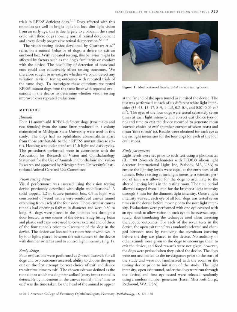
Vision testing deviceVisual performance was assessed using the vision testingdevice previously described with slight modifications.9 Asolid topped, 1.2 m square junction box, 0.9 m high, wasconstructed of wood with a wire-reinforced canvas tunnelextending from each of the four sides. These circular canvastunnels had openings 0.69 m in diameter and were 0.80 mlong. All dogs were placed in the junction box through adoor located in one corner of the device. Snug fitting foamand plastic end caps were used to cover external end of threeof the four tunnels prior to placement of the dog in thedevice. The device was located in a room free of windows, litby four lights placed between the exit tunnels of the devicewith dimmer switches used to control light intensity (Fig. 1).
Study designFour evaluations were performed at 2-week intervals for alldogs and two outcomes assessed, ability to choose the openexit on the first attempt ‘correct choice of exit’ and devicetransit time ‘time to exit’. The chosen exit was defined as thetunnel into which the dog first walked (entry into a tunnel isdetectable by movement in the canvas tunnel). The ‘time toexit’ was the time taken for the head of the animal to appear
at the far end of the open tunnel as it exited the device. Thetest was performed at each of six different white light inten-sities (35–45, 15–17, 8–9, 1–1.5, 0.2–0.4, and 0.02–0.04 cd/m2). The eyes of the four dogs were tested separately seventimes at each light intensity and correct exit choice (yes orno) and time to exit the device recorded to generate mean‘correct choice of exit’ (number correct of seven tests) andmean ‘time to exit’ (s). Results were obtained for each eye atthe six light intensities for the four dogs for each of the fourevaluations.
Study parametersLight levels were set prior to each test using a photometer(IL 1700 Research Radiometer with SED033 silicon lightdetector; International Light, Inc, Peabody, MA, USA) toensure the lighting levels were equal at the entrances of alltunnels. Before testing at each light intensity, a standard per-iod of time was allowed for the dogs to acclimate to thealtered lighting levels in the testing room. The time periodallowed ranged from 1 min for the brightest light intensitythrough 5 min for the dimmest light intensity. Once a lightintensity was set, each eye of all four dogs was tested seventimes in the device before moving onto the next light inten-sity. Evaluations were performed with one eye covered withan eye mask to allow vision in each eye to be assessed sepa-rately, thus simulating the technique used when assessingtherapeutic outcomes. For each of the seven tests in thedevice, the open exit tunnel was randomly selected and chan-ged between tests by removing the styrofoam coveringbefore the dog was placed in the device. No auditory orother stimuli were given to the dogs to encourage them toexit the device, and food rewards were not given; however,the dogs were praised when they exited the device. The dogswere not acclimated to the investigators prior to the start ofthe study and were not familiarized with the room or thetesting device prior to initiation of the study. The lightintensity, open exit tunnel, order the dogs were run throughthe device, and first eye tested were selected randomlyusing a random number generator (Excel; Microsoft Corp.,Redmond, WA, USA).
Figure 1. Modification of Gearhart et al.’s vision testing device.
r e p r o d u c i b i l i t y o f a c a n i n e v i s i o n t e s t i n g t e c h n i q u e 325
� 2012 American College of Veterinary Ophthalmologists, Veterinary Ophthalmology, 16, 324–328
Data analysisThe response variables were time to exit (s) and correctchoice of exit (n/7). The factors that could affect eachresponse variable were week, light intensity, eye, and dog.Data were analyzed using a four-factor ANOVA with the fixedfactors of week, light intensity, eye, and the random factor ofdog. The errors were assessed for normality by examinationof the histogram and normal probability plot. P values werecalculated with significance set at P < 0.05. Post hoc compari-sons were by means of Bonferroni t-test (m = 15 for lightintensity and m = 6 for week) to identify the specific time-points that were significantly different. Data were analyzedusing SAS PROC MIXED v 9.1.3 (SAS Institute Inc., Cary, NC,USA).
RESULTS
There was a significant effect of light intensity on time toexit and correct choice of exit. As lighting levels decreased,time to exit the device increased (P < 0.0001) and the num-ber of correct choices of exit declined (P < 0.0001). Therewas no significant variation in either time to exit or correctchoice of exit, between right and left eyes at any light inten-sity (P values ranged from 0.34 to 0.9 and 0.21 to 0.87,respectively). Assessment for normality of the data showed itto be unimodal and approximately symmetrical and there-fore acceptable for the analysis of variance given the robust-ness of ANOVA to departures from normality.
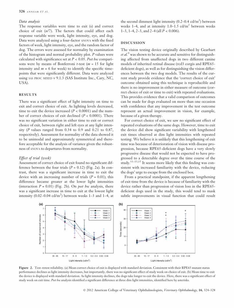
Effect of trial (week)Assessment of correct choice of exit found no significant dif-ference between the four trials (P = 0.12) (Fig. 2a). In con-trast, there was a significant increase in time to exit thedevice with an increasing number of trials (P = 0.01); thisdifference became greater at the lower light intensities(interaction P = 0.01) (Fig. 2b). On post hoc analysis, therewas a significant increase in time to exit at the lowest lightintensity (0.02–0.04 cd/m2) between weeks 1–3 and 1–4, at
the second dimmest light intensity (0.2–0.4 cd/m2) betweenweeks 1–4, and at intensity 1.0–1.5 cd/m2 between weeks1–3, 1–4, 2–3, and 2–4 (all P < 0.006).
DISCUSSION
The vision testing device originally described by Gearhartet al.9 was shown to be accurate and sensitive for distinguish-ing affected from unaffected dogs in two different caninemodels of inherited retinal disease (rcd3 corgis and RPE65-deficient dogs), as well as for distinguishing the vision differ-ences between the two dog models. The results of the cur-rent study provide evidence that the ‘correct choice of exit’outcome obtained using this technique is reproducible andthere is no improvement in either measure of outcome (cor-rect choice of exit or time to exit) with repeated evaluations.This provides evidence that a valid comparison of outcomescan be made for dogs evaluated on more than one occasionwith confidence that any improvement in the test outcomerepresent an actual improvement in vision, for example,because of a given therapy.
For correct choice of exit, we saw no significant effect ofrepeated evaluations of the same dogs. However, time to exitthe device did show significant variability with lengthenedexit times observed at dim light intensities with repeatedtesting. We believe it is unlikely that this lengthening of exittime was because of deterioration of vision with disease pro-gression, because RPE65-deficient dogs have a very slowlyprogressive disease that would not be expected to have pro-gressed to a detectable degree over the time course of thestudy.11–13,15 It seems more likely that this finding was con-sistent with increased familiarity with the device, reducingthe dogs’ urge to escape from the enclosed box.
From a practical standpoint, if the apparent lengtheningof exit time from the device is because of familiarity with thedevice rather than progression of vision loss in the RPE65-deficient dogs used in the study, this would tend to masksubtle improvements in visual function that could result
(a) (b)
Figure 2. Test–retest reliability. (a) Mean correct choice of exit is displayed with standard deviation. Consistent with their RPE65 mutant status
performance declines as light intensity decreases, but importantly, there was no significant effect of study week on choice of exit. (b) Mean time to exit
the device is displayed with standard deviation. As light intensity declines, the dogs take longer to exit the device. Here, there was a significant effect of
study week on exit time. Post hoc analysis identified a significant difference at three dim light intensities, identified here by asterisks.
326 a n n e a r E T A L .
� 2012 American College of Veterinary Ophthalmologists, Veterinary Ophthalmology, 16, 324–328
from therapy. However, if the vision testing device wasbeing used to monitor vision changes over time, the intertestintervals would probably be much greater than the 2-weekintervals in this study. Hence, familiarity with the devicecould be anticipated to play a much smaller role, and diseaseprogression may have a bearing on vision testing outcomes.For this study, very close intervals of 2 weeks between thefour evaluation weeks were chosen to increase the chance ofdetecting the influence of some nonvisual cue that mightimprove outcomes. Also of practical consideration is thegreater variation in time to exit the device at dim light inten-sities. This shows the value of including a choice-basedassessment of outcome in addition to a time-based measure,as Gearhart’s device allows.
The findings of this study support the validity of visiontesting results obtained using the Gearhart device for moni-toring response to therapy by repeated assessments of vision.Use of this objective vision testing device offers advantagesover the more subjective techniques such as obstacle course-based evaluations and can be used to test vision under scoto-pic, mesopic, or phototopic conditions. The availability ofan objective assessment of canine vision such as this is con-sidered critically important with the increasing emphasisthat is placed on the use of dog retinal dystrophy models inassessment of therapies.1–4,8 Here, the assessment of visionis important because, as shown by phase I and II clinicaltrials in RPE65 Leber Congenital Amaurosis patients, it ispossible to rescue function in sufficient photoreceptors toimprove vision, while there are not sufficient numbers res-cued to result in a measurable change in the electroretino-gram (which records a summed response originating fromphotoreceptors and resulting in inner retinal neuronresponses).16–20 The limitation of the device, as pointed outby Gearhart et al.,9 is that it is not assessing visual acuity.More sophisticated tests would be required if the goal was toassess visual acuity. Our findings in this study, while sup-porting the validity of using this device for repeated tests ofvision, should be considered in light of the fact that onlyeight eyes of four dogs were studied. Here, we sought toreduce the effect of variability between dogs with RPE65deficiency by including dogs from a single litter. This alsoexcluded the confounding effect of disease progression withage, but did result in the aforementioned limitation of a lowsample size.
The vision testing device originally described byGearhart et al.9 has previously been shown to be accuratefor discriminating affected from unaffected dogs with inher-ited retinal dystrophies and also for distinguishing betweentwo retinal dystrophies. Here, we provide evidence for thereproducibility of ‘correct exit choice’ with repeated testingusing this device. This supports the suitability of using thistechnique to assess for improvements in vision in responseto therapeutic intervention, with confidence that the changeseen is due to the given therapy. The importance of includ-ing a choice-based measure of outcome along with anassessment of device transit time was also highlighted.
ACKNOWLEDGMENTS
This work was supported by the British Retinitis Pigmen-tosa Society, The Midwest Eye Banks and TransplantationCenter Research Program and Michigan State UniversityCollege of Veterinary Medicine Purebred Dog EndowmentFund. The authors acknowledge Janice Querubin for pro-viding veterinary technician expertise in all aspects of han-dling and caring for the dogs and Dr. Cheri Johnson forassistance with canine reproduction.
REFERENCES
1. Acland GM, Aguirre GD, Bennett J et al. Long-term restoration
of rod and cone vision by single dose rAAV-mediated gene trans-
fer to the retina in a canine model of childhood blindness. Molec-
ular Therapy: The Journal of the American Society of Gene Therapy2005; 12: 1072–1082. Epub 2005/10/18.
2. Gearhart PM, Gearhart C, Thompson DA et al. Improvement of
visual performance with intravitreal administration of 9-cis-retinal
in Rpe65-mutant dogs. Archives of Ophthalmology 2010; 128:
1442–1448. Epub 2010/09/15.
3. Komaromy AM, Alexander JJ, Rowlan JS et al. Gene therapy res-
cues cone function in congenital achromatopsia. Human Molecular
Genetics 2010; 19: 2581–2593. Epub 2010/04/10.
4. Beltran WA, Cideciyan AV, Lewin AS et al. Gene therapy rescues
photoreceptor blindness in dogs and paves the way for treating
human X-linked retinitis pigmentosa. Proceedings of the National
Academy of Sciences of the United States of America 2012; 109:
2132–2137. Epub 2012/02/07.
5. Acland GM, Aguirre GD, Ray J et al. Gene therapy restores
vision in a canine model of childhood blindness. Nature Genetics
2001; 28: 92–95. Epub 2001/04/28.
6. Narfstrom K, Katz ML, Ford M et al. In vivo gene therapy in
young and adult RPE65)/) dogs produces long-term visual
improvement. The Journal of Heredity 2003; 94: 31–37. Epub
2003/04/15.
7. Garcia MM, Ying GS, Cocores CA et al. Evaluation of a behav-
ioral method for objective vision testing and identification of
achromatopsia in dogs. American Journal of Veterinary Research2010; 71: 97–102. Epub 2010/01/02.
8. Beltran WA. The use of canine models of inherited retinal degen-
eration to test novel therapeutic approaches. Veterinary Ophthal-
mology 2009; 12: 192–204. Epub 2009/04/28.
9. Gearhart PM, Gearhart CC, Petersen-Jones SM. A novel method
for objective vision testing in canine models of inherited retinal
disease. Investigative Ophthalmology & Visual Science 2008; 49:
3568–3576. Epub 2008/07/29.
10. Annear MJ, Bartoe JT, Barker SE et al. Gene therapy in the sec-
ond eye of RPE65-deficient dogs improves retinal function. GeneTherapy 2011; 18: 53–61. Epub 2010/08/13.
11. Narfstrom K, Wrigstad A, Nilsson SE. The Briard dog: a new
animal model of congenital stationary night blindness. The Brit-
ish Journal of Ophthalmology 1989; 73: 750–756. Epub 1989/09/
01.
12. Wrigstad A, Narfstrom K, Nilsson SE. Slowly progressive
changes of the retina and retinal pigment epithelium in Briard
dogs with hereditary retinal dystrophy. A morphological study.
Documenta Ophthalmologica. Advances in Ophthalmology 1994; 87:
337–354. Epub 1994/01/01.
13. Wrigstad A, Nilsson SE, Narfstrom K. Ultrastructural changes of
the retina and the retinal pigment epithelium in Briard dogs with
r e p r o d u c i b i l i t y o f a c a n i n e v i s i o n t e s t i n g t e c h n i q u e 327
� 2012 American College of Veterinary Ophthalmologists, Veterinary Ophthalmology, 16, 324–328
hereditary congenital night blindness and partial day blindness.
Experimental Eye Research 1992; 55: 805–818. Epub 1992/12/01.
14. Aguirre GD, Baldwin V, Pearce-Kelling S et al. Congenital sta-
tionary night blindness in the dog: common mutation in the
RPE65 gene indicates founder effect. Molecular Vision 1998; 4: 23.
Epub 1998/11/11.
15. Veske A, Nilsson SE, Narfstrom K et al. Retinal dystrophy of
Swedish briard/briard-beagle dogs is due to a 4-bp deletion in
RPE65. Genomics 1999; 57: 57–61. Epub 1999/04/07.
16. Bainbridge JW, Smith AJ, Barker SS et al. Effect of gene therapy
on visual function in Leber’s congenital amaurosis. The New Eng-land Journal of Medicine 2008; 358: 2231–2239. Epub 2008/04/29.
17. Hauswirth WW, Aleman TS, Kaushal S et al. Treatment of leber
congenital amaurosis due to RPE65 mutations by ocular subreti-
nal injection of adeno-associated virus gene vector: short-term
results of a phase I trial. Human Gene Therapy 2008; 19: 979–990.
Epub 2008/09/09.
18. Maguire AM, Simonelli F, Pierce EA et al. Safety and efficacy of
gene transfer for Leber’s congenital amaurosis. The New EnglandJournal of Medicine 2008; 358: 2240–2248. Epub 2008/04/29.
19. Simonelli F, Maguire AM, Testa F et al. Gene therapy for Leber’s
congenital amaurosis is safe and effective through 1.5 years after
vector administration. Molecular Therapy: The Journal of the Ameri-can Society of Gene Therapy 2010; 18: 643–650. Epub 2009/12/03.
20. Cideciyan AV, Hauswirth WW, Aleman TS et al. Human RPE65
gene therapy for Leber congenital amaurosis: persistence of early
visual improvements and safety at 1 year. Human Gene Therapy2009; 20: 999–1004. Epub 2009/07/09.
328 a n n e a r E T A L .
� 2012 American College of Veterinary Ophthalmologists, Veterinary Ophthalmology, 16, 324–328