Embed Size (px)
Citation preview
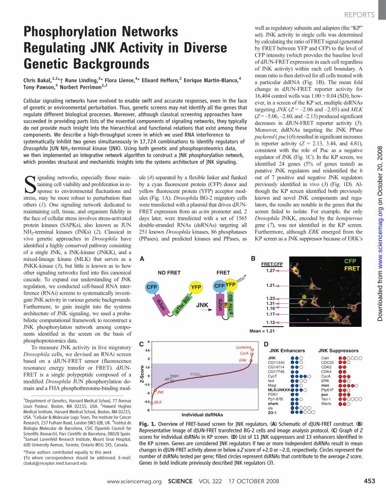
Phosphorylation NetworksRegulating JNK Activity in DiverseGenetic BackgroundsChris Bakal,1,2*† Rune Linding,3* Flora Llense,4* Elleard Heffern,2 Enrique Martin-Blanco,4Tony Pawson,5 Norbert Perrimon1,2
Cellular signaling networks have evolved to enable swift and accurate responses, even in the faceof genetic or environmental perturbation. Thus, genetic screens may not identify all the genes thatregulate different biological processes. Moreover, although classical screening approaches havesucceeded in providing parts lists of the essential components of signaling networks, they typicallydo not provide much insight into the hierarchical and functional relations that exist among thesecomponents. We describe a high-throughput screen in which we used RNA interference tosystematically inhibit two genes simultaneously in 17,724 combinations to identify regulators ofDrosophila JUN NH2-terminal kinase (JNK). Using both genetic and phosphoproteomics data,we then implemented an integrative network algorithm to construct a JNK phosphorylation network,which provides structural and mechanistic insights into the systems architecture of JNK signaling.
Signaling networks, especially those main-taining cell viability and proliferation in re-sponse to environmental fluctuations and
stress, may be more robust to perturbation thanothers (1). One signaling network dedicated tomaintaining cell, tissue, and organism fidelity inthe face of cellular stress involves stress-activatedprotein kinases (SAPKs), also known as JUNNH2-terminal kinases (JNKs) (2). Classical invivo genetic approaches in Drosophila haveidentified a highly conserved pathway consistingof a single JNK, a JNK-kinase (JNKK), and amixed-lineage kinase (MLK) that serves as aJNKK-kinase (3), but little is known as to howother signaling networks feed into this canonicalcascade. To expand our understanding of JNKregulation, we conducted cell-based RNA inter-ference (RNAi) screens to systematically investi-gate JNK activity in various genetic backgrounds.Furthermore, to gain insight into the systemsarchitecture of JNK signaling, we used a proba-bilistic computational framework to reconstruct aJNK phosphorylation network among compo-nents identified in the screen on the basis ofphosphoproteomics data.
To measure JNK activity in live migratoryDrosophila cells, we devised an RNAi screenbased on a dJUN-FRET sensor (fluorescenceresonance energy transfer or FRET). dJUN-FRET is a single polypeptide composed of amodified Drosophila JUN phosphorylation do-main and a FHA phosphothreonine-binding mod-
ule (4) separated by a flexible linker and flankedby a cyan fluorescent protein (CFP) donor andyellow fluorescent protein (YFP) acceptor mod-ules (Fig. 1A). Drosophila BG-2 migratory cellswere transfected with a plasmid that drives dJUN-FRET expression from an actin promoter and, 2days later, were transfected with a set of 1565double-stranded RNAs (dsRNAs) targeting all251 known Drosophila kinases, 86 phosphatases(PPases), and predicted kinases and PPases, as
well as regulatory subunits and adapters (the “KP”set). JNK activity in single cells was determinedby calculating the ratio of FRETsignal (generatedby FRET between YFP and CFP) to the level ofCFP intensity (which provides the baseline levelof dJUN-FRETexpression in each cell regardlessof JNK activity) within each cell boundary. Amean ratio is then derived for all cells treated witha particular dsRNA (Fig. 1B). The mean foldchange in dJUN-FRET reporter activity for16,404 control wells was 1.00 T 0.04 (SD); how-ever, in a screen of the KP set, multiple dsRNAstargeting JNK (Z = –2.06 and –2.05) and MLK(Z = –5.06, –2.60, and –2.13) produced significantdecreases in dJUN-FRET reporter activity (5).Moreover, dsRNAs targeting the JNK PPasepuckered ( puc) (6) resulted in significant increasesin reporter activity (Z = 2.13, 3.44, and 4.81),consistent with the role of Puc as a negativeregulator of JNK (Fig. 1C). In the KP screen, weidentified 24 genes (5% of genes tested) asputative JNK regulators and reidentified the 6out of 7 positive and negative JNK regulatorspreviously identified in vivo (3) (Fig. 1D). Al-though the KP screen identified both previouslyknown and novel JNK components and regu-lators, the results are notable in the genes that thescreen failed to isolate. For example, the onlyDrosophila JNKK, encoded by the hemipterousgene (7), was not identified in the KP screen.Furthermore, although ERK emerged from theKP screen as a JNK suppressor because of ERK’s
1Department of Genetics, Harvard Medical School, 77 AvenueLouis Pasteur, Boston, MA 02215, USA. 2Howard HughesMedical Institute, Harvard Medical School, Boston, MA 02215,USA. 3Cellular & Molecular Logic Team, The Institute for CancerResearch, 237 Fulham Road, London SW3 6JB, UK. 4Institut deBiologia Molecular de Barcelona, CSIC (Spanish Council forScientific Research), Parc Científic de Barcelona, 08028 Spain.5Samuel Lunenfeld Research Institute, Mount Sinai Hospital,600 University Avenue, Toronto, Ontario M5G 1X5, Canada.
*These authors contributed equally to this work†To whom correspondence should be addressed. E-mail:[email protected]
YFP
pThr
BDdJun
CFP YFP
dJu
n
CFP
pT
hr
BD
PJNK
CFPFRET1.27
1.21
1.23
1.12
1.17
1.311.16
FRET:CFP
Mean = 1.21
puckered
JNK
CycA
ERK
AKTPTEN
MLK
JNKCG11440CG14714CG17746CycTfwdMagiMLK/JNKKKPDK1Pp1-87BsharkslsZO-1
CakiCDC25CDK2CDK4CycAERKmsnPtp61FpucTao-1Warts
JNK Enhancers JNK Suppressors
NO FRET FRET
hippo
Z-S
core
Individual dsRNAs
A B
C D
Fig. 1. Overview of FRET-based screen for JNK regulators. (A) Schematic of dJUN-FRET construct. (B)Representative image of dJUN-FRET transfected BG-2 cells and image analysis protocol. (C) Graph of Zscores for individual dsRNAs in KP screen. (D) List of 11 JNK suppressors and 13 enhancers identified inthe KP screen. Genes are considered JNK regulators if two or more independent dsRNAs result in meanchanges in dJUN-FRET activity above or below a Z score of +2.0 or –2.0, respectively. Circles represent thenumber of dsRNAs tested per gene; filled circles represent dsRNAs that contribute to the average Z score.Genes in bold indicate previously described JNK regulators (3).
www.sciencemag.org SCIENCE VOL 322 17 OCTOBER 2008 453
REPORTS
on
Oct
ober
20,
200
8 w
ww
.sci
ence
mag
.org
Dow
nloa
ded
from
potential positive effects on puc transcription(fig. S1), we did not identify dsRNAs targetingother components of the ERK pathway. A highfalse-negative rate appears to be present in thisgenetic screen; therefore, we developed a combi-natorial strategy to further enhance the sensitivityof the screen.
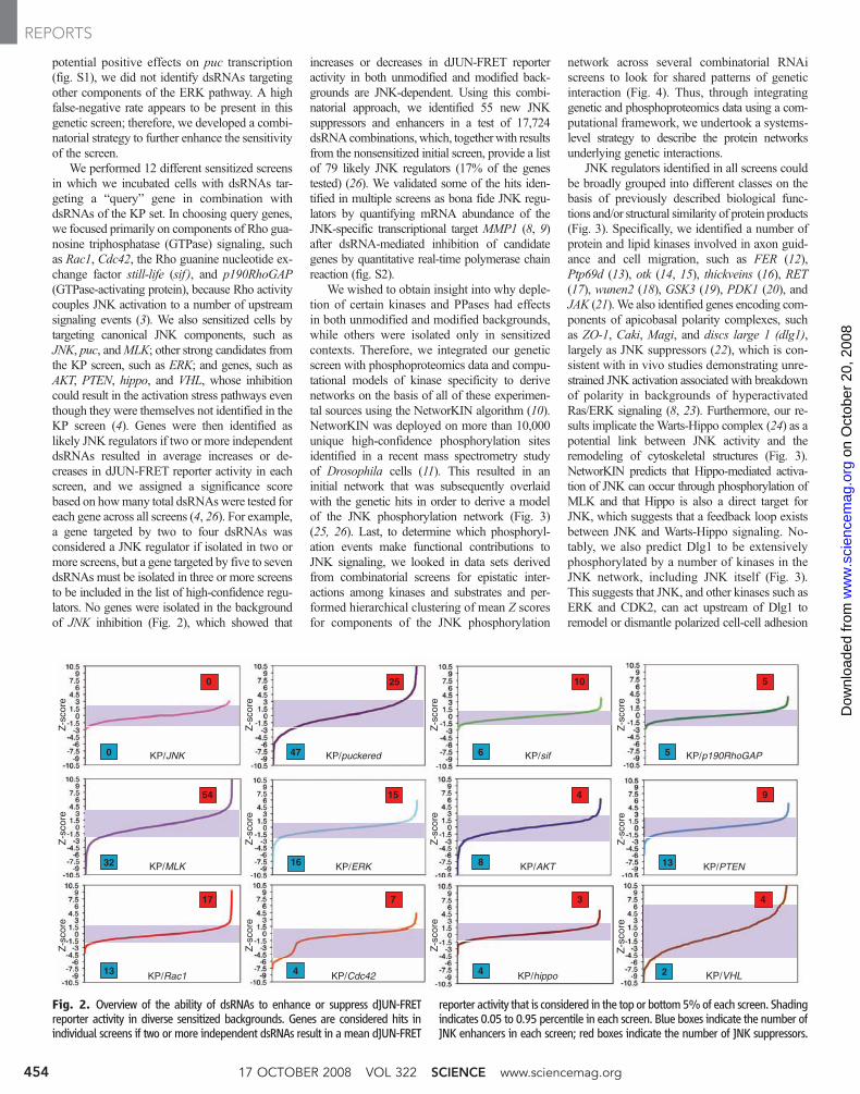
We performed 12 different sensitized screensin which we incubated cells with dsRNAs tar-geting a “query” gene in combination withdsRNAs of the KP set. In choosing query genes,we focused primarily on components of Rho gua-nosine triphosphatase (GTPase) signaling, suchas Rac1, Cdc42, the Rho guanine nucleotide ex-change factor still-life (sif ), and p190RhoGAP(GTPase-activating protein), because Rho activitycouples JNK activation to a number of upstreamsignaling events (3). We also sensitized cells bytargeting canonical JNK components, such asJNK, puc, andMLK; other strong candidates fromthe KP screen, such as ERK; and genes, such asAKT, PTEN, hippo, and VHL, whose inhibitioncould result in the activation stress pathways eventhough they were themselves not identified in theKP screen (4). Genes were then identified aslikely JNK regulators if two or more independentdsRNAs resulted in average increases or de-creases in dJUN-FRET reporter activity in eachscreen, and we assigned a significance scorebased on howmany total dsRNAswere tested foreach gene across all screens (4, 26). For example,a gene targeted by two to four dsRNAs wasconsidered a JNK regulator if isolated in two ormore screens, but a gene targeted by five to sevendsRNAs must be isolated in three or more screensto be included in the list of high-confidence regu-lators. No genes were isolated in the backgroundof JNK inhibition (Fig. 2), which showed that
increases or decreases in dJUN-FRET reporteractivity in both unmodified and modified back-grounds are JNK-dependent. Using this combi-natorial approach, we identified 55 new JNKsuppressors and enhancers in a test of 17,724dsRNAcombinations, which, togetherwith resultsfrom the nonsensitized initial screen, provide a listof 79 likely JNK regulators (17% of the genestested) (26). We validated some of the hits iden-tified in multiple screens as bona fide JNK regu-lators by quantifying mRNA abundance of theJNK-specific transcriptional target MMP1 (8, 9)after dsRNA-mediated inhibition of candidategenes by quantitative real-time polymerase chainreaction (fig. S2).
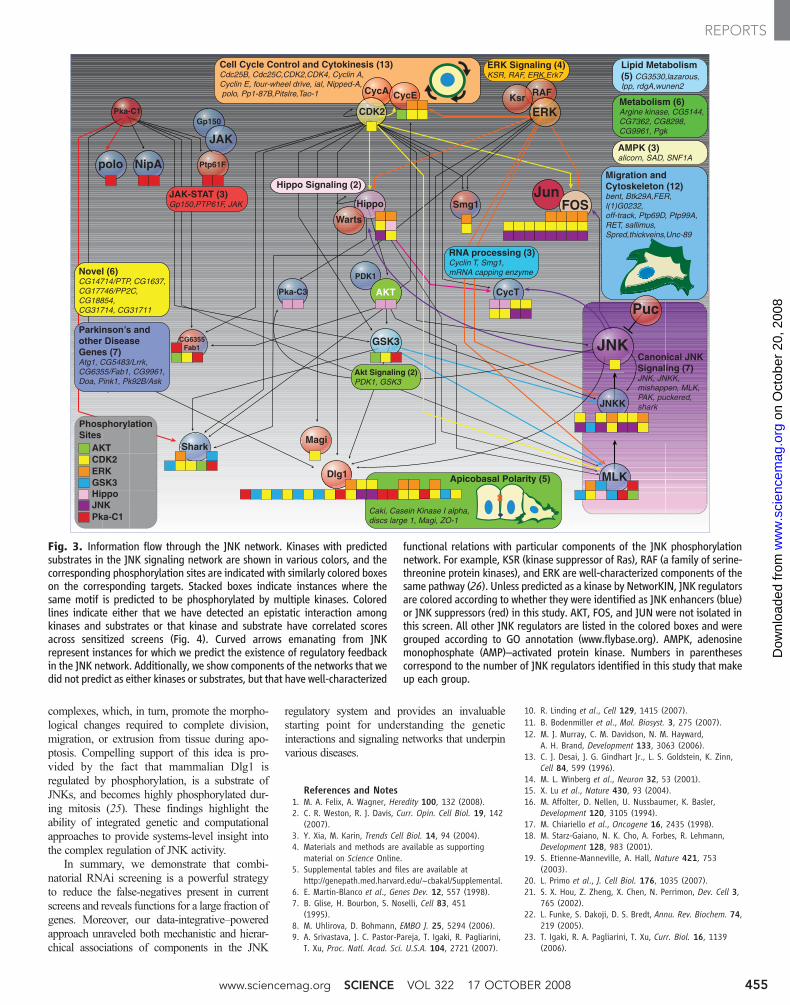
We wished to obtain insight into why deple-tion of certain kinases and PPases had effectsin both unmodified and modified backgrounds,while others were isolated only in sensitizedcontexts. Therefore, we integrated our geneticscreen with phosphoproteomics data and compu-tational models of kinase specificity to derivenetworks on the basis of all of these experimen-tal sources using the NetworKIN algorithm (10).NetworKIN was deployed on more than 10,000unique high-confidence phosphorylation sitesidentified in a recent mass spectrometry studyof Drosophila cells (11). This resulted in aninitial network that was subsequently overlaidwith the genetic hits in order to derive a modelof the JNK phosphorylation network (Fig. 3)(25, 26). Last, to determine which phosphoryl-ation events make functional contributions toJNK signaling, we looked in data sets derivedfrom combinatorial screens for epistatic inter-actions among kinases and substrates and per-formed hierarchical clustering of mean Z scoresfor components of the JNK phosphorylation
network across several combinatorial RNAiscreens to look for shared patterns of geneticinteraction (Fig. 4). Thus, through integratinggenetic and phosphoproteomics data using a com-putational framework, we undertook a systems-level strategy to describe the protein networksunderlying genetic interactions.
JNK regulators identified in all screens couldbe broadly grouped into different classes on thebasis of previously described biological func-tions and/or structural similarity of protein products(Fig. 3). Specifically, we identified a number ofprotein and lipid kinases involved in axon guid-ance and cell migration, such as FER (12),Ptp69d (13), otk (14, 15), thickveins (16), RET(17), wunen2 (18), GSK3 (19), PDK1 (20), andJAK (21).We also identified genes encoding com-ponents of apicobasal polarity complexes, suchas ZO-1, Caki, Magi, and discs large 1 (dlg1),largely as JNK suppressors (22), which is con-sistent with in vivo studies demonstrating unre-strained JNK activation associated with breakdownof polarity in backgrounds of hyperactivatedRas/ERK signaling (8, 23). Furthermore, our re-sults implicate theWarts-Hippo complex (24) as apotential link between JNK activity and theremodeling of cytoskeletal structures (Fig. 3).NetworKIN predicts that Hippo-mediated activa-tion of JNK can occur through phosphorylation ofMLK and that Hippo is also a direct target forJNK, which suggests that a feedback loop existsbetween JNK and Warts-Hippo signaling. No-tably, we also predict Dlg1 to be extensivelyphosphorylated by a number of kinases in theJNK network, including JNK itself (Fig. 3).This suggests that JNK, and other kinases such asERK and CDK2, can act upstream of Dlg1 toremodel or dismantle polarized cell-cell adhesion
Z-s
core
Z-s
core
25
Z-s
core
KP/Cdc42
0
KP/Rac113 4
Z-s
core
KP/puckered47
Z-s
core
Z-s
core
Z-s
core
KP/MLK
Z-s
core
17 7
KP/ERK
15
1632
54
0 KP/JNK
Z-s
core
Z-s
core
KP/sif6
10
KP/VHL
Z-s
core
KP/PTEN13
9
KP/p190RhoGAP5
5
8
4
2
4
KP/AKT
Z-s
core
KP/hippo4
3
Fig. 2. Overview of the ability of dsRNAs to enhance or suppress dJUN-FRETreporter activity in diverse sensitized backgrounds. Genes are considered hits inindividual screens if two or more independent dsRNAs result in a mean dJUN-FRET
reporter activity that is considered in the top or bottom5%of each screen. Shadingindicates 0.05 to 0.95 percentile in each screen. Blue boxes indicate the number ofJNK enhancers in each screen; red boxes indicate the number of JNK suppressors.
17 OCTOBER 2008 VOL 322 SCIENCE www.sciencemag.org454
REPORTS
on
Oct
ober
20,
200
8 w
ww
.sci
ence
mag
.org
Dow
nloa
ded
from
complexes, which, in turn, promote the morpho-logical changes required to complete division,migration, or extrusion from tissue during apo-ptosis. Compelling support of this idea is pro-vided by the fact that mammalian Dlg1 isregulated by phosphorylation, is a substrate ofJNKs, and becomes highly phosphorylated dur-ing mitosis (25). These findings highlight theability of integrated genetic and computationalapproaches to provide systems-level insight intothe complex regulation of JNK activity.
In summary, we demonstrate that combi-natorial RNAi screening is a powerful strategyto reduce the false-negatives present in currentscreens and reveals functions for a large fraction ofgenes. Moreover, our data-integrative–poweredapproach unraveled both mechanistic and hierar-chical associations of components in the JNK
regulatory system and provides an invaluablestarting point for understanding the geneticinteractions and signaling networks that underpinvarious diseases.
References and Notes1. M. A. Felix, A. Wagner, Heredity 100, 132 (2008).2. C. R. Weston, R. J. Davis, Curr. Opin. Cell Biol. 19, 142
(2007).3. Y. Xia, M. Karin, Trends Cell Biol. 14, 94 (2004).4. Materials and methods are available as supporting
material on Science Online.5. Supplemental tables and files are available at
http://genepath.med.harvard.edu/~cbakal/Supplemental.6. E. Martin-Blanco et al., Genes Dev. 12, 557 (1998).7. B. Glise, H. Bourbon, S. Noselli, Cell 83, 451
(1995).8. M. Uhlirova, D. Bohmann, EMBO J. 25, 5294 (2006).9. A. Srivastava, J. C. Pastor-Pareja, T. Igaki, R. Pagliarini,
T. Xu, Proc. Natl. Acad. Sci. U.S.A. 104, 2721 (2007).
10. R. Linding et al., Cell 129, 1415 (2007).11. B. Bodenmiller et al., Mol. Biosyst. 3, 275 (2007).12. M. J. Murray, C. M. Davidson, N. M. Hayward,
A. H. Brand, Development 133, 3063 (2006).13. C. J. Desai, J. G. Gindhart Jr., L. S. Goldstein, K. Zinn,
Cell 84, 599 (1996).14. M. L. Winberg et al., Neuron 32, 53 (2001).15. X. Lu et al., Nature 430, 93 (2004).16. M. Affolter, D. Nellen, U. Nussbaumer, K. Basler,
Development 120, 3105 (1994).17. M. Chiariello et al., Oncogene 16, 2435 (1998).18. M. Starz-Gaiano, N. K. Cho, A. Forbes, R. Lehmann,
Development 128, 983 (2001).19. S. Etienne-Manneville, A. Hall, Nature 421, 753
(2003).20. L. Primo et al., J. Cell Biol. 176, 1035 (2007).21. S. X. Hou, Z. Zheng, X. Chen, N. Perrimon, Dev. Cell 3,
765 (2002).22. L. Funke, S. Dakoji, D. S. Bredt, Annu. Rev. Biochem. 74,
219 (2005).23. T. Igaki, R. A. Pagliarini, T. Xu, Curr. Biol. 16, 1139
(2006).
ERK Signaling (4)KSR, RAF, ERK,Erk7
Parkinson’s and other Disease Genes (7)Atg1, CG5483/Lrrk, CG6355/Fab1, CG9961, Doa, Pink1, Pk92B/Ask
Migration and Cytoskeleton (12)bent, Btk29A,FER, l(1)G0232,off-track, Ptp69D, Ptp99A, RET, sallimus, Spred,thickveins,Unc-89
PDK1
Gp150
CycA CycE RAFKsr
JAK
Ptp61FNipApolo
CycT
Shark
Cell Cycle Control and Cytokinesis (13)Cdc25B, Cdc25C,CDK2,CDK4, Cyclin A, Cyclin E, four-wheel drive, ial, Nipped-A, polo, Pp1-87B,Pitslre,Tao-1
Magi
Dlg1
JunFOS
JNKK
MLK
CG6355Fab1
Pka-C1
Smg1
RNA processing (3)Cyclin T, Smg1,mRNA capping enzyme
Hippo
Warts
GSK3
Akt Signaling (2)PDK1, GSK3
Novel (6)CG14714/PTP, CG1637, CG17746/PP2C, CG18854,CG31714, CG31711
Lipid Metabolism (5) CG3530,lazarous,Ipp, rdgA,wunen2
AMPK (3)alicorn, SAD, SNF1A
Metabolism (6)Argine kinase, CG5144, CG7362, CG8298, CG9961, Pgk
ERKCDK2
JNK
PucAKT
Canonical JNK Signaling (7)JNK, JNKK, mishappen, MLK, PAK, puckered, shark
Caki, Casein Kinase I alpha, discs large 1, Magi, ZO-1
Apicobasal Polarity (5)
JAK-STAT (3)Gp150,PTP61F, JAK
Pka-C3
Hippo Signaling (2)
AKTCDK2ERKGSK3HippoJNKPka-C1
Phosphorylation Sites
Fig. 3. Information flow through the JNK network. Kinases with predictedsubstrates in the JNK signaling network are shown in various colors, and thecorresponding phosphorylation sites are indicated with similarly colored boxeson the corresponding targets. Stacked boxes indicate instances where thesame motif is predicted to be phosphorylated by multiple kinases. Coloredlines indicate either that we have detected an epistatic interaction amongkinases and substrates or that kinase and substrate have correlated scoresacross sensitized screens (Fig. 4). Curved arrows emanating from JNKrepresent instances for which we predict the existence of regulatory feedbackin the JNK network. Additionally, we show components of the networks that wedid not predict as either kinases or substrates, but that have well-characterized
functional relations with particular components of the JNK phosphorylationnetwork. For example, KSR (kinase suppressor of Ras), RAF (a family of serine-threonine protein kinases), and ERK are well-characterized components of thesame pathway (26). Unless predicted as a kinase by NetworKIN, JNK regulatorsare colored according to whether they were identified as JNK enhancers (blue)or JNK suppressors (red) in this study. AKT, FOS, and JUN were not isolated inthis screen. All other JNK regulators are listed in the colored boxes and weregrouped according to GO annotation (www.flybase.org). AMPK, adenosinemonophosphate (AMP)–activated protein kinase. Numbers in parenthesescorrespond to the number of JNK regulators identified in this study that makeup each group.
www.sciencemag.org SCIENCE VOL 322 17 OCTOBER 2008 455
REPORTS
on
Oct
ober
20,
200
8 w
ww
.sci
ence
mag
.org
Dow
nloa
ded
from
24. L. J. Saucedo, B. A. Edgar, Nat. Rev. Mol. Cell Biol. 8, 613(2007).
25. P. Massimi, D. Gardiol, S. Roberts, L. Banks, Exp. Cell Res.290, 265 (2003).
26. A. Friedman, N. Perrimon, Nature 444, 230 (2006).27. We are deeply indebted to the Drosophila RNAi
Screening Center and to J. Aach, S. Lee, C. Jørgensen,B. Mathey-Prevot, and B. Bodenmiller. The NetworKIN
and NetPhorest algorithms are available at http://networkin.info and http://NetPhorest.info, respectively.This work is supported by Genome Canada through theOntario Genomics Institute, the Spanish Ministerio deCiencía e Innovación (BFU/Consolider 2007), and theEuropean Union (FP6). C.B. is a Fellow of the Leukemiaand Lymphoma Society. N.P. is an Investigator of theHoward Hughes Medical Institute.
Supporting Online Materialwww.sciencemag.org/cgi/content/full/322/5900/453/DC1Materials and MethodsSOM TextFigs. S1 and S2
7 April 2008; accepted 20 August 200810.1126/science.1158739
Higher-Order CellularInformation Processing withSynthetic RNA DevicesMaung Nyan Win and Christina D. Smolke*
The engineering of biological systems is anticipated to provide effective solutions to challengesthat include energy and food production, environmental quality, and health and medicine. Ourability to transmit information to and from living systems, and to process and act on informationinside cells, is critical to advancing the scale and complexity at which we can engineer, manipulate,and probe biological systems. We developed a general approach for assembling RNA devices that canexecute higher-order cellular information processing operations from standard components. Theengineered devices can function as logic gates (AND, NOR, NAND, or OR gates) and signal filters,and exhibit cooperativity. RNA devices process and transmit molecular inputs to targeted proteinoutputs, linking computation to gene expression and thus the potential to control cellular function.
Genetically encoded technologies that per-form information processing, communi-cation, and control operations are needed
to produce new cellular functions from the di-verse molecular information encoded in the var-ious properties of small molecules, proteins, andRNA present within biological systems. For ex-
ample, genetic logic gates that process and trans-late multiple molecular inputs into prescribedamounts of signaling through new molecular out-puts would enable the integration of diverse en-vironmental and intracellular signals to a smallernumber of phenotypic responses. Basic operationssuch as signal filtering, amplification, and restora-tion would also enable expanded manipulation ofmolecular information through cellular networks.
Molecular information processing systemshave been constructed that perform computationwith biological substrates. For example, protein-based systems can perform logic operations to
convert molecular inputs to regulated transcrip-tional events (1–4). Information processing sys-tems that perform computation on small-moleculeand nucleic acid inputs can be constructed fromnucleic acid components (5–11). RNA-based sys-tems can process single inputs to regulated geneexpression events (12, 13) and integrate multipleregulatory RNAs for combinatorial gene regu-lation (14, 15). We sought to combine the richcapability of nucleic acids for performing infor-mation processing, transduction, and control op-erations with the design advantages expectedfrom the relative ease by which RNA structurescan be modeled and designed (16, 17).
We proposed a framework for the constructionof single input–single output RNA devices (18)based on the assembly of three functional compo-nents: a sensor component, made of an RNAaptamer (19); an actuator component, made of ahammerhead ribozyme (20); and a transmittercomponent, made of a sequence that couples thesensor and actuator components. The resultingdevices distribute between two primary conforma-tions: one in which the input cannot bind the sensor,and the other in which the input can bind the sensoras a result of competitive hybridization eventswithin the transmitter component. Input bindingshifts the distribution to favor the input-boundconformation as a function of increasing inputconcentration and is translated to a change in theactivity of the actuator, where a “ribozyme-active”state results in self-cleavage of the ribozyme (21).
Division of Chemistry and Chemical Engineering, CaliforniaInstitute of Technology, 1200 East California Boulevard,MC 210-41, Pasadena, CA 91125, USA.
*To whom correspondence should be addressed. E-mail:[email protected]
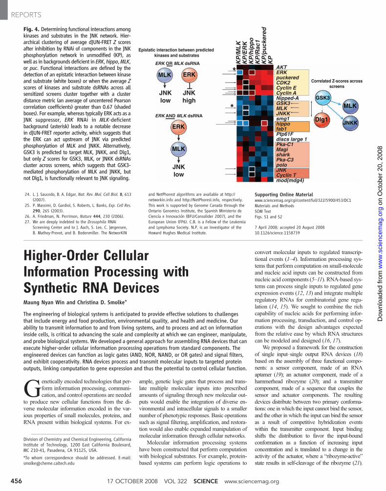
Fig. 4. Determining functional interactions amongkinases and substrates in the JNK network. Hier-archical clustering of average dJUN-FRET Z scoresafter inhibition by RNAi of components in the JNKphosphorylation network in unmodified (KP), aswell as in backgrounds deficient in ERK, hippo,MLK,or puc. Functional interactions are defined by thedetection of an epistatic interaction between kinaseand substrate (white boxes) or when the average Zscores of kinases and substrate dsRNAs across allsensitized screens cluster together with a clusterdistance metric (an average of uncentered Pearsoncorrelation coefficients) greater than 0.67 (shadedboxes). For example, whereas typically ERK acts as aJNK suppressor, ERK RNAi in MLK-deficientbackground (asterisk) leads to a notable decreasein dJUN-FRET reporter activity, which suggests thatthe ERK can act upstream of JNK via predictedphosphorylation of MLK and JNKK. Alternatively,GSK3 is predicted to target MLK, JNKK, and Dlg1,but only Z scores for GSK3, MLK, or JNKK dsRNAscluster across screens, which suggests that GSK3-mediated phosphorylation of MLK and JNKK, butnot Dlg1, is functionally relevant to JNK signaling.
KP
/ML
KK
P/E
RK
KP
/hip
po
KP
/Rac
1K
P/p
uck
ered
KP
MLK
JNKKDlg1
GSK3
*
*
JNK low
ERK OR MLK dsRNA
ERK AND MLK dsRNA
ERK
JNK high
JNK low
MLK
ERK
MLK
Epistatic interaction between predicted kinases and substrates
17 OCTOBER 2008 VOL 322 SCIENCE www.sciencemag.org456
REPORTS
on
Oct
ober
20,
200
8 w
ww
.sci
ence
mag
.org
Dow
nloa
ded
from