Embed Size (px)
Citation preview

Degree project in biotechnology Examensarbete I tillämpad bioteknologi 45 hp, Uppsala universitet 2010 Biology education centre. Department of medical biochemistry and microbiology Uppsala university Handledare: Dr. Per Jemth
Biophysical studies of E6/SAP97 interactions;
Implications in the pathogenesis of cervical
cancer
Abdul Aziz Qureshi
2
TABLE OF CONTENTS: page no.
1. SUMMARY. 3
2. INTRODUCTION. 4
2.1. Role of oncoprotein E7 in the pathogenesis of cervical cancer. 4
2.2. Contribution of oncoprotein E6 in the pathogenesis of cervical cancer. 5
2.3. Aim 1. 6
2.4. Aim 2. 6
3. RESULTS. 8
3.1. Protein purification. 8
3.2. Stability measurements. 8
3.3. Binding experiments. 10
4. DISCUSSION. 13
5. MATERIALS METHODS. 14
5.1. Description of plasmids. 14
5.2. Cloning protocol to generate K321A and S332A expression plasmids. 15
5.3. Protein over expression. 17
5.4. Protein purification. 17
5.5. Stability experiments. 20
5.6. Binding experiments. 21
6. ACKNOWLEDGEMENTS. 23
7. REFERENCES. 24
3
1. SUMMARY.
Oncoproteins E6 and E7 are encoded by the genome of human papilloma viruses (HPVs). HPVs are regarded as the main causative agents in approximately 99% cases of cervical cancer. HPV genotypes are classified as high and low risk types based on their prevalence in cervical cancer, HPV 16 being the most prevalent followed by HPV 18. E6 and E7 from high risk HPV types have been widely studied as the main players involved in the pathogenesis of cervical cancer. The major targets of E6 and E7 are p53 and pRb respectively. pRb is the regulator of transcription factors of E2F family important during S-phase of the cell cycle, binding of E7 to pRb results in the unregulated control of transcription factors that leads to the transformation of cells. p53 is a tumor suppressor protein; upon binding to E6, p53 undergoes proteasome-mediated degradation. However, E6 has shown to possess p53 independent functions most commonly the binding to SAP97 by its C-terminus. This C-terminal peptide is the characteristic feature of E6 of only high risk strains of HPVs. SAP97 from the family of MAGUK is a PDZ containing protein. It has three PDZ domains. SAP97 is associated to adherens junctions. Degradation of SAP97 upon binding to E6 results in defective cell-cell adhesions and loss of cell polarity that subsequently ends up in carcinogenesis. Recent studies involving the binding mechanism between the PDZ domains of SAP97 and full length C-terminal domain of E6 instead of the short peptide have gained more clues on the insight of their binding mechanism. Recent results from NMR studies of PDZ2/E6 binding interactions have revealed residues of PDZ2 that are not part of the binding pocket but underwent large and medium scale changes in their chemical shifts upon binding of E6. This finding implies that these residues might have some significant role in the binding of PDZ/E6 binding which may involve direct or indirect interactions to the binding pocket of PDZ2/E6 complex. In this study we aim to demonstrate the role of such residues that undergo structural rearrangements upon E6 binding in the binding interactions between E6 and PDZ2 of SAP97. For this purpose, two mutants were designed, K321A and S332A that represent the residues in close proximity to the binding pocket. There-after proteins were expressed and then purified. Stability measurements were carried out to demonstrate the effect of the mutations on PDZ2 stability. There was no significant difference seen in the stability of PDZ2 pwt and mutants. Rate constants were determined by stopped-flow binding measurements. Parameters obtained from these experiments suggest that mutants K321A and S332A do not possess any significant affect on the binding affinity of E6 to PDZ2. The results from this study proposed that these positions or residues do not involve in making direct or indirect interaction to the binding pocket and changes in their chemical shifts might bring out by some structural rearrangements in PDZ2 domain upon E6 binding or may due to intradomain allostery. Abbreviations: MAGUKs (membrane-associated guanylate kinase homologues). HPV (human papilloma viruses). PDZ (PSD-95/disc large/ ZO-1) domains. SAP97 (synapse-associated protein). PWT (pseudo-wild type) of PDZ2 domain. K321A mutant of PDZ2 (PWT). S332A mutant of PDZ2 (PWT).

4
2. INTRODUCTION.
Human papillomaviruses are small double stranded DNA viruses. They are characterized as the causative agents of over 95 % cases of cervical cancer, the second most prevailing cancer among women worldwide (Liu et al. 2007). More than 200 HPV genotypes have been identified based on genomic differences (Burd et al. 2003). These genotypes have been further grouped into high and low risk HPVs based on clinical prognosis of their associated lesions. Low risk forms include HPV types 1A, 6B and 11 that cause benign genital warts. However, types 5, 8,13,16,18 and 33 accounted for malignant lesions most prevalent being the cervix carcinoma (Bosch et al. 2003). Around 99% of cervical cancers contain HPV DNA of high-risk types, HPV16 being the most common (Woodman 2007). Nine proteins in total are encoded by the HPV genome in which E6 and E7 are regarded as the oncoproteins (Munger et al. 1989). Both E6 and E7 are consistently expressed in the cervical tumors and are the main mediators of carcinogenesis due to their interaction with various cellular targets (Munger et al. 2004).
2.1 ROLE OF ONCOPROTEIN E7 IN THE PATHOGENESIS OF CERVICAL CANCER.
E7 oncogene encodes small acidic polypeptides of approximately hundred amino acids (Figge 1988). The E7 protein has two regions at the N-terminal that are regarded as conserved regions marked as CR1 and CR2 and they both share a partial sequence and functional homology with the adenovirus E1A protein and the SV-40 large T-antigen (Dyson et al. 1992). A LXCXE conserved motif sits in the CR 2 region that is shown to be involved in bringing the interaction of E7 to its cellular targets (Singh 2005). At the C-terminal another conserved CR3 domain is present that contains two CXXC zinc- binding motifs separated by a 29/30 amino acid spacer. This region is implicated in association of pRb with a very low affinity and to other cellular proteins but most importantly in metal binding that brings out the zinc-dependent dimerization (Munger et al. 2004 and Hebner et al. 2006). In addition to pRb there are some other cellular targets of E7 including pRb activating cyclin-dependent kinase inhibitors p21 and class I histone deacetylases HDACs (Jones et al. 1997 and Lagger et al. 2003). However, pRb has been revealed and studied as the major cellular target of E7 carrying out the development and maintenance of carcinoma (Sherr 2004).
pRb is involved in controlling the cell cycle progression that is mainly done by regulating the members of the transcription factor E-2-F family (Polager et al. 2008). E-2-Fs are crucial in regulating the expression of genes that are required for entry into and then the progression through the S-phase of the cell cycle (Dimova et al. 2005). Hence, E-2-Fs are crucial in regulating the proliferation and cell-death pathways, and therefore any alteration in these pathways can result in the transformation of the cells manifested in the development of tumor (Munger 2004).
On the other hand pRb is responsible for regulating the activity of E-2-Fs by binding to E-2-F molecule and its phosphorylation dependent release bring out the transcription of S-phase genes (Harbour et al. 1999). Binding of pRb with E7 oncoprotein leads to mis-regulated release of E2F from pRb-E2F complex and thus this activates the transcription of S-phase genes and this role of E7 is mainly the actor for tumor development attributed to E7 HPVs (Munger 2004).

5
2.2. CONTRIBUTION OF ONCOPROTEIN E6 IN THE PATHOGENESIS OF CERVICAL CANCER.
Oncogene E6 of HPV encodes a small protein of about 150 amino acids with a molecular weight of 16-18 kda. E6 contains two zinc finger motifs composed of four C-X-X-C motifs which are required for E6 functions such as transcriptional activation, transformation, immortalization and association with cellular target proteins (Hebner et al. 2006, Cole et al. 1987). Even being a short length protein E6 has been known to be difficult to study due to lack of E6 stability to be isolated in native, soluble form. Presence of high contents of α-helical and β-sheet secondary structures in E6 results in the instability and insolubility of the protein once purified (Nomine et al. 2001). However, based on the sequence homology between N-terminal and C-terminal domains of the E6, structural properties of the C-terminal domain of E6 from HPV 16 have been reported. In the proposed model six non-conserved cysteins are mutated to serine. Substitutions in the proposed model not only make the purification of soluble E6 possible but the purified protein also retained the biological functions of the E6 including its ability to degrade p53 and it can also bind to its known protein partner E6-AP. Although, the proposed model for E6 does not address all the aspects of the E6 native structure such as interactions between two domains, structural appearance of two domains` margins etc but it does provide a platform to search and develop potential agents that could hamper the interaction between E6 and its cellular targets (Nomine et al. 2006, Tungteakkhun et al. 2008). Besides, available structural details of the E6 protein shows that E6 from high risk HPVs but not from low risk HPVs contains an additional conserved six residues peptide (R-R/T/N/Q-E-T-Q/E-V/L) at the C-terminus with T at -3 position and V/L at the -1 position that is the characteristic of PDZ domains binding peptides. This motif has been shown to be involved in the interaction with several PDZ domain containing proteins. This extra PDZ binding motif in E6 from high risk HPV indicates that oncogenic potential of E6 is correlated to this PDZ binding motif (Flores et al. 2006). HPV E6 protein has been shown to have multiple properties and has a variety of cellular partner proteins. These include p53, E6-AP, E6-BP (a calcium binding protein), hDlg/SAP97 and Bak (a regulator of the apoptosis pathway) (Mammas et al. 2008).
The principle activity of E6 is the formation of a complex with the cellular E6-associated ubiquitin ligase protein (E6-AP), which subsequently target p53 and carry out proteosome-mediated degradation of p53, resulting in the disruption of apoptosis, cell-cycle arrest, programmed cell death that eventually leads to viral infection and tumor development (Thomas et al.1999). In addition to other cellular targets of E6, hDlg (human homologue of the drosophila disc large tumor suppressor protein) also termed as SAP97 (synapse associated protein) has been shown to be another potent target of E6 from high risk HPVs. It is a PDZ domain containing protein (Kiyono et al. 1997). The C-terminal peptide in E6 from high risk types is not shown to be involved in p53 binding and degradation. However, this extra stretch of residues in the C-terminal is inolved in interaction to SAP97 (Gardiol 1999). SAP97 belongs to the family of proteins termed MAGUKs (membrane associated guanylate kinase homologues). MAGUKs are multifunctional proteins present at membrane-cytoskeleton interface at cell-cell junctions, fulfilling not only the structural but also the signaling purposes (Fanning et al. 1996). SAP97 is associated with adherens junctions. Degradation of SAP97 results in defective cell-cell adhesion, loss of cell polarity and unregulated cell proliferation that ultimately leads to tumor development (Nakagawa 2000). Like the down-regulation of p53, SAP97 has been shown to be targeted by E6-AP dependent, proteosome-mediated degradation. SAP97 consists of three PDZ domains which have been

6
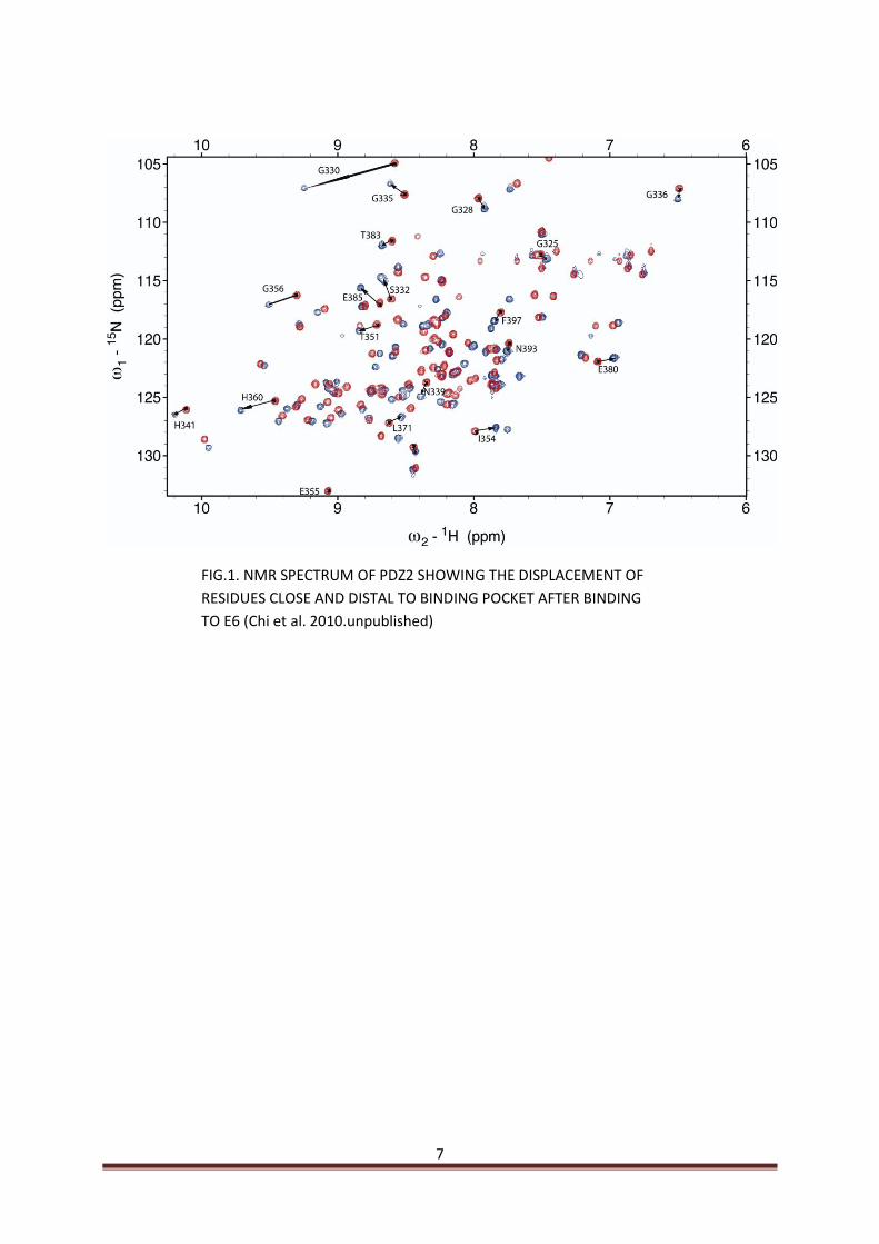
studied independently for their ability to bind diverse recognition sequences that include sequences of E6 from HPV16 (Gardiol 1999). Most of the studies to elucidate the binding mechanisms between E6 and SAP97 are limited to the use of short peptides of E6 corresponding to the C-terminal conserved region from high risk HPVs (Kiyono 1997, Gardiol 1999, Thomas 2001, Thomas 2008). For the first time the C-terminal domain from HPV16 E6 is employed by Chi in our lab in order to demonstrate binding affinities of various PDZ domains of SAP97 to E6 C-terminal domain (Chi et al. 2010.unpublished). Furthermore, NMR experiments performed on the E6-PDZ2 interaction that have revealed changes in the chemical shifts of residues of PDZ2 that are not the part of binding pocket upon binding to E6 (Fig.1). These residues are divided into two main groups on the basis of their proximity to the binding pocket. One group constitutes those residues that are close to the binding pocket and are expected to make direct or indirect interactions with the E6. The other group comprises of those residues that are not close but distal to binding pocket and are shown to exhibit large or medium scale change in their chemical shifts upon E6 binding which are not seen when the E6 C-terminal peptide is used (Chi et al. 2010.unpublished).
2.3. AIM 1.
Based on the current knowledge of the E7 protein, further study of this protein could result in targeting E7 with a suitable candidate in order to inhibit E7 activation. Therefore, E7 looks very promising as a target in order to find a suitable treatment for cervical cancer. I have selected E7 protein as the molecule to be studied during my degree project.
I have splitted my project into the following phases.
For the first phase expression and purification of E7 would be carried out, followed by its biophysical functional characterization including measurements of stability, folding and binding to ligands.
In the second phase the aim was to express and purify pRb.
In the third phase I aimed to study the binding of E7 to pRb.
However, this part of the project did not work out, since E7 could not be purified. Later on, I have decided to abandon this project.
2.4. AIM 2.
Based on the recent study by Chi I aimed to demonstrate the effect on the E6/PDZ2 binding of the residues that are close to the binding pocket of PDZ2 (Chi et al. 2010. unpublished). For this purpose two mutants were designed, K321A and S332A of PDZ2 that represent positions in close proximity of the binding pocket. Mutants and pseudo wild type (PDZ2) domains of SAP97 and E6 were purified followed by experiments to determine their stability. Later on the binding experiments were carried out to obtain the rate constants for PDZ2/E6 binding by time resolved spectroscopy.
7
FIG.1. NMR SPECTRUM OF PDZ2 SHOWING THE DISPLACEMENT OF
RESIDUES CLOSE AND DISTAL TO BINDING POCKET AFTER BINDING
TO E6 (Chi et al. 2010.unpublished)

8
3. RESULTS. 3.1. PURIFICATION. E7 was subjected to purification as a fusion with lipoyl-domain from E.coli. The fusion protein is successfully purified on anion exchanger column. However, isolated protein E7 could not be purified despite of several attempts. Therefore, I abandoned this task. Nothing is discussed any more regarding E7 in the discussion and summary sections of the project. However, all other proteins that include E6, PDZ2 domains (pwt) and mutants K321A and S332A were all successfully purified and their purity was checked on SDS-PAGE (Fig.2)
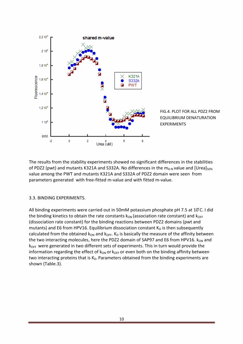
3.2. STABILITY MEASUREMENTS. Stability measurements were carried out in 50mM phosphate buffer pH 7.5. Fluorescence intensity was plotted against urea concentration and subsequently fitted to the equation for solvent denaturation of a two-state protein see materials methods (Fersht 1999). Values for mD-N and [D] 50% were generated from the equation from which ∆GD-N
[urea] 50% in H2O (free energy of denaturation when the protein is 50% denatured) was calculated, with free fitted and shared mD-N parameters (Fig.3 and 4) (Table.1 and 2).
PWT
TTT
PWT
PWT
K321A
K3K321
A
S332A E6 10kda M
FIG.2. SDS-PAGE GEL SHOWING
PURITY OF PURIFIED PROTEINS.

9
PDZ2 DOMAINS.
mD-N value. Free-fitted
kcal mol-1M-1
[Urea]50%
M ∆GD-NH2O Kcal/mol
Pseudo-wild type (PWT)
1.33±0.07 3.52±0.03 4.68±0.23
K321A 1.31±0.08 3.38±0.04 4.42±0.26
S332A 1.19±0.05 3.67±0.03 4.36±0.17
Table.1. Free-fitted parameters obtained from equilibrium denaturation experiments.
PDZ2 DOMAINS.
mD-N value. shared
kcal mol-1M-1
[Urea]50%
M ∆GD-NH2O Kcal/mol
Pseudo-wild type (PWT)
1.27±0.11 3.51±0.03 4.45±0.35
K321A 1.27±0.11 3.38±0.03 4.29±0.34
S332A 1.27±0.11 3.68±0.03 4.67±0.37
Table.2. Parameters from equilibrium denaturation experiments with shared mD-N value.
FIG.3. PLOT FOR ALL PDZ2 FROM
EQUILIBRIUM DENATURATION
EXPERIMENTS
10
The results from the stability experiments showed no significant differences in the stabilities of PDZ2 (pwt) and mutants K321A and S332A. No differences in the mD-N value and [Urea]50% value among the PWT and mutants K321A and S332A of PDZ2 domain were seen from parameters generated with free-fitted m-value and with fitted m-value. 3.3. BINDING EXPERIMENTS. All binding experiments were carried out in m otassium hos hate . at . did the binding kinetics to obtain the rate constants kON (association rate constant) and kOFF (dissociation rate constant) for the binding reactions between PDZ2 domains (pwt and mutants) and E6 from HPV16. Equilibrium dissociation constant KD is then subsequently calculated from the obtained kON and kOFF. KD is basically the measure of the affinity between the two interacting molecules, here the PDZ2 domain of SAP97 and E6 from HPV16. kON and kOFF were generated in two different sets of experiments. This in turn would provide the information regarding the effect of kON or kOFF or even both on the binding affinity between two interacting proteins that is KD. Parameters obtained from the binding experiments are shown (Table.3).
FIG.4. PLOT FOR ALL PDZ2 FROM
EQUILIBRIUM DENATURATION
EXPERIMENTS

11
PDZ2 DOMAINS
kON
µM s-1 kOFF
s-1 KD (koff/kon) µM
∆G (-RTln(Kd)) Kcal mol-1
PSEUDO WILD TYPE (PWT)
6.34±0.34 5.2±0.09 0.82±0.01 0.11±0.01
K321A 6.37±0.39 5.60±0.09 0.87±0.06 0.07±0.06
S332A 8.53±0.21 4.76±0.06 0.55±0.02 0.33±0.02
Table.3. Rate and equilibrium parameters obtained from PDZ2 and E6 interactions. Rate constants obtained from the binding experiments between PDZ2 domains and E6 from HPV16 allows a comparison on the binding affinity of E6 towards the PDZ2 domains (pwt) and PDZ2 mutants (K321A, S332). kON and kOFF were obtained in two different sets of experiments (Fig.5 and 6). KD for each interaction was calculated from the obtained kON and kOFF in each case. Data shows that there is no significant difference in the rate constants generated from the binding interactions of PDZ2 (pwt) and PDZ2 mutants, K321A and S332A suggesting that these mutations have no apparent role in the binding of E6 to PDZ2 domain. The change in the chemical shift of these residues after binding to E6 might come from intradomain allosteric interactions in the PDZ2 domain upon binding to E6 or may due to interactions to some parts of E6 C-terminus domain other than which constitutes the binding pocket.
FIG.5. PLOT OF kOBS FOR
ALL PDZ2 TO OBTAIN kON

12
FIG.6. PLOT OF kOBS FOR ALL
PDZ2 TO OBTAIN kOFF

13
4. DISCUSSION. PDZ domains (PSD-95/disc large/ZO-1) are 80-90 amino acids motif found in a few hundred of human proteins of distinct origin. They are present as either a single domain or in arrays function as specific protein recognition modules as well as have significant role in scaffolding and signal transduction (Gardiol et al. 1999, Hung et al. 2002). Structural studies revealed that PDZ domains most commonly bind to the C-terminal of their interacting proteins (Gianni et al. 2005). One of the major and direct roles of PDZ domains has been shown to be implicated in the pathogenesis of cervical cancer caused by the Human papilloma viruses (HPVs) (Bosch et al. 2003). E6 and E7 are the two oncoproteins of HPV, involved in the pathogenesis of cervical cancer. HPV`s genotypes are classified as high and low risk types based on their prevalence in the cervical cancer and their associated lesions, HPV 16 being the most common followed by HPV 18 (Burd et al. 2003, Woodman 2007). Although, the primary target of E6 is p53 with which E6 binds, E6 also binds to PDZ domain containing proteins with its conserved C-terminal peptide, characteristic of E6 from only high-risk HPVs. This conserved extra-stretch peptide at C-terminus of E6 has been implicated in its binding to hDlg also referred as SAP97 (human homologue of the drosophila disc large tumor suppressor protein Dlg) (Kiyono et al. 1997, Gardiol et al. 1999). SAP97 is found at tight junctions and play crucial role in cell-cell contact and cell rigidity. Binding of E6 to SAP97 leads to the proteasome-mediated degradation of SAP97, ultimately resulting in carcinogenesis (Nakagawa 2000). Until the recent study on E6/SAP97 by Chi (Chi et al. 2010.Unpublished), studies on E6/SAP97 have been limited to the use of short peptide from C-terminal of E6 (Kiyono 1997, Gardiol 1999, Thomas 2001, Thomas 2008). The use of short peptides to study E6/SAP97 interactions could not cover binding details completely. However, a study of Chi demonstrated some interesting features of E6/SAP97 interactions, in which the C-terminal domain of E6 with the L151V mutation is employed to investigate the binding interactions between them. NMR results in that study further elucidated the structural rearrangements of PDZ2 domain upon binding of E6 to PDZ2. Residues have been classified that undergo large and medium scale changes in their chemical shifts and are not the part of the binding pocket. These residues are divided into two main groups based on their proximity to the binding pocket of E6/PDZ2 complex, one group that is close and other is distal to the binding pocket. (Chi et al. 2010. unpublished). In this study, I aimed to study the effect of the residues K321 and S332 that are in close proximity to the binding pocket. For this purpose, mutants were designed (K321A and S332A) expressed and purified. In addition to the mutants pwt (PDZ2) and the C-terminal domain of E6 were also purified in order to carry out the binding studies. After purification of PDZ2 domains, stability measurements were performed in order to check the effect of the mutations on the stability of PDZ2 domains. Urea denaturation experiments for stability measurements suggest that the mutations imposed no such effect on the stability of PDZ2 domains with K321A and S332A mutations. Hence, the mutants are quite stable. Stability measurements were followed by binding experiments that include the determination of association and dissociation rate constants (kon and kOFF respectively). Equilibrium dissociation constant KD is subsequently obtained from the determined kon and kOFF. Rate constants are nearly the same for PDZ2 (pwt) and mutant K321A and S332A, reflecting no direct contribution of these residues or positions on the binding interaction

14
between PDZ2/E6 suggesting that change in their chemical shift might occurred by some structural rearrangements in the PDZ2 upon binding of E6. These findings in combination with the allosteric studies of PDZ2 domain of SAP97 could lead to the identification of the plausible sites that disrupt the PDZ/E6 complex due to allostery and ultimately pave the way for the identification of a competitive molecule that could serve as the antibody for E6, a potential therapeutic approach in the treatment of cervical cancer.

15
5. MATERIALS AND METHODS.
5.1. DESCRIPTION OF PLASMIDS USED DURING THE STUDY.
PDZ2 (pseudo-wild type-PWT) was provided by Celestine Chi. The construct was generated by the scheme described in one of the earlier papers by Celestine Chi (Chi et al. 2009).The cDNA construct of PDZ2 (pseudo-wild type) codes for residues 311-407 from SAP97. Isoleucine was replaced by tryptophan at position 342 (I342W) that serve as fluorescence probe in binding experiments giving a high signal to noise ratio in a stopped-flow spectrofluorimeter whereas, mutation C378A prevent the formation of disulphide bridges. The construct contains 12 extra residues along with the His-tag (MHHHHHLVPRGS) at the N-terminus.
PDZ2 SAP97 mutants K321A and S332A constructs were generated during the study using the already available PDZ2 (pseudo-wild type) plasmid as template that codes for residues 311-407 from SAP97 by employing inverted PCR.
C-terminal domain E6 provided by Chi with a L151V and four cystein to serine mutations from HPV16 used in this study. It was sub-cloned with an E.coli lipoyl-domain to enhance the expression with a thrombin digestion site and His-tag at N-terminal of lipoyl-domain by Celestine Chi in our lab using a cDNA construct coding for E6 wild type (residues 80-151) (Chi et al. 2010. unpublished).
Plasmid for E7 from HPV16 provided by Chi. also generated as fusion of E7 wild-type from HPV16 with His-tagged E.coli lipoyl-domain at N-terminal to enhance the expression; therefore in this case I had to start with expression as well. E7 fused with lipoyl-domain.
2x TY medium (5g/l NaCl, 10g/l yeast extract and 16g/l tryptone) was used for Escherichia coli cell cultures. For 2x TY plates, 15g/l agar was added. Plates and media both were supplemented with antibiotics ampicillin (100µg/ml, Astra Zeneca, Sweden) and chloramphenicol (34µg/ml, Calbiochem, USA) when desired otherwise specified.
Chemically competent Escherichia coli XL1 cells (Stratagene, USA) were used for cloning procedures. E.coli BL21 cells (DE3) PlysS (Invitrogen, USA) were used for protein over expression.
For heat-shock transformation, cells were thawed on ice before addition of 1.5 µl plasmid-DNA to 15µl and to 50µl chemically competent E.coli BL21 and XL1 cells, respectively. After incubation on ice for 15 min, cells are heat–shocked at for s and incubated on ice for another 3 min. 150 to 200µl 2x TY medium was added before incubation at for hour. Cells were plated on lates and incubated at C overnight.

16
5.2. CLONING PROTOCOL TO GENERATE K321A AND S332A EXPRESSION PLASMIDS.
PRIMERS FOR MUTANTS.
K321A_F
aaa ata atg gaa ata gcg ctc att aaa ggt cct
K321A_R
agg acc ttt aat gag cgc tat ttc cat tat ttt
S332A_F
aaa ggt ctt ggg ttt gct att gct gga ggt gtt
S332A_R
aac acc tcc agc aat agc aaa ccc aag acc ttt
PCR mixture in the PCR tube is prepared as the following scheme.
Template 1 µl
Forward primer 0.5 µl
Reverse primer 0.5 µl
DMSO(dimethyl sulfoxide)
1 µl
Pfu buffer (10x, Stratagene)
5 µl
dNTP (2.5mM stock) 2.5 µl
Pfu turbo (Stratagene) 0.5 µl
D.H2O Add to 50 µl
Table 4. PCR reaction ingredients to generate constructs for K321A and S332A PDZ2 of SAP97 using already available plasmid for PDZ2 (pseudo-wild type) of SAP97 as template.
17
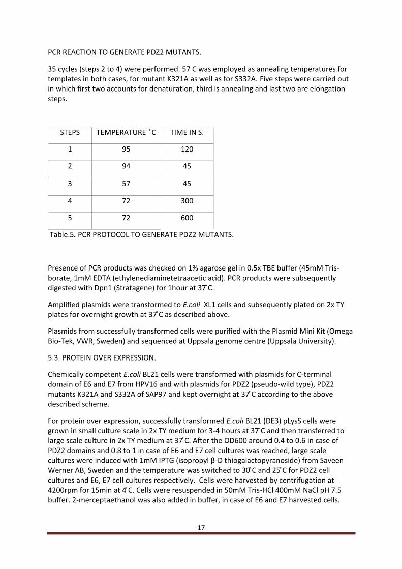
PCR REACTION TO GENERATE PDZ2 MUTANTS.
35 cycles (steps 2 to 4) were erformed. was employed as annealing temperatures for templates in both cases, for mutant K321A as well as for S332A. Five steps were carried out in which first two accounts for denaturation, third is annealing and last two are elongation steps.
STEPS TEMPERATURE C TIME IN S.
1 95 120
2 94 45
3 57 45
4 72 300
5 72 600
Table.5. PCR PROTOCOL TO GENERATE PDZ2 MUTANTS.
Presence of PCR products was checked on 1% agarose gel in 0.5x TBE buffer (45mM Tris-borate, 1mM EDTA (ethylenediaminetetraacetic acid). PCR products were subse uently digested with n tratagene for hour at .
Amplified plasmids were transformed to E.coli XL1 cells and subse uently lated on lates for overnight growth at as described above.
Plasmids from successfully transformed cells were purified with the Plasmid Mini Kit (Omega Bio-Tek, VWR, Sweden) and sequenced at Uppsala genome centre (Uppsala University).
5.3. PROTEIN OVER EXPRESSION.
Chemically competent E.coli BL21 cells were transformed with plasmids for C-terminal domain of E6 and E7 from HPV16 and with plasmids for PDZ2 (pseudo-wild type), PDZ2 mutants K321A and S332A of SAP97 and kept overnight at according to the above described scheme.
For protein over expression, successfully transformed E.coli BL21 (DE3) pLysS cells were grown in small culture scale in medium for - hours at and then transferred to large scale culture in 2x T medium at . fter the OD600 around 0.4 to 0.6 in case of PDZ2 domains and 0.8 to 1 in case of E6 and E7 cell cultures was reached, large scale cultures were induced with m P G iso ro yl β-D thiogalactopyranoside) from Saveen Werner AB, Sweden and the temperature was switched to and for P 2 cell cultures and E6, E7 cell cultures respectively. Cells were harvested by centrifugation at r m for min at . ells were resuspended in 50mM Tris-HCl 400mM NaCl pH 7.5 buffer. 2-merceptaethanol was also added in buffer, in case of E6 and E7 harvested cells.

18
5.4. PROTEIN PURIFICATION.
All purifications (E6, E7 and PDZ2 domains) were started with a general protocol for all proteins and with common purification equipments. All samples of protein expressions were thawed at room temperature and sonicated on ice for 15 minutes in total at three intervals each for 5 minutes with a Sonics Vibra Cell from chemical instruments AB. Thereafter, samples were centrifuged at 23000 rpm for one hour in a Beckman Avanti J-25 Centrifuge (Beckman Coulter AB), however in the case of PDZ2 domains 3 M urea was dissolved before centrifugation. After centrifugation cell lysates were filtered through 0.45 and then 0.2 µm filters (Sarstedt) before immobilized metal affinity chromatography (imac). Imac was carried out on a column of Chelating Sepharose Fast Flow (GE health care) with immobilized Ni+2.
E6 and E7 purification.
Cell lysate of E6 and E7 were after filtration loaded on Ni+2 columns for imac after column equilibration with 50mM Tris-HCl, 400mM, 2mM merceptaethanol and 5mM imidazole pH 7.5. Protein elution was carried out with 250mM imidazole pH 7.9 in 8ml fractions. Absorbance was measured at 280nm by Thermo Scientific Nanodrop2000c Spectrophotometer (Saveen Werner). Fractions were chosen, pooled and dialyzed overnight against 2000ml 25mM Tris-HCl, 2mM merceptaethanol pH 7.0 and 50mM Tris-HCl, 2mM merceptaethanol pH 8.5 for E7 and E6 respectively. Ion-exchange chromatography was carried out on FPLC (Pharmacia Biotech) of the dialyzed samples for further purification. Dialyzed E7 sample was loaded on an anion exchange Q column (GE Health care) with column volume of 20ml equilibrated with 25mM Tris-HCl, 2mM merceptaethanol pH 7.0. Pure lipo-E7 was then eluted at a gradient of 0-1000mM NaCl, 25mM Tris-HCl, 2mM merceptaethanol pH 7.0. Purified fractions were then sub ected for li o-domain digestion with thrombin for three to four hours at . ince, both E and li o-domain have nearly the same pI; they could not be isolated by ion-exchange chromatography. Therefore, digested samples were loaded on Ni+2column for imac. Lipo-domain contains the His-tag at its N-terminus therefore it had to bind with the Ni+2 column and the isolated E7 was expected to appear in the flow through of the loaded sample. E6 dialyzed sample was loaded on an anion exchange Source Q column (GE Health care) with column volume of 20ml equilibrated with 50mM Tris-HCl, 2mM merceptaethanol pH 8.5. Pure lipo-E6 was then eluted at a gradient of 0-500mM NaCl, 50mM Tris-HCl, 2mM merceptaethanol pH 8.5. Purified fractions were then sub ected for li o-domain digestion with thrombin for three to four hours at , filtered and loaded onto cation-exchange column S-column having 20ml column volume (GE Health care) equilibrated with 50mM Tris-HCl, 2mM merceptaethanol pH 8.5. Pure E6 was eluted at a gradient of 0-500mM NaCl, 50mM Tris-HCl, 2mM merceptaethanol pH 8.5. Purification of pure E6 was carried out on HPLC, ÄKTA explorer (Pharmacia Biotech). The purity of E6 and E7 purifications was checked on the SDS-PAGE. Gels were stained with coomassie brilliant blue.

19
Purification of PDZ2 domains. All PDZ2 domains have the same purification steps. Cell lysate of PDZ2 domains of pseudo wild type (pwt) and mutants K321A and S332A after filtration, loaded on Ni+2 columns for imac equilibrated with 50mM Tris-HCl, 400mM and 5mM imidazole pH 7.5. Protein elution was carried out with 250mM imidazole pH 7.9 in 8ml fractions. Fractions were chosen, pooled and dialyzed overnight against 2000ml 25mM Tris-HCl pH 7.5 in case of PWT and S332A and pH 7.0 in case of K321A dialysis. Dialyzed imac sample of PDZ2 (pwt) was filtered through 0.2µm filters and loaded on anion-exchange column Q column (GE Health care) equilibrated with 25mM Tris-HCl pH 7.5. This step on Q column was carried out on FPLC (Pharmacia Biotech). Protein was appeared in the flow through. The flow through was filtered through 2µm and then loaded on to cation exchange S-column equilibrated with 25mM Tris-HCl pH 7.5. Pure PDZ2 (PWT) was eluted at a gradient of 0-1000mM NaCl, 25mM Tris-HCl pH 7.5. Purification on S-column was carried out on HPLC, ÄKTA explorer (Pharmacia Biotech). The same protocol was adopted to purify PDZ2 mutants, S332A and K321A. However, in case of K321A pH of the buffers is 7.0 different from that kept in case of S332A and PWT during ion-exchange chromatography. Purity of all PDZ2 domains PWT, K321A and S332A purifications was checked on SDS-PAGE. Gels were stained with coomassie brilliant blue. Choice of the columns and pH of the buffers employed for the ion-exchange chromatography to purify the proteins was based on the theoretical pI of the proteins generated by Expasy Proteomics tool ProtParam (http://www.expasy.ch/cgi-bin/protparam). 15% Sodium Dodecyl Sulfate Polyacrylamide Gel Electrophoresis. Protein samples for SDS-PAGE (sodium dodecyl sulfate polyacrylamide gel Electrophoresis) were prepared with 3x Laemmli loading dye prepared by Maike (thanks MAIKE) according to the following table so that 15 μl sample was loaded onto the SDS-gel.
COMPONENTS AMOUNT
1M Tris-HCl pH 6.8 2.4ml
20% (w/v) SDS (Sigma, Germany)
3ml
100% (v/v) glycerol (Merck, Germany)
3ml
2-mercaptoethanol (VWR, Sweden)
1.6ml
Bromophenol blue (Fluka, Switzerland)
0.006g
H2O Add up to 10ml
Table.6. Ingredients of 3x Laemmli loading dye.

20
Solutions to prepare stacking and separating gels were prepared according to the following scheme.
Ingredients Stacking gel Separating gel
Acryl amide solution (30% acryl amide/0.8% bisacryl
amide [AppliChem, Germany])
0.75ml
5ml
80% (v/v) glycerol - 1.25ml
0.5 M Tris-HCl pH 6.8, 0.4% (w/v) SDS 1.25ml -
1.5 M Tris-HCl pH 8.8, 0.4% (w/v) SDS - 2.5ml
APS (ammonium peroxide sulfate; 40% (w/v)
8µl 20µl
E E N, N, N’, N’- Tetramethylethan-1,2-diamin;
Sigma, Germany)
8µl
20µl
H2O 3ml 1.2ml
Table.7. Table shows SDS gels preparation. After SDS-PAGE run, gels were kept for coomassie staining in a staining solution (0.1% (w/v) Coomassie [VWR, Sweden], 10% (v/v) acetic acid [Merck, Germany], 40% (v/v) ethanol [Solveco, Sweden]). Gels were kept overnight for staining. Next day stained gels were washed with distilled water and kept for destaining in water. The bands appeared in 30 to 45 minutes. 5.5. STABILITY EXPERIMENTS. Physical and chemical environment of proteins play a pivotal role in maintaining their stability. Therefore, any change in their physical and chemical environment can cause the denaturation of proteins (Fersht 1999). It is widely expected fact that protein unfolding occurs in aqueous solution by high concentrations of certain reagents like guanidine hydrochloride and urea. Denaturation with these chemicals has been one of the generally used methods to measure the conformational stability of proteins and comparing the stabilities of mutant proteins (Myers 1996). In this study, stability of proteins was measured by equilibrium denaturation experiments. In this method measurements were carried out when the native and denatured states of the protein is in equilibrium. Denaturation experiments were carried out in m otassium hos hate buffer . at . 9M urea was prepared in 50mM potassium phosphate buffer called as urea buffer. It was followed by preparing a small volume of 5µM protein in 9M urea buffer to get the highest urea concentration in 5µM protein. Highest urea concentration was 8.66M in case of PDZ2(pwt), however in case of mutants K321A and S332A urea concentration was 8.869M and 8.8M respectively. 5µM protein was gradually denatured by step-wise increase in the concentration of urea buffer. Tryptophan was excited at 280nm and the fluorescence was recorded from 300 to

21
400nm on an SLM 4800 spectrofluorimeter (SLM instruments, IL). Prior to each measurement 3 minute mixing of the solution was subjected making it sure that the equilibrium was attained. Fluorescence intensity at 345nm was then plotted against urea concentration fitted into the following equation of solvent denaturation of a two-state protein (Fersht 1999) in prism version 4.0 (Graph Pad). F = (αN + βN [denaturant] + (αD + βD [denaturant]) exp [mD-N ([denaturant] – [D] 50%)/RT]/ (1+ exp [mD-N ([denaturant] – [D] 50%) /RT]). equation.1 Where F is the observed spectroscopic signal, αN and αD are the intercepts of the native –and denatured state baselines, βN and βD are the slopes of the respective baselines, and mD-N is the dependence of the free energy of unfolding on denaturant concentration and [D] 50% is the midpoint (the concentration of denaturant at which 50% of the protein is denatured). The calculated fitting errors were given for the individual experimental measurements of mD-N and [D] 50%. The value of m reflects the cooperativity of the transition and the exposure of the denatured state relative to the native state (33). The value of mD-N is therefore assumed to be very similar for mutations that are not supposed to alter the tertiary structure of the rotein. ence, we have calculated the ∆GD-N
[urea ]50% in H2O with free-fitted mD-N and with shared mD-N values showing the dependence of free energy on denaturant concentration when the protein is 50% denatured. 5.6. BINDING EXPERIMENTS. Binding studies include stopped-flow measurements were conducted in m otassium hos hate . at on a -20MV stopped-flow spectrometer (Applied Photophysics, Leatherhead, UK). Stopped-flow kON (association rate constants) measurements were carried out for the binding reactions between PDZ2 domains (pwt, mutants K321A, S332A) of SAP97 and E6 from HPV16. E6 with fixed concentration of 3µM was mixed with varying concentrations (0-10 µM) of PDZ2 domains (pwt and mutants K321A, S332A) in the stopped-flow (Fig.5). Increase in tryptophan fluorescence was monitored between 300-360 nm after excitation at 280nm. Binding traces were fitted to the following single exponential equation to obtain observed rate constants. (y = A· (1-exp (kobst)) +C. equation.2 where k is the observed rate constant, A is the amplitude and C is the end point. Association rate constant kON were then obtained by plotting the observed rate constants kOBS against the PDZ2 concentration and data was then fitted to the following equation generating kON. kobs = sqrt ((k2
on* (n-[A]O)2 + k2off + 2* kon * koff * (n+[A]O)) equation.3
where [A]O and n are the initial concentrations of the varied and constant species, respectively.

22
Dissociation rate constants kOFF were determined by displacement reaction in which complex solution of 2µM PDZ2 with 4µM E6 fixed was mixed with varying concentrations (0,2.5,5,10,15,20,25,30,35,40,45) of dansylated peptide (D-YKQTSV) in the stopped-flow (Fig6). Increase in dansyl group fluorescence due to FRET was monitored above 475nm after tryptophan excitation at 280nm upon displacement of E6 from PDZ2 domains of SAP97. Kinetic traces were fitted to the equation.2 to get the observed rate constants. Obtained kOBS
were then plotted against the peptide concentration. However, in this case we did not fit the plot into the equation instead we took the average of kOBS at highest four concentration of the peptide. Since, at the high concentrations of peptide the observed rate constant approached the overall off-rate constant kOFF
(app) for the binding reaction between PDZ2 domains and E6. All concentrations were after mixing in the stopped flow in 50:50 ratios (Fig.5 and 6). Equilibrium dissociation constant was calculated as Kd=koff/kon. Free energy ∆G was calculated from Kd by the formula ∆G=-RTln(Kd) where R is the general gas constant, T is the temperature in Kelvin. Value of RT is 0.56, hence ∆G= -0.56ln(Kd).

23
6. ACKNOWLEDGEMENTS.
There are so many people that have been there by me during my thesis and studies here in Uppsala.
First of all I would like to thank my supervisor, a keen runner Per Jemth for giving me the opportunity to work in his lab and introducing me from the very basics of protein science research. He has always been there to my questions and to my worries regarding the project. For his smiles to even my stupid questions and most important that is above of all for providing me such a wonderful environment in which anyone can enjoy to work and learn.
One more thanks for providing me a chance to experience skating for the very first time in my life. Thank you very much Per for your kindness and encouragement.
I would like to pay my special gratitude to my lab mates Maike, Chi, Andreas and Raza. They all have been contributed very well during my work. Thanks Maike for her very good company in the lab for “fikas” and for her lab tips. Chi has been there to help in executing the project and letting me understand the aim of the project since this study is the follow up of his recent study. Especially Raza, thank you very much bhai for your kindness, your patience and teaching me various techniques and very much thanks for making the inductions in large cultures when I left for Stockholm.
I am very much grateful to my relatives in Stockholm for their support during my entire studies in Uppsala and for making my life comfortable without worries here in Sweden. I must appreciate Iqbal mamu, Javed khalu and most importantly Imran khalu and Salman bhai (chacha). I am thankful of Javed khalu for his valuable suggestions, God bless you very good health and life. My sincere thanks to Shahana, Shaista and Seema (my all loving khalas) for their interesting chats and serving me very tasty Pakistani food (love u all). All bacha party here in Stockholm for sharing very good time with me that should be remembered throughout my life. To Owais, for sharing valuable thoughts about life. To Danish my best buddy and to Haris with his discussions about his Julie. To Aeman for her sudden baajaas. Aisha and Amna thanks for always being there to help me out. Adnan and Faizan for their innocent friendship. Nana nani for their kindness. God bless you all with every happiness of life.
Now I would like to pay my whole hearted gratitude to my family members back home. First of all my parents (ammi, abbu) for their efforts to let me understand the meanings of life and for providing me every good thing in life (I miss you both, I need you both). Thank you very much Salman mamu (guruji), the words simply cannot explain your contribution. My mamu mumani for having chats on skype and their good wishes that always give me strength. My grandfathers (nana and my belated dada) for their contributions to bring up such a nice family and letting me understand the worth of knowledge and giving a direction in life from my very childhood. At last but not least, I must pay my sincere appreciations, gratitude, affection and love to my prettiest part of life and reason of my motivations Nadia (min moti) for her sweet chats, glittering eyes and anger to my stupid questions (love you).
24
7. REFERENCES.
Bosch FX, de Sanjosé S. Human papillomavirus and cervical cancer--burden and assessment
of causality. J Natl Cancer Inst Monogr. 2003;(31):3-13. Review.
Burd EM. Human papillomavirus and cervical cancer. Clin Microbiol Rev. 2003 Jan;16(1):1-17.
Review.
Chi CN, Bach A, Engström A, Wang H, Strømgaard K, Gianni S, Jemth P. A sequential binding
mechanism in a PDZ domain. Biochemistry. 2009 Aug 4;48(30):7089-97.
Chi CN, Bach A, Engström A, Wang H, Strømgaard K, Patrik Lund ström, Neil Ferguson Jemth
P. Submitted 2010.
Clarke J, Fersht AR. Engineered disulfide bonds as probes of the folding pathway of barnase:
increasing the stability of proteins against the rate of denaturation. Biochemistry. 1993 Apr
27;32(16):4322-9.
Cole ST, Danos O. Nucleotide sequence and comparative analysis of the human
papillomavirus type 18 genome. Phylogeny of papillomaviruses and repeated structure of
the E6 and E7 gene products. J Mol Biol. 1987 Feb 20;193(4):599-608.
Dimova DK, Dyson NJ. The E2F transcriptional network: old acquaintances with new faces.
Oncogene. 2005 Apr 18;24(17):2810-26. Review.
Dyson N, Guida P, Münger K, Harlow E. Homologous sequences in adenovirus E1A and
human papillomavirus E7 proteins mediate interaction with the same set of cellular proteins.
J Virol. 1992 Dec;66(12):6893-902
Fanning AS, Anderson JM. Protein-protein interactions: PDZ domain networks. Curr Biol.
1996 Nov 1;6(11):1385-8. Review.
Fersht, A. (1999). Structure and Mechanism in Potein Science: A Guide to Enzyme Catalysis
and Protein Folding (New York, Freeman).
Figge J, Webster T, Smith TF, Paucha E. Prediction of similar transforming regions in simian
virus 40 large T, adenovirus E1A, and myc oncoproteins. J Virol. 1988 May;62(5):1814-8
Flores R, Papenfuss M, Klimecki WT, Giuliano AR. Cross-sectional analysis of oncogenic HPV
viral load and cervical intraepithelial neoplasia. Int J Cancer. 2006 Mar 1;118(5):1187-93.
Gardiol D, Kühne C, Glaunsinger B, Lee SS, Javier R, Banks L. Oncogenic human
papillomavirus E6 proteins target the discs large tumour suppressor for proteasome-
mediated degradation. Oncogene. 1999 Sep 30;18(40):5487-96

25
Gianni S, Engström A, Larsson M, Calosci N, Malatesta F, Eklund L, Ngang CC, Travaglini-
Allocatelli C, Jemth P. The kinetics of PDZ domain-ligand interactions and implications for the
binding mechanism. J Biol Chem. 2005 Oct 14;280(41):34805-12. Epub 2005 Jul 27.
Harbour JW, Luo RX, Dei Santi A, Postigo AA, Dean DC. Cdk phosphorylation triggers
sequential intramolecular interactions that progressively block Rb functions as cells move
through G1. Cell. 1999 Sep 17;98(6):859-69.
Hung AY, Sheng M. PDZ domains: structural modules for protein complex assembly. J Biol
Chem. 2002 Feb 22;277(8):5699-702. Epub 2001 Dec 10. Review.
Jones MW, Kounelis S, Papadaki H, Bakker A, Swalsky PA, Finkelstein SD. The origin and
molecular characterization of adenoid basal carcinoma of the uterine cervix. Int J Gynecol
Pathol. 1997 Oct;16(4):301-6.
Kiyono T, Hiraiwa A, Fujita M, Hayashi Y, Akiyama T, Ishibashi M. Binding of high-risk human
papillomavirus E6 oncoproteins to the human homologue of the Drosophila discs large
tumor suppressor protein. Proc Natl Acad Sci U S A. 1997 Oct 14;94(21):11612-6.
Lagger G, Doetzlhofer A, Schuettengruber B, Haidweger E, Simboeck E, Tischler J, Chiocca S,
Suske G, Rotheneder H, Wintersberger E, Seiser C. The tumor suppressor p53 and histone
deacetylase 1 are antagonistic regulators of the cyclin-dependent kinase inhibitor
p21/WAF1/CIP1 gene. Mol Cell Biol. 2003 Apr;23(8):2669-79.
Liu Y, Henry GD, Hegde RS, Baleja JD. Solution structure of the hDlg/SAP97 PDZ2 domain and
its mechanism of interaction with HPV-18 papillomavirus E6 protein. Biochemistry. 2007 Sep
25; 46(38):10864-74. Epub 2007 Aug 22.
Mammas IN, Sourvinos G, Giannoudis A, Spandidos DA. Human papilloma virus (HPV) and
host cellular interactions. Pathol Oncol Res. 2008 Dec;14(4):345-54. Epub 2008 May 21.
Review
Münger K, Baldwin A, Edwards KM, Hayakawa H, Nguyen CL, Owens M, Grace M, Huh K.
Mechanisms of human papillomavirus-induced oncogenesis. J Virol. 2004 Nov;78(21):11451-
60. Review.
Münger K, Phelps WC, Bubb V, Howley PM, Schlegel R.The E6 and E7 genes of the human
papillomavirus type 16 together are necessary and sufficient for transformation of primary
human keratinocytes. J Virol. 1989 Oct;63(10):4417-21.

26
Nakagawa S, Huibregtse JM. Human scribble (Vartul) is targeted for ubiquitin-mediated
degradation by the high-risk papillomavirus E6 proteins and the E6AP ubiquitin-protein
ligase. Mol Cell Biol. 2000 Nov;20(21):8244-53
Nominé Y, Masson M, Charbonnier S, Zanier K, Ristriani T, Deryckère F, Sibler AP, Desplancq
D, Atkinson RA, Weiss E, Orfanoudakis G, Kieffer B, Travé G. Structural and functional
analysis of E6 oncoprotein: insights in the molecular pathways of human papillomavirus-
mediated pathogenesis. Mol Cell. 2006 Mar 3;21(5):665-78
Nominé Y, Ristriani T, Laurent C, Lefèvre JF, Weiss E, Travé G. Formation of soluble inclusion
bodies by hpv e6 oncoprotein fused to maltose-binding protein. Protein Expr Purif. 2001
Oct;23(1):22-32.
Polager S, Ginsberg D. E2F - at the crossroads of life and death. Trends Cell Biol. 2008
Nov;18(11):528-35. Epub 2008 Sep 18. Review.
Sherr CJ. Principles of tumor suppression. Cell. 2004 Jan 23;116(2):235-46. Review.
Singh M, Krajewski M, Mikolajka A, Holak TA. Molecular determinants for the complex
formation between the retinoblastoma protein and LXCXE sequences. J Biol Chem. 2005 Nov
11;280(45):37868-76. Epub 2005 Aug 23.
Thomas M, Dasgupta J, Zhang Y, Chen X, Banks L. Analysis of specificity determinants in the
interactions of different HPV E6 proteins with their PDZ domain-containing substrates.
Virology. 2008 Jul 5;376(2):371-8. Epub 2008 May 2.
Thomas M, Glaunsinger B, Pim D, Javier R, Banks L. HPV E6 and MAGUK protein interactions:
determination of the molecular basis for specific protein recognition and degradation.
Oncogene. 2001 Sep 6;20(39):5431-9
Thomas M, Pim D, Banks L. The role of the E6-p53 interaction in the molecular pathogenesis
of HPV. Oncogene. 1999 Dec 13;18(53):7690-700. Review
Tungteakkhun SS, Duerksen-Hughes PJ. Cellular binding partners of the human
papillomavirus E6 protein. Arch Virol. 2008;153(3):397-408. Epub 2008 Jan 3. Review
Woodman CB, Collins SI, Young LS. The natural history of cervical HPV infection: unresolved
issues. Nat Rev Cancer. 2007 Jan;7(1):11-22. Review.

27

28


![FACIT - Uppsala universitet · ` Antikolinergika Kompetitiv nikotinreceptorantagonist Muskarinreceptoragonist Q17: [1 pt.] u I A) distributionsvolym B) absorption ` A](https://img.pdfslide.us/doc/110x75/5cdf262388c993d22e8b8993/facit-uppsala-universitet-antikolinergika-kompetitiv-nikotinreceptorantagonist.jpg)