Embed Size (px)
Citation preview
Proc. Nat. Acad. Sci. USAVol. 71, No. 5, pp. 1743-1747, May 1974
Replication and Transcription of Eukaryotic DNA in Escherichia coli(restriction/plansmid/transformation/recombination/ribosomal DNA)
JOHN F. MORROW*tt, STANLEY N. COHENt, ANNIE C. Y. CHANGt, HERBERT W. BOYER§,HOWARD M. GOODMAN[, AND ROBERT B. HELLING§11Departments of * Biochemistry and t Medicine, Stanford University School of Medicine, Stanford, California 94305; andDepartments of § Microbiology and ¶F Biochemistry and Biophysics, University of California, San Francisco, Calif. 94143
Communicated by Joshua Lederberg, January 4, 1974
ABSTRACT Fragments of amplified Xenopus laevisDNA, coding for 18S and 28S ribosomal RNA and generatedby EcoRI restriction endonuclease, have been linked invitro to the bacterial plasmid pSCl01; and the recombinantmolecular species have been introduced into E. coli bytransformation. These recombinant plasmids, containingboth eukaryotic and prokaryotic DNA, replicate stably inE. coli. RNA isolated from E. coli minicells harboring theplasmids hybridizes to amplified X. laevis rDNA.
Recombinant DNA molecules constructed in vitro from sepa-rate plasmids (1, 2) by the joining of DNA fragments havingcohesive termini (3, 4) generated by the EcoRI restrictionendonuclease (5, 6) can form biologically functional repliconswhen introduced into Escherichia coli by transformation (7).The E. coli tetracycline resistance plasmid, pSC101 (1, 8)(molecular weight 5.8 X 10⁶), is useful for selection of recom-binant plasmids in E. coli transformants, since insertion of aDNA segment at its single EcoRI cleavage site does not inter-fere with expression of its tetracycline resistance gene(s)or with the replication functions of the plasmid (1, 2).
This report describes the in vitro linkage of pSC101 andeukaryotic DNA cleaved by EcoRI endonuclease, and sub-sequent recovery of recombinant DNA molecules from trans-formed E. coli in the absence of selection for genetic propertiesexpressed by the eukaryotic DNA. The amplified rDNA(coding for 18S and 28S ribosomal RNA) of Xenopus laeviswas used as a source of eukaryotic DNA, since it has beenwell characterized and can be isolated in quantity (9, 10).Recombinant plasmids containing both X. laevis and pSC101DNA replicate stably in E. coli, where they are capable ofsynthesizing RNA complementary to X. laevi8 rDNA.
MATERIALS AND METHODS
DNA coding for ribosomal RNA of X. laevis, isolated by CsCl-gradient centrifugation, and 82P-labeled 18S and 28S X.laevis ribosomal RNA were the generous gifts of Dr. D. D.Brown. Bacterial strains and the tetracycline resistance plas-mid pSC101 have been described (1, 2, 8). Covalently-closedcircular plasmid DNA was isolated as described (8, 11), or
Abbreviations: rRNA, ribosomal RNA; rDNA, amplified DNAcontaining the genes for 18S and 28S rRNA; EcoRI, the RI re-
striction and modification host specificity of E. coli controlled bythe fi + plasmid, pHB1.JPresent address: Carnegie Institution of Washington, Depart-ment of Embryology, 115 W. University Parkway, Baltimore,Maryland 21210.I Present address: Department of Botany, University of Michigan,Ann Arbor, Mich. 48104.
1743
by an adaptation of a NaCl-sodium dodecyl sulfate cleared-lysate procedure (12, 13). Transformation of E. coli by plasmidDNA (7), isolation of E. coli minicells (14), heteroduplexanalysis by electron microscopy (15), DNA RNA hybridiza-tion (16, 17), and analysis of fragments generated by EcoRIendonuclease by agarose gel electrophoresis (refs. 1, 6, and18; Helling, Goodman and Boyer, in preparation) have beendescribed elsewhere. Molecular weights of fragments werecalculated from their mobility in gels relative to the mobilityof fragments of X DNA cleaved by EcoRI endonuclease. Radio-active labeling of RNA in E. coli minicells was accordingto Roozen et al. (19); 3H-Labeled RNA was isolated fromminicells by a modification of a procedure described (17).
Purification of EcoRI restriction endonuclease (20) andE. coli ligase (the generous gift of Drs. P. Modrich and I. R.Lehman) (21) have been described. E. coli-X. laevis recom-binant plasmids were constructed in vitro as follows: the re-action mixture (60 .A) contained 100 mM Tris- HCO (pH 7.5),50 mM NaCl, 5 mM MgCl2, 1.0 jgg of pSC101 plasmid DNA,2.5,g of X. laevis rDNA, and excess EcoRI restriction endo-nuclease (1 ,l, 2 units). After a 15-min incubation at 370,the reaction mixture was placed at 630 for 5 min to inactivatethe EcoRI endonuclease. A 3-,ul sample was examined byelectron microscopy to assess digestion. The remainder wasrefrigerated at 0.50 for 24 hr to allow association of the shortcohesive termini; melting temperature (Tm) was 5-6' (3).The reaction mixture for ligation of phosphodiester bonds
was adjusted to a total volume of 100 ,ul and contained, inaddition to the components of the endonuclease reaction,30 mM Tris-HCl (pH 8.1), 1 mM sodium EDTA, 5 mMMgCl2, 3.2 nM NAD, 10 mM (NH4)2SO4, 5 ug of bovine-serum albumin, and 9 units of E. coli DNA ligase (21). Allcomponents were chilled to 0.50 before their addition to thereaction mixture. Ligase reactions were incubated at 14°for 45 min, and returned to 0.5° for 48 hr. Additional NADand ligase were added, and the mixture was incubated at 150for 30 min and then for 15 min at 37°. A 3-,1 sample of themixture was examined by electron microscopy for reassocia-tion of fragments. Ligated DNA was used directly in the plas-mid transformation procedure (7).
RESULTS
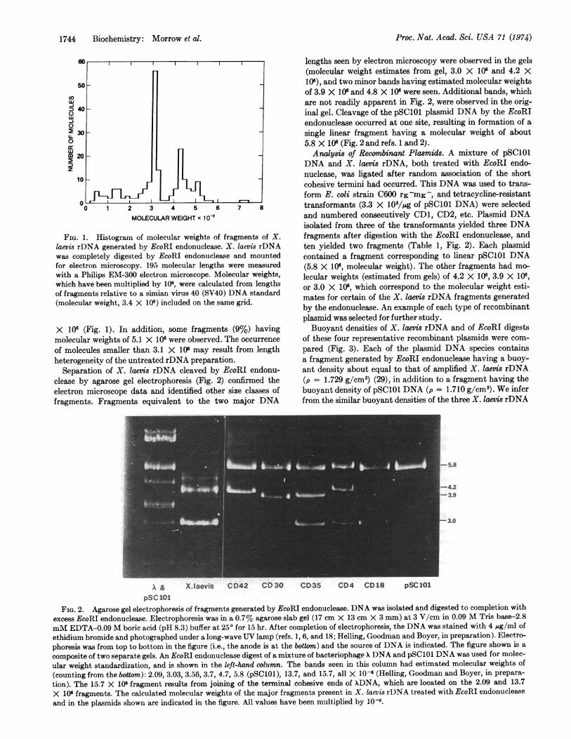
Cleavage ofrDNA of X. laevis. Linear molecules of X. laevisrDNA (molecular weight about 50 X 106, as determined byelectron microscopy) were treated with excess EcoRI endo-nuclease. After complete digestion, about 44% of the mole-cules had a molecular weight of 3.1 X 10⁶ (Fig. 1) and a secondmajor class (25%) of fragments had a molecular weight of 4.3
1744 Biochemistry: Morrow et al.
50
w
D 40Uw
0
2 30U.
0
w
CO20
z
lO1
0 1 2 3 4 5 6
MOLECULAR WEIGHT x 10-67 a
FIG. 1. Histogram of molecular weights of fragments of X.laevis rDNA generated by EcoRI endonuclease. X. laevis rDNAwas completely digested by EcoRI endonuclease and mountedfor electron microscopy. 195 molecular lengths were measuredwith a Philips EM-300 electron microscope. Molecular weights,which have been multiplied by 106, were calculated from lengthsof fragments relative to a simian virus 40 (SV40) DNA standard(molecular weight, 3.4 X 10⁶) included on the same grid.
X 106 (Fig. 1). In addition, some fragments (9%) havingmolecular weights of 5.1 X 10⁶ were observed. The occurrence
of molecules smaller than 3.1 X 10⁶ may result from lengthheterogeneity of the untreated rDNA preparation.
Separation of X. laevis rDNA cleaved by EcoRI endonu-clease by agarose gel electrophoresis (Fig. 2) confirmed theelectron microscope data and identified other size classes offragments. Fragments equivalent to the two major DNA
lengths seen by electron microscopy were observed in the gels(molecular weight estimates from gel, 3.0 X 10⁶ and 4.2 X10⁶), and two minor bands having estimated molecular weightsof 3.9 X 10⁶ and 4.8 X 10 were seen. Additional bands, whichare not readily apparent in Fig. 2, were observed in the orig-inal gel. Cleavage of the pSC101 plasmid DNA by the EcoRIendonuclease occurred at one site, resulting in formation of asingle linear fragment having a molecular weight of about5.8 X 106 (Fig. 2 and refs. 1 and 2).Analysis of Recombinant Plasmids. A mixture of pSC101
DNA and X. laevis rDNA, both treated with EcoRI endo-nuclease, was ligated after random association of the shortcohesive termini had occurred. This DNA was used to trans-form E. coli strain C600 rK-mK-, and tetracycline-resistanttransformants (3.3 X 10/,gg of pSC101 DNA) were selectedand numbered consecutively CD1, CD2, etc. Plasmid DNAisolated from three of the transformants yielded three DNAfragments after digestion with the EcoRI endonuclease, andten yielded two fragments (Table 1, Fig. 2). Each plasmidcontained a fragment corresponding to linear pSC101 DNA(5.8 X 10⁶, molecular weight). The other fragments had mo-lecular weights (estimated from gels) of 4.2 X 10⁶, 3.9 X 10⁶,or 3.0 X 10⁶, which correspond to the molecular weight esti-mates for certain of the X. laevis rDNA fragments generatedby the endonuclease. An example of each type of recombinantplasmid was selected for further study.Buoyant densities of X. laevis rDNA and of EcoRI digests
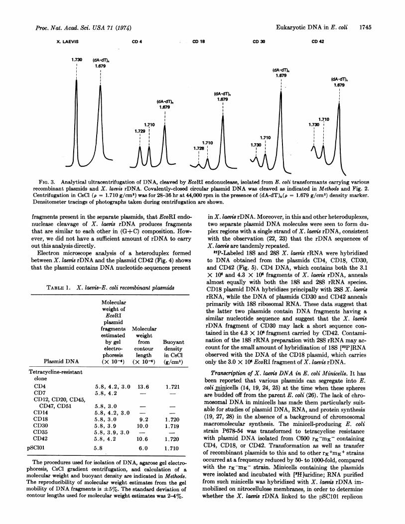
of these four representative recombinant plasmids were com-pared (Fig. 3). Each of the plasmid DNA species containsa fragment generated by EcoRI endonuclease having a buoy-ant density about equal to that of amplified X. laevis rDNA(p = 1.729 g/cm3) (29), in addition to a fragment having thebuoyant density of pSC101 DNA (p = 1.710 g/cm3). We inferfrom the similar buoyant densities of the three X. laevis rDNA
_^ ^ _ Z _~~~~~~~~~~~~~~~-5.8
I ~~~~~~~~-.2-3.9~~~~~~~- .
I~~~~~~~~~~~~~~I
\ & X.Iaevis CD42 CD30 CD35 CD4 CD18 pSCI0lPSC 101
FIG. 2. Agarose gel electrophoresis of fragments generated by EcoRI endonuclease. DNA was isolated and digested to completion with
excess EcoRI endonuclease. Electrophoresis was in a 0.7% agarose slab gel (17 cm X 13 cm X 3 mm) at 3 V/cm in 0.09 M Tris base-2.8
mM EDTA-0.09 M boric acid (pH 8.3) buffer at 250 for 15 hr. After completion of electrophoresis, the DNA was stained with 4 ,ug/ml of
ethidium bromide and photographed under a long-wave UV lamp (refs. 1, 6, and 18; Helling, Goodman and Boyer, in preparation). Electro-
phoresis was from top to bottom in the figure (i.e., the anode is at the bottom) and the source of DNA is indicated. The figure shown is a
composite of two separate gels. An EcoRI endonuclease digest of a mixture of bacteriophage X DNA and pSC101 DNA was used for molec-
ular weight standardization, and is shown in the left-hand column. The bands seen in this column had estimated molecular weights of
(counting from the bottom): 2.09, 3.03, 3.56, 3.7, 4.7, 5.8 (pSC101), 13.7, and 15.7, all X 10- (Helling, Goodman and Boyer, in prepara-
tion). The 15.7 X 10⁶ fragment results from joining of the terminal cohesive ends of xDNA, which are located on the 2.09 and 13.7
X 106 fragments. The calculated molecular weights of the major fragments present in X. laevis rDNA treated with EcoRI endonuclease
and in the plasmids shown are indicated in the figure. All values have been multiplied by 10-6.
,<4I
Proc. Nat. Acad. Sci. USA 71 (1974)
Proc. Nat. Acad. Sci. USA 71 (1974)
X. LAEVIS CD 4 CD 18 CD 30
1.730 (dA-dT),1.679
CD 42
(dA-dT).1.679
(dA-dT),1.679
(dA-dT)h1.679
1.710
(dA-dT)n
FIG. 3. Analytical ultracentrifugation of DNA, cleaved by EcoRI endonuclease, isolated from E. coli transformants carrying variousrecombinant plasmids and X. laevis rDNA. Covalently-closed circular plasmid DNA was cleaved as indicated in Methods and Fig. 2.Centrifugation in CsCl (p = 1.710 g/cm') was for 28-36 hr at 44,000 rpm in the presence of (dA-dT)n(p = 1.679 g/cm3) density marker.Densitometer tracings of photographs taken during centrifugation are shown.
fragments present in the separate plasmids, that EcoRI endo-nuclease cleavage of X. laevis rDNA produces fragmentsthat are similar to each other in (G+C) composition. How-ever, we did not have a sufficient amount of rDNA to carryout this analysis directly.
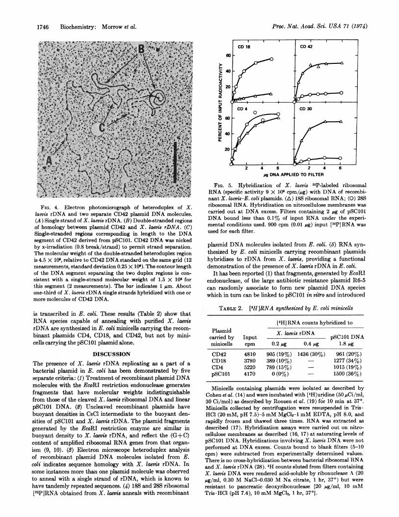
Electron microscope analysis of a heteroduplex formedbetween X. laevis rDNA and the plasmid CD42 (Fig. 4) showsthat the plasmid contains DNA nucleotide sequences present
TABLE 1. X. laevis-E. coli recombinant plasmids
Molecularweight ofEcoRIplasmid
fragments Molecularestimated weightby gel from Buoyantelectro- contour densityphoresis length in CsCl
Plasmid DNA (X 10-6) (X 106) (g/cm3)
Tetracycline-resistantcloneCD4 5.8, 4.2, 3.0 13.6 1.721CD7 5.8, 4.2CD12, CD20, CD45,CD47, CD51 5.8, 3.0 -
CD14 5.8, 4.2, 3.0 -CD18 5.8, 3.0 9.2 1.720CD30 5.8, 3.9 10.0 1.719CD35 5.8, 3.9, 3.0CD42 5.8, 4.2 10.6 1.720
pSClO1 5.8 6.0 1.710
The procedures used for isolation of DNA, agarose gel electro-phoresis, CsCl gradient centrifugation, and calculation of amolecular weight and buoyant density are indicated in Methods.The reproducibility of molecular weight estimates from the gelmobility of DNA fragments is ±5%. The standard deviation ofcontour lengths used for molecular weight estimates was 2-4%.
in X. laevis rDNA. Moreover, in this and other heteroduplexes,two separate plasmid DNA molecules were seen to form du-plex regions with a single strand of X. laevis rDNA, consistentwith the observation (22, 23) that the rDNA sequences ofX. laevis are tandemly repeated.
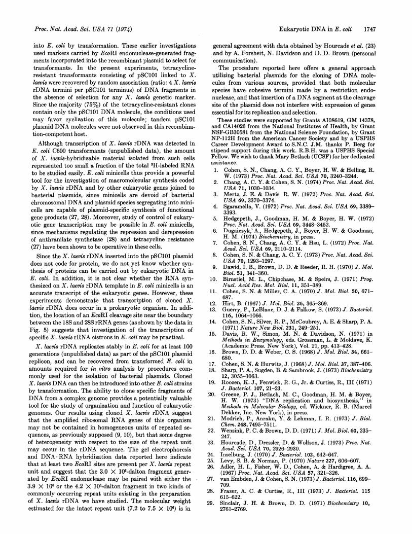
32P-Labeled 18S and 28S X. laevis rRNA were hybridizedto DNA obtained from the plasmids CD4, CD18, CD30,and CD42 (Fig. 5). CD4 DNA, which contains both the 3.1X 10⁶ and 4.3 X 10⁶ fragments of X. laevis rDNA, annealsalmost equally with both the 18S and 28S rRNA species.CD18 plasmid DNA hybridizes principally with 28S X. laevisrRNA, while the DNA of plasmids CD30 and CD42 annealsprimarily with 18S ribosomal RNA. These data suggest thatthe latter two plasmids contain DNA fragments having asimilar nucleotide sequence and suggest that the X. laevisrDNA fragment of CD30 may lack a short sequence con-tained in the 4.3 X 10⁶ fragment carried by CD42. Contami-nation of the 18S rRNA preparation with 28S rRNA may ac-count for the small amount of hybridization of 18S ['2P]RNAobserved with the DNA of the CD18 plasmid, which carriesonly the 3.0 X 10⁶ EcoRI fragment of X. laevis rDNA.
Tranwription of X. laevis DNA in E. coli Minicells. It hasbeen reported that various plasmids can segregate into E.coli minicells (14, 19, 24, 25) at the time when these spheresare budded off from the parent E. coli (26). The lack of chro-mosomal DNA in minicells has made them particularly suit-able for studies of plasmid DNA, RNA, and protein synthesis(19, 27, 28) in the absence of a background of chromosomalmacromolecular synthesis. The minicell-producing E. colistrain P678-54 was transformed to tetracycline resistancewith plasmid DNA isolated from C600 rK-mK- containingCD4, CD18, or CD42. Transformation as well as transferof recombinant plasmids to this and to other rK+mK+ strainsoccurred at a frequency reduced by 50- to 1000-fold, comparedwith the rK-mK- strain. Minicells containing the plasmidswere isolated and incubated with ['Hjuridine; RNA purifiedfrom such minicells was hybridized with X. laevis rDNA im-mobilized on nitrocellulose membranes, in order to determinewhether the X. taevis rDNA linked to the pSC101 replicon
Eukaryotic DNA in E. coli 1745
1746 Biochemistry: Morrow et al.
FIG. 4. Electron photomicrograph of heteroduplex of X.laevis rDNA and two separate CD42 plasmid DNA molecules.(A) Single strand of X. laevis rDNA. (B) Double-stranded regionsof homology between plasmid CD42 and X. Iaevis rDNA. (C)Single-stranded regions corresponding in length to the DNAsegment of CD42 derived from pSC101. CD42 DNA was nickedby x-irradiation (0.8 break/strand) to permit strand separation.The molecular weight of the double-stranded heteroduplex regionis 4.5 X 10⁶, relative to CD42 DNA standard on the same grid (12measurements, standard deviation 0.25 X 10⁶). The contour lengthof the DNA segment separating the two duplex regions is con-
sistent with a single-strand molecular weight of 1.5 X 10⁶ forthis segment (2 measurements). The bar indicates 1 pm. Aboutone-third of X. laevis rDNA single strands hybridized with one or
more molecules of CD42 DNA.
is transcribed in E. coli. These results (Table 2) show thatRNA species capable of annealing with purified X. laevis
rDNA are synthesized in E. coli minicells carrying the recom-
binant plasmids CD4, CD18, and CD42, but not by mini-cells carrying the pSC101 plasmid alone.
DISCUSSIONThe presence of X. laevis rDNA replicating as a part of a
bacterial plasmnid in E. coli has been demonstrated by fiveseparate criteria: (1) Treatment of recombinant plasmid DNAmolecules with the EcoRI restriction endonuclease generatesfragments that have molecular weights indistinguishablefrom those of the cleaved X. laevis ribosomal DNA and linearpSC101 DNA. (2) Uncleaved recombinant plasmids havebuoyant densities in CsCl intermediate to the buoyant den-sities of pSC101 and X. laevis rDNA. The plasmid fragmentsgenerated by the EcoRI restriction enzyme are similar inbuoyant density to X. laevis rDNA, and reflect the (G+C)content of amplified ribosomal RNA genes from that organ-
ism (9, 10). (3) Electron microscope heteroduplex analysisof recombinant plasmid DNA molecules isolated from E.coli indicates sequence homology with X. laevis rDNA. Insome instances more than one plasmid molecule was observedto anneal with a single strand of rDNA, which is known tohave tandemly repeated sequences. (4) 18S and 28S ribosomal["2PIRNA obtained from X. laevis anneals with recombinant
4e
° 2C
0 aI--
zU.
zuJU
20
20
CD 18 CD 42
0
1
2 4 6 2 4 6
pg DNA APPLIED TO FILTER
FIG. 5. Hybridization of X. laevis 32P-labeled ribosomalRNA (specific activity 9 X 10⁶ cpm/jug) with DNA of recombi-nant X. laevis-E. coli plasmids. (A) 18S ribosomal RNA; (0) 28Sribosomal RNA. Hybridization on nitrocellulose membranes wascarried out at DNA excess. Filters containing 2 pug of pSC101DNA bound less than 0.1% of input RNA under the experi-mental conditions used. 900 cpm (0.01 ug) input [2P]RNA wasused for each filter.
plasmid DNA molecules isolated from E. coli. (5) RNA syn-thesized by E. coli minicells carrying recombinant plasmidshybridizes to rDNA from X. laevis, providing a functionaldemonstration of the presence of X. laevis rDNA in E. coli.
It has been reported (1) that fragments, generated by EcoRIendonuclease, of the large antibiotic resistance plasmid R6-5can randomly associate to form new plasmid DNA specieswhich in turn can be linked to pSC1O1 in vitro and introduced
TABLE 2. ['H]RNA synthesized by E. coli minicells
[3H] RNA counts hybridized toPlasmid X. laevis rDNA
carried by Input pSC101 DNAminicells cpm 0.2.ug 0.4 ug 1.8 pg
CD42 4810 905 (19%) 1436 (30%) 961(20%)CD18 3780 389 (10%) 1277 (34%)CD4 5220 789 (15%) 1015 (19%)pSC101 4170 0 (0%) 1500 (36%)
Minicells containing plasmids were isolated as described byCohen et al. (14) and were incubated with ['H] uridine (50 ,Ci/ml,30 Ci/mol) as described by Roozen et al. (19) for 10 min at 37°.Minicells collected by centrifugation were resuspended in Tris-HOC (20 mM, pH 7.5)-5 mM MgCl2-1 mM EDTA, pH 8.0, andrapidly frozen and thawed three times. RNA was extracted asdescribed (17). Hybridization assays were carried out on nitro-cellulose membranes as described (16, 17) at saturating levels ofpSC101 DNA. Hybridizations involving X. laevis DNA were notperformed at DNA excess. Counts bound to blank filters (5-10cpm) were subtracted from experimentally determined values.There is no cross-hybridization between bacterial ribosomal RNAand X. laevis rDNA (28). 'H counts eluted from filters containingX. laevis DNA were rendered acid-soluble by ribonuclease A (20pg/ml, 0.30 M NaCl-0.030 M Na citrate, 1 hr, 370) but wereresistant to pancreatic deoxyribonuclease [20 pg/ml, 10 mMTris HCl (pH 7.4), 10mM MgCl2, 1 hr, 37°].
Proc. Nat. Acad. Sci. USA 71 (1974)
Proc. Nat. Acad. Sci. USA 71 (1974)
into E. coli by transformation. These earlier investigationsused markers carried by EcoRI endonuclease-generated frag-ments incorporated into the recombinant plasmid to select fortransformants. In the present experiments, tetracycline-resistant transformants consisting of pSC101 linked to X.laevis were recovered by random association (ratio: 4 X. laevisrDNA termini per pSC101 terminus) of DNA fragments inthe absence of selection for any X. laevis genetic marker.Since the majority (75%) of the tetracycline-resistant clonescontain only the pSC101 DNA molecule, the conditions usedmay favor cyclization of this molecule; tandem pSC101plasmid DNA molecules were not observed in this recombina-tion-competent host.Although transcription of X. laevis rDNA was detected in
E. coli C600 transformants (unpublished data), the amountof X. laevis-hybridizable material isolated from such cellsrepresented too small a fraction of the total 3H-labeled RNAto be studied easily. E. coli minicells thus provide a powerfultool for the investigation of macromolecular synthesis codedby X. laevis rDNA and by other eukaryotic genes joined tobacterial plasmids, since minicells are devoid of bacterialchromosomal DNA and plasmid species segregating into mini-cells are capable of plasmid-specific synthesis of functionalgene products (27, 28). Moreover, study of control of eukary-otic gene transcription may be possible in E. coli minicells,since mechanisms regulating the repression and derepressionof anthranilate synthetase (28) and tetracycline resistance(27) have been shown to be operative in these cells.
Since the X. laevis rDNA inserted into the pSC101 plasmiddoes not code for protein, we do not yet know whether syn-thesis of proteins can be carried out by eukaryotic DNA inE. coli. In addition, it is not clear whether the RNA syn-thesized on X. laevis rDNA template in E. coli minicells is anaccurate transcript of the eukaryotic genes. However, theseexperiments demonstrate that transcription of cloned X.laevis rDNA does occur in a prokaryotic organism. In addi-tion, the location of an EcoRI cleavage site near the boundarybetween the 18S and 28S rRNA genes (as shown by the data inFig. 5) suggests that investigation of the transcription ofspecific X. laevis rRNA cistrons in E. coli may be practical.X. laevis rDNA replicates stably in E. coli for at least 100
generations (unpublished data) as part of the pSC101 plasmidreplicon, and can be recovered from transformed E. coli inamounts required for in vitro analysis by procedures com-monly used for the isolation of bacterial plasmids. ClonedX. laevis DNA can then be introduced into other E. coli strainsby transformation. The ability to clone specific fragments ofDNA from a complex genome provides a potentially valuabletool for the study of organization and function of eukaryoticgenomes. Our results using cloned X. laevis rDNA suggestthat the amplified ribosomal RNA genes of this organismmay not be contained in homogeneous units of repeated se-quences, as previously supposed (9, 10), but that some degreeof heterogeneity with respect to the size of the repeat unitmay occur in the rDNA sequence. The gel electrophoresisand DNA-RNA hybridization data reported here indicatethat at least two EcoRI sites are present per X. laevis repeatunit and suggest that the 3.0 X 10⁶-dalton fragment gener-ated bv EcoRI endonuclease may be paired with either the3.9 X 106 or the 4.2 X 10⁶-dalton fragment in two kinds ofcommonly occurring repeat units existing in the preparationof X. laevis rDNA we have studied. The molecular weightestimated for the intact repeat unit (7.2 to 7.5 X 10⁶) is in
general agreement with data obtained by Hourcade et al. (23)and by A. Forsheit, N. Davidson and D. D. Brown (personalcommunication).The procedure reported here offers a general approach
utilizing bacterial plasmids for the cloning of DNA mole-cules from various sources, provided that both molecularspecies have cohesive termini made by a restriction endo-nuclease, and that insertion of a DNA segment at the cleavagesite of the plasmid does not interfere with expression of genesessential for its replication and selection.These studies were supported by Grants AI08619, GM 14378,
and CA14026 from the National Institutes of Health, by GrantNSF-GB30581 from the National Science Foundation, by GrantNP-112H from the American Cancer Society and by a USPHSCareer Development Award to S.N.C. J.M. thanks P. Berg forstipend support during this work. R.B.H. was a USPHS SpecialFellow. We wish to thank Mary Betlach (UCSF) for her dedicatedassistance.1. Cohen, S. N., Chang, A. C. Y., Boyer, H. W. & Helling, R.
W. (1973) Proc. Nat. Acad. Sci. USA 70, 3240-3244.2. Chang, A. C. Y. & Cohen, S. N. (1974) Proc. Nat. Acad. Sci.
USA 71, 1030-1034.3. Mertz, J. E. & Davis, R. W. (1972) Proc. Nat. Acad. Sci.
USA 69, 3370-3374.4. Sgaramella, V. (1972) Proc. Nat. Acad. Sci. USA 69, 3389-
3393.5. Hedgepeth, J., Goodman, H. M. & Boyer, H. W. (1972)
Proc. Nat. Acad. Sci. USA 69, 3448-3452.6. Dugaiczyk,' A., Hedgepeth, J., Boyer, H. W. & Goodman,
H. M. (1974) Biochemistry, in press.7. Cohen, S. N., Chang, A. C. Y. & Hsu, L. (1972) Proc. Nat.
Acad. Sci. USA 69, 2110-2114.8. Cohen, S. N. & Chang, A. C. Y. (1973) Proc. Nat. Acad. Sci.
USA 70, 1293-1297.9. Dawid, I. B., Brown, D. D. & Reeder, R. H. (1970) J. Mol.
Biol. 51, 341-360.10. Birnstiel, M. L., Chipchase, M. & Speirs, J. (1971) Prog.
Nucl. Acid Res. Mol. Biol. 11, 351-389.11. Cohen, S. N. & Miller, C. A. (1970) J. Mol. Biol. 50, 671-
687.12. Hirt, B. (1967) J. Mol. Biol. 26, 365-369.13. Guerry, P., LeBlanc, D. J. & Falkow, S. (1973) J. Bacteriol.
116, 1064-1066.14. Cohen, S. N., Silver, R. P., McCoubrey, A. E. & Sharp, P. A.
(1971) Nature New Biol. 231, 249-251.15. Davis, R. W., Simon, M. N. & Davidson, N. (1971) in
Methods in Enzymology, eds. Grossman, L. & Moldave, K.(Academic Press. New York), Vol. 21, pp. 413-428.
16. Brown, D. D. & Weber, C. S. (1968) J. Mol. Biol. 34, 661-680.
17. Cohen, S. N. & Hurwitz, J. (1968) J. Mol. Biol. 37, 387-406.18. Sharp, P. A., Sugden, B. & Sambrook, J. (1973) Biochemistry
12, 3055-3063.19. Roozen, K. J., Fenwick, R. G., Jr. & Curtiss, R., III (1971)
J. Bacteriol. 107, 21-23.20. Greene, P. J., Betlach, M. C., Goodman, H. M. & Boyer,
H. W. (1973) "DNA replication and biosynthesis," inMethods in Molecular Biology, ed. Wickner, R. B. (MarcelDekker, Inc. New York), in press.
21. Modrich, P., Anraku, Y. & Lehman, I. R. (1973) J. Biol.Chem. 248, 7495-7511.
22. Wensink, P. C. & Brown, D. D. (1971) J. Mol. Biol. 60, 235-247.
23. Hourcade, D., Dressler, D. & Wotfson, J. (1973) Proc. Nat.Acad. Sci. USA 70, 2926-2930.
24. Inselburg, J. (1970) J. Bacteriol. 102, 642-647.25. Levy, S. B. & Norman, P. (1970) Nature 227, 606-607.26. Adler, H. I., Fisher, W. D., Cohen, A. & Hardigree, A. A.
(1967) Proc. Nat. Acad. Sci. USA 57, 321-326.27. van Embden, J. & Cohen, S. N. (1973) J. Bacteriol. 116, 699-
709.28. Frazer, A. C. & Curtiss, R., III (1973) J. Bacteriol. 115
615-622.29. Sinclair, J. H. & Brown, D. D. (1971) Biochemistry 10,
2761-2769.
Eukaryotic DNA in E. coli 1747