Embed Size (px)
Citation preview

REPLICATION-COMPETENT NON-INDUCED PROVIRUSES IN THE
LATENT RESERVOIR INCREASE BARRIER TO HIV-1 CURE
by
Ya-Chi Ho
A dissertation submitted to the Johns Hopkins University School of Medicine in
conformity with the requirements for the degree of Doctor of Philosophy
Baltimore, Maryland
September 2013
© Ya-Chi Ho 2013
All rights reserved
ii
ABSTRACT
Antiretroviral therapy (ART) fails to cure HIV-1 infection because latent
proviruses persist in resting CD4+ T cells. T cell activation reverses latency, but >99% of
proviruses are not induced to release infectious virus after maximum in vitro T cell
activation under standard viral outgrowth assay conditions. These non-induced proviruses
are generally considered defective but have never been characterized.
Using limiting dilution near-full length nested PCR and Poisson distribution
analysis, we characterized 213 clones of non-induced proviruses from eight aviremic
patients on suppressive ART. Most (88.3%) of the non-induced proviruses are defective,
including 45.5% large internal deletions, 32.4% APOBEC-mediated G→A
hypermutations, 6.6% mutations/deletions in the cis-acting element, and 3.8%
insertions/nonsense mutations.
Strikingly, 11.7% of the non-induced proviruses have intact genomes. Using
direct sequencing and de novo genome synthesis, we reconstructed six full-length non-
induced proviral clones and demonstrated growth kinetics comparable to four
reconstructed induced proviruses from the same patients.
Using luciferase assay to measure non-induced proviral LTR activity, we found
that non-induced proviruses have intact promoter function unless they are hypermutated.
Using limiting dilution bisulfite sequencing, we found that non-induced proviruses have
unmethylated promoters. Using inverse PCR, we found that non-induced proviruses are

iii
integrated into active transcription units. We demonstrated that these non-induced
proviruses, though not induced after in vitro maximum T cell activation, can be
reactivated after repeated stimuli. We propose that maximum T cell activation does not
lead to maximum non-induced provirus activation. Rather, activation of non-induced
proviruses is stochastic.
Thus, it cannot be excluded that non-induced proviruses may become activated in
vivo. The discovery of replication-competent non-induced proviruses indicates that the
size of the latent reservoir, and hence the barrier to cure, may be ~60-fold greater than
previously estimated.
Underestimation of intact proviruses by viral outgrowth assays could be reflected
in delayed viral rebound after an apparent “cure”, and overestimation of latent reservoir
size resulting from detection of defective proviruses by PCR assays could result in
prolonged, excessive exposure to toxic latency reversing agents. Thus, the molecular
analysis of non-induced proviruses contributes in an important way to HIV-1 eradication
efforts.
Advisor:
Robert F. Siliciano, M.D., Ph.D.
Professor of Medicine
Johns Hopkins University School of Medicine.
Reader:
Joel L. Pomerantz, Ph.D.
Associate professor of Biological Chemistry
Johns Hopkins University School of Medicine
iv
PREFACE
I would like to express my deepest appreciation to my advisor, Dr. Robert F.
Siliciano, for his inspirational guidance and full support to my intellectual interests and
career. He envisions the most significant issues in the field and asks important questions.
He not only guided me to solve difficult problems, but even more importantly, provided
me the freedom to ask different questions and conduct experiments on my own. He works
as a perfectionist – creative, enthusiastic, industrious and conscientious and insightful.
But he is extremely kind, encouraging and generous to me, even for negative results and
inadequate performance. His dedication to science and education makes him an
exceptional physician-scientist and great mentor. It is my privilege to learn from him,
who led me all the way to this exciting scientific adventure.
I would like to thank my unofficial advisor, Dr. Janet Siliciano, for her effort and
advises on scientific discussions as well as family management. I would like to thank my
thesis committee members, Dr. Stuart Ray, Dr. Joel Blankson and Dr. Joel Pomerantz. Dr.
Stuart Ray is very smart and kind. Each time after I presented my work to Dr. Stuart Ray,
he always provided insightful and important suggestions immediately. These suggestions
eventually answered the critical questions that other world experts in the field asked. Dr.
Joel Blankson is always available for discussions and insightful for my directions. He
guided me from the smallest technical problems to career planning. Dr. Joel Pomerantz is
a distinguished scientist whose enthusiasm in research is so contagious and influential. I
had the privilege to work closely with him during my rotation, which built the foundation
of my scientific curiosity and molecular biology skills.
v
I appreciate all the members the Siliciano lab, who constantly give me vibrant
scientific discussions and help in a very pleasant atmosphere. Particularly, Dr. Liang
Shan and Seyed Alireza Rabi gave me so many great ideas. They are extremely smart,
innovative, hardworking, outstanding but humble. I thank Dr. Hung-Chih Yang, Dr. Sifei
Xing, Dr. Maria Salgado, Kai Deng, Robert Buckheit III, Christopher Pohlmeyer and
Sarah Laskey for their encouragement and friendship. I keep learning from their diligence,
multi-tasking ability and altruism. I thank Dr. Daniel Rosenbloom and Sarah Laskey for
their comprehensive mathematical estimation and modeling of the size of the latent
reservoir. I thank Ms. Bonita Grant and Ms. Jun Lai for their effort meeting all the needs
from me and this big lab. I would like to thank my collaborators, including Dr. Eitan
Halper-Stromberg and Dr. Haiping Hao for their help for deep sequencing.
Special thanks go to Dr. Pierre Coulombe, Dr. M. Christine Zink, Ms. Leslie
Lichter and Ms. Colleen Graham of the Cellular and Molecular Medicine PhD training
program for giving me the chance to study in this prestigious institute.
I would like to thank the Ministry of Education of Taiwan who offered me a one-
year fellowship to study in the United States. I also thank the Howard Hughes Medical
Institutions International Student Research Fellowship program who supported my thesis
project and two years of research.
The completion of this PhD means tremendous support from my family. My
parents spent the past five years helping me taking care of my daughters so that I can
concentrate on my research. They could have lived a much better and easier life, but
instead they chose to stay with us in the States with restricted access to their friends.

vi
I dedicate all my work to my husband, Dr. Yu-Min Chuang. He is the very person
who understands and fully supports my immense interest in scientific research. Although
we work on different fields, he often provides insightful suggestions and asks key
questions regarding my project. His love, kindness and dedication to both our family and
research supported me during the most desperate period of my life.
I am grateful to all my friends, near or far, who supported me regarding science
and life. I look forward to working as a physician-scientist, using basic research tools to
solve clinically important questions.

vii
TABLE OF CONTENTS
Title i
Abstract ii
Preface iv
Table of Contents vi
List of Tables viii
List of Figures ix
Chapter 1. Introduction 1
Chapter 2. Viral outgrowth assay conditions achieves maximum in vitro
T cell activation 4
Chapter 3. Characterization of non-induced proviral clones 11
Chapter 4. Intact non-induced proviruses are replication competent 30
Chapter 5. Non-induced proviruses lack epigenetic silencing markers 44
Chapter 6. Intact non-induced proviruses may increase latent reservoir size
by ~60 fold 53
Chapter 7. Stochastic activation of non-induced proviruses 62
References 69
Curriculum vitae 77

viii
LIST OF TABLES
Table 1. Patient Characteristics 19
Table 2. PCR primers and conditions 20
Table 3. Estimation of intact non-induced provirus frequency 59

ix
LIST OF FIGURES
Figure 1. Viral outgrowth assay conditions achieve maximum in vitro
activation and outgrowth 9
Figure 2. Characterization of non-induced proviruses 21
Figure 3. APOBEC3G-mediated G to A hypermutation renders some
non-induced proviruses defective 23
Figure 4. Mapping of large internal deletions in non-induced proviruses 26
Figure 5. Deletions and mutations in cis elements 28
Figure 6. Growth kinetics of reconstructed non-induced viruses 35
Figure 7. Phylogenetic analysis for reconstruction of the 108 bp sequence 37
Figure 8. LTR activity of non-induced proviruses 42
Figure 9. Integration sites of non-induced proviruses 49
Figure 10. CpG methylation of non-induced proviruses 51
Figure 11. Quantification of intact non-induced proviruses 61
Figure 12. Stochastic activation of non-induced proviruses 66
Figure 13. Mechanism of HIV-1 latency reactivation 68

1
Chapter 1. Introduction
Despite prolonged antiretroviral therapy (ART), HIV-1 persists as
transcriptionally inactive proviruses in resting memory CD4+ T cells (Chun et al., 1997;
Finzi et al., 1997; Wong et al., 1997). This latent reservoir has a long half-life,
preventing cure by ART alone (Finzi et al., 1997; Siliciano et al., 2003; Strain et al.,
2003). In resting CD4+ T cells, the lack of active forms of key cellular transcription
factors (Bohnlein et al., 1988; Duh et al., 1989; Ganesh et al., 2003; Kinoshita et al., 1997;
Nabel and Baltimore, 1987; West et al., 2001) and of HIV-1 Tat and its cellular cofactors
(Cujec et al., 1997; Herrmann and Rice, 1995; Jones and Peterlin, 1994; Kao et al., 1987;
Selby and Peterlin, 1990; Tyagi et al., 2010) limits the initiation and elongation,
respectively, of viral transcription (Lassen et al., 2004; Williams and Greene, 2007). The
latent reservoir may thus be established when activated CD4+ T cells become infected as
they revert back to a resting memory state. In addition, DNA methylation and repressive
histone modifications may promote silencing of proviruses (Blazkova et al., 2009; Coull
et al., 2000; He and Margolis, 2002; Kauder et al., 2009; Van Lint et al., 1996; Verdin et
al., 1993; Williams et al., 2006).
A major approach to eradicating HIV-1 involves reversing latency in patients on
ART (Deeks, 2012; Richman et al., 2009). Cells harboring induced proviruses could then
be lysed by HIV-1-specific cytolytic T lymphocytes (CTL) (Shan et al., 2012), while new
rounds of infection are blocked by ART. Clinical trials exploring this strategy have used
the histone deacetylase inhibitors valproic acid (Lehrman et al., 2005), vorinostat (Archin
et al., 2009; Archin et al., 2012; Contreras et al., 2009) and panobinostat (Katlama et al.,
2013).
2
Accurate measurement of the latent reservoir is essential for evaluating
eradication strategies. If the latent reservoir is completely eradicated, ART can be
discontinued without rebound viremia. Interruption before complete eradication will
likely result in rebound (Davey et al., 1999) and repopulation of the latent reservoir,
nullifying the eradication effort.
The standard assay for latent reservoir size is a viral outgrowth assay (viral
outgrowth assay (Finzi et al., 1997; Siliciano and Siliciano, 2005), which measures the
frequency of resting CD4+ T cells that produce infectious virus after a single round of
maximum in vitro T cell activation. Limiting dilutions of resting CD4+ T cells are
stimulated with the mitogen phytohemagglutinin (PHA), which reverses latency by
inducing T cell activation. Released viruses are expanded by addition of CD4+ T
lymphoblasts from HIV-1-negative donors. Culture supernatants are examined for
exponential viral growth by enzyme-linked immunosorbant assay (ELISA) for HIV-1 p24.
With this assay, the mean frequency of latently infected cells in patients on ART is
~1/106 resting CD4
+ T cells (Eriksson et al., 2013).
It has been assumed that latent reservoir size can be assessed with agents like
PHA that induce uniform activation of CD4+ T cells (Hermankova et al., 2003; Patel et
al., 1988). However, the frequency of latently infected cells detected in the viral
outgrowth assay is on average 300 fold lower than the frequency of resting CD4+ T cells
that harbor proviruses detectable by PCR (Eriksson et al., 2013). Thus at limiting
dilution in the viral outgrowth assay, negative wells contain many proviruses, which we
designate non-induced proviruses. The non-induced proviruses are generally considered
defective. However, they have not been molecularly characterized. The magnitude of

3
the challenge presented by the latent reservoir depends on whether non-induced
proviruses can be induced in vivo. We present here the first molecular characterization of
non-induced proviruses, the results of which provide disturbing new insights into the
difficulty of curing HIV-1 infection.
The latent reservoir in resting CD4+ T cells is the major barrier to HIV-1
eradication, and as efforts to cure the infection proceed, accurate measurement of latent
reservoir size will be essential. This study provides a molecular basis for understanding
measures of the latent reservoir. Through an analysis of proviruses that did not give rise
to infectious virus following a single round of T cell activation (non-induced proviruses),
we have provided a definitive explanation for the large discrepancy between results of
PCR and culture assays of latent reservoir size. In addition, the discovery of intact non-
induced proviruses indicates that the size of the latent reservoir may be much greater than
previously thought. We reconstructed full-length, intact non-induced proviruses from
multiple patients, and all showed growth kinetics comparable to induced proviruses from
the same patient and a reference isolate. These intact non-induced proviruses are not
detected in standard culture assays but may nevertheless prevent cure. Thus the present
study provides new insights into the extent of the challenge posed by the latent reservoir
and may lead to novel strategies that target intact non-induced proviruses.
4
Chapter 2. Viral outgrowth assay conditions achieves maximum in vitro T cell
activation
Introduction
For determining the size of the latent reservoir, the current standard is the viral
outgrowth assay (86 Siliciano,J.D. 2005; 220 Finzi,D. 1997). This assay measures the
frequency of resting CD4+ T cells that produce replication-competent virus after as single
round of maximum in vitro T cell activation. Limiting dilutions of resting CD4+ T cells
purified from patients on suppressive ART are activated with PHA. PHA, a potent
mitogenic lectin, cross-links T cell receptor/CD3 complex, activates phospholipase C and
protein kinase C (Mustelin et al., 1990), up-regulates interleukin-2 (IL-2) and IL-2
receptor (CD25) expression (Depper et al., 1985), resulting in an autocrine stimulation
and T cell proliferation. PHA provides 100% colony forming ability in CD4+ T cells
(Hermankova et al., 2003; Patel et al., 1988) and reverses latency by global T cell
activation, as one of the most potent reagent used in vitro to reverse latency (Yang et al.,
2009b), in a greater efficiency than most latency reversing agents tested to date. PHA
was washed out after one day of culture to reduce cytotoxicity. Viruses released into the
supernatant following PHA activation are further amplified by co-culture with two
additions of allogeneic CD4+ T lymphoblasts. Culture supernants are examined for
exponential viral growth p24 ELISA for HIV-1 Gag antigen on day 14 – 21 of culture,
and positive wells are considered to have contained at least one latently infected cells
harboring inducible, replication competent virus. Poisson statistics are used to establish
5
the frequency of latently infected cells (Siliciano and Siliciano, 2005). In patients on
ART, it is typically on the order of one cell per 106 resting CD4
+ T cells.
Unlike human endogenous retroviruses, which are silenced during embryogenesis
(Rowe et al., 2010) and generally thought to be harmless, HIV-1 integrates into
terminally differentiated CD4+ T cells and does not follow silencing mechanisms seen in
endogenous retroviruses (Rowe et al., 2010). Although they were not induced by in vitro
activation using PHA, it does not mean that they would never be reactivated in vivo.
Unlike PCR based methods, which solely represent the presence of DNA
fragment regardless of being intact or defective, the relatively high detection limit of
viruses in the supernatant indicate the presence of multiple rounds of exponential viral
propagation, reflecting presence of functional and replication competent proviruses. The
detection limit of the viral outgrowth assay is 104,167 HIV-1 RNA copies/mL, which
was calculated as: 3 fg HIV-1 p24/50 RNA copies (Barletta et al., 2004) x 6.25 pg/mL
detection limit = 104,167 copies/mL. However, with this relatively high detection limit,
p24 negative wells are not merely a reflection of false negative results of an insensitive
detection assay. Rather, it has been confirmed that all p24 negative wells remain
negative after a more sensitive mRNA detection method (Laird et al., 2013) down to 555
copies/mL (which was calculated by the fact that detecting one copy/well of quantitative
PCR equals to 1.8 µL of culture supernatant in this assay).
The main obstable to isolate full-length proviral DNA from the viral outgrowth
cultures is that the patient resting CD4+ T cells (for example 0.2 or 0.04 million per well)
are outnumbered by the healthy donor lymphoblasts (1 million per well x 2 additions = 2
million per well). Isolating proviral DNA from this large amount of human chromosomal

6
DNA is technically challenging. An alternative way is to separate patient cells and
healthy donor lymphoblasts by a transwell culture system, with a 0.4 µm membrane
permeable for viruses and nutrients but impermeable for cells, to obtain a pure population
of patient cells for further proviral DNA isolation. This separation, however, prevents
direct intercellular contact, which may or may not reduce the efficiency of viral
propagation, and hence the sensitivity of the viral outgrowth assay. We here
demonstrated that our transwell viral outgrowth assay provides maximum T cell
activation and comparable sensitivity with the standard viral outgrowth assay.
Methods
Viral outgrowth assay
Viral outgrowth assays were performed as described (Finzi et al., 1997; Siliciano
and Siliciano, 2005). Briefly, resting CD4+ T cells were isolated from patients under
suppressive ART with undetectable plasma viral load for >6 months prior to enrollment
to ensure no preintegration latency. Using magnetic microbead selection (Miltenyi
Biotec), CD4+ T cells were first isolated using negative selection, and CD25, CD69 and
HLA-DR expressing cells were removed by positive selection. Standard and transwell
cultures were preformed side by side using the same population of patient resting CD4+ T
cells, irradiated allogeneic healthy donor peripheral blood mononuclear cells (PBMCs)
and CD8-deplted healthy donor lymphoblasts. The volume of culture medium and the
total number of cells per well was equal for these two culture conditions. In transwell
cultures, patient resting CD4+ T cells and irradiated PBMC from healthy donors were
incubated at the upper chamber on a cell-impermeable polyester membrane with 0.4 μM

7
pores. The irradiated PBMCs are necessary for optimal PHA activation but die during
the culture period, leaving the upper chamber containing a pure population of patient
CD4+ T cells. CD8-depleted allogeneic lymphoblasts were added to the lower chamber
of the transwell culture in the same numbers as for standard cultures.
After 21 days of culture, 180 µL of culture supernatant were subjected to p24
ELISA (Perkin Elmer). P24 ELISA >6.25 ng/µL were determined as positive.
Results
To analyze proviruses that did not give rise to infectious virus in the viral
outgrowth assay (non-induced proviruses), we first established that the conditions were
sufficient to activate 100% of resting CD4+ T cells. Resting CD4
+ T cells from patients
on suppressive ART for >6 months (range, 19–108) were labeled with carboxyfluorescein
succinimidyl ester (CFSE) and stimulated with PHA and irradiated allogeneic PBMC
under conditions used in the viral outgrowth assay. By day 7, >99.8% of patient cells had
divided at least once (Figure 1A), confirming that PHA causes uniform T cell activation.
In the viral outgrowth assay, viruses released after reversal of latency replicate in
healthy donor CD4+ lymphoblasts added to the cultures. To facilitate cloning of non-
induced proviruses, we explored whether comparable levels of activation and viral
outgrowth could be achieved in transwell cultures in which patient cells were separated
from donor lymphoblasts by a cell-impermeable membrane (Figure 1B). In side-by-side
comparison with standard viral outgrowth assay cultures from ten patients, transwell
cultures showed comparable cellular activation in both p24 positive and negative wells,
as >95% of patient cells expressed HLA-DR and/or CD25 on day 21 (Figure 1C).

8
Transwell cultures also showed viral outgrowth comparable to that observed for standard
viral outgrowth assay cultures (Figure 1D). Non-induced proviruses were thus cloned
from p24-negative wells of limiting dilution transwell and standard viral outgrowth assay
cultures.
Discussion
Although difficult and time-consuming, the viral outgrowth assay (Eriksson et al.,
2013; Finzi et al., 1997), which has recently been simplified (Laird et al., 2013), does
allow detection of cells harboring replication-competent virus. We first explored to make
sure this is not simply an issue of assay sensitivity. We showed that the PHA stimulation
activates all resting CD4+ T cells as assessed by proliferation and activation marker
expression. Using prolonged culture and sensitive RT-PCR assays, we also verified that
wells from which non-induced proviruses were obtained were truly negative for viral
outgrowth. It is also unlikely that these p24 negative wells remained negative because of
reduced viral fitness, as we showed that intact non-induced proviruses had growth
kinetics comparable to induced viruses from p24 positive wells (see below). Taken
together, these results confirm that we are examining a population of intact proviruses
that were not induced to produce infectious virus after a single round of maximum in
vitro activation.

9
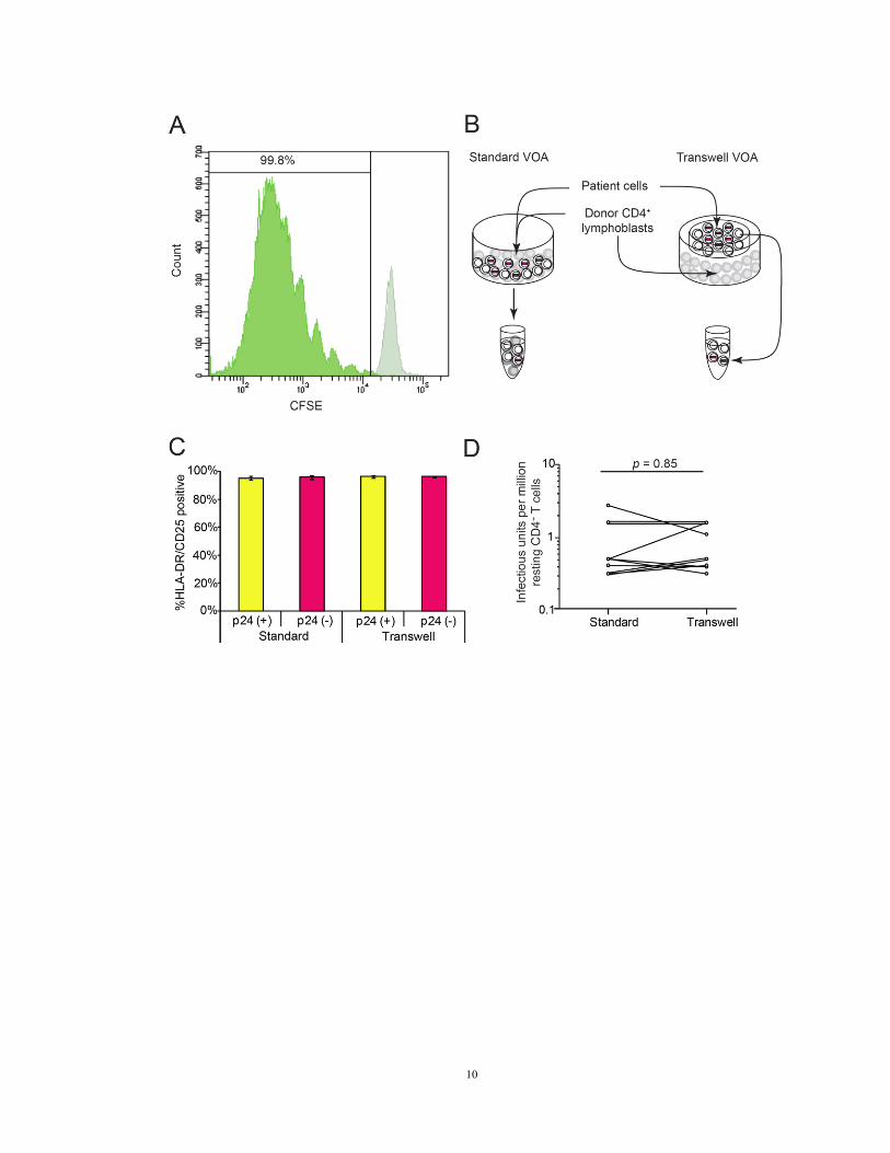
Figure 1. Viral outgrowth assay conditions achieve maximum in vitro activation and
outgrowth
(A) CFSE fluorescence in labeled patient resting CD4+ T cells on day 0 (light green) and
day seven (dark green) of the viral outgrowth assay.
(B) Separation of patient cells from healthy donor CD4+ lymphoblasts in a transwell
version of the viral outgrowth assay. Patient cells (white) were activated with PHA and
irradiated allogeneic PBMC (not shown). To expand the viruses released from infected
cells, CD4+ T lymphoblasts from healthy donors (grey) were added directly to the culture
(standard viral outgrowth assay) or were separated from patient cells by a cell-
impermeable polyester membrane with 0.4 μm pores (transwell viral outgrowth assay).
(C) Comparison of activation status of patient cells in standard and transwell viral
outgrowth assay cultures, and in p24 ELISA positive and negative wells on day 21.
HLA-A2 negative patient cells were co-cultured with HLA-A2 positive healthy donor
CD4+ lymphoblasts and irradiated allogeneic PBMC. The viable HLA-A2 negative
population was gated as patient cells for surface activation marker analysis.
(D) Comparison of viral outgrowth assay results by p24 ELISA of supernatants from
parallel standard and transwell viral outgrowth assay cultures from ten patients.
10

11
Chapter 3. Characterization of non-induced proviral clones
Introduction
The non-induced proviruses had been considered to be defective through various
mechanisms, including apolipoprotein B mRNA editing enzyme (APOBEC) mediated G-
to-A hypermutations, recombination during reverse transcription (Sanchez et al., 1997;
Temin, 1993) and low fidelity of reverse transcriptase (3.4 x10⁻⁵
mutations/bp/cycle
(Mansky and Temin, 1995)). The incoming virus is infectious and intact upon entry, but
becomes defective during reverse transcription. As long as the very ends of the reverse-
transcribed proviral DNA are intact, these defective proviruses will be integrated into the
human genome. Based on the analysis of proviruses from acutely infected patients, at
least one third are overtly defective (Salazar-Gonzalez et al., 2009), not including large
internal deletions. Cells harboring these defective proviruses, which will not produce
viral proteins to be presented and recognized by CTLs, continue to expand through
homeostatic proliferation. However, these defective proviral genomes remain detectable
by PCR-based methods, and are being used in evaluating eradication trials (Katlama et al.,
2013). Characterization of these non-induced proviruses is essential in the eradication
effort, to avoid prolonged patient exposure to toxic latency reversing agents for
overestimating the latent reservoir by detection of defective “graveyard” proviruses.
The main barrier to identify full-length proviral clones is the rarity of the
proviruses in resting CD4+ T cells. Identification of full-length HIV-1 genome has been
reported, but all from viremic patients with predominant clones in the circulation
(Ehrenberg and Michael, 2005; Li et al., 2007; Li et al., 1991; Sahu et al., 2010; Salazar-

12
Gonzalez et al., 2009). Identification of clonal non-induced proviral clones requires
limiting dilution PCR to avoid complementing different proviral clones in one reaction,
causing a hybrid of different proviral genomes. Full-length proviral PCR amplification
of single clones has been inefficient and challenging, due to the sequence variability in
different patients and the bulk of the human genome in PCR reactions. We here
established a near-full-length nested PCR to identify single clones of non-induced
proviruses and characterized the defects in the proviral genome.
Methods
Study subjects
Peripheral blood was obtained from healthy volunteers and HIV-1 infected donors (Table
1) who had suppression of viremia to <50 copies HIV-1 RNA/ml for >6 months on ART
to avoid preintegration latency(Blankson et al., 2000). This study was approved by the
Johns Hopkins Institutional Review Board. Written informed consent was obtained from
all participants.
Characterization of full-length non-induced proviruses
Genomic DNA was extracted from cells of p24 negative wells using Gentra Puregene
Cell Kit (Qiagen). Genomic DNA isolated from p24 negative wells seeded with 4 x 10⁴
or 2 x 10⁵ patient resting CD4
+ T cells was subjected to limiting dilution prior to
amplification with an initial near full genome length outer PCR (U5 to U5) followed
nested amplification of a segment of the gag gene. Aliquots were serially diluted and
distributed into 96 well plates. Each well was filled up to a final volume of 50 μL with

13
PCR reaction mixture solutions including outer PCR primers (BLOuterF and BLOuterR).
Touchdown cycling conditions with decreasing annealing temperature were used to
increase specificity (Table 2). Then, 2 μL aliquots from each outer PCR well were
subjected to nested gag PCR and 1% agarose gel electrophoresis. If <20% of the gag
PCR wells were positive, the corresponding outer PCR wells contained one template with
>90% of possibility, based on the Poisson distribution. From these corresponding
positive outer PCR wells of such plates, 2 μL aliquots were taken for each of four nested
PCRs (A, B, C and D). The products were directly sequenced without cloning.
Sequences were aligned using CodonCode Sequence Assembly and Alignment Software.
The presence of double peaks on sequencing chromatograms was taken as evidence that
more than one template may have been present initially, and the sequence was discarded.
Direct sequencing results were analyzed by the Los Alamos Hypermut program and
HIVAlign program to identify hypermutation and nonsense mutations.
Results
Clonal amplification and sequencing of non-induced proviruses
We obtained near full-length clonal sequences of non-induced proviruses from
eight patients on suppressive ART. Patient characteristics are in Table 1. Non-induced
proviruses were obtained from wells that were seeded with 4 x 10⁴ or 2 x 10
5 resting
CD4+ T cells and that were negative for p24 on day 21. In clonal viral outgrowth assay
cultures, wells with replicating virus are p24-positive by day 10 – 14 (Laird et al., 2013).
Even with a more sensitive RT-PCR assay for HIV-1 mRNA (Laird et al., 2013), none of

14
the p24 negative wells showed exponential growth. Thus, the non-induced proviruses
were obtained from wells with no replicating virus despite maximal T cell activation.
Non-induced proviruses were amplified in limiting dilution PCRs to avoid in vitro
recombination. A near full-length 9.1 kb outer PCR (Li et al., 2007) spanning U5 to U5
(positions 623 – 9,686 by HXB2 coordinates) was followed by nested inner PCRs.
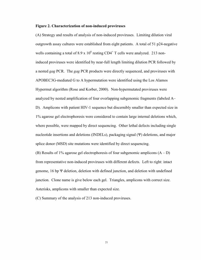
Aliquots from the outer PCR were first amplified in a nested inner gag PCR (Figure 2A).
Cell dilutions for which <20% of the inner reactions were positive were selected because
positive wells at these dilutions have >90% probability of being clonal. Aliquots from
the gag-positive, clonal outer PCRs were amplified using four sets of inner PCR primers
to obtain fragments overlapping by 150 – 3,173 bp (Figure 2A). Importantly, instead of
cloning PCR products, we directly sequenced them. This dramatically reduces PCR
errors since errors occurring after the first or second cycle are present in too small a
fraction of the final products to be observed. Sequences with double peaks or non-
identical overlap regions were discarded. We identified 213 non-induced proviruses from
eight patients.
Hypermutation and large internal deletions render most non-induced proviruses
defective
Most (88.3%) non-induced proviruses had obvious defects precluding replication
(Figure 1C). Direct sequencing of the nested gag PCR product revealed that ~1/3 (32.4%)
of non-induced proviruses had APOBEC3G-mediated G→A hypermutation occurring in
the expected sequence context (GG or GGG) (Yu et al., 2004), with the first G being
mutated. Hypermutated proviruses are replication-defective due to start codon mutations

15
and numerous tryptophan→stop codon mutations (Figure 3). Although the gag gene was
analyzed here, other regions of the genome show even greater hypermutation (Yu et al.,
2004). Importantly, it is unlikely that hypermutated proviruses could produce functional
viral proteins due to stop codons in most open reading frames (ORFs).
Non-induced proviruses that were not hypermutated were further analyzed by
nested amplification of four overlapping subgenomic fragments (Figure 2A). Of the 144
clonal non-induced proviruses that were not hypermutated, 97 had large internal deletions
identified by smaller amplicon size in agarose gel electrophoresis (Figure 2B). We
mapped the precise deletion junctions in 58 of these clones (Figure 4A). For example,
clone 10CB7_48H1 (Figure 2B) gave a smaller amplicon for the nested C reaction, and
amplification of fragments A, B, and D failed due to deletion of nucleotides 4,869 –
9,533. All 58 mapped deletions would affect expression of the essential regulatory
proteins Tat and Rev (Figure 4A) because the deletions encompass the tat and rev exons,
the splice sites, and the Rev-responsive element (RRE).
Deletions are not unexpected. HIV-1 is prone to recombination due to
pseudodiploidity (two RNA copies per virion with physical proximity for recombination).
Frequent template switching events occur during reverse transcription (Simon-Loriere
and Holmes, 2011). Switching between short repeats in a single genome results in
deletion of the intervening sequence and one repeat (Temin, 1993). Frequent large
deletions have been observed in unfractionated PBMC from viremic patients (Sanchez et
al., 1997). Several lines of evidence in our analysis suggest that these deletions occur in
vivo rather than during in vitro analysis. First, deletions were observed following direct
sequencing of uncloned PCR products. Second, for a given provirus, precisely the same

16
deletion junctions were observed in different nested PCRs using different primers. Third,
short amplicons were not seen in control experiments with plasmids carrying the
reference proviral genomes NL4-3 and BaL. Plasmids were mixed, diluted to eight
copies per 100,000 human genome equivalents, and amplified under the same conditions.
No deletion or recombination was observed. Fourth, short sequence repeats were
identified at some deletion junctions (Figure 2B), consistent with a single polymerase
jump due to copy choice recombination during reverse transcription of the minus strand
(Sanchez et al., 1997). Taken together, these results demonstrate that a large fraction of
non-induced proviruses are non-functional due to large internal deletions, likely
introduced during reverse transcription.
The precise fraction of proviruses with deletions could be underestimated by this
analysis because deletions could affect PCR primer binding sites. For 39 clones, the
exact deletion junction could not be identified, probably because the deletions
encompassed some of the binding sites for primers used in the nested PCRs. For these
clones, we obtained at least two sequences to ensure that the amplicons contained non-
hypermutated, patient-specific sequences. For 12 mapped deletions, the deletion
included the reverse gag primer binding site, but mapping was possible because other
nested reactions were successful.
Mutations in cis elements render some non-induced proviruses defective
Proviruses with correct amplicon size were directly sequenced. A small fraction
(8/213) had non-sense mutations and/or frame-shifting insertions or deletions in one or
more ORFs. Small deletions (8 – 98 bp) were found in the packaging signal (Ψ) in 12

17
sequences (Figure 5A). The deletions encompassed the major splice donor (MSD) site.
Point mutations in the MSD site were found in two proviruses. Full genome sequencing
showed that seven of the non-induced proviruses with Ψ or MSD mutations were
otherwise intact. To determine whether these mutations rendered non-induced proviruses
defective, we reconstructed three clones by genome synthesis as described below. The
reconstructed proviruses included two clones with short (8 bp and 16 bp) Ψ deletions in
packaging stem loop two and one with a MSD site mutation (TG|GT→TG|GG).
Although these clones had intact ORFs, they did not replicate in healthy donor CD4+
lymphoblasts (Figure 5B) under conditions in which other reconstructed proviruses
replicated well (see below). Thus mutations in cis elements render otherwise intact
proviruses defective.
Discussion
We show here that most non-induced proviruses were rendered defective during
reverse transcription by APOBEC3G-induced hypermutation (Yu et al., 2004), by
internal deletions caused by copy choice recombination during reverse transcription
(Sanchez et al., 1997), or by frame-shift or nonsense mutations caused by the error-prone
reverse transcriptase (Bebenek et al., 1989). The resulting defective viral genomes can
still integrate because only defects at the ends of the genome affect integration. The
defective genomes will be detected in most PCR-based assays of proviral DNA provided
that the primer binding sites are intact. Many of the defective proviruses have large
internal deletions encompassing the Tat and Rev ORFs and the RRE. Tat-mediated
transactivation is required for effective transcriptional elongation (Kao et al., 1987) and

18
the production of virus particles requires that singly spliced and unspliced HIV-1 mRNAs
be exported from the nucleus in a Rev-dependent fashion (Malim et al., 1989). Thus
these deleted proviruses may not produce viral proteins even after successful induction of
transcription. The same is true for hypermutated proviruses, which have stop codons in
every ORF. Importantly, eradication strategies depend on the production of viral proteins
which allows recognition of the infected cells by HIV-1 specific CTL (Shan et al., 2012).
Defective proviruses with large internal deletions and/or APOBEC3G-induced
hypermutation may not be eliminated even by strategies that effectively eliminate cells
carrying replication-competent virus. These considerations highlight the difficulty of
assessing eradication strategies with PCR-based assays.

19
Table 1. Patient Characteristics

20
Table 2. PCR primers and conditions
21
Figure 2. Characterization of non-induced proviruses
(A) Strategy and results of analysis of non-induced proviruses. Limiting dilution viral
outgrowth assay cultures were established from eight patients. A total of 51 p24-negative
wells containing a total of 8.9 x 10⁶ resting CD4
+ T cells were analyzed. 213 non-
induced proviruses were identified by near-full length limiting dilution PCR followed by
a nested gag PCR. The gag PCR products were directly sequenced, and proviruses with
APOBEC3G-mediated G to A hypermutation were identified using the Los Alamos
Hypermut algorithm (Rose and Korber, 2000). Non-hypermutated proviruses were
analyzed by nested amplification of four overlapping subgenomic fragments (labeled A–
D). Amplicons with patient HIV-1 sequence but discernibly smaller than expected size in
1% agarose gel electrophoresis were considered to contain large internal deletions which,
where possible, were mapped by direct sequencing. Other lethal defects including single
nucleotide insertions and deletions (INDELs), packaging signal (Ψ) deletions, and major
splice donor (MSD) site mutations were identified by direct sequencing.
(B) Results of 1% agarose gel electrophoresis of four subgenomic amplicons (A – D)
from representative non-induced proviruses with different defects. Left to right: intact
genome, 16 bp Ψ deletion, deletion with defined junction, and deletion with undefined
junction. Clone name is give below each gel. Triangles, amplicons with correct size.
Asterisks, amplicons with smaller than expected size.
(C) Summary of the analysis of 213 non-induced proviruses.
22
23
Figure 3. APOBEC3G-mediated G to A hypermutation renders some non-induced
proviruses defective
(A) Nucleotide sequences of the gag gene (representing nucleotides 790 – 2,292, HXB2
coordinates) of hypermutated non-induced proviruses from a representative patient (#20).
Sequence changes representing probable G to A hypermutation are shaded. Sequences
are aligned to the reference sequence HXB2 and an intact, non-induced provirus from the
same patient.
(B) Amino acid sequences of Gag from the hypermutated non-induced proviruses from
(A). Sequence changes resulting from probable G to A hypermutation are shaded.
Sequences are aligned to the reference sequence HXB2 and an intact, non-induced
provirus from the same patient. Note that the methionine start codon of Gag is frequently
mutated to isoleucine (ATG → ATA). This is because the second amino acid of Gag is
glycine (GGX), which results in an APOBEC3G recognition site ATGGGX, allowing
mutation of the third position of the start codon. Hypermutation also affects many
tryptophan codons which contain the APOBEC3G recognition site. Mutation of either or
both of the G nucleotides to A generates a stop codon (TGG → TGA, TAG, or TAA,
designated with an *).

24

25

26
Figure 4. Mapping of large internal deletions in non-induced proviruses
(A) Locations of deletions. Dark blue horizontal bars: continuous sequencing reads
interrupted by deletions. White horizontal bars: deletions. Light blue vertical bars, tat
and rev ORFs. Gray vertical bar: RRE element.
(B) Representative sequences at deletion junctions. Short repeats (underlined) at the
deletion junctions suggest a single reverse transcriptase jump due to copy choice
recombination (Sanchez et al., 1997; Temin, 1993). Hypermutated sequences (see Figure
S2) were not analyzed for deletions.

27

28
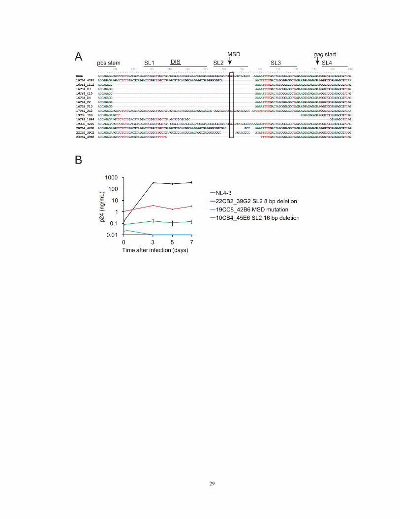
Figure 5. Deletions and mutations in cis elements
(A) Location of cis element deletions and mutations in 14 clones from five patients. pbs,
tRNA primer binding site; SL, packaging stem loop; DIS, dimerization initiation site;
MSD, major splice donor site, typically TG|GT (arrow and box).
(B) Growth kinetics of three reconstructed clones from three patients with MSD site
deletion or mutation but intact ORFs for all viral proteins, in comparison with the NL4-3
reference strain. Each viral inoculum was normalized to 200 ng p24/mL.
29

30
Chapter 4. Intact non-induced proviruses are replication competent
Introduction
Clonal analysis of full-length HIV-1 genomes from untreated patients has
contributed to current understanding of HIV-1 evolution and antiviral immune responses
(272 Li,B. 2007; 236 Salazar-Gonzalez,J.F. 2009; 274 Sahu,G.K. 2010; 297 Li,Y. 1991).
Reconstruction of near-full-length proviruses had be reported (Gao et al., 1998)(Sahu et
al., 2010), but existing reconstruction strategies involve combining two cloned PCR
products, which raises the questions of combining different clones into one and
introducing PCR errors. Bulk PCR sequencing without cloning will only pick up the true
sequences, as PCR induced errors will not be reported in the sequencing results. We
therefore choose de novo gene synthesis to these avoid cloning induced errors.
The replication competence of the reconstructed proviruses depends not only on
intact open reading frames of all viral genomes, but also intact promoter (long terminal
repeat, LTR) function and cis-acting elements, including the packaging signal (Ψ), trans-
activation response elements (TAR), ribosomal frameshift signal, polypurine tracks and
Rev response element (RRE). The function of these elements, with extensive secondary
structures, may not be fully evaluated by sequence analysis alone. Therefore,
reconstruction of the full-length genome becomes the only way to evaluate the
competence of non-induced proviruses, not only to understand the impact of missense
mutations but also the function of the essential elements.
Methods
31
Reconstruction of full-length non-induced proviruses
Direct sequencing results were used for de novo genome synthesis. The resulting
sequences were cloned into pNL4-3 vector (GenScript Inc USA and Bio Basic Inc) by
BssHII and NgoMIV. To correct the 5’LTR to 100% patient derived, the plasmid
fragments between the AatII and BssHII sites were cloned into Zero Blunt TOPO vector
(Invitrogen) and subjected to site directed mutagenesis by QuikChange Site-Directed
Multi Kit (Agilent Technologies, Inc). Reconstructed plasmids were transformed into
Stbl3 chemically competent cells (Invitrogen) and cultured at 30°C to reduce
recombination. The reconstructed plasmids were confirmed by restriction digestion and
sequencing. Induced proviruses from end-dilution viral outgrowth cultures were
reconstructed using similar methods. Plasmids were transfected to HEK 293T cells using
Lipofectamine 2000 (Invitrogen). Viruses released into the supernatants were collected
by ultracentrifugation 48 – 72 hours after transfection. Viruses were titered and
normalized to 200 ng p24/mL for growth kinetics experiments. Healthy donor CD4+
lymphoblasts were infected using spinoculation (1,200 g, 25°C for two hours), and
growth was monitored by p24 ELISA of culture supernatants.
Measurement of LTR function
Patient-derived LTR sequences were cloned into wt-LTR-Luc reporter and transfected
into resting CD4+ T cells from healthy donors using nucleofection (Amaxa) (Yang et al.,
2009a). TK-RLuc was used as internal control. After 48 hours, basal luciferase activity
was measured. We also measured luciferase activity four hours after stimulation with
phorbol myristate acetate (PMA) (10 ng/mL) and ionomycin (1 µM).

32
Results
Of 213 non-induced proviruses, 25 (11.7%) had intact ORFs and cis elements.
When sequences were compared with corresponding sequences from induced proviruses
from the same patient, no known lethal mutations were seen. To determine whether these
intact non-induced proviruses are replication-competent, we used the direct sequencing
results to reconstruct full-length non-induced proviral clones by de novo genome
synthesis (Figure 6A). This strategy avoids PCR and cloning-induced errors. We
reconstructed six non-induced proviruses from four patients by inserting the synthesized
sequence into a plasmid carrying the reference isolate NL4-3. Not captured in our PCR
strategy is a 108 bp segment of the provirus, representing nucleotides (nt) 565 – 672
(HXB2 coordinates). This segment in the 5’ untranslated region includes part of U5 and
the primer binding site (pbs) (Figure 6A). Although U5 deletions may not affect
replication-competence (Vicenzi et al., 1994), we took additional steps to make the
reconstructed clones fully patient-derived, without any NL4-3 sequence. We used
limiting dilution PCR to amplify the LTR-gag region from cells in p24 negative wells in
separate reactions. Using a 424 bp segment from the 5’ U3-R-U5 region (HXB2 nt 140–
564), we constructed phylogenetic trees (Figure 7). Then, using site-directed
mutagenesis, we corrected the 108 bp segment from NL4-3 to the phylogenetically
closest sequence from the same patient (Figure 6A). This process results in proviruses
that are 100% patient-derived and 98.2 % equivalent to specific proviruses present in vivo
with the remaining 1.8% equivalent to a very closely related provirus from the same
patient. Given the high sequence conservation in the 108 bp segment (Figure 7), we

33
estimate that the reconstructed clones could differ from the parent clones by at most 3 nt,
or 0.03% of the genome. For each patient, we also reconstructed an induced viral clone
from a p24 positive well.
Strikingly, all six reconstructed non-induced proviruses from four different
patients showed replication fitness comparable to that of the reference isolate NL4-3 and
reconstructed induced proviruses from the same patients (Figure 6B). It is formally
possible that these intact non-induced proviruses could have actually been defective, with
an inactivating mutation in the 108 bp region that was corrected when the NL4-3
sequence was reverted to the most closely related patient-specific sequence. Such a
mutation would have had to arise during reverse transcription in cell from which the
proviral sequence was obtained. Based on measured in vivo mutation rate of 3.4x10⁻⁵
mutations/bp/cycle (Mansky and Temin, 1995), the probability of a mutation arising in
the 108 bp segment during a single cycle is only 0.0037, and only a subset of these
mutations would be inactivating. Importantly, all of the proviruses that we reconstructed
were functional. The probability that all of these actually had defects in the 108 bp
region is (0.00324)6 = 1.16 x 10
-15. Thus we feel that it is extremely unlikely that the
intact non-induced proviruses described here are in fact defective. This conclusion is
further supported by experiments showing that an additional round of PHA stimulation
causes some non-induced proviruses to produce replication-competent virus (see below).
Taken together, these results indicate that a substantial fraction of the non-induced
proviruses are intact and capable of generating infectious virus if induced in vivo.
Non-induced proviruses have intact promoter function unless hypermutated
34
The ability of intact non-induced proviruses to produce infectious virus suggests
that, at the primary sequence level, their promoters are functional. To confirm this, we
cloned LTRs from representative non-induced proviruses into a luciferase reporter
construct (Yang et al., 2009a). We measured luciferase activity in transfected resting
CD4+ T cells before and four hours after stimulation with PMA and ionomycin. We also
analyzed LTRs from induced proviruses from the same patients and NL4-3. In general,
LTRs from non-induced proviruses showed basal (Figure 8A, C) and stimulated (Figure
8B, D) activity comparable to LTRs from induced proviruses and NL4-3. Decreased
LTR function was only observed for hypermutated clones. This likely reflects G→A
hypermutation in binding sites for the transcription factors NF-κB and Sp1 (Figure 8E).
Thus, most non-induced proviruses have LTRs that are intact at the primary sequence
level.

35
Figure 6. Growth kinetics of reconstructed non-induced viruses
(A) Reconstruction of full-length non-induced proviruses. Pink, clonal patient-derived
sequence. Blue, pNL4-3. Purple, sequence from the most closely related patient-specific
virus. See text for details.
(B) Growth kinetics of reconstructed non-induced viruses from p24 negative wells (pink
and purple), reconstructed induced viruses from p24 positive wells (yellow), and NL4-3
(black).

36

37
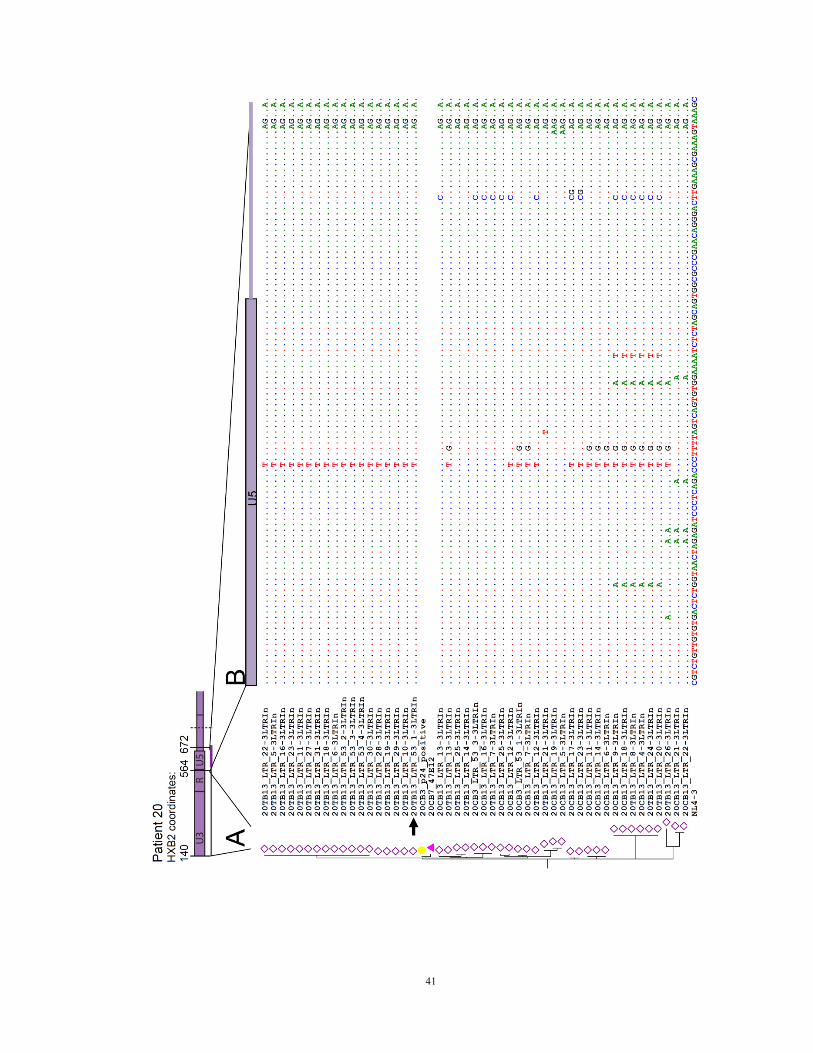
Figure 7. Phylogenetic analysis for reconstruction of the 108 bp sequence
(A) Use of phylogenetic analysis to reconstruct full-length patient-derived proviruses for
six non-induced proviral clones and four induced proviral clones. We corrected the 108
bp segment (565 – 672, HXB2 coordinates) missing from the original PCRs (Figure 3A)
to the most closely related sequence from the same patient. For this purpose, we used
limiting dilution PCR to amplify the LTR-gag region from cells of p24 negative wells.
An average of 31 clonal sequences were obtained for each patient. Using a 424 bp
segment from the U3-R-U5 region (HXB2 positions 140 – 564), we constructed
phylogenetic trees and calculated the phylogenetic distance between reconstructed clones
and each of these LTR-gag PCR sequences using a maximum likelihood estimate. Pink
triangles, intact non-induced proviruses to be reconstructed. Yellow circles, induced
proviruses from p24 positive wells from the same patient. Open diamonds, non-induced
proviral LTR sequences from the same patient. Arrows, exact sequences used for
correction, chosen based on smallest pairwise distance in a maximum likelihood
phylogenic analysis using MEGA 5.10 software.
(B) Nucleotide sequences of the 108 bp segment (nt 565 – 672). Nucleotide differences
between NL4-3 (bottom sequence) and the patient-derived sequence that was most
closely related to the reconstructed clone (arrow) were corrected by site directed
mutagenesis in the reconstructed clones (blank sequences).

38

39

40
41
42
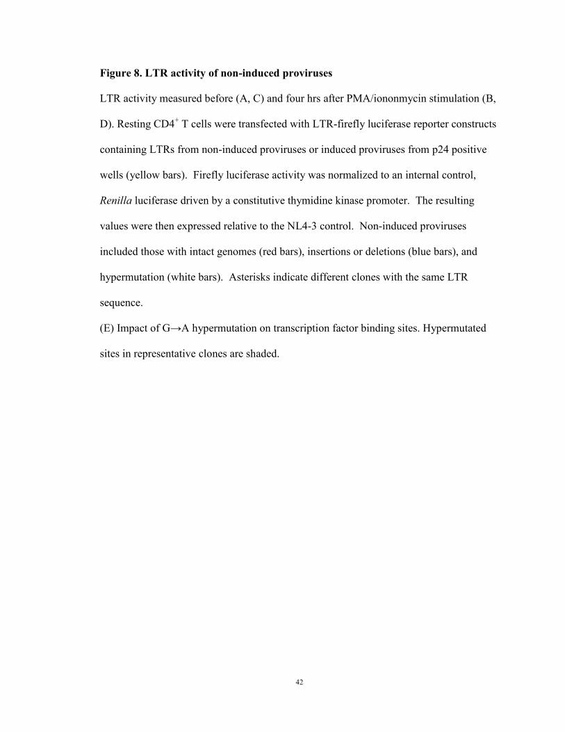
Figure 8. LTR activity of non-induced proviruses
LTR activity measured before (A, C) and four hrs after PMA/iononmycin stimulation (B,
D). Resting CD4+ T cells were transfected with LTR-firefly luciferase reporter constructs
containing LTRs from non-induced proviruses or induced proviruses from p24 positive
wells (yellow bars). Firefly luciferase activity was normalized to an internal control,
Renilla luciferase driven by a constitutive thymidine kinase promoter. The resulting
values were then expressed relative to the NL4-3 control. Non-induced proviruses
included those with intact genomes (red bars), insertions or deletions (blue bars), and
hypermutation (white bars). Asterisks indicate different clones with the same LTR
sequence.
(E) Impact of G→A hypermutation on transcription factor binding sites. Hypermutated
sites in representative clones are shaded.
43
44
Chapter 5. Non-induced proviruses lack epigenetic silencing markers
Introduction
The presence of intact genome and replication competence indicates intact non-
induced proviruses contain required viral components to become infectious once
reactivated. We then explored whether these non-induced proviruses are within favorable
cellular conditions for reactivation. First, the compact heterochromatin needs to be
released into relaxed euchromatin for transcription factors to access binding sites
(Tamaru, 2010). Although several groups reported HIV-1 latency in cell-line models is
associated with integration into heterochromatin and alphoids repeats (Blazkova et al.,
2009; Jordan et al., 2003; Verdin et al., 1993), latent clone selection process in cell line
models may represent a biased population of integration into heterochromatin and may
not reflect latency conditions in vivo. Integration site analysis in patient cells suggests
integration into active transcription units in vivo (Han et al., 2004). Whether non-induced
proviruses are a biased population which are integrated into heterochromatin needs to be
investigated to understand whether they could potentially be reactivated.
Second, local chromatin structure is essential for transcriptionally permissive state
(Deaton and Bird, 2011). Unmethylated CpG at promoter region is associated with
nucleosome displacement and constitutive RNA polymerase II binding, allowing
transcription factors to bind (Deaton and Bird, 2011). Methylated promoter CpG inhibits
transcription factor binding or recruits methyl-binding domain proteins, which further
recruit corepressor complexes and promotes histone deacetylation and therefore
transcriptional repression (Deaton and Bird, 2011). Although HIV-1 promoter is

45
associated with heavy CpG methylation in some cell-line models (Blazkova et al., 2009;
Kauder et al., 2009), analysis of HIV-1 promoter CpG methylation led to contradictory
results in patient samples (Blazkova et al., 2009; Blazkova et al., 2012b). Here we
analyzed CpG methylation of HIV-1 promoter in non-induced proviruses to understand
whether non-induced proviruses have favorable or unfavorable local chromatin
environment for transcription and reactivation.
Methods
Integration site and CpG methylation
Integration site analysis was carried out by inverse PCR as previously described
(Han et al., 2004). Transcription levels of the host genes containing integration sites
were determined using a previously described serial analysis of gene expression (SAGE)
analysis of Bcl-2-transduced primary resting and activated CD4+
T cells (Shan et al.,
2011). CpG methylation analysis was carried out at limiting dilution by bisulfite
sequencing as described by Blazkova et al. (Blazkova et al., 2012b).
Results
Most non-induced proviruses are integrated into active transcription units
We determined the locations of non-induced proviruses in the host genome to
understand whether they were integrated into sites unfavorable for transcription.
Pioneering studies by Bushman et al. showed that HIV-1 typically integrates into
transcription units (Schroder et al., 2002). However, in some model systems, integration
into regions of heterochromatin is associated with latency (Jordan et al., 2003). Using

46
inverse PCR at limiting dilution, we found that 92.9% of 70 non-induced proviruses
resided in transcription units (Figure 9A), consistent with previous observations in patient
resting CD4+ T cells (91%) (Han et al., 2004). Using transcript levels measured in a
SAGE library from a primary cell model of HIV-1 latency (Shan et al., 2011), we found
that most non-induced proviruses were integrated into genes that were transcribed at
moderate levels in both resting and activated CD4+ T cells (Figure 9B). Non-induced
proviruses were found in both orientations with respect to the host genes (Figure 9C).
Overall these results indicate that non-induced proviruses are not integrated into
chromosomal regions that are repressive for transcription, and thus other factors must
have prevented expression of these proviruses.
Lack of CpG methylation in the LTR of non-induced proviruses
We next examined whether non-induced proviruses were epigenetically silenced.
Unlike histone modifications, which can only be analyzed for a population, promoter
CpG methylation status can be analyzed at the single provirus level. CpG islands are
present in the HIV-1 genome (Chavez et al., 2011), including one in the U3 region of the
LTR which contains critical transcription factor binding sites (Figure 10). CpG
methylation in the LTR could lead to recruitment of methyl-CpG binding proteins which
mediate transcriptional silencing by recruiting nucleosome remodeling and histone
deacetylation complexes (Kauder et al., 2009). Therefore, DNA from freshly isolated
resting CD4+ T cells and from cells in p24 negative wells was treated with bisulfite, and
then the LTR region was amplified under limiting dilution conditions (Blazkova et al.,
2012b). Direct sequencing was used to determine the extent of CpG methylation. Only

47
3.1% of the LTR CpGs were methylated in resting CD4+ T cells from study patients.
Even fewer (0.9%) of the CpGs in the LTRs of non-induced proviruses were methylated
(Figure 10). In contrast, we readily detected methylation at a CpG island in the env
region (75.5%), indicating that this method did not selectively amplify non-methylated
sites. Although CpG methylation at the LTR clearly is well documented in some models
of HIV-1 latency (Kauder et al., 2009), our results indicate that non-induced proviruses
are not silenced through CpG methylation at the 5’ LTR.
Discussion
Understanding why intact non-induced proviruses did not produce infectious virus
after maximum in vitro T cell activation is critical for determining their clinical
significance. Possible explanations include silencing by repressive chromatin
modifications or transcriptional interference. We analyzed CpG methylation of the LTRs
of non-induced proviruses at the clonal level. In contrast to some in vitro models of HIV-
1 latency (Kauder et al., 2009), we found that in patient CD4+ T cells, there was little
CpG methylation at the LTR, consistent with another recent study (Blazkova et al.,
2012b). We also examined whether non-induced proviruses are silenced by integration
into heterochromatin. We found that most of the non-induced proviruses were integrated
into active transcription units, consistent with previous studies showing that most HIV-1
proviruses are integrated into introns of actively transcribed genes in cell lines (Schroder
et al., 2002) and patient resting CD4+ T cells (Han et al., 2004). Another potential
explanation for the non-induced proviruses is transcriptional interference (Han et al.,
2008; Lenasi et al., 2008). Since T cell activation may overcome transcriptional
48
interference due to the high affinity of NF-κB for its binding sites in the LTR (Lenasi et
al., 2008), transcriptional interference may not be a major cause of silencing of the non-
induced proviruses.

49
Figure 9. Integration sites of non-induced proviruses
(A) Distribution of integration sites of non-induced proviruses. Intron and exon
boundaries determined as previously described (Shan et al., 2011). Refseq, EST, Human
mRNA and Unigene databases were used for intron and exon boundary determination.
Other, integration sites matched with other gene or transcription databases but not Refseq.
(B) Transcription level of host genes in which non-induced proviruses were integrated.
Transcription levels were determined by SAGE analysis of Bcl-2 transduced activated
and resting primary CD4+
T cells as previously described (Shan et al., 2011). Blue circles,
transcript levels in resting and activated CD4+ T cells. Red squares, transcript levels for
the subset of genes in which integration sites of non-induced proviruses were mapped.
(C) Transcriptional orientation of non-induced proviruses relative to the host gene.

50

51
Figure 10. CpG methylation of non-induced proviruses
(A) Positions of CpGs analyzed.
(B) CpG methylation in the 5’ LTR of proviruses in resting CD4+ T cells from the
indicated patients. Each row represents a single provirus amplified by limiting dilution
PCR after bisulfite treatment. Open circles, nonmethylated CpGs. Closed circles,
methylated CpGs. Missing circles, no CpG present due to sequence polymorphism.
(C) CpG methylation in the env gene of proviruses from resting CD4+ T cells.
(D) CpG methylation in the 5’ LTR of non-induced proviruses from p24 negative wells.
(E) CpG methylation in the env gene of non-induced proviruses from p24 negative wells.
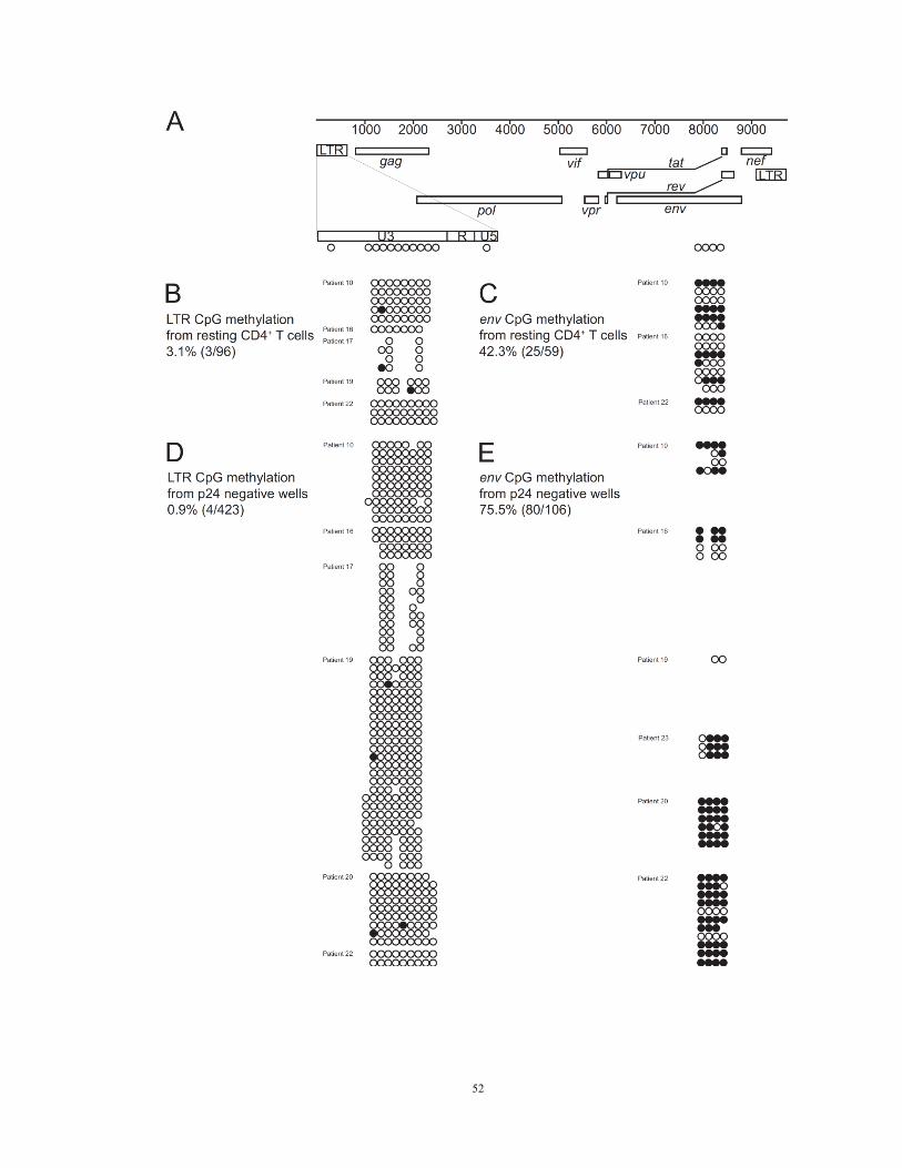
52

53
Chapter 6. Intact non-induced proviruses may increase latent reservoir size by ~60
fold
Introduction
Our discovery of intact non-induced proviruses implies potential underestimation
of the HIV-1 latent reservoir. The viral outgrowth assay has been used as the standard
measurement of the size of the latent reservoir, although all the latency reversing agents
to date can only reactivate a much smaller fraction of the latent reservoir, compared with
that of the single round of PHA activation used in viral outgrowth assay (Blazkova et al.,
2012a). We demonstrated that intact non-induced proviruses may be an underestimated
clinical threat in addition to the inducible reservoir by PHA activation. Identification of
this increased barrier challenges the shock-and-kill HIV-1 eradication strategy.
Characterization of non-induced proviruses provides the molecular basis of HIV-1
measurement. Proviral DNA measurements have been used to measure the size of the
latent reservoir. However, we found that 88% of the proviruses are defective. Measuring
and monitoring defective proviruses does not reflect the true size of the clinically
important latent reservoirs. Rather, using PCR-based proviral DNA measurements to
monitor the size of the latent reservoir may overestimate, leading to unnecessary and
prolonged exposure to latency reversing agents in patients.
Various assays had been used to measure the size of the latent reservoir with
highly diverse results (Eriksson et al., 2013). We demonstrated here the individual
variability and comparison between different estimates of the most reasonable
measurement of the latent reservoir.

54
Methods
Quantitative DNA PCR
Total proviral DNA in resting CD4+ T cells was quantified using a real-time PCR
assay (Durand et al., 2012; Palmer et al., 2003). Copy numbers of the human RNaseP
gene copies were measured in separate reactions to quantify cell number (TaqMan Copy
Number Reference Assay, RNaseP, Invitrogen) (Spivak et al., 2011).
Statistical analysis
The correlation between viral outgrowth and intact non-induced provirus
frequency was analyzed using MedCalc software. Bayesian inference was used to
estimate the proportion of intact non-induced genomes in each patient. An empirical
Bayesian prior, among five models (Table 3), was chosen to reflect the distribution of the
observed data. These proportions were multiplied by total HIV-1 DNA copies per 106
resting CD4+ T cells to yield an estimate of intact non-induced proviruses per million
cells.
Estimate the frequency of intact non-induced proviruses
The number of intact proviral genomes per million cells was estimated for each
patient by Bayesian inference, in collaboration with Sarah B. Laskey and Dr. Daniel I. S.
Rosenbloom. The clones examined were modeled as binomial data, with an unknown
probability of being intact. The probability was estimated for each patient based on five
different prior distributions, in order to emphasize the effect of the choice of prior

55
distribution on results. The beta distribution is the conjugate prior for a binomial
distribution and describes the probabilities of failure and success for a binomial
experiment, i.e., the probability of an examined clone being intact versus defective, so all
five priors are beta distributions with varying parameters.
The uniform and Jeffreys prior distributions are uninformative priors, chosen to
minimize the effect of initial assumptions (Brown et al., 2001). The empirical Bayesian
prior distribution was determined by calculating the sample mean and variance of the
observed proportions of intact non-induced clones and calculating the beta distribution
that fits those parameters. The weighted empirical Bayesian prior has a weighted mean
and sample variance, using the number of clones tested for each patient as the weight.
The weighting decreases the sample variance and makes the prior stronger. By using the
weighting, we acknowledge the consequently strong prior and claim that if we were to
double or triple the number of clones tested, we would probably find at least one intact
non-induced provirus in every patient. The three patients for whom no intact clones were
found are also the three patients in whom the fewest clones were tested. The iterative
median approach started with an arbitrary prior, used the posterior median to create an
empirical Bayes prior for the next round, and iterated until convergence. (Using the
empirical Bayes prior as the initial prior, 40 iterations were sufficient for convergence.)
The empirical Bayesian priors reflect the distribution of the observed data, so
these are not fully Bayesian analyses. However, empirical Bayesian analysis can be
understood as an approximation of the fully Bayesian hierarchical approach (Gelman et
al., 2004).

56
The median and 95% credible intervals for each approach are reported in Table 3.
To estimate the number of cells harboring intact non-induced proviruses per million
resting CD4+ T cells, the median and 95% interval were multiplied by the number of cells
carrying HIV-1 DNA per million resting CD4+ T cells.
Results
Although most non-induced proviruses have identifiable lethal defects, a
substantial fraction are intact and replication-competent at the primary sequence level.
Analysis of LTR function, integration sites, and methylation status suggests that these
intact non-induced proviruses could be induced in vivo, thereby increasing latent
reservoir size. We compared the frequency of induced proviruses (defined using the viral
outgrowth assay) and intact non-induced proviruses (quantitated as the product of total
proviral DNA frequency and the fraction of non-induced proviruses that are intact)
among the total pool of proviruses (measured by quantitative real-time PCR). Bayesian
analysis was chosen instead of maximum likelihood estimation because the former
provides nonzero point estimates for patients from whom no clones with intact genomes
were identified. The positive viral outgrowth assay results in every patient and the
successful detection of intact non-induced proviruses in patients for whom >20 clones
were analyzed suggest that intact non-induced proviruses could be detected in every
patient if enough clones are examined. The fraction of intact non-induced proviruses was
calculated as the median of an empirical Bayesian posterior, the most conservative of five
models tested (Table 3), with a prior distribution chosen to reflect the observed data.
Both the fraction of intact non-induced proviruses and the total number of proviruses per

57
million resting CD4+ T cells varied dramatically from patient to patient (Figure 11A).
There was no correlation between the viral outgrowth assay and the frequency of intact
non-induced proviruses (Figure 11B). All statistical models (Table 3) indicated that the
median frequency of intact non-induced proviruses was at least ~60 fold higher than the
frequency of induced proviruses detected in the viral outgrowth assay. If the intact non-
induced proviruses described here can be induced in vivo, then the size of the latent
reservoir is much greater than previously thought (Figure 11C).
Discussion
To prove replication-competence, we reconstructed six intact, non-induced
proviruses from six different p24 negative wells from four patients. Surprisingly, all
reconstructed viruses replicated as well as the standard reference isolate and control
viruses reconstructed from p24 positive wells. A sterilizing cure requires elimination of
all replication-competent HIV-1, and therefore the discovery that intact non-induced
proviruses are replication-competent means that the number of proviruses that must be
eliminated is much higher than previously thought. We conservatively estimate that the
number may be ~60 fold higher than estimates based on the viral outgrowth assay. Some
statistical models suggest an even higher number (medians of 97 – 273 fold).
Importantly, there is large inter-patient variation in this and other measures of latent
reservoir size. Overall, our results indicate that the “shock and kill” strategy (Archin et al.,
2012; Deeks, 2012) is challenged with a large but unmeasured hidden population of
replication-competent proviruses. Interestingly, despite the intense search for novel
latency reversing agents, none of the drugs tested to date reaches the robust level of in
58
vitro HIV-1 induction achieved by PHA. Thus the finding that the true size of the latent
reservoir may be ~60 fold greater than that estimated using PHA activation is particularly
disturbing. However, it is also important to point out that the even a low level of virus
gene expression may be sufficient to allow the elimination of infected cells by an
appropriately primed CTL response (Shan et al., 2012), and that the critical variable may
the fraction of latently infected cells induced to express HIV-1 genes.

59
Tab
le 3
. E
stim
ati
on
of
inta
ct n
on
-in
du
ced
pro
vir
us
freq
uen
cy

60
Figure 11. Quantification of intact non-induced proviruses
(A) Comparison of the frequency of cells detected in the viral outgrowth assay, cells
carrying intact non-induced proviruses, and cells carrying HIV-1 DNA. The frequency of
cells with HIV-1 DNA was measured by quantitative PCR on freshly isolated resting
CD4+ T cells. The frequency of cells with intact, non-induced proviruses was calculated
as frequency of cells with HIV-1 DNA times the proportion of intact non-induced
proviruses estimated for each patient using an empirical Bayesian model. Open symbols,
no intact proviruses detected, empirical Bayesian estimate plotted. Bars, median value.
(B) Correlation between the frequency of cells detected in the viral outgrowth assay and
the frequency of cells with intact non-induced proviruses. Open symbols, see (A).
(C) Scale representation of the frequencies of the infected resting CD4+ T cell
populations. Volume reflects population size. Yellow circles, minimum size of the latent
reservoir as measured by viral outgrowth assay. Magenta, frequency of cells with intact
proviruses, calculated as the frequency of cells detected in the viral outgrowth assay plus
the frequency of cells with intact non-induced proviruses. This is the potential size of the
latent reservoir if intact non-induced proviruses can be induced in vivo. Blue, cells with
HIV-1 DNA.

61

62
Chapter 7. Stochastic activation of non-induced proviruses
Introduction
Although non-induced proviruses were not reactivated after one round of
maximum T cell activation in vitro, it does not mean that it will never be induced in vivo.
Whether and why non-induced proviruses can be reactivated in vivo is the key question to
HIV-1 eradication.
It has been believed that maximum T cell activation leads to maximum HIV-1
activation. Theoretically, our demonstration of 100% T cell activation by PHA should
lead to 100% HIV-1 provirus reactivation, and therefore none of the non-induced
proviruses can be reactivated. However, HIV-1 activation may not follow T cell
activation mechanisms. Variable levels of Tat, which is required to form positive
transcriptional elongation factor b (pTEFb) to released stalled RNA polymerase II from
LTR, may lead to stochastic fluctuations and phenotypic variability(Weinberger et al.,
2005). Viral fate determination has been proposed to be determined by transcriptional
bursting of Tat (Singh et al., 2010; Weinberger et al., 2008). If stochasticity is the major
mechanism of HIV-1 reactivation, it means that every single non-induced proviruses
might become activated due to stochasticity, and cure cannot be achieved before
eradicating all the intact non-induced proviruses.
To determine whether these non-induced proviruses can be reactivated by
stochasticity, we examined whether repeated PHA activation can activate additional non-
induced proviruses.

63
Methods
Resting CD4+ T cells from four aviremic donors were plated at 0.2 million/well
and subjected to activation with PHA and irradiated allogeneic PBMC for one day.
Patient cells were then cultured in a transwell viral outgrowth assay, with patient cells in
the upper chamber along with irradiated allogeneic PBMC and donor lymphoblasts in the
lower chamber. On day seven, viability of cells in the upper chamber was examined by
forward and side scattered flow cytometry. On day seven of culture, the cells in the
upper and lower chambers of each well were combined and then separated equally into
duplicate culture wells. As all patient cells had divided at least once by day seven, each
pair of split wells should contain an approximately equal number of daughter cells
derived from the patient cells in the original well. One set of the split culture wells was
subjected to an additional round of stimulation with PHA and irradiated allogeneic
PBMC. The other set of wells were not stimulated. Both sets of wells were cultured for
an additional 14 days with two more additions of donor lymphoblasts. Levels of HIV-1
p24 antigen were measured in the supernatant on day 21.
Results
To determine whether intact non-induced proviruses are permanently silenced or
potentially inducible under certain conditions, we tested whether repeated PHA
stimulation can induce additional non-induced proviruses (Figure 12A). We stimulated
multiple replicate cultures of 2 x 10⁵ patient resting CD4
+ T cells with PHA in viral
outgrowth assay conditions. We then split each culture well equally into two wells on
day seven. As all patient cells have divided by day seven (Figure 1), each split well

64
contained daughter cells derived from cells activated in the original well. One set of the
split cultures wells were activated again with PHA while the other set was cultured
without additional stimulation. We then compared supernatant p24 levels after another
14 days of culture. Among 126 p24 negative wells from four patients, 31 (24.6%)
became p24 positive after the additional round of PHA stimulation, while their paired
culture well that did not receive an additional round of stimulation remained p24 negative
(Figure 12B). These results demonstrate that at least some intact non-induced proviruses
can be induced under repeated stimulation.
Discussion
We propose that despite maximum T cell activation, the induction of latent
proviruses is stochastic. Cellular gene expression levels may follow a digital or analogue
distribution after T cell receptor activation, as a result of stochastic and dynamic
processes (Chakraborty and Das, 2010). Elegant experimental and theoretical studies
have shown that HIV-1 proviruses may show stochastic fluctuations in expression
depending on levels of Tat (Burnett et al., 2009; Weinberger et al., 2005; Weinberger et
al., 2005; Weinberger et al., 2008). We propose that intact proviruses undergo stochastic
induction even after maximum cellular activation (Figure 13). Some will be induced by
one round of activation, while others will remain silent but retain the potential to be
activated subsequently. These results indicate an increased barrier to cure, as all intact
non-induced proviruses need to be eradicated. Underestimation of intact proviruses by
viral outgrowth assays could be reflected in delayed viral rebound after an apparent
“cure”, and overestimation of latent reservoir size resulting from detection of defective
65
proviruses by PCR assays could result in prolonged, excessive exposure to toxic latency
reversing agents. Thus, the molecular analysis of non-induced proviruses contributes in
an important way to HIV-1 eradication efforts.
66
Figure 12. Stochastic activation of non-induced proviruses
(A) Scheme for repeated stimulation. To understand whether non-induced proviruses
may be induced by repeated stimuli, resting CD4+ T cells from four aviremic donors were
plated at 0.2 million/well and subjected to activation with PHA and irradiated allogeneic
PBMC for one day. Patients cells were then cultured in a transwell viral outgrowth assay,
with patient cells in the upper chamber along with irradiated allogeneic PBMC and donor
lymphoblasts in the lower chamber. On day seven, viability of cells in the upper chamber
was examined. On day seven of culture, the cells in the upper and lower chambers of
each well were combined and then separated equally into duplicate culture wells. As all
patient cells had divided at least once by day seven, each pair of split wells should
contain an approximately equal number of daughter cells derived from the patient cells in
the original well. One set of the split culture wells was subjected to an additional round
of stimulation with PHA and irradiated allogeneic PBMC. The other set of wells were
not stimulated. Both sets of wells were cultured for an additional 14 days with two more
additions of donor lymphoblasts. Levels of HIV-1 p24 antigen were measured in the
supernatant on day 21.
(B) Comparison of p24 results for of each pair of standard and repeated stimulation
culture wells. Red bars: number of wells containing non-induced proviruses induced to
produce replication competent viruses after repeated PHA stimulation. Magenta bars:
number of wells containing non-induced proviruses which are not induced after repeated
PHA stimulation. Similar results were obtained in experiments in which patient resting
CD4+ T cells were plated at one million/well except that the fraction of positive wells
was higher.
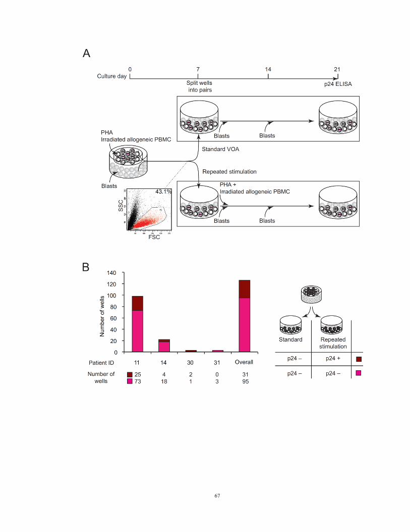
67

68
Figure 13. Mechanism of HIV-1 latency reactivation

69
REFERENCES
Archin, N.M., Espeseth, A., Parker, D., Cheema, M., Hazuda, D., and Margolis, D.M.
(2009). Expression of latent HIV induced by the potent HDAC inhibitor suberoylanilide
hydroxamic acid. AIDS Res. Hum. Retroviruses 25, 207-212.
Archin, N.M., Liberty, A.L., Kashuba, A.D., Choudhary, S.K., Kuruc, J.D., Crooks, A.M.,
Parker, D.C., Anderson, E.M., Kearney, M.F., Strain, M.C., et al. (2012). Administration
of vorinostat disrupts HIV-1 latency in patients on antiretroviral therapy. Nature 487,
482-485.
Barletta, J.M., Edelman, D.C., and Constantine, N.T. (2004). Lowering the detection
limits of HIV-1 viral load using real-time immuno-PCR for HIV-1 p24 antigen. Am. J.
Clin. Pathol. 122, 20-27.
Bebenek, K., Abbotts, J., Roberts, J.D., Wilson, S.H., and Kunkel, T.A. (1989).
Specificity and mechanism of error-prone replication by human immunodeficiency virus-
1 reverse transcriptase. J. Biol. Chem. 264, 16948-16956.
Blankson, J.N., Finzi, D., Pierson, T.C., Sabundayo, B.P., Chadwick, K., Margolick, J.B.,
Quinn, T.C., and Siliciano, R.F. (2000). Biphasic decay of latently infected CD4+ T cells
in acute human immunodeficiency virus type 1 infection. J. Infect. Dis. 182, 1636-1642.
Blazkova, J., Chun, T.W., Belay, B.W., Murray, D., Justement, J.S., Funk, E.K., Nelson,
A., Hallahan, C.W., Moir, S., Wender, P.A., and Fauci, A.S. (2012a). Effect of histone
deacetylase inhibitors on HIV production in latently infected, resting CD4(+) T cells
from infected individuals receiving effective antiretroviral therapy. J. Infect. Dis. 206,
765-769.
Blazkova, J., Murray, D., Justement, J.S., Funk, E.K., Nelson, A.K., Moir, S., Chun,
T.W., and Fauci, A.S. (2012b). Paucity of HIV DNA methylation in latently infected,
resting CD4+ T cells from infected individuals receiving antiretroviral therapy. J. Virol.
86, 5390-5392.
Blazkova, J., Trejbalova, K., Gondois-Rey, F., Halfon, P., Philibert, P., Guiguen, A.,
Verdin, E., Olive, D., Van Lint, C., Hejnar, J., and Hirsch, I. (2009). CpG methylation
controls reactivation of HIV from latency. PLoS Pathog. 5, e1000554.
Bohnlein, E., Lowenthal, J.W., Siekevitz, M., Ballard, D.W., Franza, B.R., and Greene,
W.C. (1988). The same inducible nuclear proteins regulates mitogen activation of both
the interleukin-2 receptor-alpha gene and type 1 HIV. Cell 53, 827-836.
Brown, L.D., Cai, T.T., and DasGupta, A. (2001). Interval Estimation for a Binomial
Proportion. Statistical Science 16(2), 101.
70
Burnett, J.C., Miller-Jensen, K., Shah, P.S., Arkin, A.P., and Schaffer, D.V. (2009).
Control of stochastic gene expression by host factors at the HIV promoter. PLoS Pathog.
5, e1000260.
Chakraborty, A.K., and Das, J. (2010). Pairing computation with experimentation: a
powerful coupling for understanding T cell signalling. Nat. Rev. Immunol. 10, 59-71.
Chavez, L., Kauder, S., and Verdin, E. (2011). In vivo, in vitro, and in silico analysis of
methylation of the HIV-1 provirus. Methods 53, 47-53.
Chun, T.W., Stuyver, L., Mizell, S.B., Ehler, L.A., Mican, J.A., Baseler, M., Lloyd, A.L.,
Nowak, M.A., and Fauci, A.S. (1997). Presence of an inducible HIV-1 latent reservoir
during highly active antiretroviral therapy. Proc. Natl. Acad. Sci. U. S. A. 94, 13193-
13197.
Contreras, X., Schweneker, M., Chen, C.S., McCune, J.M., Deeks, S.G., Martin, J., and
Peterlin, B.M. (2009). Suberoylanilide hydroxamic acid reactivates HIV from latently
infected cells. J. Biol. Chem. 284, 6782-6789.
Coull, J.J., Romerio, F., Sun, J.M., Volker, J.L., Galvin, K.M., Davie, J.R., Shi, Y.,
Hansen, U., and Margolis, D.M. (2000). The human factors YY1 and LSF repress the
human immunodeficiency virus type 1 long terminal repeat via recruitment of histone
deacetylase 1. J. Virol. 74, 6790-6799.
Cujec, T.P., Okamoto, H., Fujinaga, K., Meyer, J., Chamberlin, H., Morgan, D.O., and
Peterlin, B.M. (1997). The HIV transactivator TAT binds to the CDK-activating kinase
and activates the phosphorylation of the carboxy-terminal domain of RNA polymerase II.
Genes Dev. 11, 2645-2657.
Davey, R.T.,Jr, Bhat, N., Yoder, C., Chun, T.W., Metcalf, J.A., Dewar, R., Natarajan, V.,
Lempicki, R.A., Adelsberger, J.W., Miller, K.D., et al. (1999). HIV-1 and T cell
dynamics after interruption of highly active antiretroviral therapy (HAART) in patients
with a history of sustained viral suppression. Proc. Natl. Acad. Sci. U. S. A. 96, 15109-
15114.
Deaton, A.M., and Bird, A. (2011). CpG islands and the regulation of transcription.
Genes Dev. 25, 1010-1022.
Deeks, S.G. (2012). HIV: Shock and kill. Nature 487, 439-440.
Depper, J.M., Leonard, W.J., Drogula, C., Kronke, M., Waldmann, T.A., and Greene,
W.C. (1985). Interleukin 2 (IL-2) augments transcription of the IL-2 receptor gene. Proc.
Natl. Acad. Sci. U. S. A. 82, 4230-4234.
Duh, E.J., Maury, W.J., Folks, T.M., Fauci, A.S., and Rabson, A.B. (1989). Tumor
necrosis factor alpha activates human immunodeficiency virus type 1 through induction

71
of nuclear factor binding to the NF-kappa B sites in the long terminal repeat. Proc. Natl.
Acad. Sci. U. S. A. 86, 5974-5978.
Durand, C.M., Ghiaur, G., Siliciano, J.D., Rabi, S.A., Eisele, E.E., Salgado, M., Shan, L.,
Lai, J.F., Zhang, H., Margolick, J., et al. (2012). HIV-1 DNA is detected in bone marrow
populations containing CD4+ T cells but is not found in purified CD34+ hematopoietic
progenitor cells in most patients on antiretroviral therapy. J. Infect. Dis. 205, 1014-1018.
Ehrenberg, P.K., and Michael, N.L. (2005). PCR amplification, cloning, and construction
of HIV-1 infectious molecular clones from virtually full-length HIV-1 genomes. Methods
Mol. Biol. 304, 387-398.
Eriksson, S., Graf, E.H., Dahl, V., Strain, M.C., Yukl, S.A., Lysenko, E.S., Bosch, R.J.,
Lai, J., Chioma, S., Emad, F., et al. (2013). Comparative analysis of measures of viral
reservoirs in HIV-1 eradication studies. PLoS Pathog. 9, e1003174.
Finzi, D., Hermankova, M., Pierson, T., Carruth, L.M., Buck, C., Chaisson, R.E., Quinn,
T.C., Chadwick, K., Margolick, J., Brookmeyer, R., et al. (1997). Identification of a
reservoir for HIV-1 in patients on highly active antiretroviral therapy. Science 278, 1295-
1300.
Ganesh, L., Burstein, E., Guha-Niyogi, A., Louder, M.K., Mascola, J.R., Klomp, L.W.,
Wijmenga, C., Duckett, C.S., and Nabel, G.J. (2003). The gene product Murr1 restricts
HIV-1 replication in resting CD4+ lymphocytes. Nature 426, 853-857.
Gao, F., Robertson, D.L., Carruthers, C.D., Morrison, S.G., Jian, B., Chen, Y., Barre-
Sinoussi, F., Girard, M., Srinivasan, A., Abimiku, A.G., et al. (1998). A comprehensive
panel of near-full-length clones and reference sequences for non-subtype B isolates of
human immunodeficiency virus type 1. J. Virol. 72, 5680-5698.
Gelman, A., Carlin, J.B., Stern, H.S., and Rubin, D.B. (2004). Bayesian Data Analysis,
Second Edition.(Boca Raton, FL: Chapman and Hall/CRC).
Han, Y., Lassen, K., Monie, D., Sedaghat, A.R., Shimoji, S., Liu, X., Pierson, T.C.,
Margolick, J.B., Siliciano, R.F., and Siliciano, J.D. (2004). Resting CD4+ T cells from
human immunodeficiency virus type 1 (HIV-1)-infected individuals carry integrated
HIV-1 genomes within actively transcribed host genes. J. Virol. 78, 6122-6133.
Han, Y., Lin, Y.B., An, W., Xu, J., Yang, H.C., O'Connell, K., Dordai, D., Boeke, J.D.,
Siliciano, J.D., and Siliciano, R.F. (2008). Orientation-dependent regulation of integrated
HIV-1 expression by host gene transcriptional readthrough. Cell. Host Microbe 4, 134-
146.
He, G., and Margolis, D.M. (2002). Counterregulation of chromatin deacetylation and
histone deacetylase occupancy at the integrated promoter of human immunodeficiency

72
virus type 1 (HIV-1) by the HIV-1 repressor YY1 and HIV-1 activator Tat. Mol. Cell.
Biol. 22, 2965-2973.
Hermankova, M., Siliciano, J.D., Zhou, Y., Monie, D., Chadwick, K., Margolick, J.B.,
Quinn, T.C., and Siliciano, R.F. (2003). Analysis of human immunodeficiency virus type
1 gene expression in latently infected resting CD4+ T lymphocytes in vivo. J. Virol. 77,
7383-7392.
Herrmann, C.H., and Rice, A.P. (1995). Lentivirus Tat proteins specifically associate
with a cellular protein kinase, TAK, that hyperphosphorylates the carboxyl-terminal
domain of the large subunit of RNA polymerase II: candidate for a Tat cofactor. J. Virol.
69, 1612-1620.
Jones, K.A., and Peterlin, B.M. (1994). Control of RNA initiation and elongation at the
HIV-1 promoter. Annu. Rev. Biochem. 63, 717-743.
Jordan, A., Bisgrove, D., and Verdin, E. (2003). HIV reproducibly establishes a latent
infection after acute infection of T cells in vitro. EMBO J. 22, 1868-1877.
Kao, S.Y., Calman, A.F., Luciw, P.A., and Peterlin, B.M. (1987). Anti-termination of
transcription within the long terminal repeat of HIV-1 by tat gene product. Nature 330,
489-493.
Katlama, C., Deeks, S.G., Autran, B., Martinez-Picado, J., van Lunzen, J., Rouzioux, C.,
Miller, M., Vella, S., Schmitz, J.E., Ahlers, J., Richman, D.D., and Sekaly, R.P. (2013).
Barriers to a cure for HIV: new ways to target and eradicate HIV-1 reservoirs. Lancet 381,
2109-2117.
Kauder, S.E., Bosque, A., Lindqvist, A., Planelles, V., and Verdin, E. (2009). Epigenetic
regulation of HIV-1 latency by cytosine methylation. PLoS Pathog. 5, e1000495.
Kinoshita, S., Su, L., Amano, M., Timmerman, L.A., Kaneshima, H., and Nolan, G.P.
(1997). The T cell activation factor NF-ATc positively regulates HIV-1 replication and
gene expression in T cells. Immunity 6, 235-244.
Laird, G.M., Eisele, E.E., Rabi, S.A., Lai, J., Chioma, S., Blankson, J.N., Siliciano, J.D.,
and Siliciano, R.F. (2013). Rapid Quantification of the Latent Reservoir for HIV-1 Using
a Viral Outgrowth Assay. PLoS Pathog. 9, e1003398.
Lassen, K., Han, Y., Zhou, Y., Siliciano, J., and Siliciano, R.F. (2004). The multifactorial
nature of HIV-1 latency. Trends Mol. Med. 10, 525-531.
Lehrman, G., Hogue, I.B., Palmer, S., Jennings, C., Spina, C.A., Wiegand, A., Landay,
A.L., Coombs, R.W., Richman, D.D., Mellors, J.W., et al. (2005). Depletion of latent
HIV-1 infection in vivo: a proof-of-concept study. Lancet 366, 549-555.

73
Lenasi, T., Contreras, X., and Peterlin, B.M. (2008). Transcriptional interference
antagonizes proviral gene expression to promote HIV latency. Cell. Host Microbe 4, 123-
133.
Li, B., Gladden, A.D., Altfeld, M., Kaldor, J.M., Cooper, D.A., Kelleher, A.D., and Allen,
T.M. (2007). Rapid reversion of sequence polymorphisms dominates early human
immunodeficiency virus type 1 evolution. J. Virol. 81, 193-201.
Li, Y., Kappes, J.C., Conway, J.A., Price, R.W., Shaw, G.M., and Hahn, B.H. (1991).
Molecular characterization of human immunodeficiency virus type 1 cloned directly from
uncultured human brain tissue: identification of replication-competent and -defective
viral genomes. J. Virol. 65, 3973-3985.
Malim, M.H., Hauber, J., Le, S.Y., Maizel, J.V., and Cullen, B.R. (1989). The HIV-1 rev
trans-activator acts through a structured target sequence to activate nuclear export of
unspliced viral mRNA. Nature 338, 254-257.
Mansky, L.M., and Temin, H.M. (1995). Lower in vivo mutation rate of human
immunodeficiency virus type 1 than that predicted from the fidelity of purified reverse
transcriptase. J. Virol. 69, 5087-5094.
Mustelin, T., Coggeshall, K.M., Isakov, N., and Altman, A. (1990). T cell antigen
receptor-mediated activation of phospholipase C requires tyrosine phosphorylation.
Science 247, 1584-1587.
Nabel, G., and Baltimore, D. (1987). An inducible transcription factor activates
expression of human immunodeficiency virus in T cells. Nature 326, 711-713.
Palmer, S., Wiegand, A.P., Maldarelli, F., Bazmi, H., Mican, J.M., Polis, M., Dewar, R.L.,
Planta, A., Liu, S., Metcalf, J.A., Mellors, J.W., and Coffin, J.M. (2003). New real-time
reverse transcriptase-initiated PCR assay with single-copy sensitivity for human
immunodeficiency virus type 1 RNA in plasma. J. Clin. Microbiol. 41, 4531-4536.
Patel, S.S., Duby, A.D., Thiele, D.L., and Lipsky, P.E. (1988). Phenotypic and functional
characterization of human T cell clones. J. Immunol. 141, 3726-3736.
Richman, D.D., Margolis, D.M., Delaney, M., Greene, W.C., Hazuda, D., and Pomerantz,
R.J. (2009). The challenge of finding a cure for HIV infection. Science 323, 1304-1307.
Rose, P.P., and Korber, B.T. (2000). Detecting hypermutations in viral sequences with an
emphasis on G --> A hypermutation. Bioinformatics 16, 400-401.
Rowe, H.M., Jakobsson, J., Mesnard, D., Rougemont, J., Reynard, S., Aktas, T., Maillard,
P.V., Layard-Liesching, H., Verp, S., Marquis, J., et al. (2010). KAP1 controls
endogenous retroviruses in embryonic stem cells. Nature 463, 237-240.

74
Sahu, G.K., Sarria, J.C., and Cloyd, M.W. (2010). Recovery of replication-competent
residual HIV-1 from plasma of a patient receiving prolonged, suppressive highly active
antiretroviral therapy. J. Virol. 84, 8348-8352.
Salazar-Gonzalez, J.F., Salazar, M.G., Keele, B.F., Learn, G.H., Giorgi, E.E., Li, H.,
Decker, J.M., Wang, S., Baalwa, J., Kraus, M.H., et al. (2009). Genetic identity,
biological phenotype, and evolutionary pathways of transmitted/founder viruses in acute
and early HIV-1 infection. J. Exp. Med. 206, 1273-1289.
Sanchez, G., Xu, X., Chermann, J.C., and Hirsch, I. (1997). Accumulation of defective
viral genomes in peripheral blood mononuclear cells of human immunodeficiency virus
type 1-infected individuals. J. Virol. 71, 2233-2240.
Schroder, A.R., Shinn, P., Chen, H., Berry, C., Ecker, J.R., and Bushman, F. (2002).
HIV-1 integration in the human genome favors active genes and local hotspots. Cell 110,
521-529.
Selby, M.J., and Peterlin, B.M. (1990). Trans-activation by HIV-1 Tat via a heterologous
RNA binding protein. Cell 62, 769-776.
Shan, L., Deng, K., Shroff, N.S., Durand, C.M., Rabi, S.A., Yang, H.C., Zhang, H.,
Margolick, J.B., Blankson, J.N., and Siliciano, R.F. (2012). Stimulation of HIV-1-
specific cytolytic T lymphocytes facilitates elimination of latent viral reservoir after virus
reactivation. Immunity 36, 491-501.
Shan, L., Yang, H.C., Rabi, S.A., Bravo, H.C., Shroff, N.S., Irizarry, R.A., Zhang, H.,
Margolick, J.B., Siliciano, J.D., and Siliciano, R.F. (2011). Influence of host gene
transcription level and orientation on HIV-1 latency in a primary-cell model. J. Virol. 85,
5384-5393.
Siliciano, J.D., Kajdas, J., Finzi, D., Quinn, T.C., Chadwick, K., Margolick, J.B., Kovacs,
C., Gange, S.J., and Siliciano, R.F. (2003). Long-term follow-up studies confirm the
stability of the latent reservoir for HIV-1 in resting CD4+ T cells. Nat. Med. 9, 727-728.
Siliciano, J.D., and Siliciano, R.F. (2005). Enhanced culture assay for detection and
quantitation of latently infected, resting CD4+ T-cells carrying replication-competent
virus in HIV-1-infected individuals. Methods Mol. Biol. 304, 3-15.
Simon-Loriere, E., and Holmes, E.C. (2011). Why do RNA viruses recombine? Nat. Rev.
Microbiol. 9, 617-626.
Singh, A., Razooky, B., Cox, C.D., Simpson, M.L., and Weinberger, L.S. (2010).
Transcriptional bursting from the HIV-1 promoter is a significant source of stochastic
noise in HIV-1 gene expression. Biophys. J. 98, L32-4.
75
Spivak, A.M., Salgado, M., Rabi, S.A., O'Connell, K.A., and Blankson, J.N. (2011).
Circulating monocytes are not a major reservoir of HIV-1 in elite suppressors. J. Virol.
85, 10399-10403.
Strain, M.C., Gunthard, H.F., Havlir, D.V., Ignacio, C.C., Smith, D.M., Leigh-Brown,
A.J., Macaranas, T.R., Lam, R.Y., Daly, O.A., Fischer, M., et al. (2003). Heterogeneous
clearance rates of long-lived lymphocytes infected with HIV: intrinsic stability predicts
lifelong persistence. Proc. Natl. Acad. Sci. U. S. A. 100, 4819-4824.
Tamaru, H. (2010). Confining euchromatin/heterochromatin territory: jumonji crosses the
line. Genes Dev. 24, 1465-1478.
Temin, H.M. (1993). Retrovirus variation and reverse transcription: abnormal strand
transfers result in retrovirus genetic variation. Proc. Natl. Acad. Sci. U. S. A. 90, 6900-
6903.
Tyagi, M., Pearson, R.J., and Karn, J. (2010). Establishment of HIV latency in primary
CD4+ cells is due to epigenetic transcriptional silencing and P-TEFb restriction. J. Virol.
84, 6425-6437.
Van Lint, C., Emiliani, S., Ott, M., and Verdin, E. (1996). Transcriptional activation and
chromatin remodeling of the HIV-1 promoter in response to histone acetylation. EMBO J.
15, 1112-1120.
Verdin, E., Paras, P.,Jr, and Van Lint, C. (1993). Chromatin disruption in the promoter of
human immunodeficiency virus type 1 during transcriptional activation. EMBO J. 12,
3249-3259.
Vicenzi, E., Dimitrov, D.S., Engelman, A., Migone, T.S., Purcell, D.F., Leonard, J.,
Englund, G., and Martin, M.A. (1994). An integration-defective U5 deletion mutant of
human immunodeficiency virus type 1 reverts by eliminating additional long terminal
repeat sequences. J. Virol. 68, 7879-7890.
Weinberger, L.S., Burnett, J.C., Toettcher, J.E., Arkin, A.P., and Schaffer, D.V. (2005).
Stochastic gene expression in a lentiviral positive-feedback loop: HIV-1 Tat fluctuations
drive phenotypic diversity. Cell 122, 169-182.
Weinberger, L.S., Dar, R.D., and Simpson, M.L. (2008). Transient-mediated fate
determination in a transcriptional circuit of HIV. Nat. Genet. 40, 466-470.
West, M.J., Lowe, A.D., and Karn, J. (2001). Activation of human immunodeficiency
virus transcription in T cells revisited: NF-kappaB p65 stimulates transcriptional
elongation. J. Virol. 75, 8524-8537.

76
Williams, S.A., Chen, L.F., Kwon, H., Ruiz-Jarabo, C.M., Verdin, E., and Greene, W.C.
(2006). NF-kappaB p50 promotes HIV latency through HDAC recruitment and
repression of transcriptional initiation. EMBO J. 25, 139-149.
Williams, S.A., and Greene, W.C. (2007). Regulation of HIV-1 latency by T-cell
activation. Cytokine 39, 63-74.
Wong, J.K., Hezareh, M., Gunthard, H.F., Havlir, D.V., Ignacio, C.C., Spina, C.A., and
Richman, D.D. (1997). Recovery of replication-competent HIV despite prolonged
suppression of plasma viremia. Science 278, 1291-1295.
Yang, H.C., Shen, L., Siliciano, R.F., and Pomerantz, J.L. (2009a). Isolation of a cellular
factor that can reactivate latent HIV-1 without T cell activation. Proc. Natl. Acad. Sci. U.
S. A. 106, 6321-6326.
Yang, H.C., Xing, S., Shan, L., O'Connell, K., Dinoso, J., Shen, A., Zhou, Y., Shrum,
C.K., Han, Y., Liu, J.O., et al. (2009b). Small-molecule screening using a human primary
cell model of HIV latency identifies compounds that reverse latency without cellular
activation. J. Clin. Invest. 119, 3473-3486.
Yu, Q., Konig, R., Pillai, S., Chiles, K., Kearney, M., Palmer, S., Richman, D., Coffin,
J.M., and Landau, N.R. (2004). Single-strand specificity of APOBEC3G accounts for
minus-strand deamination of the HIV genome. Nat. Struct. Mol. Biol. 11, 435-442.

77
Ya-Chi Ho Last revised: September 2013
EDUCATION
Ph.D. 2013 Cellular and Molecular Medicine Program
Johns Hopkins University School of Medicine
Mentor: Robert F. Siliciano, M.D., Ph.D.
M.M.S 2007 Graduate Institute of Clinical Medicine
National Taiwan University School of Medicine
Mentors: Shan-Chwen Chang, M.D., Ph.D.
Wei-Kung Wang, M.D., Sc.D.
M.D. 2002 National Cheng Kung University School of Medicine
PROFESSIONAL EXPERIENCE
Teaching 2009 Pollard Scholar of Fundamentals of Genetics
assistant Cellular and Molecular Medicine PhD Program
Johns Hopkins School of Medicine
Attending 2007-2008 Division of Infectious Diseases
physician Department of Internal Medicine
National Taiwan University Hospital
Clinical 2005-2007 Division of Infectious Diseases
fellow Department of Internal Medicine
National Taiwan University Hospital
Resident 2002-2005 Department of Internal Medicine
National Taiwan University Hospital
Clinical 2001 Visiting Medical Student
rotation Clerkship in Pulmonary Medicine, Consultative Cardiology
Emergency Surgical Care and Neurology
Duke University Medical Center
Clinical 2000 Visiting Medical Student
rotation Clerkship in Medical Oncology
Yale-New Haven Hospital
Research 1999 Summer Research Student
rotation Laboratory of Lien-I Hor, Ph.D.
Department of Microbiology and Immunology
National Cheng Kung University School of Medicine
American Bureau for Medical Advancement in China and
Schering Plough Summer Research Grant

78
HONORS AND AWARDS
2013 First prize, Graduate Student Poster Competition
Johns Hopkins University School of Medicine
2013 Young Investigator Award
The 20th Conference on Retroviruses and Opportunistic
Infections (CROI)
2011 Howard Hughes Medical Institute International Student
Research Fellowship
2009 American Association for the Advancement of Science
(AAAS)/Science Program for Excellence in Science
2008 Scholarship for clinicians doing research
National Health Research Institute, Taiwan
2007 Scholarship for PhD Studentship Abroad
Ministry of Education, Taiwan
2006 Best Teaching Resident Award
National Taiwan University Hospital
2003-2005 Best Resident Award, Department of Internal Medicine
National Taiwan University Hospital
2002 Dean’s Award on graduation from National Cheng Kung
University School of Medicine
2002 Best Intern Award
National Cheng Kung University Hospital
2002 Doctor Tsong-Ming Tu’s award
2002 Phi Tau Phi Scholastic Honor Society Award
2001 Dr. Andrew T. Huang Medical Education Promotion Grant
1999-2001 Presidential Award, National Cheng Kung University
1999 American Bureau for Medical Advancement in China and
Schering Plough Summer Research Grant
1998 Tzu-Chi Charity Foundation Medical Scholarship
1996 Presidential Award, National Cheng Kung University
RESEARCH GRANT PARTICIPATION
Research 2011 Analysis of non-induced HIV-1 provirus in latent reservoirs:
fellow a critical but neglected threat to HIV-1 eradication
Howard Hughes Medical Institute International Student
Research Fellowship
Junior 2013 Characterization of non-inducible proviruses
Co-Principle Principle Investigator: Robert F. Siliciano
Investigator CFAR-MDC NIH supplement working group
National Institute of Health
Co-Principle 2006 Clinical manifestations and treatment result of patients with
Investigator severe influenza infection in Taiwan
Principle Investigator: Shan-Chwen Chang
Centers for Disease Control, Taiwan
79
Assistant 2006 A clinical and epidemiological study of type A influenza in
Investigator Taiwan
Principle Investigator: Shan-Chwen Chang
National Science Council, Taiwan
PUBLICATIONS
Peer reviewed articles
Ho Y.C., Liang S., Hosmane N.H., Wang J., Laskey S.B., Rosenbloom D. I. S., Lai J.,
Blankson J.N., Siliciano R.F. Replication-competent non-induced proviruses in the latent
reservoir increase barrier to HIV-1 cure. Cell. In press (2013).
Yukl S.A., Busch M., Chun T.W., Douek D., Bentsen C., Eisele E., Haase A., Ho Y.C.,
Hütter G., Lee T.H., Justement J.S., Keating S., Li P., Murray D., Palmer S., Pilcher C.,
Pillai S., Price R.W., Rothenberger M., Schacker T., Siliciano J.D., Siliciano R.F.,
Sinclair E., Strain M., Wong J., Richman D.D., Deeks S.G. Challenges in detecting HIV
persistence during potentially curative interventions: a study of the Berlin patient. PLoS
Pathog. 9(5):e1003347 (2013).
Thayil S.M., Ho Y.C., Bollinger R.C., Blankson J.N., Siliciano R.F., Karakousis P.C.,
Page K.R. Mycobacterium tuberculosis complex enhances susceptibility of CD4 T cells
to HIV through a TLR2-mediated pathway. PLoS One. 7(7):e41093 (2012).
Wu U.I., Wang J.T., Ho Y.C., Pan S.C., Chen Y.C., Chang S.C. Factors associated with
development of complications among adults with influenza: A 3-year prospective
analysis. J. Formos. Med. Assoc. 111(7):364-9 (2012).
Ho Y.C., Wang J.L., Wang J.T., Wu U.I., Chang C.W., Wu H.S., Chen C.H., Chuang
Y.M., Chang S.C. Prognostic factors for fatal adult influenza pneumonia. J. Infect.
58(6):439-45 (2009).
Ho Y.C., Chang S.C., Lin S.R., Wang W.K. High levels of mecA DNA detected by a
quantitative real-time PCR assay are associated with mortality in patients with
methicillin-resistant Staphylococcus aureus bacteremia. J. Clin. Microbiol. 47(5):1443-
51 (2009).
Sun H.Y., Kung H.C., Ho Y.C., Chien Y.F., Chen M.Y., Sheng W.H., Hsieh S.M., Wu
C.H., Liu W.C., Hung C.C., Chang S.C. Seroprevalence of hepatitis A virus infection in
persons with HIV infection in Taiwan: implications for hepatitis A vaccination. Int. J.
Infect. Dis. 13(5):e199-205 (2009).
Ho Y.C., Sun H.Y., Chen M.Y., Hsieh S.M., Sheng W.H., Chang S.C.. Clinical
presentations and outcome of toxoplasmic encephalitis in patients with human
immunodeficiency virus type one infection. J. Microbiol. Immunol. Infect.. 41(5):386-
92 (2008).

80
Ho Y.C., Shih T.F., Lin Y.H., Hiao C.F., Chen M.Y., Hsieh S.M., Sheng W.H., Hung
C.C., Chang S.C. Osteonecrosis in patients with human immunodeficiency virus type one
infection in Taiwan. Jpn. J. Infecti. Dis. 60(6):382-6 (2007).
Chuang Y.M., Tseng S.P., Teng L.J., Ho Y.C., Hsueh P.R. Emergence of cefotaxime
resistance in Citrobacter freundii causing necrotizing fasciitis and osteomyelitis. J.
Infect. 53(3): e161-3 (2006).
Fang J.J., Shao C.P., Ho Y.C. and Hor L.I. Isolation and characterization of a Vibrio
vulnificus mutant deficient in both extracellular metalloprotease and cytolysin. Infect.
Immun. 69(9): 5943-8 (2001).
Oral Abstracts and Posters
Ho Y.C., Shan L., Wang J., Hosmane N.N., Blankson J.N., Silciano R.F. Replication-
competent non-induced HIV-1 proviruses in the latent reservoir increase the barrier to
HIV-1 cure. Johns Hopkins Center for AIDS Research (CFAR) Annual Meeting,
Baltimore, United States. Poster presentation (2013).
Ho Y.C., Shan L., Wang J., Hosmane N.N., Blankson J.N., Silciano R.F.
Characterization of non-induced HIV-1 proviruses dampens the hope for HIV-1
eradication. The 20th
Conferences on Retroviruses and Opportunistic Infections. Atlanta,
United States. Oral presentation (2013).
Ho Y.C., Blankson J.N., Siliciano R.F. Transwell limiting dilution co-culture as a novel
and effective method for HIV-1 latent reservoir analysis. The 1114th
General Meeting of
the American Society of Microbiology, San Francisco, United States. Poster presentation
(2012).
Yukl S., Chun T.W., Strain M., Siliciano J.D., Eisele E., Buckeit R., Ho Y.C., Wong J.,
Busch M., Hutter G., Richman D.D., Siliciano R.F., Deeks S.G. Challenges Inherent in
Detecting HIV Persistence During Potentially Curative Interventions. International
Workshop on HIV & Hepatitis Virus Drug Resistance and Curative Strategies. Melia
Sitges, Spain. Poster presentation (2012).
Ho Y.C., Siliciano R.F. Suberoylanilide hydroxamic acid (SAHA) does not reactivate
additional HIV-1 proviruses from latent reservoirs in addition to those reactivated by
phytohemagglutinin (PHA). Keystone symposia, Frontiers in HIV Pathogenesis, Therapy
and Eradication (X8), Whistler, Canada. Poster presentation (2012).
Ho Y.C., Chang S.C. Prognostic factors for fatal pediatric influenza pneumonia. The
47th
Annual Meeting of the Infectious Disease of America, Philadelphia, United States.
Poster presentation (2009).
81
Ho Y.C., Wang W.K., Chang S.C. Quantification of methicillin-resistantStaphylococcus
aureus in blood by a real-time polymerase chain reaction assay and its application in
monitoring the treatment of methicillin-resistant Staphylococcus aureus bacteremia.
Infectious Disease Society of Taiwan Annual Meeting, Taipei, Taiwan, oral presentation.
The 108th
General Meeting of American Society for Microbiology, Toronto, Canada,
poster presentation (2007).
Ho Y.C., Wang J.T., Hung C.C., Chang S.C. Clinical profile of pathologically-proven
toxoplasma lymphadenitis in non-HIV infected patients. Infectious Disease Society of
Taiwan Annual Meeting. Oral presentation. (2006).
Ho Y.C., Hor L.I. Roles of protease and cytolysin in tissue damage caused by Vibrio
vulnificus. American Bureau for Medical Advancement in China and Schering-Plough
Summer Research Scholarship Conference, Taipei, Taiwan. Oral presentation (1999).


![EndogenousMurineLeukemiaViruses:Relationshipto ...downloads.hindawi.com/journals/av/2011/940210.pdfline less than 1.5 million years ago [3, 4], and generation of new endogenous proviruses](https://img.pdfslide.us/doc/110x75/5e80085e1f5d4f27dd45f8fe/endogenousmurineleukemiavirusesrelationshipto-line-less-than-15-million-years.jpg)