Embed Size (px)
Citation preview

Repetitive N-WASP–Binding Elements of theEnterohemorrhagic Escherichia coli Effector EspFU
Synergistically Activate Actin AssemblyKenneth G. Campellone1, Hui-Chun Cheng2, Douglas Robbins3, Anosha D. Siripala1, Emma J. McGhie4,
Richard D. Hayward4, Matthew D. Welch1, Michael K. Rosen2, Vassilis Koronakis4, John M. Leong3*
1 Department of Molecular and Cell Biology, University of California Berkeley, Berkeley, California United States of America, 2 Department of Biochemistry and Howard
Hughes Medical Institute, University of Texas Southwestern Medical Center, Dallas, Texas, United States of America, 3 Department of Molecular Genetics and Microbiology,
University of Massachusetts Medical School, Worcester, Massachusetts, United States of America, 4 Department of Pathology, University of Cambridge, Cambridge, United
Kingdom
Abstract
Enterohemorrhagic Escherichia coli (EHEC) generate F-actin–rich adhesion pedestals by delivering effector proteins intomammalian cells. These effectors include the translocated receptor Tir, along with EspFU, a protein that associates indirectlywith Tir and contains multiple peptide repeats that stimulate actin polymerization. In vitro, the EspFU repeat region iscapable of binding and activating recombinant derivatives of N-WASP, a host actin nucleation-promoting factor. In spite ofthe identification of these important bacterial and host factors, the underlying mechanisms of how EHEC so potentlyexploits the native actin assembly machinery have not been clearly defined. Here we show that Tir and EspFU are sufficientfor actin pedestal formation in cultured cells. Experimental clustering of Tir-EspFU fusion proteins indicates that the centralrole of the cytoplasmic portion of Tir is to promote clustering of the repeat region of EspFU. Whereas clustering of a singleEspFU repeat is sufficient to bind N-WASP and generate pedestals on cultured cells, multi-repeat EspFU derivatives promoteactin assembly more efficiently. Moreover, the EspFU repeats activate a protein complex containing N-WASP and the actin-binding protein WIP in a synergistic fashion in vitro, further suggesting that the repeats cooperate to stimulate actinpolymerization in vivo. One explanation for repeat synergy is that simultaneous engagement of multiple N-WASP moleculescan enhance its ability to interact with the actin nucleating Arp2/3 complex. These findings define the minimal set ofbacterial effectors required for pedestal formation and the elements within those effectors that contribute to actin assemblyvia N-WASP-Arp2/3–mediated signaling pathways.
Citation: Campellone KG, Cheng H-C, Robbins D, Siripala AD, McGhie EJ, et al. (2008) Repetitive N-WASP–Binding Elements of the Enterohemorrhagic Escherichiacoli Effector EspFU Synergistically Activate Actin Assembly. PLoS Pathog 4(10): e1000191. doi:10.1371/journal.ppat.1000191
Editor: Jorge E. Galan, Yale University School of Medicine, United States of America
Received April 9, 2008; Accepted September 30, 2008; Published October 31, 2008
Copyright: � 2008 Campellone et al. This is an open-access article distributed under the terms of the Creative Commons Attribution License, which permitsunrestricted use, distribution, and reproduction in any medium, provided the original author and source are credited.
Funding: This work was supported by National Institutes of Health (NIH) grants R01-AI46454 to JML, R01-GM56322 to MKR, and R01-GM59609 to MDW, aHoward Hughes Medical Institute Investigatorship and Welch Foundation Grant (I-1544) to MKR, a Wellcome Trust programme grant and a Medical ResearchCouncil project grant to VK, and a Chilton Foundation Fellowship to H-CC. RDH is a Royal Society University Research Fellow. KGC visited the Koronakis laboratorywith support from a Human Frontier Science Program international fellowship.
Competing Interests: The authors have declared that no competing interests exist.
* E-mail: [email protected]
Introduction
Enterohemorrhagic Escherichia coli (EHEC) O157:H7 colonize the
intestinal tract of cattle and other reservoir hosts without inducing
disease, but cause severe diarrheal illness in humans that ingest
contaminated materials [reviewed in 1–3]. The mode of epithelial
colonization by EHEC reflects its membership in the attaching and
effacing (AE) family of pathogens. These bacteria, which include
enteropathogenic E. coli (EPEC) and Citrobacter rodentium, attach
tightly to the intestinal epithelium, efface microvilli, and generate
filamentous (F-)actin pedestals beneath sites of adherence. The
formation of AE lesions is critical for pathogenesis, because
mutations that abolish their biogenesis severely impair colonization
[4–7]. Moreover, an EHEC mutant that forms AE lesions but
possesses a diminished capacity to stimulate actin assembly is
defective at expanding the initial infectious niche [8].
During infection, EHEC expresses a type III secretion system
capable of translocating more than 30 effector proteins from the
bacterium into the mammalian cell [9]. This delivery system is
encoded by the locus of enterocyte effacement (LEE), which also
contains several of the substrates for injection [reviewed in 10–11].
Among these LEE-encoded effectors is the translocated intimin
receptor (Tir), which is essential for AE lesion formation. Tir is
delivered into the host cell, where it localizes to the plasma
membrane in a hairpin conformation that includes a central
extracellular region that binds to intimin, a LEE-encoded adhesin.
Intimin-Tir interaction promotes intimate attachment to the host
cell, and also results in clustering of the N- and C-terminal
cytoplasmic domains of Tir [12], which are capable of interacting
with host proteins.
The Tir molecules from EHEC and EPEC both trigger actin
assembly pathways that involve N-WASP, an actin nucleation-
promoting factor [13–15]. N-WASP utilizes a C-terminal WH2/
verprolin-connector-acidic (VCA) segment to activate the Arp2/3
complex, a major actin nucleator in cells [reviewed in 16–17].
Normally, N-WASP adopts an autoinhibited conformation in
PLoS Pathogens | www.plospathogens.org 1 October 2008 | Volume 4 | Issue 10 | e1000191

which its VCA domain is sequestered by an intramolecular
interaction with a central GTPase binding domain (GBD). It can
be activated by several stimuli, including Nck, an adaptor protein
that binds to its proline-rich domain (PRD), and Cdc42, a small
GTPase that binds the Cdc42-Rac binding (CRIB) sequence
within the GBD [18–19]. When assayed using purified proteins in
vitro, either Cdc42 or Nck is sufficient to stimulate N-WASP-
Arp2/3–mediated actin assembly. However, under physiological
conditions, N-WASP regulation is significantly more complex,
since several proteins including WIP (WASP-interacting protein),
bind to its N-terminal WH1 domain and influence its activation
[20]. In fact, Cdc42 is insufficient to stimulate the native N-
WASP/WIP complex [21].
EPEC pedestal formation involves activation of N-WASP by
signaling initiated from the C-terminal cytoplasmic domain of its
Tir protein, which is phosphorylated by host tyrosine kinases [22–
24]. In fact, Tir is the only EPEC effector required for pedestal
formation, since clustering of its ectopically expressed C-terminus
in mammalian cells is sufficient to generate pedestals with high
efficiency [25]. The dominant pathway for N-WASP stimulation
by EPEC involves recruitment of Nck to one site of Tir tyrosine
phosphorylation [reviewed in 26–27].
In contrast to EPEC, EHEC requires a second effector to trigger
pedestal formation. This protein, EspFU (also known as TccP),
localizes beneath bound bacteria after delivery into host cells, co-
precipitates with Tir, and promotes phosphotyrosine- and Nck-
independent actin assembly [28–29]. Residues 456 to 458 in the
cytoplasmic C-terminus of Tir are required for EspFU recruitment
and efficient pedestal formation [30–31], but because direct
interactions between Tir and EspFU have not been detected,
additional factors are assumed to mediate their association.
EspFU contains an N-terminal secretion signal followed by a C-
terminus consisting of multiple nearly-identical 47-residue proline-
rich peptide repeats [32]. EspFU derivatives containing these
repeats bind to a segment of N-WASP encompassing the GBD
[27–28,32]. Mutants containing the EspFU N-terminus and as few
as two repeats were shown to be capable of stimulating actin
assembly using purified N-WASP and Arp2/3 in vitro and also
could promote some degree of pedestal formation during infection
of cultured cells, whereas derivatives containing only a single
repeat did not [32].
These observations provide a framework for understanding the
mechanisms by which EHEC triggers actin pedestal formation:
Tir and EspFU are central to actin assembly, the Tir C-terminus is
critical for recruitment of EspFU and other putative factors that
may contribute to actin nucleation, and multiple proline-rich
repeats of EspFU are required for maximal signaling. However,
important questions remain unanswered. For example, the
potential roles for EHEC effectors other than Tir and EspFU
during actin assembly have not been defined, nor have the precise
roles of the Tir C-terminus and/or the putative factors that
mediate Tir-EspFU interactions. In addition, whereas EspFU can
bind and activate purified recombinant N-WASP derivatives in
vitro, it is unknown how accurately these assays might reflect N-
WASP-stimulating activities in the context of the complex
intracellular milieu, where other N-WASP binding partners like
WIP likely modulate its autoinhibited state.
In the current study, we show that Tir and EspFU are sufficient
to trigger pedestal formation in the absence of any other EHEC
factors. By analyzing EspFU derivatives for the ability to bind
native and recombinant N-WASP and stimulate N-WASP/WIP-
mediated actin assembly in vitro and pedestal formation in cells,
we arrive at a model in which the critical function of Tir is to
promote clustering of the C-terminal repeats of EspFU. These
repeats, in turn, activate N-WASP synergistically and lead to the
formation of an Arp2/3-containing multi-protein complex that
promotes robust actin polymerization.
Results
Intimin-mediated clustering of Tir and EspFU is sufficientto promote actin pedestal formation
During infection, EHEC is capable of translocating more than
30 effector proteins into the host cell [9]. At least two of these
effectors, EspFU and EspF, are known to directly activate N-
WASP [28–29,33], while additional proteins stimulate signaling
pathways that may also lead to N-WASP-mediated actin
polymerization [34]. However, only two effectors, Tir and EspFU,
have been shown to be crucial for EHEC pedestal formation in
genetic deletion studies. Moreover, KC12, an EPEC strain in
which EPEC tir has been replaced by EHEC tir, is defective at
actin pedestal formation, whereas expression of EspFU by KC12
allows pedestal formation at high efficiency in manner indistin-
guishable from that of EHEC [28]. These results are consistent
with the possibility that Tir and EspFU are the only effectors
essential for EHEC-mediated actin pedestal formation. To
definitively test whether EspFU is the only effector in addition to
Tir that is required for actin pedestal assembly, mammalian cells
were transfected with plasmids encoding derivatives of EHEC Tir
and EspFU in the absence of all other bacterial factors. Tir was N-
terminally tagged with an HA epitope, and its first transmembrane
domain replaced with the transmembrane segment of the New-
castle Disease Virus HN surface protein to promote efficient
plasma membrane localization [31]. EspFU was fused to GFP at its
N-terminus and a 5myc tag at its C-terminus to allow detection by
fluorescence microscopy and immunoblotting, respectively
(Figure 1A). In addition to full-length EspFU, we also generated
GFP-fusions carrying only its N-terminal 88 residues (EspFU-N) or
its C-terminal repeats (EspFU-R1-6) (Figure 1B). To cluster Tir in
the plasma membrane, transfected cells were treated with a non-
Author Summary
Enterohemorrhagic Escherichia coli (EHEC) O157:H7 is afood-borne pathogen that causes diarrhea and life-threatening systemic illnesses. EHEC colonizes the intestineby adhering tightly to host cells and injecting bacterialmolecules that trigger the formation of a ‘‘pedestal’’ belowbound bacteria. These pedestals are generated byreorganizing the actin cytoskeleton into densely packedfilaments beneath the plasma membrane. Pedestal forma-tion is therefore not only important for EHEC disease, itprovides a means to study how mammalian cells controltheir shape. We show here that two EHEC proteins, Tir andEspFU, are sufficient to trigger pedestal formation. Tirlocalizes to the mammalian plasma membrane, and itscentral function is to promote clustering of EspFU. EspFU
contains multiple repeat sequences that stimulate actinpolymerization by binding N-WASP, a host protein thatinitiates actin assembly. Although a single repeat of EspFU
can generate pedestals, multi-repeat variants promoteactin assembly cooperatively. One explanation for thissynergy is that tandem repeats can potently trigger theformation of a complex of mammalian proteins thatmodulate the actin cytoskeleton. These findings define theminimal set of EHEC effectors required for pedestalformation and the elements within those effectors thatconfer their ability to alter cell shape.
EspFU, N-WASP, and Actin Assembly
PLoS Pathogens | www.plospathogens.org 2 October 2008 | Volume 4 | Issue 10 | e1000191

pathogenic strain of E. coli that was engineered to express intimin
(Figure 1A), and binds selectively to Tir-expressing cells [31].
To evaluate actin pedestal formation on cells additionally co-
expressing EspFU, only those cells exhibiting GFP fluorescence were
examined. Adherent bacteria were identified by DAPI-staining and
F-actin was visualized using fluorescent phalloidin. Bacteria that
bound to cells co-expressing Tir and full-length EspFU were
associated with robust localized actin assembly, indicating that
clustering of Tir in the presence of EspFU is sufficient to trigger
pedestal formation (Figure 1C). In addition, pedestals were formed
on cells expressing the C-terminus of EspFU, but not cells expressing
the N-terminus, indicating that the activity of EspFU in pedestal
formation resides entirely within the repeat region.
Experimental clustering of a single EspFU repeatbypasses the requirement for the Tir C-terminus duringactin pedestal formation
While deletion of the N-terminal cytoplasmic domain of Tir has
a modest effect on actin assembly [31], the C-terminus of EHEC
Tir contains a tripeptide sequence that is critical for both
recruitment of EspFU and pedestal formation [30]. Given that
Tir and EspFU do not appear to bind one another directly [28–
32], and that EspFU can activate N-WASP, the simplest model for
pedestal formation is that the C-terminus of Tir serves to recruit a
host factor that mediates the Tir-EspFU interaction, thereby
indirectly clustering EspFU beneath the plasma membrane. If the
major role of such a host protein is to act as an adaptor between
Tir and EspFU, then artificial clustering of EspFU at the plasma
membrane should bypass the requirement for the C-terminus of
Tir during pedestal formation. To test this possibility, we replaced
the C-terminal cytoplasmic domain of HN-Tir with the C-
terminal repeats of EspFU, expressed this Tir-EspFU fusion in
mammalian cells, and clustered it at the plasma membrane using
antibodies directed against the extracellular region of Tir and
formalin-fixed Staphylococcus aureus particles to engage Tir-bound
antibodies (Figure 2A). We found that clustering of HN-Tir-
EspFU-R1-6, which encompasses all 6 repeats, resulted in high
levels of actin pedestal formation. Measurement of the fraction of
cells harboring five or more pedestals yielded a pedestal ‘‘index’’ of
Figure 1. Clustering of Tir and EspFU is sufficient to promote actin pedestal formation. (A) A full-length derivative of EHEC Tir (HN-TirFL) isdepicted, featuring an HA-tag, N- and C-terminal cytoplasmic regions, an extracellular intimin-binding domain, and two transmembrane segments,including one derived from the Newcastle Disease Virus HN surface protein. A derivative of EspFU tagged with GFP at its N-terminus and 5myc at itsC-terminus (GFP-EspFU) is also shown. Treatment of transfected cells with a non-pathogenic strain of E. coli expressing EHEC intimin can promoteclustering of membrane-localized HN-TirFL. (B) An alignment of EspFU and the EHEC pseudogene EspFM is shown, featuring an N-terminal EspFU
secretion signal and six nearly identical proline-rich peptide repeats plus one partial repeat at its C-terminus. The N-terminal boundary of the EspFU
repeats was assigned based upon alignment with the sequence of EspFM, which is missing the N-terminal translocation signal. (C) HeLa cells co-transfected with plasmids encoding HN-TirFL and GFP-EspFU derivatives were treated with intimin-expressing E. coli, fixed, and stained with DAPI toidentify bacteria and phalloidin to detect F-actin. All scalebars are 1 mm in length.doi:10.1371/journal.ppat.1000191.g001
EspFU, N-WASP, and Actin Assembly
PLoS Pathogens | www.plospathogens.org 3 October 2008 | Volume 4 | Issue 10 | e1000191
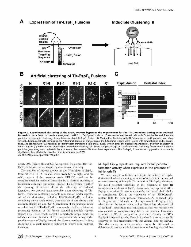
nearly 90% (Figure 2B and 2C). As expected, the control HN-Tir-
EspFU-N fusion did not trigger significant actin assembly.
The number of repeats present in the C-terminus of EspFU
from different EHEC isolates varies from two to eight, and an
espFU mutant of the prototype strain EDL933 could not be
complemented for pedestal formation by a plasmid encoding a
truncation with only one repeat [32,35]. To determine whether
the quantity of repeats affects the efficiency of pedestal
formation, we assessed actin assembly upon clustering of Tir-
EspFU chimeras containing variable numbers of EspFu repeats.
All of the derivatives, including HN-Tir-EspFU-R1, a fusion
containing only a single repeat, were capable of stimulating actin
assembly (Figure 2B and 2C). Quantitation of the pedestal index
revealed that HN-Tir-EspFU-R1 was roughly half as efficient at
generating pedestals as the fusions containing multiple repeats
(Figure 2C). These results suggest a remarkably simple model in
which the central function of Tir is to promote clustering of the
peptide repeats of EspFU beneath the plasma membrane. In fact,
clustering of a single repeat is sufficient to trigger actin pedestal
formation.
Multiple EspFU repeats are required for full pedestalformation activity when expressed in the presence offull-length Tir
We next sought to further investigate the activity of EspFU
derivatives harboring varying numbers of repeats in experimental
systems involving full-length Tir instead of Tir-EspFU chimeras.
To avoid potential variability in the efficiency of type III
translocation of different EspFU derivatives, we expressed GFP-
EspFU truncations in mammalian cells, and tested their ability
to complement KC12, the equivalent of an EHECDespFU
mutant [28], for actin pedestal formation. As expected [36],
KC12 generated pedestals on cells expressing GFP-EspFU-R1-6,
which carries the entire repeat region (Figure 3A). Moreover, all
of the EspFU derivatives that contained at least two repeats were
also capable of complementing KC12 for pedestal formation.
However, KC12 did not generate pedestals efficiently on GFP-
EspFU-R1-expressing cells. Only 1–4 pedestals were occasionally
observed on cells transfected with this variant (data not shown).
This discrepancy in actin assembly cannot be attributed to
differences in protein levels, because immunoblotting revealed that
Figure 2. Experimental clustering of the EspFU repeats bypasses the requirement for the Tir C-terminus during actin pedestalformation. (A) A fusion of membrane-targeted HN-TirFL to EspFU-myc is shown. Treatment of transfected cells with Tir antibodies and S. aureusparticles can promote clustering of membrane-localized Tir-EspFU fusions. (B) Murine fibroblast-like cells (FLCs) transfected with plasmids encodingTir-EspFU fusion constructs comprising the N-terminal domain or truncations of the C-terminal repeats were treated with Tir antibodies and S. aureus,fixed, and stained with HA antibodies to identify both transfected cells and S. aureus (which binds the fluorescent antibodies) and with phalloidin todetect F-actin. (C) Pedestal formation indices were determined by calculating the percentage of transfected cells harboring five or more S. aureusparticles generating actin pedestals. Data represent the mean+/2SD from three experiments. The Tir-EspFU R1 construct triggered actin assemblysignificantly less efficiently than the other truncations (p,0.05).doi:10.1371/journal.ppat.1000191.g002
EspFU, N-WASP, and Actin Assembly
PLoS Pathogens | www.plospathogens.org 4 October 2008 | Volume 4 | Issue 10 | e1000191
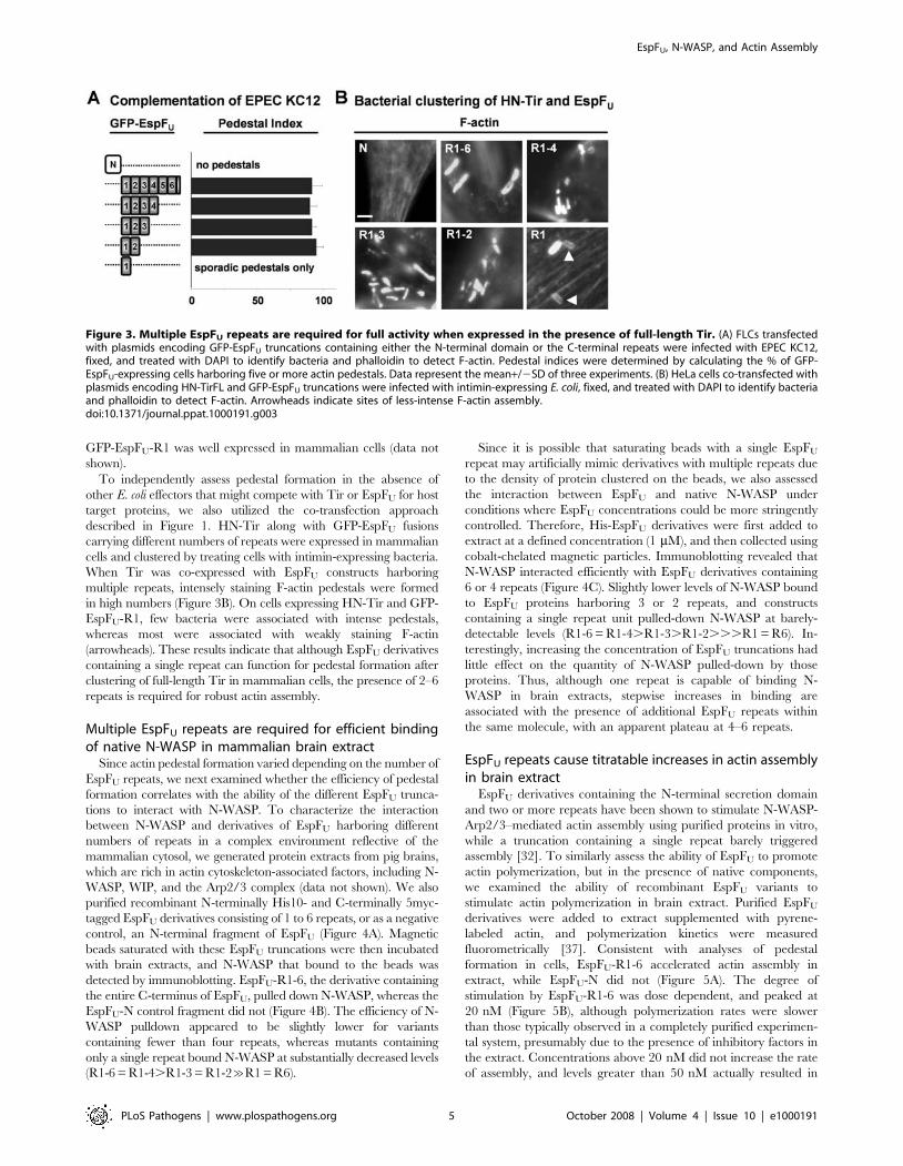
GFP-EspFU-R1 was well expressed in mammalian cells (data not
shown).
To independently assess pedestal formation in the absence of
other E. coli effectors that might compete with Tir or EspFU for host
target proteins, we also utilized the co-transfection approach
described in Figure 1. HN-Tir along with GFP-EspFU fusions
carrying different numbers of repeats were expressed in mammalian
cells and clustered by treating cells with intimin-expressing bacteria.
When Tir was co-expressed with EspFU constructs harboring
multiple repeats, intensely staining F-actin pedestals were formed
in high numbers (Figure 3B). On cells expressing HN-Tir and GFP-
EspFU-R1, few bacteria were associated with intense pedestals,
whereas most were associated with weakly staining F-actin
(arrowheads). These results indicate that although EspFU derivatives
containing a single repeat can function for pedestal formation after
clustering of full-length Tir in mammalian cells, the presence of 2–6
repeats is required for robust actin assembly.
Multiple EspFU repeats are required for efficient bindingof native N-WASP in mammalian brain extract
Since actin pedestal formation varied depending on the number of
EspFU repeats, we next examined whether the efficiency of pedestal
formation correlates with the ability of the different EspFU trunca-
tions to interact with N-WASP. To characterize the interaction
between N-WASP and derivatives of EspFU harboring different
numbers of repeats in a complex environment reflective of the
mammalian cytosol, we generated protein extracts from pig brains,
which are rich in actin cytoskeleton-associated factors, including N-
WASP, WIP, and the Arp2/3 complex (data not shown). We also
purified recombinant N-terminally His10- and C-terminally 5myc-
tagged EspFU derivatives consisting of 1 to 6 repeats, or as a negative
control, an N-terminal fragment of EspFU (Figure 4A). Magnetic
beads saturated with these EspFU truncations were then incubated
with brain extracts, and N-WASP that bound to the beads was
detected by immunoblotting. EspFU-R1-6, the derivative containing
the entire C-terminus of EspFU, pulled down N-WASP, whereas the
EspFU-N control fragment did not (Figure 4B). The efficiency of N-
WASP pulldown appeared to be slightly lower for variants
containing fewer than four repeats, whereas mutants containing
only a single repeat bound N-WASP at substantially decreased levels
(R1-6 = R1-4.R1-3 = R1-2&R1 = R6).
Since it is possible that saturating beads with a single EspFU
repeat may artificially mimic derivatives with multiple repeats due
to the density of protein clustered on the beads, we also assessed
the interaction between EspFU and native N-WASP under
conditions where EspFU concentrations could be more stringently
controlled. Therefore, His-EspFU derivatives were first added to
extract at a defined concentration (1 mM), and then collected using
cobalt-chelated magnetic particles. Immunoblotting revealed that
N-WASP interacted efficiently with EspFU derivatives containing
6 or 4 repeats (Figure 4C). Slightly lower levels of N-WASP bound
to EspFU proteins harboring 3 or 2 repeats, and constructs
containing a single repeat unit pulled-down N-WASP at barely-
detectable levels (R1-6 = R1-4.R1-3.R1-2...R1 = R6). In-
terestingly, increasing the concentration of EspFU truncations had
little effect on the quantity of N-WASP pulled-down by those
proteins. Thus, although one repeat is capable of binding N-
WASP in brain extracts, stepwise increases in binding are
associated with the presence of additional EspFU repeats within
the same molecule, with an apparent plateau at 4–6 repeats.
EspFU repeats cause titratable increases in actin assemblyin brain extract
EspFU derivatives containing the N-terminal secretion domain
and two or more repeats have been shown to stimulate N-WASP-
Arp2/3–mediated actin assembly using purified proteins in vitro,
while a truncation containing a single repeat barely triggered
assembly [32]. To similarly assess the ability of EspFU to promote
actin polymerization, but in the presence of native components,
we examined the ability of recombinant EspFU variants to
stimulate actin polymerization in brain extract. Purified EspFU
derivatives were added to extract supplemented with pyrene-
labeled actin, and polymerization kinetics were measured
fluorometrically [37]. Consistent with analyses of pedestal
formation in cells, EspFU-R1-6 accelerated actin assembly in
extract, while EspFU-N did not (Figure 5A). The degree of
stimulation by EspFU-R1-6 was dose dependent, and peaked at
20 nM (Figure 5B), although polymerization rates were slower
than those typically observed in a completely purified experimen-
tal system, presumably due to the presence of inhibitory factors in
the extract. Concentrations above 20 nM did not increase the rate
of assembly, and levels greater than 50 nM actually resulted in
Figure 3. Multiple EspFU repeats are required for full activity when expressed in the presence of full-length Tir. (A) FLCs transfectedwith plasmids encoding GFP-EspFU truncations containing either the N-terminal domain or the C-terminal repeats were infected with EPEC KC12,fixed, and treated with DAPI to identify bacteria and phalloidin to detect F-actin. Pedestal indices were determined by calculating the % of GFP-EspFU-expressing cells harboring five or more actin pedestals. Data represent the mean+/2SD of three experiments. (B) HeLa cells co-transfected withplasmids encoding HN-TirFL and GFP-EspFU truncations were infected with intimin-expressing E. coli, fixed, and treated with DAPI to identify bacteriaand phalloidin to detect F-actin. Arrowheads indicate sites of less-intense F-actin assembly.doi:10.1371/journal.ppat.1000191.g003
EspFU, N-WASP, and Actin Assembly
PLoS Pathogens | www.plospathogens.org 5 October 2008 | Volume 4 | Issue 10 | e1000191

slower kinetics (not shown), implying that polymerization in
extract is refractory in the presence of high levels of EspFU.
To characterize the relationship between the number of EspFU
repeats and the ability to stimulate actin assembly, pyrene-actin
polymerization was also measured when the recombinant EspFU
truncations were added to extract. When tested at 20 nM, the
optimal concentration for actin assembly mediated by the EspFU
derivative containing all 6 repeats, EspFU variants containing 2, 3,
or 4 repeats accelerated actin assembly, and the rate of
polymerization positively correlated with the number of repeats
that were present (Figure 5C). In contrast, neither EspFU-R1 nor
EspFU-R6, the truncations containing a single repeat, caused a
significant acceleration of actin assembly in this assay system.
Notably, the kinetics of actin assembly in these studies correlated
with the ability of the EspFU constructs to interact with N-WASP
in the extract (Figure 4). Consistent with such results, increasing
the concentration of EspFU variants harboring less than 3 repeats
did not enhance actin assembly (data not shown). Collectively,
these data indicate that the presence of multiple repeats within the
same polypeptide is important for triggering actin polymerization
in an extract system designed to mimic a complex native
environment.
Figure 4. Multiple EspFU repeats are required for efficientbinding of N-WASP in brain extract. (A) N-terminally His10-taggedand C-terminally 5myc-tagged EspFU derivatives were expressed in E.coli, purified, resolved by SDS-PAGE, and stained with Coomassie blue.(B) Cobalt-chelated magnetic particles were coated with saturatingconcentrations of EspFU derivatives and subsequently incubated withporcine brain extract. The association of native N-WASP with EspFU-coated beads was assessed by SDS-PAGE followed by immunoblottingof bead eluates with antibodies to N-WASP and staining EspFU withPonceau S. (C) His-EspFU-myc constructs were added to brain extract atthe indicated concentrations and collected using cobalt-chelatedmagnetic particles. The association of native N-WASP and EspFU withthe beads was assessed by immunoblotting of bead eluates withantibodies to N-WASP and staining EspFU with Ponceau S.doi:10.1371/journal.ppat.1000191.g004 Figure 5. EspFU repeats cause titratable increases in actin
assembly in brain extract. (A) His-EspFU-myc constructs (20 nM each)were added to Arp2/3-enriched brain extract supplemented with 2.5 mMG-actin (10% pyrene labeled), and fluorescent actin polymerization, inarbitrary units (AU), was measured over time. EspFU did not trigger actinassembly in the absence of extract (data not shown). (B) Pyrene-actinpolymerization in the presence of brain extract and various concentra-tions of EspFU-R1-6 was measured over time. (C) Pyrene-actin polymer-ization in the presence of brain extract and EspFU truncations containingdifferent repeat segments (20 nM each) was measured over time.doi:10.1371/journal.ppat.1000191.g005
EspFU, N-WASP, and Actin Assembly
PLoS Pathogens | www.plospathogens.org 6 October 2008 | Volume 4 | Issue 10 | e1000191

EspFU repeats synergistically activate N-WASP/WIP–mediated actin assembly in vitro
Under physiological conditions, N-WASP stably associates with
WIP or it homologs CR16 and WIRE/WICH [21,38]. In addition,
WIP is capable of inhibiting Cdc42-mediated activation of N-WASP
[20] and, unlike recombinant N-WASP, the native N-WASP/WIP
complex is insensitive to Cdc42 treatment [21], suggesting that
regulation of N-WASP in isolation may not accurately reflect
physiological N-WASP regulation in vivo. We therefore examined
whether EspFU can trigger Arp2/3-mediated actin assembly in the
presence of an N-WASP/WIP complex. We purified a recombinant
complex containing N-WASP and WIP at 1:1 stoichiometry and also
contained a trace amount of insect cell actin (see Materials and
Methods; Figure 6A). We also purified recombinant Arp2/3
complex [39], and examined the basal activity of the N-WASP/
WIP complex using the pyrene-actin polymerization assay. Consis-
tent with the predicted autoregulation of N-WASP, the recombinant
N-WASP/WIP complex caused only a small increase in actin
assembly kinetics when compared to Arp2/3 alone (Figure 6B).
To determine if EspFU could trigger actin assembly in this
purified system, pyrene-actin polymerization was measured in the
presence of recombinant EspFU-R1-6, N-WASP/WIP, and Arp2/
3 complex. Consistent with analyses of pedestal formation in cells
and actin assembly in extracts, EspFU-R1-6 accelerated actin
assembly in this reconstituted system (Figure 6B). In the presence
of 20 nM N-WASP/WIP and 20 nM Arp2/3, activation ap-
peared to saturate at approximately 35 nM EspFU-R1-6.
We next examined the abilities of the EspFU truncations to
stimulate actin assembly. By identifying the length of time each
reaction took to reach half of the maximal F-actin concentration
and measuring the rates of actin polymerization at these times, we
directly compared the activity of each construct (Figure 6C). These
experiments revealed that EspFU-R1-6 and EspFU-R1-4 were
nearly indistinguishable at stimulating actin assembly, as the
concentrations of these proteins that were required for reaching
half of the maximal polymerization rate were approximately
4.0 nM and 5.7 nM, respectively. EspFU-R1-3 was less active than
either the 6- or 4-repeat constructs at all concentrations tested, and
Figure 6. EspFU repeats synergistically activate actin assembly mediated by recombinant N-WASP/WIP complex in vitro. (A) N-terminally His6-Flag-tagged N-WASP and His6-Myc-tagged WIP were co-expressed in insect cells, purified as a stoichiometric complex, resolved bySDS-PAGE, and stained with Coomassie blue. (B) Actin (2 mM) was polymerized in the presence of Arp2/3 complex, N-WASP/WIP complex, and theindicated concentrations of EspFU. F-actin fluorescence was measured in arbitrary units (AU). (C) Actin polymerization was examined in the presenceof 20 nM Arp2/3 complex, 20 nM N-WASP/WIP complex, and the indicated concentrations of EspFU derivatives. Polymerization rates at half-maximalF-actin concentrations were measured relative to the rate of polymerization in control Arp2/3+N-WASP/WIP samples lacking EspFU. Curves were fitusing Prism software. (D) Actin polymerization was measured as in (C), except that EspFU concentrations have been scaled to the number of repeatsin each protein.doi:10.1371/journal.ppat.1000191.g006
EspFU, N-WASP, and Actin Assembly
PLoS Pathogens | www.plospathogens.org 7 October 2008 | Volume 4 | Issue 10 | e1000191

it stimulated half-maximal assembly at a concentration of
19.4 nM. EspFU-R1-2 was substantially less active than the 3-repeat
derivative (half-maximal at 78 nM), while the single repeat
constructs EspFU-R1 and EspFU-R6 were even less active (half-
maximal at 99–117 nM). Even when present at very high
concentrations (.500 nM), the EspFU derivatives harboring 1–2
repeats could not accelerate polymerization to rates comparable to
those elicited by 35 nM EspFU-R1-6 (data not shown). Thus, in the
presence of purified N-WASP/WIP and Arp2/3, the number of
EspFU repeats positively correlates with the rate of actin assembly.
To compare the activity of the EspFU derivatives on the basis of
repeat numbers, we normalized protein concentrations to the
quantity of repeat units within each polypeptide. For example,
4.17 nM of the 6-repeat construct was considered equivalent to
8.33 nM of a 3-repeat truncation and 25 nM of a single repeat.
Scaling of the data in this manner revealed that EspFU derivatives
containing greater numbers of repeats within the same protein had
substantially higher activity than smaller constructs (Figure 6D).
For example, when protein concentrations were normalized to
25 nM of repeats, the 6-repeat protein was actually twice as active
as the 3-repeat protein, which was more than twice as active as the
2-repeat protein, which was roughly twice as active a 1-repeat
protein. A similar trend was apparent at higher concentrations
(50–200 nM), as longer EspFU constructs always had greater levels
of activity than smaller derivatives containing equimolar amounts
of repeats. Hence, EspFU repeats cooperate when present within
the same protein to promote synergistic activation of N-WASP/
WIP and Arp2/3-mediated actin assembly.
The GBD of N-WASP is a dominant negative inhibitor ofEHEC pedestal formation and binds with high efficiencyto a single EspFU repeat in yeast two-hybrid assays
Insight into the ability of multi-repeat EspFU derivatives to
pulldown N-WASP from brain extracts and stimulate N-WASP/
WIP-mediated actin assembly in vitro would be enhanced by a
better understanding of EspFU binding by N-WASP, which is
mediated by the GBD [28–29]. To further test the functional
significance of this interaction during pedestal formation, we
transfected mammalian cells with constructs expressing Flag-
tagged N-WASP variants, each encompassing a different combi-
nation of N-WASP domains (Figure 7A), and then examined
pedestal formation upon infection with EHEC. Consistent with
previous observations showing that N-WASP residues 226–274
within the GBD mediate recruitment of N-WASP to sites of
EHEC attachment [14], all tagged N-WASP derivatives contain-
ing the GBD (residues 151–273) were recruited to sites of bacterial
adherence (Figure 7B, top row). In addition, quantitation of
pedestal formation on transfected cells revealed that neither the N-
terminal WH1 domain nor full-length N-WASP had a substantial
effect on actin polymerization, whereas overexpression of each of
the 4 GBD-containing derivatives that lacked the VCA domain,
which is necessary for Arp2/3 activation, effectively abolished
pedestal formation (Figure 7B, bottom row). This inhibition was
specific, because overexpression of the GBD did not affect actin
assembly triggered by an EPEC strain that generates pedestals
independently of EspFU (Figure 7C). As predicted by mapping of
requirements for N-WASP recruitment to EHEC [14], an H208D
point mutation that abrogates N-WASP binding by the GTPase
Cdc42 had no effect on the inhibitory activity of the GBD. These
results indicate that the interaction between EspFU and the C-
terminal portion of the N-WASP GBD is important for pedestal
formation in mammalian cells.
Next, to begin to assess whether the positive correlation between
the number of repeats within an EspFU derivative and its ability to
activate actin assembly is reflected in binding to the N-WASP GBD,
we examined this interaction in yeast two-hybrid assays. As
demonstrated previously [28], the N-WASP GBD interacted with
the C-terminus of EspFU that contains the repeat sequences, but not
the EspFU N-terminus (Figure 7D). In addition, all of the C-terminal
subfragments of EspFU, even those containing only a single repeat,
interacted with the GBD in these assays. However, the degree of
interaction did not positively correlate with increasing numbers of
repeats. Rather, the number of repeats in EspFU inversely correlated
with the degree of expression of the lacZ reporter. Although reporter
activity in the yeast two-hybrid assay reflects a number of
parameters, including levels of expression and nuclear import of
the binding partners, these results provide no evidence that the more
efficient activation by multi-repeat EspFU derivatives is a conse-
quence of cooperativity in GBD binding.
Increasing the number of EspFU repeats does not alteraffinity for the GBD, but promotes the formation of anArp2/3-containing complex
To further assess potential cooperativity in GBD binding by
EspFU, we examined the interactions of EspFU derivatives with the
GBD of WASP, the hematopoetic-specific homologue of N-WASP
that has been shown to also promote actin pedestal formation in
cells [14]. Recombinant EspFU proteins containing 5, 2, or 1
repeats (R91-5, R94-5, and R95, respectively) were generated with
N- and C-terminal repeat boundaries based on recent structural
studies that defined the GBD-binding sequences of EspFU [40–
42]. The affinity of the WASP GBD for these EspFU constructs
was determined using isothermal titration calorimetry and yielded
predicted differences in stoichiometry (i.e., 5.7, 1.8, and 1.0 for the
5, 2, and 1 repeat derivatives, respectively) but similar dissociation
constants (i.e., 83 nM, 95 nM, and 89 nM, respectively)
(Figure 8A). Thus, variation in the number of EspFU repeats is
not associated with differences in affinity for the GBD.
We next tested whether oligomerization of N-WASP by
adjacent EspFU repeats instead might promote binding of the
Arp2/3 complex. N-WASPC, a C-terminal fragment of N-WASP
that contains the GBD, PRD, and VCA domain, is a fragment
previously shown to function in pedestal formation [14]. To
determine whether EspFU derivatives differing in repeat number
also differed in their ability to form a complex containing N-
WASPC and Arp2/3, EspFU variants containing 1 or 2 repeats,
fluorescently labeled at their N-termini with AlexaFluor647, were
incubated with equivalent molar concentrations of N-WASPC in
the absence or presence of the Arp2/3 complex. The relative size
of EspFU-containing complexes, detected by absorbance at
650 nM, was then determined by gel filtration chromatography.
The single EspFU repeat bound N-WASP at essentially stoichio-
metric levels when mixed in equimolar (2 mM) quantities, as
determined by an increase in the apparent size (i.e., earlier elution
profile) of EspFU (Figure 8B left, red vs. blue peaks). The addition
of 2 mM (i.e., a two-fold higher molar concentration) Arp2/3
caused ,60% of the EspFU to shift to an even more rapidly eluting
fraction indicative of an EspFU-N-WASP-Arp2/3 complex
(Figure 8B, yellow profile). Very little of this complex was detected
using 1 mM Arp2/3 (Figure 8B left, small shoulder in gray profile).
When 1 mM N-WASPC was incubated with 0.5 mM of the 2-
repeat EspFU derivative (i.e., containing a normalized repeat
concentration), the two proteins bound one another at essentially
stoichiometric levels (Figure 8B, right, compare light blue and
green profiles). However, in contrast to the requirement for 2 mM
Arp2/3 to observe appreciable complex formation with a single
repeat, the addition of as little as 0.5 mM Arp2/3 to N-WASPC
and the two repeat derivative resulted in the formation of an
EspFU, N-WASP, and Actin Assembly
PLoS Pathogens | www.plospathogens.org 8 October 2008 | Volume 4 | Issue 10 | e1000191

Arp2/3-containing complex (Figure 8B right, blue or pink profiles,
respectively). The greater complex formation even at lower Arp2/
3 concentration indicates that the ability of tandem EspFU repeats
to assemble N-WASP dimers (or by implication higher order
multimers) may facilitate binding of the Arp2/3 complex,
providing a likely source of inter-repeat cooperativity.
Since the assay demonstrating inter-repeat cooperation of EspFU
for the formation of an Arp2/3-containing complex described above
involved the addition of only EspFU, N-WASPC, and Arp2/3, N-
WASP-associated proteins such as WIP should not be required for
cooperativity between the EspFU repeats in actin assembly.
Furthermore, domains of WASP (or N-WASP) other than the
GBD (which binds EspFU) and VCA (which binds Arp2/3) should
also be dispensable. We therefore tested whether the EspFU repeats
were capable of synergistically activating a minimized (and normally
autoinhibited [40]) derivative of WASP containing only the GBD
Figure 7. The GTPase-binding domain (GBD) of N-WASP is a dominant negative inhibitor of EHEC pedestal formation and bindswith high efficiency to a single EspFU repeat in yeast two-hybrid assays. (A) The modular structure of N-WASP is depicted, featuring WASPhomology-1 (WH1), GTPase-binding (GBD), proline-rich (PRD), and WH2/verprolin-connector-acidic (VCA) domains. Several N-WASP-binding partnersare shown above their interacting domains. (B) HeLa cells transfected with plasmids encoding Flag-N-WASP constructs were infected with EHECDdam(a mutant that binds to mammalian cells and generates pedestals with considerably higher efficiency than wild type EHEC [44]), fixed, and treatedwith DAPI to identify bacteria, a Flag antibody to visualize tagged N-WASP (top panels), and phalloidin to detect F-actin (bottom panels). Flag-N-WASP recruitment was only evaluated in cells expressing low levels of these tagged proteins (top panels), while effects on actin pedestal formationwere only assessed in cells expressing high levels of Flag-N-WASP (bottom panels). Pedestal formation indices were determined by calculating thepercentage of mock-transfected or Flag-N-WASP overexpressing cells harboring five or more actin pedestals. Data represent the mean+/2SD of threeexperiments. (C) HeLa cells expressing GFP alone, a GFP-tagged GBD, or a GFP-tagged GBD H208D point mutant were infected with EHECDdam orEPEC and treated with DAPI to identify bacteria and phalloidin to detect F-actin. Pedestal formation indices were determined as in (B). (D) Plasmidsencoding the N-WASP GBD fused to the LexA DNA-binding domain and EspFU fragments fused to the Gal4 transcriptional activation domain were co-transformed into a yeast two-hybrid reporter strain. Data represent the mean+/2SD of b-galactosidase activity for three co-transformants for eachpairwise combination.doi:10.1371/journal.ppat.1000191.g007
EspFU, N-WASP, and Actin Assembly
PLoS Pathogens | www.plospathogens.org 9 October 2008 | Volume 4 | Issue 10 | e1000191

and VCA domain. Similar to results using EspFU truncations and an
N-WASP/WIP complex, pyrene actin assays using WASP GBD-
VCA revealed that the number of EspFU repeats correlated with
increased actin polymerization rates, even when protein concentra-
tions were normalized to repeat units (Figure 8C). These results
indicate that synergy in actin assembly require no WASP sequences
other than the GBD and VCA domain.
Discussion
In order to better understand EHEC pedestal formation, we
first sought to define the minimal set of bacterial effectors essential
to this process. We found that EspFU is likely the only EHEC
effector besides Tir that is required for pedestal formation, because
pedestals can be induced on mammalian cells that express these
two factors in the absence of any other bacterial proteins. In
addition, the cytoplasmic C-terminus of Tir could be functionally
replaced with the C-terminus of EspFU, indicating that the only
critical function of this Tir domain is to recruit the EspFU repeats
to sites of EHEC attachment. The C-terminus of the prototype
EspFU protein consists of six 47-residue proline-rich repeats, and a
fusion containing a single repeat unit retained significant ability to
generate pedestals when clustered at the plasma membrane. This
is consistent with a recent report describing actin recruitment to
the plasma membrane after artificially clustering a single repeat
with antibodies [42]. That study, as well as the recent report of the
structure of a single EspFU repeat bound to the WASP GBD [41],
revealed that EspFU binds to the autoinhibitory region within the
GBD that normally interacts with the VCA domain, a finding
consistent with previous mapping studies [14,27,32]. Therefore,
the bare minimum components required for pedestal formation
are the domains of Tir that facilitate its membrane localization
and clustering by intimin, and a single GBD-binding EspFU
peptide repeat.
Figure 8. Increasing the number of EspFU repeats does not alter affinity for the GBD, but promotes the formation of an Arp2/3-containing complex. (A) Isothermal titration calorimetry analyses of the interactions between WASP GBD and EspFU fragments are shown. The GBDwas titrated into R95 (left), R94-5 (middle), or R91-5 (right). Raw and integrated heats of injections are shown in upper and lower panels, respectively.Black lines in the lower panels show fits of data into a single-affinity, multi-site binding model. Fits of the data for R94-5 and R91-5 to models with twodifferent affinities were not statistically improved over the single-affinity model. (B) Interactions between Arp2/3 complex and N-WASPC in complexwith Alexa647-labeled EspFU R95 or R94-5 were examined by gel filtration chromatography. The A650 profile is shown. (C) Actin (4 mM) waspolymerized by itself (black curve), or in the presence of 10 nM Arp2/3 complex plus either 0.5 mM WASP GBD-VCA (purple, control), 0.5 mM GBD-VCA+1 mM R95 (yellow), or 0.5 mM GBD-VCA+0.5 mM R94-5 (blue), or 0.5 mM GBD-VCA+0.2 mM R91-5 (red). F-actin fluorescence was measured inarbitrary units (AU). Note that R95, R94-5, and R91-5 were used at the same total repeat concentration.doi:10.1371/journal.ppat.1000191.g008
EspFU, N-WASP, and Actin Assembly
PLoS Pathogens | www.plospathogens.org 10 October 2008 | Volume 4 | Issue 10 | e1000191
Within the Tir C-terminal domain, a tripeptide sequence
(NPY458) is required for recruitment of EspFU and actin pedestal
formation [30], but a direct interaction between Tir and EspFU has
not been detected, suggesting that they interact indirectly. The
delineation of Tir and EspFU as the only bacterial effectors required
for pedestal formation indicates that the putative adaptor that
mediates this interaction is of host origin. Efficient association of
EspFU with the putative adaptor linking it to Tir may require
multiple EspFU repeats, because when Tir and EspFU were
expressed separately rather than as a single fusion protein, at least
two repeats were required for robust EspFU function. Consistent
with these results, a translocated 2-repeat EspFU derivative, but not a
1-repeat derivative, localized to sites of bacterial attachment [32].
Aside from promoting Tir-EspFU interactions, this host factor
may itself promote some level of actin assembly, because residual
pedestals can form at low levels in the absence of EspFU [28].
Cortactin, which has the ability to stimulate Arp2/3 and contributes
to pedestal formation via an unknown mechanism, can interact with
both Tir and EspFU, but binds the Tir N-terminus [36], a domain
that is largely dispensable for actin assembly [31]. Regardless of the
identity of the adaptor, the finding that a hybrid protein containing
Tir fused directly to the EspFU repeats is fully functional for pedestal
formation indicates that neither the adaptor nor the C-terminus of
Tir play any essential role in actin assembly other than to recruit
EspFU. Thus, EspFU is the primary effector that signals to the actin
cytoskeleton during pedestal formation.
In addition to promoting more efficient recruitment to Tir, the
presence of multiple repeat units in EspFU provides more robust
signaling function, because a Tir-EspFU fusion carrying two or
more repeats triggered pedestal formation at levels 2-fold higher
than a fusion carrying a single repeat. Recently, a full-length
repeat region was shown to recruit GFP-actin to the plasma
membrane somewhat (,20%) more frequently than a single
repeat [42]. That report and others have also shown that multi-
repeat EspFU derivatives promote more efficient actin assembly in
vitro than a single-repeat derivative [32,42]. Such studies have
relied upon analyses of recombinant N-WASP or small N-WASP
fragments containing the minimal autoinhibitory module, a GBD-
VCA fusion. However, in recent years it has become apparent that
in the cell cytoplasm the intrinsic activity of N-WASP may be
significantly modulated through interactions with other factors
[20–21]. We therefore examined EspFU-induced actin polymer-
ization in brain extracts, and found that in this complex
environment, the number of repeats correlated both with the
ability of EspFU derivatives to interact with native N-WASP and to
stimulate actin assembly. Single-repeat constructs did not
accelerate polymerization in these assays, possibly due to
competition with endogenous N-WASP activators or the presence
of inhibitory factors in the extract.
Under physiological conditions, N-WASP is stably associated
with WIP, a protein that binds to its N-terminal WH1 domain and
influences its activation [20–21]. We found that EspFU constructs
are capable of potently activating Arp2/3-mediated actin assembly
in the presence of a recombinant N-WASP/WIP complex, and as
predicted based upon N-WASP pulldown assays and measure-
ments of actin polymerization in extracts, EspFU derivatives
containing greater numbers of repeats stimulate actin assembly
more rapidly. Interestingly, when we normalized the EspFU
derivatives to the concentration of repeats and quantitated actin
assembly rates, we found that increasing the number of repeats
within individual proteins does not simply result in additive
increases in actin polymerization. Instead, the presence of multiple
repeats in the same derivative enhances N-WASP/WIP-mediated
actin assembly synergistically.
It is possible that this enhancement phenotype is due to
cooperativity among the repeats in EspFU-N-WASP binding.
Indeed, an earlier study reported that the dissociation constants
(Kd) for the EspFU-N-WASP interaction varied depending on the
numbers of repeats (e.g., 3.6 nM for 6 repeats, 6.4 nM for 4
repeats, and 11 nM for 2 repeats, and no measurable binding for 1
repeat) [32]. However, these values were calculated on the basis of
in vitro actin assembly assays that involve the formation of an N-
WASP-Arp2/3 complex, and may not simply reflect the
interaction between EspFU and N-WASP. Similarly, the more
efficient pulldown of N-WASP by multi-repeat EspFU derivatives
in brain extracts that we observed could be influenced by other
endogenous factors. Although a previous study found that 2
repeats is the minimal N-WASP-binding module within EspFU in
gel overlay assays [32], we found that a single repeat bound to the
GBD with high efficiency in yeast two-hybrid assays. Furthermore,
the Kd for binding of purified 1-, 2-, or 5-repeat EspFU derivatives
to the WASP GBD were all similar to one another (i.e., 83–
95 nM), and within an order of magnitude of that measured for
the N-WASP GBD (18 nM; [42]). These results provide
convincing evidence that the EspFU repeats do not bind to the
GBD in a cooperative fashion.
An alternative explanation for inter-repeat cooperativity during
actin polymerization is that an EspFU-N-WASP-Arp2/3 complex
can be formed more efficiently. The major threshold for pedestal-
forming function apparently resides within 2 EspFU repeats, and
we were unable to detect substantial differences in the frequency
or intensity of actin pedestal formation promoted by EspFU
proteins harboring 2, 3, 4, or 6 repeats (although in vitro actin
assembly assays indicate that the number of repeats present in an
EspFU derivative correlates positively with function). Similarly,
analysis of a diverse collection of espFU-containing E. coli strains
showed that all EspFU proteins contain at least 2 repeats [35].
Therefore, we compared the ability of EspFU derivatives
containing 1 versus 2 repeats to enter into a complex with
recombinant N-WASP and Arp2/3 complex. As predicted from
the affinities of EspFU fragments for the GBD, the single and two
repeat derivatives bound to N-WASP indistinguishably. However,
the 2-repeat protein formed a complex with both N-WASP and
Arp2/3 with much greater efficiency. These experiments suggest
that N-WASP multimers assembled by tandem EspFU repeats may
have higher affinity for Arp2/3 complex. As predicted from such a
model, the inter-repeat cooperativity was revealed during in vitro
actin assembly assays using a WASP derivative containing only the
EspFU-binding GBD and the Arp2/3-binding VCA domain.
Further study will be required to illuminate the molecular basis of
this enhanced interaction.
Interestingly, the multivalency that is clearly a critical property
of EspFU is likely an important feature of other Arp2/3-mediated
processes that are triggered by microbial pathogens. Pedestal
formation by EPEC, which occurs in an EspFU-independent
manner, requires clustering of Tir, a step that may mimic
multivalent binding. The critical EPEC Tir phosphopeptide has
no activity when added in soluble form in standard pyrene actin
assays [S. Rankin, unpub. obs.], but potently stimulates actin
assembly when clustered on a bead [25]. EspF, an EHEC effector
that is 35% similar to EspFU but not involved in pedestal
formation [28], also uses peptide repeats to activate N-WASP in
vitro [33]. Finally, the Shigella IcsA (VirG) protein uses repetitive
sequences to bind N-WASP and promote actin-based intracellu-
lar motility [43]. Future investigation into how the multivalent
nature of EspFU promotes actin assembly is likely to provide
insight into related phenomena central to multiple pathogenic
processes.
EspFU, N-WASP, and Actin Assembly
PLoS Pathogens | www.plospathogens.org 11 October 2008 | Volume 4 | Issue 10 | e1000191

Materials and Methods
Bacteria, plasmids, and cell linesThe EHEC dam mutant used in this study was derived from
TUV93-0, a Shiga toxin-deficient version of the prototype
O157:H7 strain EDL933 [44]. The EPEC bacterium was the
prototype O127:H6 strain, JPN15/pMAR7. EPEC KC12, which
contains the EPEC tir-cesT-eae operon replaced with the corre-
sponding EHEC operon, has also been described [45]. A non-
pathogenic strain of E. coli (MC1061) expressing EHEC intimin
was also described elsewhere [31]. For yeast two-hybrid analyses,
espFU derivatives were generated by PCR from EDL933 genomic
DNA and cloned into the EcoRI and BamHI sites of pGAD424
[28] to create fusions between an N-terminal GAL4AD and C-
terminal 5myc tag. For transfections, espFU fragments were
subcloned into the KpnI and BamHI sites of pKC425 [46] to
generate fusions between an N-terminal GFP-tag and C-terminal
5myc tag. For protein expression and purification, most espFU
derivatives were subcloned into the NdeI and XbaI sites of the
vector pET16b (Novagen) to create fusions between an N-terminal
His10-tag and C-terminal 5myc tag. Some EspFU constructs
contained only an N-terminal His-tag (Figure 8) [41]. For Tir-
EspFU hybrids, espFU-myc derivatives were subcloned into the
KpnI and XbaI sites of the HN-Tir-related vector pKC689 [31].
EspFU truncations contained amino acids 1-88 (N), 80-384 (R1-6),
80-275 (R1-4), 80-228 (R1-3), 80-181 (R1-2), 80-134 (R1), 229-
384 (R4-6), 276-384 (R5-6), 323-384 (R6), 80-314 (R91-5), 221-
314 (R94-5), and 268-314 (R95). For dominant negative transfec-
tions, rat N-WASP derivatives were generated by PCR with an N-
terminal Flag-tag using domain boundaries described previously
[28] and cloned into the KpnI-EcoRI sites of the vector pCDNA3
(Invitrogen). N-terminally GFP-tagged derivatives of the GBD
were generated by subcloning into pKC425. Plasmids for
expression of membrane-targeted HN-TirFL in mammalian cells
and expression of the N-WASP GBD fused to the LexA DBD in
pBTM116 have been described previously [28,31]. Plasmids for
expression of His-Flag-N-WASP and His-Myc-WIP in insect cells
are described elsewhere (ADS and MDW, submitted). N-
terminally His- or GST-tagged WASP GBD (residues 242–310),
N-WASPC (residues 193-501) and WASP GBD-VCA (residues
230–310 and 420–502 with a GGSGGS linker) constructs are
described elsewhere [41]. For routine passage, all E. coli strains
were grown in LB media at 37uC. Prior to infections, EHEC was
cultured in DMEM+100 mM HEPES pH 7.4 in 5% CO2 to
enhance type III secretion. HeLa, Cos7, and murine fibroblast-like
cells (FLCs) [47] were used interchangeably and cultured in
DMEM+10% FBS at 37uC in 5% CO2.
Transfections and infectionsAll transfections were performed as described previously [31].
Infections for 3 h with non-pathogenic E. coli expressing intimin
[31] and EHEC or EPEC strains [44–45] have also been
described. To cluster Tir, cells were treated with antibodies that
recognize its extracellular domain prior to the addition of
formalin-fixed S. aureus Pansorbin particles (Calbiochem) [25].
Immunofluorescence microscopyInfected cells were fixed in 2.5% paraformaldehyde for
35 minutes and permeabilized with 0.1% Triton-X-100 in PBS
as described previously [45]. Bacteria were identified with 1 mg/
ml DAPI (Sigma), and F-actin was detected using 4 U/ml
Alexa568-phalloidin (Molecular Probes). To visualize N-WASP
derivatives, cells were treated with an anti-Flag M5 antibody
(Sigma) and Alexa488 goat anti-mouse antibodies (Molecular
Probes). To visualize HN-Tir derivatives, cells were treated with
an HA.11 antibody (Covance) and Alexa488 or Alexa350 goat
anti-mouse antibodies. To quantify the pedestal formation index in
cells expressing high levels of N-WASP derivatives, which were
identified by bright anti-Flag or GFP fluorescence, the percentage
of cells harboring at least 10 adherent bacteria and 5 actin
pedestals was measured. To quantify the pedestal index in cells
infected with KC12, the percentage of cells harboring at least 10
adherent bacteria and 5 actin pedestals was measured. To quantify
the pedestal index in cells treated with pansorbin particles, the
percentage of HA-fluorescing cells harboring at least 10 adherent
particles (the latter identified by virtue of their ability to bind
fluorescently labeled secondary antibodies) and 5 actin pedestals
was measured. At least 50 cells were examined per sample per
experiment. Our previous work suggests that scoring of a pedestal
index accurately reflects similar quantification methods that
measure the fraction of bound bacteria that generate pedestals
[25]. Cells expressing extremely high fluorescence levels of EspFU
were refractory to pedestal formation and were not included in
these analyses. All scalebars are 1 mm in length.
EspFU interaction assaysFor yeast two-hybrid assays. EspFU variants consisting of
different combinations of the 6 repeats were fused to the Gal4
transcriptional activation domain, whereas the N-WASP GBD was
fused to the LexA DNA-binding domain. Pairwise combinations of
these fusion proteins were tested for interactions by measuring
activation of a lacZ reporter, as described [28]. For some pulldown
assays (Figure 4B), cobalt-chelate conjugated magnetic Talon
particles (Invitrogen) were saturated with His-EspFU-myc deriva-
tives by incubating them with 75 mg/ml of each recombinant
fragment for 1 h. After removal of unbound EspFU, beads were
incubated with brain extract for 1 h in 50 mM NaPO4 pH 7.4,
150 mM NaCl, and 0.015% Triton X-100. Bound proteins were
eluted by boiling in SDS-PAGE sample buffer. For other pulldown
assays (Figure 4C), His-EspFU-myc derivatives were incubated in
brain extract at specific concentrations for 0.5 h and collected
using an excess of Talon particles. Bound proteins were again
eluted by boiling in SDS-PAGE sample buffer.
ImmunoblottingTo prepare mammalian cell lysates, transfected cells were
collected in PBS+2 mM EDTA and lysed in 50 mM Tris-HCl,
pH 7.6, 50 mM NaCl, 1% Triton X-100, 1 mM Na3VO4, 1 mM
PMSF, and 10 mg/ml each of aprotinin, leupeptin, pepstatin, and
chymostatin (Sigma)), prior to mixing with SDS-PAGE sample
buffer. Protein samples were boiled for 10 minutes, centrifuged,
and analyzed by 10% SDS-PAGE prior to staining with
Coomassie blue or transferring to nitrocellulose membranes and
staining with Ponceau S. Membranes were blocked in PBS+5%
milk (PBSM) before probing with N-WASP or GFP antibodies, as
described previously [28]. Following washes, membranes were
treated with secondary antibodies conjugated to alkaline phos-
phatase or horseradish peroxidase and developed using BCIP/
NBT [45] or enhanced chemiluminescence [48].
EspFU expression and purificationHis-EspFU-myc fusion proteins were expressed in E. coli BL21-
Rosetta (Novagen) at 37uC in the presence of 0.1 mM IPTG for
3 h. Bacteria were lysed in 10 mM Tris pH 7.4, 150 mM NaCl,
and protease inhibitor cocktail (Roche) using a cell disruptor
(30kpsi, Constant Cell Systems). EspFU was purified first using
His-tag affinity for Ni-NTA-agarose beads (Qiagen) and eluted in
lysis buffer containing 400 mM imidazole. To remove EspFU
EspFU, N-WASP, and Actin Assembly
PLoS Pathogens | www.plospathogens.org 12 October 2008 | Volume 4 | Issue 10 | e1000191

degradation products, an anion exchange purification step,
facilitated by the negatively charged 5myc-tag, was performed.
EspFU was bound to a HiTrap Q column (GE Healthcare) and
eluted in 10 mM Tris pH 9.5 containing 1 M NaCl. Other EspFU
constructs (R95, R94-5, R1-5; Figure 8) with just an N-terminal
His-tag were expressed in E. coli BL21-DE3 at 20uC in the
presence of 1 mM IPTG for 16 h, and after Ni-NTA affinity
purification the tag was cleaved with thrombin. Cleaved proteins
were subjected to ion exchange and gel filtration chromatography
to remove the tag and other impurities. Protein concentrations
were estimated by Bradford assay (Bio-Rad).
Preparation of brain extractsBrains obtained from freshly slaughtered pigs (Dalehead Foods
Ltd, Linton, UK) were cleaned, sectioned, and resuspended in
extraction buffer (0.1 M MES pH 6.8, 1 mM EGTA, 0.5 mM
MgCl2, 0.1 mM EDTA, 1 mM DTT, protease inhibitor cocktail),
prior to homogenization using a Waring blender (2615 s, 4uC).
After clarification (7,000 g, 20 min, 4uC), extracts were filtered
through cheesecloth and further clarified (11,000 g, 40 min, 4uC)
prior to storage (280uC). Extract to be subfractionated (60 ml) was
dialysed against 265 L 20 mM Tris-Cl pH8, 2 mM MgCl2,
5 mM EGTA, 1 mM EDTA, 0.5 mM DTT, 0.2 mM ATP, 2.5%
glycerol at 4uC, prior to loading onto tandem HiTrap Heparin
columns (265 ml), pre-equilibrated with dialysis buffer. Bound
proteins were eluted using a 0–0.5 M KCl gradient in dialysis
buffer (2 ml/min) and 20 3 ml fractions collected. Fractions 11–17
contained both Arp2/3 complex and N-WASP upon immuno-
blotting, and were combined, dialysed (as above), concentrated 5-
fold (Centricon), and stored at 280uC.
N-WASP, WIP, and Arp2/3 expression and purificationHis-tagged human N-WASP and WIP were expressed using
recombinant baculovirus-infected High5 insect cells, as described
in detail elsewhere (ADS and MDW, submitted). A recombinant
N-WASP/WIP complex was purified by Nickel affinity chroma-
tography followed by gel filtration chromatography to separate the
complex from N-WASP or WIP alone. To maintain an
autoinhibited N-WASP/WIP complex, freeze-thaw cycles and
prolonged periods of storage on ice were kept to a minimum.
Recombinant human Arp2/3 complex and native bovine Arp2/3
complex were purified as described previously [39,41].
Pyrene-actin assembly assaysAssays utilizing brain extract contained samples supplemented
with 2.5 mM skeletal muscle actin (10% pyrene-labeled), and
polymerization was measured as described previously [49]. Assays
using N-WASP/WIP complex contained 2.0 mM actin (7%
pyrene-labeled) and 20 nM recombinant Arp2/3 complex, and
polymerization was measured as described previously [48]. Assays
using the N-WASP GBD-VCA contained 4.0 mM actin (5%
pyrene-labeled) and 10 nM bovine Arp2/3 complex, and
polymerization was measured as described elsewhere [41].
Isothermal titration calorimetry and gel filtrationchromatography
Isothermal titration calorimetry was performed as described
elsewhere [41]. Briefly, the WASP GBD was titrated into R95,
R94-5, or R91-5 in KMEI buffer (50 mM KCl, 1 mM MgCl2,
1 mM EGTA, and 10 mM imidazole pH 7.0) plus 5 mM b-
mercaptoethanol. Prior to gel filtration analyses, the N-termini of
EspFU fragments R95 and R94-5 were fluorescently labeled by
dialysis against 100 mM sodium bicarbonate pH 8.3 and
treatment with a five-fold molar excess of AlexaFluor647-
carboxylic acid, succinimidyl ester (Invitrogen) at 4uC for
16 hours. Extra dye was removed by desalting chromatography
and subsequent dialysis. Conjugation was confirmed by mass
spectrometry. Labeling efficiency was estimated as .97% by the
ratio of absorbance at 280 and 650 nm. Interactions between
Arp2/3 complex and N-WASPC in complex with EspFU were
examined using a Superdex 200 10/300 GL column (GE
Healthcare) equilibrated in KMEI plus 1 mM dithiothreitol
buffer.
Acknowledgments
We thank M. Brady, D. Tipper, and S. Padrick for discussions and critique
of the manuscript, Susannah Rankin for communication of unpublished
results, and Marc Kirschner for providing reagents.
Author Contributions
Conceived and designed the experiments: KGC HCC MKR. Performed
the experiments: KGC HCC DR. Analyzed the data: KGC HCC DR
EJM RDH MKR VK JML. Contributed reagents/materials/analysis
tools: HCC ADS EJM RDH MW MKR VK JML. Wrote the paper: KGC
JML.
References
1. Donnenberg MS, Whittam TS (2001) Pathogenesis and evolution of virulence in
enteropathogenic and enterohemorrhagic Escherichia coli. J Clin Invest 107:
539–548.
2. Kaper JB, Nataro JP, Mobley HL (2004) Pathogenic Escherichia coli. Nat Rev
Microbiol 2: 123–140.
3. Spears KJ, Roe AJ, Gally DL (2006) A comparison of enteropathogenic and
enterohaemorrhagic Escherichia coli pathogenesis. FEMS Microbiol Lett 255:
187–202.
4. Deng W, Puente JL, Gruenheid S, Li Y, Vallance BA, et al. (2004) Dissecting
virulence: systematic and functional analyses of a pathogenicity island. Proc Natl
Acad Sci U S A 101: 3597–3602.
5. Marches O, Nougayrede JP, Boullier S, Mainil J, Charlier G, et al. (2000) Role
of tir and intimin in the virulence of rabbit enteropathogenic Escherichia coli
serotype O103:H2. Infect Immun 68: 2171–2182.
6. Ritchie JM, Thorpe CM, Rogers AB, Waldor MK (2003) Critical roles for stx2,
eae, and tir in enterohemorrhagic Escherichia coli-induced diarrhea and
intestinal inflammation in infant rabbits. Infect Immun 71: 7129–7139.
7. Tacket CO, Sztein MB, Losonsky G, Abe A, Finlay BB, et al. (2000) Role of
EspB in experimental human enteropathogenic Escherichia coli infection. Infect
Immun 68: 3689–3695.
8. Ritchie JM, Brady MJ, Riley KN, Ho TD, Campellone KG, et al. (2007)
EspF(U), a type III-translocated effector of actin assembly, fosters epithelial
association and late-stage intestinal colonization by E. coli O157:H7. Cell
Microbiol 10: 836–847.
9. Tobe T, Beatson SA, Taniguchi H, Abe H, Bailey CM, et al. (2006) An
extensive repertoire of type III secretion effectors in Escherichia coli O157 and
the role of lambdoid phages in their dissemination. Proc Natl Acad Sci U S A
103: 14941–14946.
10. Dean P, Maresca M, Kenny B (2005) EPEC’s weapons of mass subversion. Curr
Opin Microbiol 8: 28–34.
11. Garmendia J, Frankel G, Crepin VF (2005) Enteropathogenic and enterohe-
morrhagic Escherichia coli infections: translocation, translocation, translocation.
Infect Immun 73: 2573–2585.
12. Touze T, Hayward RD, Eswaran J, Leong JM, Koronakis V (2004) Self-
association of EPEC intimin mediated by the beta-barrel-containing anchor
domain: a role in clustering of the Tir receptor. Mol Microbiol 51: 73–87.
13. Kalman D, Weiner OD, Goosney DL, Sedat JW, Finlay BB, et al. (1999)
Enteropathogenic E. coli acts through WASP and Arp2/3 complex to form actin
pedestals. Nat Cell Biol 1: 389–391.
14. Lommel S, Benesch S, Rohde M, Wehland J, Rottner K (2004) Enterohaemor-
rhagic and enteropathogenic Escherichia coli use different mechanisms for actin
pedestal formation that converge on N-WASP. Cell Microbiol 6: 243–254.
15. Lommel S, Benesch S, Rottner K, Franz T, Wehland J, et al. (2001) Actin
pedestal formation by enteropathogenic Escherichia coli and intracellular
motility of Shigella flexneri are abolished in N-WASP-defective cells. EMBO
Rep 2: 850–857.
16. Goley ED, Welch MD (2006) The ARP2/3 complex: an actin nucleator comes
of age. Nat Rev Mol Cell Biol 7: 713–726.
EspFU, N-WASP, and Actin Assembly
PLoS Pathogens | www.plospathogens.org 13 October 2008 | Volume 4 | Issue 10 | e1000191

17. Stradal TE, Scita G (2006) Protein complexes regulating Arp2/3-mediated actin
assembly. Curr Opin Cell Biol 18: 4–10.
18. Rohatgi R, Ma L, Miki H, Lopez M, Kirchhausen T, et al. (1999) The
interaction between N-WASP and the Arp2/3 complex links Cdc42-dependent
signals to actin assembly. Cell 97: 221–231.
19. Rohatgi R, Nollau P, Ho HY, Kirschner MW, Mayer BJ (2001) Nck and
phosphatidylinositol 4,5-bisphosphate synergistically activate actin polymeriza-
tion through the N-WASP-Arp2/3 pathway. J Biol Chem 276: 26448–26452.
20. Martinez-Quiles N, Rohatgi R, Anton IM, Medina M, Saville SP, et al. (2001)
WIP regulates N-WASP-mediated actin polymerization and filopodium
formation. Nat Cell Biol 3: 484–491.
21. Ho HY, Rohatgi R, Lebensohn AM, Le M, Li J, et al. (2004) Toca-1 mediates
Cdc42-dependent actin nucleation by activating the N-WASP-WIP complex.
Cell 118: 203–216.
22. Bommarius B, Maxwell D, Swimm A, Leung S, Corbett A, et al. (2007)
Enteropathogenic Escherichia coli Tir is an SH2/3 ligand that recruits and
activates tyrosine kinases required for pedestal formation. Mol Microbiol 63:
1748–1768.
23. Phillips N, Hayward RD, Koronakis V (2004) Phosphorylation of the
enteropathogenic E. coli receptor by the Src-family kinase c-Fyn triggers actin
pedestal formation. Nat Cell Biol 6: 618–625.
24. Swimm A, Bommarius B, Li Y, Cheng D, Reeves P, et al. (2004)
Enteropathogenic Escherichia coli use redundant tyrosine kinases to form actin
pedestals. Mol Biol Cell 15: 3520–3529.
25. Campellone KG, Rankin S, Pawson T, Kirschner MW, Tipper DJ, et al. (2004)
Clustering of Nck by a 12-residue Tir phosphopeptide is sufficient to trigger
localized actin assembly. J Cell Biol 164: 407–416.
26. Frankel G, Phillips AD (2008) Attaching effacing Escherichia coli and paradigms
of Tir-triggered actin polymerization: getting off the pedestal. Cell Microbiol 10:
549–556.
27. Hayward RD, Leong JM, Koronakis V, Campellone KG (2006) Exploiting
pathogenic Escherichia coli to model transmembrane receptor signalling. Nat
Rev Microbiol 4: 358–370.
28. Campellone KG, Robbins D, Leong JM (2004) EspF(U) is a translocated EHEC
effector that interacts with Tir and N-WASP and promotes Nck-independent
actin assembly. Dev Cell 7: 217–228.
29. Garmendia J, Phillips AD, Carlier MF, Chong Y, Schuller S, et al. (2004) TccP
is an enterohaemorrhagic Escherichia coli O157:H7 type III effector protein
that couples Tir to the actin-cytoskeleton. Cell Microbiol 6: 1167–1183.
30. Brady MJ, Campellone KG, Ghildiyal M, Leong JM (2007) Enterohaemor-
rhagic and enteropathogenic Escherichia coli Tir proteins trigger a common
Nck-independent actin assembly pathway. Cell Microbiol 9: 2242–2253.
31. Campellone KG, Brady MJ, Alamares JG, Rowe DC, Skehan BM, et al. (2006)
Enterohaemorrhagic Escherichia coli Tir requires a C-terminal 12-residue
peptide to initiate EspF(U)-mediated actin assembly and harbours N-terminal
sequences that influence pedestal length. Cell Microbiol 8: 1488–1503.
32. Garmendia J, Carlier MF, Egile C, Didry D, Frankel G (2006) Characterization
of TccP-mediated N-WASP activation during enterohaemorrhagic Escherichia
coli infection. Cell Microbiol 8: 1444–1455.
33. Alto NM, Weflen AW, Rardin MJ, Yarar D, Lazar CS, et al. (2007) The type III
effector EspF coordinates membrane trafficking by the spatiotemporal activationof two eukaryotic signaling pathways. J Cell Biol 178: 1265–1278.
34. Alto NM, Shao F, Lazar CS, Brost RL, Chua G, et al. (2006) Identification of a
bacterial type III effector family with G protein mimicry functions. Cell 124:133–145.
35. Garmendia J, Ren Z, Tennant S, Midolli Viera MA, Chong Y, et al. (2005)Distribution of tccP in clinical enterohemorrhagic and enteropathogenic
Escherichia coli isolates. J Clin Microbiol 43: 5715–5720.
36. Cantarelli VV, Kodama T, Nijstad N, Abolghait SK, Nada S, et al. (2007)Tyrosine phosphorylation controls cortactin binding to two enterohaemorrhagic
Escherichia coli effectors: Tir and EspFu/TccP. Cell Microbiol 9: 1782–1795.37. Pollard TD (2007) Regulation of actin filament assembly by Arp2/3 complex
and formins. Annu Rev Biophys Biomol Struct 36: 451–477.38. Ho HY, Rohatgi R, Ma L, Kirschner MW (2001) CR16 forms a complex with
N-WASP in brain and is a novel member of a conserved proline-rich actin-
binding protein family. Proc Natl Acad Sci U S A 98: 11306–11311.39. Goley ED, Rodenbusch SE, Martin AC, Welch MD (2004) Critical
conformational changes in the Arp2/3 complex are induced by nucleotideand nucleation promoting factor. Mol Cell 16: 269–279.
40. Kim AS, Kakalis LT, Abdul-Manan N, Liu GA, Rosen MK (2000)
Autoinhibition and activation mechanisms of the Wiskott-Aldrich syndromeprotein. Nature 404: 151–158.
41. Cheng HC, Skehan BM, Campellone KG, Leong JM, Rosen MK (2008)Structural mechanism of WASP activation by the enterohaemorrhagic E. coli
effector EspF(U). Nature; In press.42. Sallee NA, Rivera GM, Dueber JE, Vasilescu D, Mullins RD, et al. (2008) The
pathogen protein EspF(U) hijacks actin polymerization using mimicry and
multivalency. Nature; In press.43. Suzuki T, Miki H, Takenawa T, Sasakawa C (1998) Neural Wiskott-Aldrich
syndrome protein is implicated in the actin-based motility of Shigella flexneri.EMBO J 17: 2767–2776.
44. Campellone KG, Roe AJ, Lobner-Olesen A, Murphy KC, Magoun L, et al.
(2007) Increased adherence and actin pedestal formation by dam-deficiententerohaemorrhagic Escherichia coli O157:H7. Mol Microbiol 63: 1468–1481.
45. Campellone KG, Giese A, Tipper DJ, Leong JM (2002) A tyrosine-phosphorylated 12-amino-acid sequence of enteropathogenic Escherichia coli
Tir binds the host adaptor protein Nck and is required for Nck localization toactin pedestals. Mol Microbiol 43: 1227–1241.
46. Campellone KG, Leong JM (2005) Nck-independent actin assembly is mediated
by two phosphorylated tyrosines within enteropathogenic Escherichia coli Tir.Mol Microbiol 56: 416–432.
47. Snapper SB, Takeshima F, Anton I, Liu CH, Thomas SM, et al. (2001) N-WASP deficiency reveals distinct pathways for cell surface projections and
microbial actin-based motility. Nat Cell Biol 3: 897–904.
48. Campellone KG, Webb NJ, Znameroski EA, Welch MD (2008) WHAMM is anArp2/3 complex activator that binds microtubules and functions in ER to Golgi
transport. Cell 134: 148–161.49. McGhie EJ, Hayward RD, Koronakis V (2001) Cooperation between actin-
binding proteins of invasive Salmonella: SipA potentiates SipC nucleation andbundling of actin. EMBO J 20: 2131–2139.
EspFU, N-WASP, and Actin Assembly
PLoS Pathogens | www.plospathogens.org 14 October 2008 | Volume 4 | Issue 10 | e1000191