Embed Size (px)
Citation preview

Repeated doses of methylone, a new drug of abuse, induce
changes in serotonin and dopamine systems in the mouse.
Journal: Psychopharmacology
Manuscript ID: Psych-2013-00681.R2
Manuscript Type: Original Investigation
Date Submitted by the Author: n/a
Complete List of Authors: López-Arnau, Raúl; University of Barcelona, Pharmacology and Therapeutic Chemistry Martínez-Clemente, José; University of Barcelona, Pharmacology and Therapeutic Chemistry Abad, Sonia; University of Barcelona, Pharmacology and Therapeutic Chemistry Pubill, David; University of Barcelona, Pharmacology and Therapeutic Chemistry Camarasa, Jorge; University of Barcelona, Pharmacology and Therapeutic Chemistry Escubedo, Elena; University of Barcelona, Pharmacology and Therapeutic Chemistry
Keywords: ABUSE, DOPAMINE, DOPAMINE TRANSPORTER, NEUROTOXICITY, SEROTONIN, SEROTONERGIC
Psychopharmacology
1
Repeated doses of methylone, a new drug of abuse, induce changes in serotonin
and dopamine systems in the mouse.
Raúl López-Arnau, José Martínez-Clemente, Sonia Abad, David Pubill, Jorge
Camarasa*, Elena Escubedo
Department of Pharmacology and Therapeutic Chemistry (Pharmacology Section) and
Institute of Biomedicine (IBUB). Faculty of Pharmacy. University of Barcelona. Spain.
(*) Corresponding author:
Jorge Camarasa
Department of Pharmacology and Therapeutic Chemistry.
Faculty of Pharmacy. University of Barcelona.
Av. Joan XXIII s/n
08028 Barcelona. Spain
Tel: +34 934024530
Fax: +34 934035982
E-mail: [email protected]
Page 1 of 29 Psychopharmacology
123456789101112131415161718192021222324252627282930313233343536373839404142434445464748495051525354555657585960

2
Abstract
Rationale Methylone, a new drug of abuse sold as “bath salts" has similar effects to
ecstasy or cocaine.
Objective We have investigated changes in dopaminergic and serotoninergic markers,
indicative of neuronal damage, induced by methylone in the frontal cortex,
hippocampus and striatum of mice and according two different treatment schedules.
Methods Methylone was given subcutaneously to male Swiss CD1 mice and at an
ambient temperature of 26ºC. Treatment A: three doses of 25 mg/Kg at 3.5 h interval
between doses for two consecutive days. Treatment B: four doses of 25 mg/Kg at 3 h
interval in one day.
Results Repeated methylone administration induced hyperthermia and a significant
loss in body weight. Following treatment A, methylone induced transient dopaminergic
(frontal cortex) and serotoninergic (hippocampus) impairment. Following treatment B,
transient dopaminergic (frontal cortex) and serotonergic (frontal cortex and
hippocampus) changes 7 days after treatment were found. We found evidence of
astrogliosis in the CA1 and the dentate gyrus of the hippocampus following treatment
B. The animals also showed an increase in immobility time in the forced swim test,
pointing to a depressive-like behavior. In cultured cortical neurons, methylone (for 24
and 48 h) did not induce a remarkable cytotoxic effect.
Conclusions The neural effects of methylone differ depending upon the treatment
schedule. Neurochemical changes elicited by methylone are apparent when
administered at an elevated ambient temperature, four times per day at 3 h intervals,
which is in accordance with its short half-life.
Keywords: Methylone. Neurotoxicity. Striatum. Frontal cortex. Hippocampus. Mice
Page 2 of 29Psychopharmacology
123456789101112131415161718192021222324252627282930313233343536373839404142434445464748495051525354555657585960
3
Introduction
The illegal status of the classic psychostimulants (particularly MDMA and cocaine) has
encouraged users to seek newer drugs that have become increasingly available
through the Internet, which allows effective marketing, sale and distribution and is the
major reason for the increase in their availability (Brandt et al., 2010; McElrath and O’
Neill, 2011; Karila and Reynaud, 2010).
Synthetic modifications of cathinone, structurally similar to amphetamine and
extracted from the leaves of khat, have led to a number of so-called designer cathinone
derivatives sold as “bath salts”. The most commonly available cathinones appear to be
mephedrone (4-methyl-methcathinone) and methylone (3,4-methylene-
dioxymethcathinone) (Brunt et al., 2011). Methylone was first synthesized as an
antidepressant but, around 2004, emerged as a recreational drug under the trade
name “explosion”, and was one of the first products of this nature to be marketed on-
line (Bossong et al., 2005). It is taken by the oral or intranasal route.
Methylone shows a strong structural and pharmacological similarity to MDMA, but
little is known about its particular pharmacology. Results from in vitro studies
hypothesized that methylone acted similarly to d-amphetamine (Cozzi et al., 1999;
Baumann et al., 2012), by binding to monoamine transporters (Nagai et al., 2007;
Simmler et al., 2013). Recently, studies have been published on the pharmacological
targets of cathinones by our group (Martínez-Clemente et al., 2012; López-Arnau et al.,
2012) and others (Kehr et al., 2011; Hadlock et al., 2011; Motbey et al., 2011)
demonstrating that methylone acts on monoaminergic systems. Moreover, in vitro
studies reveal that cathinone derivatives are non-selective substrates for monoamine
transporters, which lead to reuptake blockage and enhancement of the release of
monoamines by reversing the flow of the transporter, which results in elevated synaptic
neurotransmitter levels (Sogawa et al., 2011; Baumann et al., 2012; Simmler et al.,
2013; Eshleman et al., 2013). This is a critical point since only transporter substrates
(and not uptake inhibitors) are capable of causing long-term deficits in monoamine cells
(Fleckenstein et al., 2007).
Human data on methylone abuse are obtained from consumer reports (Shimizu et
al., 2007; Boulanger-Gobeil et al., 2012). On-line reports indicate that 100-250 mg in
each intake is a common oral dose of methylone and users evidenced a desire to re-
dose, leading them to ingest large quantities of the drug. Fatal intoxications due to
methylone have been described (Pearson et al., 2012) with elevated body temperature
and symptoms similar to sympathomimetic toxicity, including metabolic acidosis,
rhabdomyolysis, acute renal failure and disseminated intravascular coagulation. The
Page 3 of 29 Psychopharmacology
123456789101112131415161718192021222324252627282930313233343536373839404142434445464748495051525354555657585960

4
authors concluded that peripheral blood methylone concentrations greater than 0.5
mg/L may be lethal.
Not only is the rise in abuse of methylone of concern, but so is the lack of
experimental data on the neurotoxicity of methylone in rodents. MDMA produces
species-dependent neurotoxicity (Logan et al., 1988), and there is reason to suspect
that methylone would display a similar effect. In keeping with this, some species
differences in the sensitivity to long-term neurochemical effects of methylone have
been shown. Hollander et al. (2013) described no effects of methylone on serotonin (5-
HT) levels in mice, but a widespread depletion of 5-HT and 5-HT transporter levels in
rats was evidenced. Moreover, Baumann et al. (2012) found no effect of methylone on
monoamine levels two weeks after treatment, also in rats, although they concluded that
the effects of methylone and other cathinones should be evaluated in assays
measuring 5-HT deficits, for example, with high-dose administrations.
The aim of this paper is to investigate methylone-induced neurochemical changes in
mice that are indicative of neurotoxicity, addressing some of the limitations found in the
literature on the subject to date. Any information that may lead to suspicion regarding
the neurotoxicity or safety of methylone is critical. Experiments were carried out at a
high ambient temperature simulating hot conditions found in dance clubs where
amphetamine derivatives are usually consumed (Senn et al., 2007). We have
evaluated the in vivo effect of this cathinone using different dosage schedules and in
different brain areas assessed by decreases in the density of DA or 5-HT uptake sites
and the enzyme that catalyzes the first and rate-limiting step in the biosynthesis of both
neurotransmitters. Other outcome measures included drug-induced changes in body
weight, core body temperature, depressive behavior and glial activation. Although mice
or rats do not provide an exact model of methylone-induced neurotoxicity for humans,
the present study intends to broaden our understanding of the adverse effects of this
cathinone derivative.
Materials and methods
Drugs and reagents
Racemic and pure methylone HCl was synthetized and characterized by us in our
department’s organic chemistry laboratory, under authorization from the University of
Barcelona as described previously (López-Arnau et al., 2012). The rest of drugs were
obtained from Sigma-Aldrich (St. Louis, MO, USA). [3H]ketanserin, [3H]paroxetine and
[3H]WIN35428 were from Perkin Elmer (Boston, MA, USA). All buffer reagents were of
analytical grade.
Page 4 of 29Psychopharmacology
123456789101112131415161718192021222324252627282930313233343536373839404142434445464748495051525354555657585960

5
Animals
The experimental protocols for the use of animals in this study were approved by the
Animal Ethics Committee of the University of Barcelona under the supervision of the
Autonomous Government of Catalonia, following the guidelines of the European
Community Council (86/609/EEC). Male Swiss CD-1 mice (Charles River, Spain)
weighing 25-30 g and aged 4-5 weeks were used. Animals were housed at 22 ± 1 ºC
under a 12 h light/dark cycle with free access to food and drinking water. All the
endpoints were taken in different animals.
In vivo neurotoxicity assays
The doses used in the present study were chosen according to available Internet
information (www.erowid.org) and literature (Hollander et al., 2013; Simmler et al.,
2013) and were calculated following to the FDA (Food and Drug Administration, Center
for Drug Evaluation and Research, 2005) guidelines. No information is available on
subcutaneous doses in humans. A dose of 150-200 mg in a 60-70 kg human yields a
2-3 mg/kg dose equivalent to 25-35 mg/kg in mice.
Mice (8-12 animals per group) were treated with methylone applying a regimen of
three subcutaneous doses of 25 mg/kg, with a 3.5 h interval between each injection for
2 consecutive days (treatment A; total daily dose: 75 mg/kg); or four subcutaneous
injections of 25 mg/kg in one day with a 3 h interval between each injection (treatment
B; total daily dose: 100 mg/kg). Another group also received saline (5 ml/kg). Rectal
temperatures were measured by inserting a lubricated, flexible rectal probe (1.5 cm)
attached to a digital thermometer (0331 Panlab, Barcelona, Spain) into the rectum. In
preliminary studies we found that maximum hyperthermia was achieved 45 min after
administration. However, in order to reduce animal stress we chose to record body
temperature after the second dose of each day’s treatment. Mice were lightly restrained
by hand during the procedure, with a steady read-out of temperature obtained
approximately 40 s after the probe insertion. During the treatments, the animals were
maintained in an ambient temperature of 26 ± 2ºC and kept under these conditions
until 1 h after the last daily dose.
Tissue sample preparation
Crude membrane preparations were prepared as described elsewhere (Escubedo et
al., 2005) with minor modifications. Mice were killed by cervical dislocation at 3 or 7
days post-treatment, and the brains were rapidly removed from the skull.
Hippocampus, striatum, frontal or parietal cortex were quickly dissected out, frozen on
dry ice, and stored at -80°C until use. When required, tissue samples were thawed and
Page 5 of 29 Psychopharmacology
123456789101112131415161718192021222324252627282930313233343536373839404142434445464748495051525354555657585960
6
homogenized at 4°C in 20 volumes of buffer consisting of 5 mM Tris-HCl, 320 mM
sucrose, and protease inhibitors (aprotinin 4.5 µg/µl, 0.1 mM phenylmethylsulfonyl
fluoride, and 1 mM sodium orthovanadate), pH 7.4. The homogenates were centrifuged
at 1,000 x g for 15 min at 4°C. Aliquots of the resulting supernatants were stored at -
80ºC until use for Western blot experiments. The rest of the samples were
resuspended and centrifuged at 15,000 x g for 30 min at 4°C. The pellets were
resuspended in buffer and incubated at 37ºC for 5 min to remove endogenous
neurotransmitters. The protein samples were then recentrifuged and washed two more
times. The final pellets (crude membrane preparation) were resuspended in the
appropriate buffer and stored at -80°C until use in radioligand binding experiments.
Protein content was determined using the Bio-Rad Protein Reagent.
DA and 5-HT transporter densities
The density of DA transporters in striatal or frontal cortex membranes was measured
by [3H]WIN35428 binding assays. Assays were performed in glass tubes containing
250 or 500 µl of [3H]WIN35428 diluted in phosphate-buffered 0.32 M sucrose (final
radioligand concentration, 5 nM) and 50 or 100 µg of membranes, respectively.
Incubation was done for 2 h at 4°C and non-specific binding was determined in the
presence of 30 µM bupropion. All incubations were finished by rapid filtration under
vacuum through GF-B glass fiber filters (Whatman, Maidstone, UK) pre-soaked in 0.5%
polyethyleneimine. Tubes and filters were washed rapidly three times with 4 ml of ice-
cold buffer, and the radioactivity in the filters was measured by liquid scintillation
spectrometry.
The density of 5-HT transporters in the hippocampal and frontal cortex membranes
was quantified by measuring the specific binding of 0.05 nM [3H]paroxetine after
incubation with 150 µg of protein at 25ºC for 2 h in a Tris-HCl buffer (50 mM, pH 7.4),
containing 120 mM NaCl and 5 mM KCl to a final volume of 1.6 ml. Clomipramine (100
µM) was used to determine non-specific binding.
5-HT2A receptor density
The density of 5-HT2A receptors in mice parietal or frontal cortex membranes was
measured by [3H]ketanserin binding assays. Membranes were resuspended in 50 mM
Tris–HCl buffer, pH 7.4 at 4 °C to a concentration of 1 µg/µl. Assays were performed in
glass tubes containing 1 nM [3H]ketanserin and 100 µg of membranes. Incubation was
carried out at 37 °C for 30 min in a 50 mM Tris–HCl buffer to a final volume of 0.5 ml.
Methysergide (10 µM) was used to determine non-specific binding.
Page 6 of 29Psychopharmacology
123456789101112131415161718192021222324252627282930313233343536373839404142434445464748495051525354555657585960
7
Western blotting and immunodetection
A general Western blotting and immunodetection protocol was used to determine
tyrosine hydroxylase (TH) and tryptophan hydroxylase 2 (TPH2) levels. For each
sample, 20 µg of protein was mixed with sample buffer [0.5 M Tris-HCl, pH 6.8, 10%
glycerol, 2% (w/v) SDS, 5% (v/v) 2-β-mercaptoethanol, 0.05% bromophenol blue, final
concentrations], boiled for 5 min, and loaded onto a 10% acrylamide gel. Proteins were
then transferred to polyvinylidene fluoride (PVDF) sheets (Immobilon-P; Millipore,
USA). PVDF membranes were blocked overnight with 5% defatted milk in Tris-buffered
saline buffer plus 0.05% Tween-20 and incubated for 2 h at room temperature with a
primary mouse monoclonal antibody against TH (Transduction Laboratories, Lexington,
KY, USA) diluted 1:5000 or with a primary rabbit polyclonal antibody against TPH2
(Millipore, USA) diluted 1:1000. After washing, membranes were incubated with a
peroxidase-conjugated antimouse IgG antibody (GE Healthcare, Buckinghamshire, UK)
diluted 1:2500 or with a peroxidase-conjugated antirabbit IgG antibody (GE Healthcare)
diluted 1:5000. Immunoreactive protein was visualized using a chemoluminescence-
based detection kit following the manufacturer's protocol (Immobilon Western, Millipore,
USA) and a BioRad ChemiDoc XRS gel documentation system (BioRad
Labs.,Hercules, CA, USA). Scanned blots were analyzed using BioRad Image Lab
software and dot densities were expressed as a percentage of those taken from the
control. Immunodetection of β-actin (mouse monoclonal antibody, dil.1:2500) served as
a control of load uniformity for each lane and was used to normalize differences in TH
or TPH2 expression due to protein content.
Immunohistochemistry
Seven days after treatment B animals were anaesthetized with pentobarbital sodium
(60 mg/kg) and perfused through the heart with 4% paraformaldehyde in 0.1 M
phosphate buffer (1 ml/g of body weight). Brains were removed and postfixed for 2 h in
the same solution, cryoprotected by immersion in 30% sucrose/phosphate buffer
solution for 24 h and frozen in dry ice-cooled isopentane. Serial coronal sections (30
µm thick) through the whole brain were cut in a cryostat and collected in phosphate
buffer solution. Free-floating coronal sections were incubated for 15 min at room
temperature in H2O2 (0.3% in phosphate buffer with 10% methanol). Thereafter,
sections were incubated in a blocking solution (1% of fetal bovine serum, and 0.2 M
glycine plus 0.5% Triton X-100 in phosphate buffer). After blocking with 10% normal
serum and 0.2% bovine serum albumin, sections were rinsed and incubated overnight
at 4°C using a monoclonal antibody against fibrillary acidic protein (GFAP, 1:1000)
Page 7 of 29 Psychopharmacology
123456789101112131415161718192021222324252627282930313233343536373839404142434445464748495051525354555657585960
8
(Dako, Denmark). Following this, sections were washed and incubated with a
biotinylated secondary antibody (1:200 Sigma-Aldrich) for 2 h at room temperature.
Afterwards sections were incubated with avidin-biotin-peroxidase complex (ABC;
1:200; Vector, Burlingame, CA). Peroxidase reaction was developed with 0.05%
diaminobenzidine in 0.1 M phosphate buffer and 0.02% H2O2, and immunoreacted
sections were mounted on gelatinized slides. Stained sections were examined under a
light microscope (Olympus BX61).
Neuronal cell cultures
Primary neuronal cultures of cerebral cortex were obtained from mouse embryos (E-
16-18). The cerebral cortex was dissected, meninges were removed, and tissue was
incubated for 20 min in trypsin (0.05%) at 37°C. Trypsin was inactivated with fetal
bovine serum and tissue was triturated with a fire-polished Pasteur pipette. Dissociate
cells were washed with phosphate buffer containing 0.6% glucose and centrifuged at
500 x g for 5 min to remove debris. The cells were redissociated in Neurobasal medium
(Invitrogen, Carlsbad, CA, USA) with 0.5 mM L-glutamine, sodium bicarbonate (0.04%)
and 1 µg/ml penicillin and streptomycin, containing B27 supplement and 10% horse
serum. Neurons were plated at 0.4 million cells/ml in 96-well plates precoated with 1
mg/mL poly-L-lysine. Cultures were maintained at 37°C in a humidified incubator with
5% CO2 and 95% air. Twenty-four hours later, cells were treated with
arabinosylcytosine (10µM) to prevent the growth of glial cells. The culture medium was
changed every 3 days to Neurobasal medium with B27 without antioxidants and the
concentration of horse serum was reduced gradually down to 1%. The cultures were
used for experiments after 8-9 days in vitro with different concentrations of methylone
(80-1000 µM) and different times of drug exposure (24 and 48 h).
Cell Viability Assay
Cell viability was assessed by the MTT (3-(4,5-dimethylthiazole-2-yl)-2,5-
diphenyltetrazoliumbromide) assay. MTT was added to the cells to a final concentration
of 250 µM and incubated for 2 h (Hansen et al., 1989). The media were removed and
cells were dissolved in dimethylsulphoxide. Formation of formazan was tested by
measuring the amount of reaction product by absorbance change (595 nm) using a
microplate reader (BioRad Laboratories, CA, U.S.A.). Viability results were expressed
as a percentage of the absorbance measured in untreated cells.
Forced swimming test (FST)
Page 8 of 29Psychopharmacology
123456789101112131415161718192021222324252627282930313233343536373839404142434445464748495051525354555657585960
9
The immobility time in the FST was measured by an observer blind to the treatment
using the procedure described by Porsolt et al. (1978). Briefly, mice were placed
individually in a glass cylinder (height 21 cm, diameter 12 cm) containing water at 25 ±
1°C up to a height of 15 cm. Mice do not try to dive or explore the water surface, which
explains the ease in use of mice that do not need a previous session (Petit-Demouliere
et al., 2005). Animals were randomly divided into two groups (12-16 animals per group)
and treated with saline or methylone and tested 3 or 7 days after treatment. Each
animal was recorded for 6 min and the total period of immobility, in seconds, was
measured. A mouse was judged to be immobile when it remained floating in water,
making only the necessary movements to keep its head above water. Each mouse was
only tested once (Calapai et al., 2001). Increase in the duration of immobility was
considered to reflect a depressant-like effect of the drug.
Statistical Analysis
All data are expressed as mean ± standard error of the mean (S.E.M.). Differences
between groups were compared using one-way ANOVA or Student-t test for
independent samples where appropriate. Significant (P<0.05) differences were then
analyzed by Tukey’s post hoc test for multiple means comparisons where appropriate.
All statistic calculations were performed using Graph Pad Instat (GraphPad Software,
San Diego, USA).
Results
Lethality
Initial experiments were carried out with 4-6 animals per cage (48x25x13 cm). Under
these conditions, lethality was at an approximately 75% level. Given that, all the
treatments carried out in this study were performed with a single animal per cage
(35x14x13 cm) housed 4 days before dosing.
The number of fatalities of methylone-treated mice was similar in treatment A (of
about 25%) and treatment B (of about 20%) and occurred 1-2 h after the third or the
fourth dose. To obtain an accurate cause of death a veterinarian necropsy was
performed. Immediately after the animal's death, the veterinarian did an overall
examination of some of the animals, as well as looking at individual organs within the
body. Final diagnostics evidenced hepatomegaly, and acute hemorrhagic pericarditis
as the cause of death.
Effect of methylone on body temperature and body weight
Page 9 of 29 Psychopharmacology
123456789101112131415161718192021222324252627282930313233343536373839404142434445464748495051525354555657585960

10
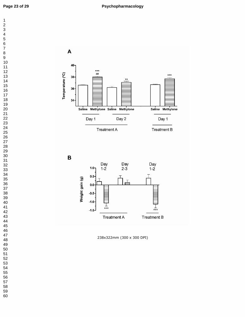
Mehylone induced a significant increase in the body temperature in treatment A and
treatment B (Fig. 1A). Since the hyperthermia induced by methylone at the second day
of treatment A was significantly lower than that induced at the first day, tolerance to
hyperthermic effects of methylone can be concluded.
In both treatments, methylone produced significant loss in body weight (Fig. 1B).
At the end of treatment A methylone-treated animals showed an overall decrease in
body weight of -0.60 ± 0.16 g.
Effect of methylone on different in vivo markers of DA and 5-HT terminals.
Treatment A: methylone-treated mice showed a transient decrease in [3H]WIN35428
specific binding in the frontal cortex that resumed a normal level four days later (Fig.
2A). This decrease was not accompanied by a change in TH expression (3 days:
saline: 100.00 ± 22.60%; methylone: 110.06 ± 12.70%; 7 days: saline: 100.00 ±
11.72%; methylone: 116.00 ± 9.49%).
In contrast, in the striatum, methylone neither affected [3H]WIN35428 binding
(saline: 100.00 ± 10.27%; methylone: 94.38 ± 8.20%) nor TH expression (saline:
100.00 ± 11.10%; methylone: 82.40 ± 4.20%), even after 3 days following the end of
exposure. Thus no further determinations were performed at 7 days.
With regards to 5-HT transporters, methylone did not modify [3H]paroxetine binding
in the frontal cortex (Fig 2B). However, in the hippocampus, methylone induced a slight
reduction in [3H]paroxetine binding, measured 3 days after treatment, which was
reverted 4 days later (Fig 2C). The levels of THP2 remained unchanged after the
treatment (saline: 100.00 ± 3.80%; methylone: 83.20 ± 5.70%, P>0.05).
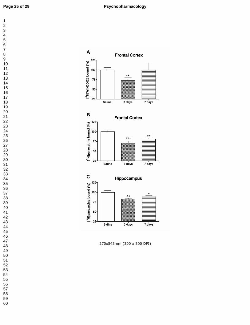
Treatment B: As above, methylone treatment induced a transient decrease in the
specific binding of [3H]WIN35428 of about 27% in the frontal cortex that returned to
control values four days later (Fig. 3A). Similarly, no changes in TH expression were
evidenced (3 days: saline: 100.00 ± 24.47%; methylone: 94.75 ± 15.47%; 7 days:
saline: 100.00 ± 10.52%; methylone: 97.43 ± 29.88%). As in treatment A, methylone
did not affect any of these dopaminergic markers in the striatum ([3H]WIN35428
binding: saline, 100.00 ± 7.57%; methylone, 93.91 ± 4.26%. TH expression: saline:
100.00 ± 3.53%; methylone: 97.06 ± 4.70%; 3 days after drug exposure).
As regards serotonergic markers, at three and seven days post-treatment,
methylone induced a diminution of 5-HT reuptake sites of about 30-20% in frontal
cortex and 20-12% in hippocampus respectively (Fig. 3B and 3C). Additionally, 7 days
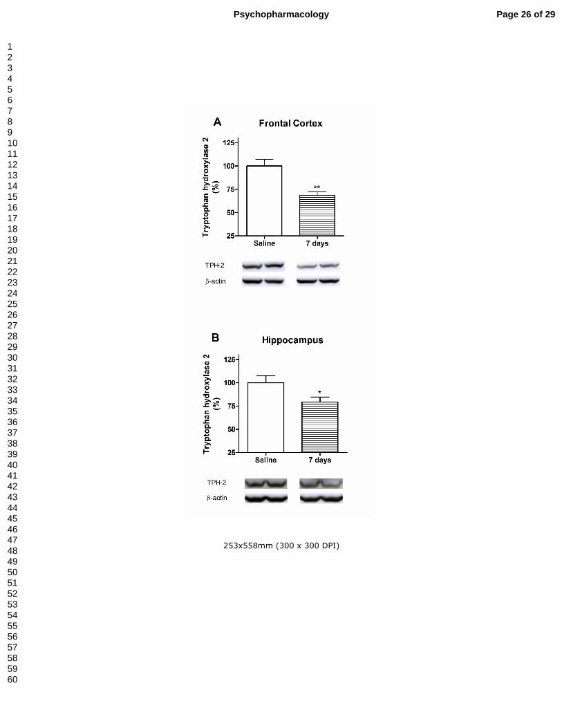
after treatment, TPH2-immunoreactivity levels were decreased in both brain areas in
the methylone-treated mice that correlates with the diminution of 5-HT reuptake sites
(Fig. 4A and 4B).
Page 10 of 29Psychopharmacology
123456789101112131415161718192021222324252627282930313233343536373839404142434445464748495051525354555657585960
11
Effect of methylone on 5-HT2A receptor density.
According to the treatment A schedule, methylone-treated, animals showed a decrease
in the number of 5-HT2A receptors in the frontal (saline: 100.00 ± 4.80%; methylone:
81.69 ± 2.65%, P<0.01) and parietal cortex (saline: 100.00 ± 4.75%; methylone: 80.60
± 5.35%, P<0.05), measured as [3H]ketanserin binding, 3 days after treatment, which
returned to basal values four days later (105.26 ± 10.23% and 119.13 ± 8.49%,
respectively), pointing to a homeostatic process. In contrast, following schedule B,
methylone did not modify the density of 5-HT2A receptors in the two cortical areas either
at 3 (frontal cortex: 85.79 ± 5.40%; parietal cortex: 96.40 ± 3.58%) or 7 days after drug
exposure (frontal cortex: 111.23 ± 12.96%; parietal cortex: 113.16 ± 3.45%).
Effect of methylone on astroglial activation.
Because methylone induced neuronal damage 7 days after treatment B schedule, the
next experiment was carried out to assess the presence of astroglial activation.
Accordingly, immunohistochemistry studies were performed with the glial-specific
marker GFAP in brains from animals killed 7 days after the treatment. There were no
signs of striatal or cortical astroglial activation in methylone-treated animals. However,
in the hippocampus, an increase in GFAP immunoreactivity was observed in the CA1
and dentate gyrus of the methylone group, compared with that from saline-tested mice.
This suggests the presence of a slight reactive astrocytosis (Fig. 5).
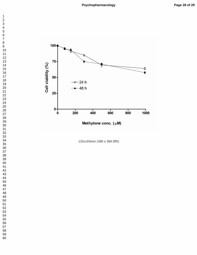
Effect of methylone on cultured cortical neuron viability
The exposure of cultured cortical mouse cells to various concentrations of methylone
(from 80 µM to 1 mM) for 24 h or 48 h caused a weak concentration- or time-dependent
decrease in metabolically active cells, as assessed by the MTT assay (Fig. 6). Cell
viability was only affected by methylone concentrations above 300 µM and the
corresponding calculated LD50 value for methylone after 24 h or 48 h exposure was
over 1 mM, ruling out cell toxicity in cortical neurons.
Depressant-like effect of methylone
Methylone administration (treatments A and B) increased the immobility time in the
FST 3-day post-treatment as compared with the saline group. This time period
coincided with the observed impairment of both dopaminergic and serotonergic
markers. These effects vanished in both schedules one week following drug exposure,
when only the serotonergic markers remained decreased (Fig. 7).
Page 11 of 29 Psychopharmacology
123456789101112131415161718192021222324252627282930313233343536373839404142434445464748495051525354555657585960

12
Discussion
There is little information regarding methylone and its potential toxicity. The initial
status of cathinones as legal highs may have contributed to their increasing popularity
as drugs of abuse. Because of the relatively short history of the use of cathinones as
recreational drugs, their long-term effects have not yet been determined.
Very few studies exist, even in rodents, on the dosing schedules or doses required
to induce damage (Baumann et al., 2012; Hollander et al., 2013). Therefore, the
primary goal of this study was to evaluate the risk of neuronal changes linked to
methylone abuse in mice. Methylone is a close structural analogue of MDMA, differing
only by the addition of a β-ketone group. Consequently, it is also known as beta-keto-
MDMA. As with MDMA, it might affect the DA or 5-HT system differently, depending on
the animal species used for the experiment. Most authors described the maximum
neurotoxic effects of methamphetamine three days after treatment (Pu and Vorhees,
1993) and those of MDMA seven days after treatment (Battaglia et al., 1988). Thus, we
examined the neurotoxic injury induced by methylone at 3 and 7 days after following
the end of the treatment. In addition, a close relationship was already established
between the hyperthermic response and the severity of the brain lesion induced by
amphetamines (Sánchez et al., 2004), supporting the hypothesis that MDMA is
neurotoxic when a binge dosing schedule is employed and the animals are in a hot
environment. Accordingly, present experiments were carried out at a high ambient
temperature simulating hot conditions found in dance clubs. We administered the drug
at 3-3.5h interval, in accordance with our previous paper characterizing the
pharmacokinetics of methylone, distinguished by its short half-life (López-Arnau et al.,
2013). To model recreational methylone use, we simulated the widespread practices of
“stacking” (taking multiple doses at once in order to increase the desired effect and/or
offset tolerance from prior use) and “boosting” (taking supplemental doses over time in
order to maintain the drug’s effect). Thus, we chose to administer multiple doses/day of
methylone during each treatment.
Overall, our data demonstrate a slight serotonergic toxicity of methylone, one week
after treatment, only when four doses are administered in a day. This toxicity is
substantiated by decreases in density of 5-HT terminal marker and reduction in TPH2
expression, both more apparent in frontal cortex than in hippocampus. Post-mortem
necropsies showed evident signs of hepatomegaly and hemorrhagic pericarditis that
could be the cause of death. Further pathology studies are needed to affirm that these
Page 12 of 29Psychopharmacology
123456789101112131415161718192021222324252627282930313233343536373839404142434445464748495051525354555657585960
13
signs resulted directly from methylone toxicity but methylone has been described as a
cause of cardiac arrest leading to human death (Cawrse et al., 2012).
Our initial experiments indicated that methylone toxicity is exacerbated in group-
housed animals like other amphetamines (Fantigrossi et al., 2003). Baumann et al.,
(2013) already described that, in humans, adverse effects of bath salts could be
intensified in hot crowded spaces, such as rave party venues where these drugs are
often used. Present experiments were performed in singly-housed mice. We have
studied the evolution of body weight during methylone treatments. Like other
amphetamine derivatives such MDMA, the animals treated with methylone, but not with
saline, showed a weight loss, probably due to an anorectic effect of the drug.
Several factors, and particularly hyperthermia, contribute to MDMA-induced
neurotoxicity. In this regard, the influence of ambient temperature on MDMA-induced
thermal responses has been shown in earlier studies that noted a hyperthermic
response when experiments were carried out at high ambient temperatures (26-28°C).
Hyperthermia is, in fact, a commonly reported acute adverse effect of beta-
ketoamphetamine ingestion in humans (Borek and Holstege, 2012; Prosser and
Nelson, 2012). In the present study, experiments were performed at ambient
temperature of 26 ± 2ºC. Under these conditions, methylone induced hyperthermia.
This effect was more apparent on the first day of treatment and diminished when the
drug was administered on a second day, indicating the possibility of tolerance that
could be due to a depletion of neurotransmitter stores.
Following the two treatments performed in this study, methylone induced a
transient loss of the DA transporter in the frontal cortex. The initial decline and later
recovery of DA transporter points to a biochemical down-regulation in the absence of
tissue damage but we can also assume that a methylone-induced dopamine
transporter structural modification could occur, explaining the reduction in binding
experiment. This hypothesis is in agreement with our previous results, which
demonstrate that methylone inhibits [3H]DA uptake after drug withdrawal, pointing to
alterations in the transporter that are more complex than a simple blocking of the
carrier (López-Arnau et al., 2013). Unlike MDMA (Chipana et al., 2006), methylone did
not alter DA transporter radioligand binding or TH levels in striatum in any of the
performed treatments. The main difference between treatments was found in 5-HT
terminal markers. When exposure to methylone was performed over two consecutive
days with three doses per day, we registered a transient reduction of these markers,
but when treatment consisted of four doses in a single day, a more persistent effect
appeared, affecting frontal cortex and hippocampus. The final reduction in 5-HT
Page 13 of 29 Psychopharmacology
123456789101112131415161718192021222324252627282930313233343536373839404142434445464748495051525354555657585960

14
transporter marker correlates with the decrease in TPH2 expression. Astrocytes
stabilize and maintain homeostatic tissue repair and contribute to early wound repair
(Eddleston and Mucke, 1993). In the present study methylone-treated animals with
schedule B showed an increase in GFAP immunoreactivity in hippocampal CA1 and
dentate gyrus that corresponds to real terminal injury in these areas.
Only one study has been published concerning the neurotoxic effect of methylone
in DA and 5-HT systems in mice (Hollander et al., 2013). Authors demonstrated that
methylone exposure (30 mg/kg, twice daily for 4 days) had no effect on
neurotransmitter levels in C57BL/J6 mice two weeks after treatment. Our results
demonstrate that methylone neurotoxicity in mice depends on the number of doses and
intervals between each dose, as occurs with other cathinones. Furthermore, we
suspect that the high room temperature, used in this study, could play an important role
in methylone-induced neurotoxicity if we compare our findings with those of Hollander
et al. (2013). Nonetheless, further research is necessary in order to assess whether the
role of hyperthermia and room temperature is complementary or essential in the advent
of methylone-induced neurotoxicity. Some differences among neurotoxicity cathinone
literature can be partially due to differences in the employed dosing-regimen and the
time of sacrifice. However, due to the mechanism of action and structural similarity
between methylone and MDMA, we used similar doses and time of sacrifice assessed
in MDMA neurotoxicity studies (Chipana et al., 2006; Granado et al., 2008; Sánchez et
al., 2003; Mueller et al., 2013).
Mice differ from other animal species because they display deficits in DA
neurotransmission greater than 5-HT after binge MDMA exposure. Present results
demonstrated that methylone acts on contrary. The methylone neurochemical profile
could be explained by the fact that this drug acts preferentially as an inhibitor for the 5-
HT transporter than for the DA transporter (Baumann et al., 2012; López-Arnau et al.,
2012; Sogawa et al., 2011), implying a better access of this drug to the 5-HT nerve
terminals leading to the corresponding injury. Moreover, a well-recognized hypothesis
of MDMA neurotoxicity involves some metabolite participation that has not been
demonstrated for methylone.
Methylone increased the immobility time in the FST following both treatments,
which indicates an increase in stress-related depressive behavior. This effect was
evidenced 3 days after treatment and correlates with the reduction of DA and 5-HT
markers assayed. This is in accordance with results from McGregor et al. (2003), who
demonstrated that MDMA-treated animals show a higher immobility and fewer active
escape attempts in the forced swimming model. To our knowledge, the present studies
provide the first preclinical data to shed light on this issue, suggesting that mice
Page 14 of 29Psychopharmacology
123456789101112131415161718192021222324252627282930313233343536373839404142434445464748495051525354555657585960

15
exposed to a stacking and boosting regime of methylone could be more prone to
suffering from depressive-like symptoms. This effect disappeared 7 days after
treatment, when only serotonergic neurotransmission remained impaired. It must be
noted that depression pathophysiology may also involve changes in 5-HT2 receptor in
brain regions selectively implicated in mood regulation. In this regard, in treatment A
we found a significant transient decrease in the number of cortical 5-HT2A receptors
three days after administration, possibly resulting from a neuroadaptative response to
the massive 5-HT release induced by methylone. These results hark back to those
published by Scheffel et al. (1992) regarding MDMA. In treatment B, we detected a
similar, but non-significant, reduction in frontal cortex.
The impairment induced by methylone on 5-HT and DA terminals is limited to
frontal cortex and hippocampus when exposure is clustered in four doses in a day. This
mild cathinone neurotoxicity correlates with results of our in vitro studies in cortical
cultured cells, where we describe that methylone did not show concentration- and time-
dependent deleterious effect on neuronal viability. The data reveal that doses up to
1000 µM for 24 to 48 h do not appreciably affect cell viability. This is a remarkable
finding, which confirms previous studies that found methylone alone is not cytotoxic
even at high doses (Nakagawa et al., 2009; Sogawa et al., 2011). In this regard, other
studies assessing the effects of MDMA on cortical or hippocampal cultured cell viability
reported no or low cell death following exposure to high MDMA concentrations (Capela
et al., 2006).
In conclusion, our results demonstrate that methylone-induced brain
consequences differ according to treatment schedule (dose, number of doses and dose
interval). Neurochemical changes elicited by methylone are apparent when
administered at an elevated ambient temperature, four times per day at 3h intervals.
This schedule is related with patterns used by humans and agree with methylone’s
half-life in rodents (López-Arnau et al., 2013). Following this, we found decrease in
frontal cortex and hippocampal serotoninergic nerve ending markers around 20-25%
together with hippocampal astrogliosis suggesting nerve ending injuries. No effect in
striatum was evidenced. Methylone did not show a cytotoxic effect in cortical cultured
neurons. The limited neurotoxicity found in this study, however, should not preclude
advice concerning the high risk of acute fatal effects affecting the cardiovascular
system and thermoregulation.
Acknowledgements. Authors acknowledge C. Roberts for language revision of the
manuscript. This study was supported by grants from the Plan Nacional sobre Drogas
(2010/005 and 2012/102); the Ministerio de Ciencia e Innovación (SAF2010-15948)
Page 15 of 29 Psychopharmacology
123456789101112131415161718192021222324252627282930313233343536373839404142434445464748495051525354555657585960
16
and the Generalitat de Catalunya (SGR977). López-Arnau is a recipient of a fellowship
from Generalitat de Catalunya. Martínez-Clemente is a recipient of a fellowship from
the Plan Nacional sobre Drogas and Abad is a recipient of a fellowship from IBUB.
Conflicts of interest. The authors declare that they have no financial or commercial
conflicts of interest.
Page 16 of 29Psychopharmacology
123456789101112131415161718192021222324252627282930313233343536373839404142434445464748495051525354555657585960
17
References
Battaglia G, Yeh SY, De Souza EB (1988) MDMA-induced neurotoxicity: parameters of
degeneration and recovery of brain serotonin neurons. Pharmacol. Biochem. Behav.
29:269-274.
Baumann MH, Ayestas MA, Partilla, JS, Sink JR, Shulgin AT, Daley PF, Brandt SD,
Rothman RB, Ruoho AE, Cozzi NV (2012) The designer methcathinone analogs,
mephedrone and methylone, are substrates for monoamine transporters in brain tissue.
Neuropsychopharmacology 37:1192–1203.
Baumann MH, Partilla JS, Lehner KR (2013) Psychoactive "bath salts": not so
soothing. Eur J Pharmacol. 698:1-5..
Borek HA, Holstege CP (2012) Hyperthermia and multiorgan failure after abuse of
"bath salts" containing 3,4-methylenedioxypyrovalerone. Ann. Emerg. Med. 60:103-
105.
Bossong MG, Van Dijk JP, Niesink RJ (2005) Methylone and mCPP, two new drugs of
abuse? Addict. Biol. 10:321-323.
Boulanger-Gobeil C, St-Onge M, Laliberté M, Auger PL (2012) Seizures and
hyponatremia related to ethcathinone and methylone poisoning. J. Med. Toxicol. 8:59-
61.
Brandt SD, Sumnall HR, Measham F, Cole J (2010) Analyses of second generation
“legal highs” in the UK: initial findings. Drug Test. Anal. 2:377–382.
Brunt TM, Poortman A, Niesink RJ, Van den Brink W (2011) Instability of the ecstasy
market and a new kid on the block: mephedrone. J. Psychopharmacol. 25:1543-1547.
Calapai G, Crupi A, Firenzuoli F, Inferrera G, Squadrito F, Parisi A, De Sarro G, Caputi
A (2001) Serotonin, norepinephrine and dopamine involvement in the antidepressant
action of hypericum perforatum. Pharmacopsychiatry 34:45-49.
Capela JP, Meisel A, Abreu AR, Branco PS, Ferreira LM, Lobo AM, Remião F, Bastos
ML, Carvalho F (2006) Neurotoxicity of Ecstasy metabolites in rat cortical neurons, and
influence of hyperthermia. J. Pharmacol. Exp. Ther. 316:53-61.
Cawrse BM, Levine B, Jufer RA, Fowler DR, Vorce SP, Dickson AJ, Holler JM (2012)
Distribution of methylone in four postmortem cases. J. Anal. Toxicol. 36:434-439.
Chipana C, Camarasa J, Pubill D, Escubedo E (2006) Protection against MDMA-
induced dopaminergic neurotoxicity in mice by methyllycaconitine: Involvement of
nicotinic receptors. Neuropharmacology 51:885-895.
Cozzi NV, Sievert MK, Shulgin AT, Jacob P, Ruoho AE (1999) Inhibition of plasma
membrane monoamine transporters by beta-ketoamphetamines. Eur. J. Pharmacol.
381:63-69.
Eddleston M, Mucke L (1993) Molecular profile of reactive astrocytes--implications for
their role in neurologic disease. Neuroscience 54:15-36.
Page 17 of 29 Psychopharmacology
123456789101112131415161718192021222324252627282930313233343536373839404142434445464748495051525354555657585960

18
Escubedo E, Chipana C, Pérez-Sanchez M, Camarasa J, Pubill D (2005).
Methyllycaconitine prevents methamphetamine-induced effects in mouse striatum:
involvement of alpha7 nicotinic receptors. J. Pharmacol. Exp. Ther. 315:658-667.
Eshleman AJ, Wofrum KM, Hatfield MG, Johnson RA, Murphy KV, Janowsky A (2013).
Subtituted methcathinones differ in transporter and receptor interactions. Biochem.
Pharmacol. 85:1803-1815.
Fantigrossi WE, Godlewski T, Karabenick RL, Stephens JM, Ullrich T, Rice KC, Woods
JH (2003). Pharmacological characterization of the effects of 3,4-
methylenedioxymethamphetamine ("ecstasy") and its enantiomers on lethality, core
temperature, and locomotor activity in singly housed and crowded mice.
Psychopharmacology (Berl). 166:202-211.
Fleckenstein AE, Volz TJ, Riddle EL, Gibb JW, Hanson GR (2007). New insights into
the mechanism of action of amphetamines. Annu. Rev. Pharmacol. Toxicol. 47:681-
698.
Food and Drug Administration Center for Drug Evaluation and Research (CDER).
Guidance for Industry. Estimating the Maximum Safe Starting Dose in Initial Clinical
Trials for Therapeutics in Adult Healthy Volunteers (2005)
http://www.fda.gov/cder/guidance/index.htm (last accessed May, 14, 2013).
Granado N, Escobedo I, O’Shea E, Colado I, Moratalla R (2008) Early loss of
dopaminergic terminals in striosomes after MDMA administration to mice. Synapse
62:80-84.
Hadlock GC, Webb KM, McFadden LM, Chu PW, Ellis JD, Allen SC, Andrenyak DM, Vieira-Brock PL, German CL, Conrad KM, Hoonakker AJ, Gibb JW, Wilkins DG, Hanson GR, Fleckenstein AE (2011) 4-methylmethcathinone (mephedrone): neuropharmacological effects of a designer stimulant of abuse. J. Pharmacol. Exp. Ther. 339: 530-536. Hansen MB, Nielsen SE, Berg K (1989) Re-examination and further development of a precise and rapid dye method for measuring cell growth/cell kill. J Immunol. Meth. 119:203-210. Hollander B den, Rozov S, Linden AM, Uusi-Oukari M, Ojanperä I, Korpi ER (2013)
Long-term cognitive and neurochemical effects of "bath salt" designer drugs methylone
and mephedrone. Pharmacol. Biochem. Behav. 103:501-509.
Karila L, Reynaud M (2010) GHB and synthetic cathinones: clinical effects and
potential consequences. Drug Test Anal. 3:552-559.
Kehr J, Ichinose F, Yoshitake S, Goiny M, Sievertsson T, Nyberg F, Yoshitake T (2011)
Mephedrone, compared to MDMA (ecstasy) and amphetamine, rapidly increases both
dopamine and serotonin levels in nucleus accumbens of awake rats. Br. J. Pharmacol.
164:1949-1958.
Logan BJ, Laverty R, Sanderson WD, Yee YB (1988) Differences between rats and
mice in MDMA (methylenedioxymethylamphetamine) neurotoxicity. Eur. J. Pharmacol.
152:227-234.
Page 18 of 29Psychopharmacology
123456789101112131415161718192021222324252627282930313233343536373839404142434445464748495051525354555657585960

19
López-Arnau R, Martínez-Clemente J, Carbó M, Pubill D, Escubedo E, Camarasa J
(2013) An integrated pharmacokinetic and pharmacodynamic study of a new drug of
abuse, methylone, a synthetic cathinone sold as "bath salts". Prog.
Neuropsychopharmacol. Biol. Psychiatr. 45:64-72.
López-Arnau R, Martínez-Clemente J, Pubill D, Escubedo E, Camarasa J (2012)
Comparative neuropharmacology of three psychostimulant cathinone derivatives:
butylone, mephedrone and methylone. Br. J. Pharmacol. 167:407-420.
Martínez-Clemente J, Escubedo E, Pubill D, Camarasa J (2012) Interaction of
mephedrone with dopamine and serotonin targets in rats. Eur. Neuropsychopharmacol.
22:231-236.
McElrath K, O’Neill C (2011) Experiences with mephedrone pre- and post-legislative
control: perceptions of safety and sources of supply. Int. J. Drug Policy 22:120–127.
McGregor IS, Gurtman CG, Morley KC, Clemens KJ, Blokland A, Li KM, Cornish JL,
Hunt GE (2003) Increased anxiety and "depressive" symptoms months after MDMA
("ecstasy") in rats: drug-induced hyperthermia does not predict long-term outcomes.
Psychopharmacology (Berl). 168:465-474.
Motbey CP, Hunt GE, Bowen MT, Artiss S, McGregor IS (2011) Mephedrone (4-
methylmethcathinone, 'meow'), acute behavioural effects and distribution of Fos
expression in adolescent rats. Addict. Biol. 7:409-422.
Mueller M, Maldonado-Adrian C, Yuan J, McCann UD, Ricaurte GA (2013) Studies of
(±)-3,4-methylenedioxymethamphetamine (MDMA) metabolism and disposition in rats
and mice: relationship to neuroprotection and neurotoxicity profile. J. Pharmacol. Exp.
Ther. 344:479-488.
Nagai F, Nonaka R, Satoh HKK (2007) The effects of non-medically used psychoactive
drugs on monoamine neurotransmission in rat brain. Eur. J. Pharmacol. 559:132-137.
Nakagawa Y, Suzuki T, Tayama S, Ishii H, Ogata A (2009) Cytotoxic effects of 3,4-
methylenedioxy-N-alkylamphetamines, MDMA and its analogues, on isolated rat
hepatocytes. Arch. Toxicol. 83:69-80.
Pearson JM, Hargraves TL, Hair KLS, Massucci CJ, Frazee CC, Garg U, Pietak BR
(2012) Three fatal intoxications due to methylone. J. Anal. Toxicol. 36:444–451.
Petit-Demouliere B, Chenu F, Bourin M (2005) Forced swimming test in mice: a review
of antidepressant activity. Psychopharmacology (Berl). 177:245-255.
Porsolt RD, Anton G, Blavet N, Jalfre M (1978) Behavioural despair in rats: a new
model sensitive to antidepressant treatments. Eur. J. Pharmacol. 47:379-391.
Prosser JM, Nelson LS (2012) The toxicology of bath salts: a review of synthetic
cathinones. J. Med. Toxicol. 8:33-42.
Pu C, Vorhees CV (1993) Developmental dissociation of methamphetamine-induced
depletion of dopaminergic terminals and astrocyte reaction in rat striatum. Brain Res.
Dev. Brain Res. 72:325-328.
Page 19 of 29 Psychopharmacology
123456789101112131415161718192021222324252627282930313233343536373839404142434445464748495051525354555657585960

20
Sánchez V, Camarero J, O’Shea E, Green AR, Colado MI (2003) Differential effect of
dietary selenium on the long-term neurotoxicity induced by MDMA in mice and rats.
Neuropharmacology 44:449-461.
Sánchez V, O'Shea E, Saadat KS, Elliott JM, Colado MI, Green AR (2004) Effect of
repeated ('binge') dosing of MDMA to rats housed at normal and high temperature on
neurotoxic damage to cerebral 5-HT and dopamine neurons. J. Psychopharmacol.
(Berl). 18:412-416.
Scheffel U, Lever JR, Stathis M, Ricaurte GA (1992) Repeated administration of MDMA
causes transient down-regulation of serotonin 5-HT2 receptors. Neuropharmacology
31:881-893.
Senn Ch, Bücheli A, Schaub M, Stohler R (2007) Club drugs. Ther. Umsch. 64:109-
113.
Shimizu E, Watanabe H, Kojima T, Hagiwara H, Fujisaki M, Miyatake R, Hashimoto K,
Iyo M (2007) Combined intoxication with methylone and 5-MeO-MIPT. Prog.
Neuropsychopharmacol. Biol. Psychiatr. 31:288-291.
Simmler LD, Buser TA, Donzelli M, Schramm Y, Dieu LH, Huwyler J, Chaboz S,
Hoener MC, Liechti ME (2013) Pharmacological characterization of designer
cathinones in vitro. Br. J. Pharmacol. 168:458-470.
Sogawa C, Sogawa N, Ohyama K, Kikura-Hanajiri R, Goda Y, Sora I, Kitayama S
(2011) Methylone and monoamine transporters: Correlation with toxicity. Curr.
Neuropharmacol. 9:58-62.
Page 20 of 29Psychopharmacology
123456789101112131415161718192021222324252627282930313233343536373839404142434445464748495051525354555657585960
21
Legends of Figures
Figure 1.- Effect of methylone treatments A and B in body temperature measured after
the second dose of each day’s treatment (panel A) and in body weight (panel B; empty
bars represent saline and striped bars represent methylone). Results are expressed as
mean ± S.E.M. from 8-10 animals. **P<0.01 and ***P<0.001 vs. saline. ## P<0.01 vs.
methylone treatment A day 2.
Figure 2.- Effect of a methylone treatment (3 doses of 25 mg/kg, sc at 3.5 h interval for
2 days) in dopamine transporter density, measured as [3H]WIN35428 binding in mouse
frontal cortex (panel A) and serotonin transporter density, measured as [3H]paroxetine
binding in frontal cortex (panel B) and hippocampus (panel C). Results are expressed
as mean ± S.E.M. from 8-10 animals. *P<0.05 and **P<0.01 vs. saline.
Figure 3.- Effect of a methylone treatment (4 doses of 25 mg/kg, sc at 3 h interval) in
dopamine transporter density, measured as [3H]WIN35428 binding in mouse frontal
cortex (panel A) and serotonin transporter density, measured as [3H]paroxetine binding
in frontal cortex (panel B) and hippocampus (panel C). Results are expressed as mean
± S.E.M. from 8-10 animals. *P<0.05; **P<0.01 and ***P<0.001 vs. saline.
Figure 4.- Effect of a methylone treatment (4 doses of 25 mg/kg, sc at 3 h interval) on
tryptophan hydroxylase 2 expression in mouse frontal cortex (panel A) and
hippocampus (panel B) 7 days after treatment. Below each bar graph, representative
Western blots of TPH-2 expression in frontal cortex and hippocampus, respectively.
**P<0.01 and *P<0.05 vs. saline.
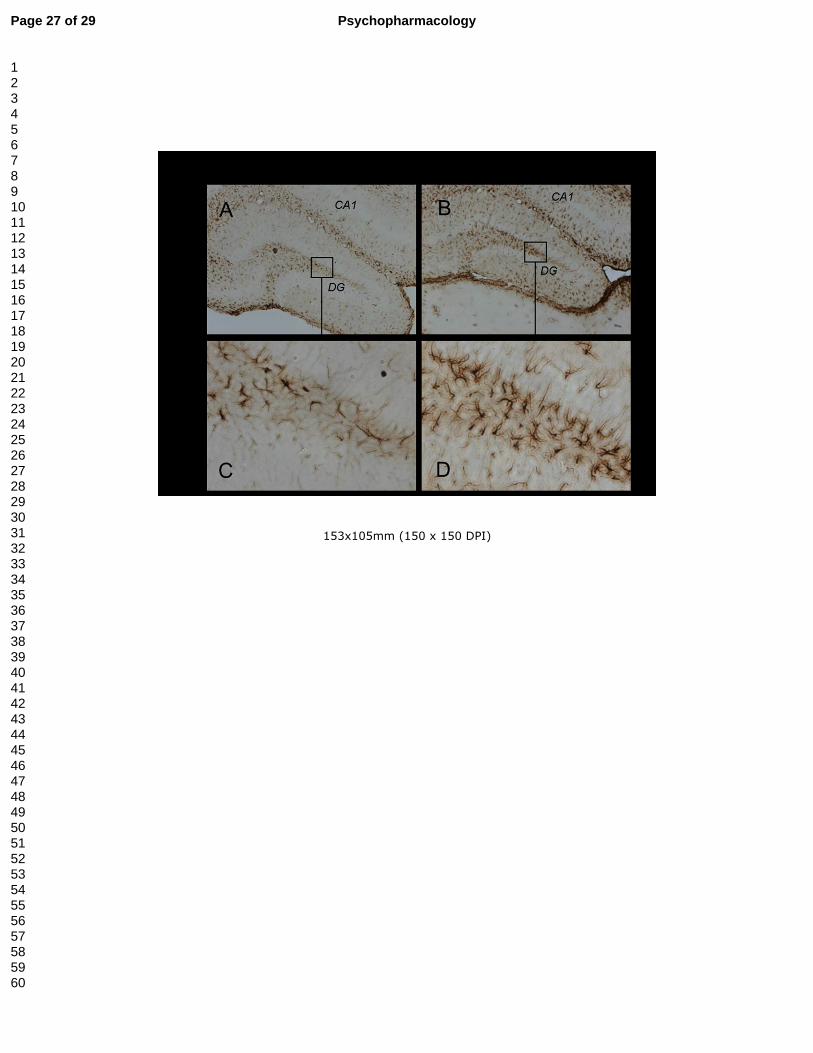
Figure 5.- Representative hippocampal expression of glial fibrilliary acidic protein
(GFAP). Sections of the dentate gyrus from mice treated with saline (panels A and C)
or methylone (4 doses of 25 mg/kg, sc 3 h interval) (panels B and D). The animals
were sacrificed 7 days after treatment.
Figure 6.- Effect in cell viability of methylone on mouse cortical cultured neurons. Cells
were exposed to different concentrations of methylone for 24 or 48 h and cell viability
was assessed by the MTT assay. Data are expressed as mean ± S.E.M. from 3
different cultures.
Page 21 of 29 Psychopharmacology
123456789101112131415161718192021222324252627282930313233343536373839404142434445464748495051525354555657585960
22
Figure 7.- Effect of methylone on immobility time in mouse forced swim test. Animals
(12-16 animals/group) were randomly divided and treated subcutaneously with saline
(5 ml/kg) or methylone (3 doses of 25 mg/kg, sc at 3.5 h interval for 2 days (panel A) or
4 doses of 25 mg/kg, sc 3 h interval (panel B)) and tested 3 or 7 days after treatment.
Each animal was recorded for 6 min and the total period of immobility was registered.
Each mouse was used only once for each experimental session. Each bar represents
mean ± S.E.M. immobility time in seconds. **P<0.01 and *P<0.05 as compared with
respective saline-treated group (one-way ANOVA and post hoc Tukey’s test).
Page 22 of 29Psychopharmacology
123456789101112131415161718192021222324252627282930313233343536373839404142434445464748495051525354555657585960
238x322mm (300 x 300 DPI)
Page 23 of 29 Psychopharmacology
123456789101112131415161718192021222324252627282930313233343536373839404142434445464748495051525354555657585960
283x582mm (300 x 300 DPI)
Page 24 of 29Psychopharmacology
123456789101112131415161718192021222324252627282930313233343536373839404142434445464748495051525354555657585960
270x543mm (300 x 300 DPI)
Page 25 of 29 Psychopharmacology
123456789101112131415161718192021222324252627282930313233343536373839404142434445464748495051525354555657585960
253x558mm (300 x 300 DPI)
Page 26 of 29Psychopharmacology
123456789101112131415161718192021222324252627282930313233343536373839404142434445464748495051525354555657585960
153x105mm (150 x 150 DPI)
Page 27 of 29 Psychopharmacology
123456789101112131415161718192021222324252627282930313233343536373839404142434445464748495051525354555657585960
132x103mm (300 x 300 DPI)
Page 28 of 29Psychopharmacology
123456789101112131415161718192021222324252627282930313233343536373839404142434445464748495051525354555657585960

263x412mm (300 x 300 DPI)
Page 29 of 29 Psychopharmacology
123456789101112131415161718192021222324252627282930313233343536373839404142434445464748495051525354555657585960