Embed Size (px)
Citation preview
RepD-mediated recruitment of PcrA helicase atthe Staphylococcus aureus pC221 plasmidreplication origin, oriDC. Machon1, G. P. Lynch2, N. H. Thomson2, D. J. Scott3, C. D. Thomas2 and
P. Soultanas1,*
1Centre for Biomolecular Sciences, School of Chemistry, University of Nottingham University Park, NottinghamNG7 2RD, 2Astbury Centre for Structural Molecular Biology, University of Leeds, Leeds LS2 9JT and3National Centre for Molecular Hydrodynamics, School of Biosciences, University of Nottingham,Sutton Bonington, Leics LE12 5RD, UK
Received August 28, 2009; Revised November 6, 2009; Accepted November 23, 2009
ABSTRACT
Plasmid encoded replication initiation (Rep) proteinsrecruit host helicases to plasmid replication origins.Previously, we showed that RepD recruits direction-ally the PcrA helicase to the pC221 oriD, remainsassociated with it, and increases its processivityduring plasmid unwinding. Here we show thatRepD forms a complex extending upstream anddownstream of the core oriD. Binding of RepDcauses remodelling of a region upstream from thecore oriD forming a ‘landing pad’ for the PcrA. PcrAis recruited by this extended RepD–DNA complexvia an interaction with RepD at this upstream site.PcrA appears to have weak affinity for this regioneven in the absence of RepD. Upon binding ofADPNP (non-hydrolysable analogue of ATP), byPcrA, a conformational rearrangement of theRepD–PcrA–ATP initiation complex confines itstrictly within the boundaries of the core oriD. Weconclude that RepD-mediated recruitment of PcrAat oriD is a three step process. First, an extendedRepD–oriD complex includes a region upstreamfrom the core oriD; second, the PcrA is recruitedto this upstream region and thirdly uponATP-binding PcrA relocates within the core oriD.
INTRODUCTION
R-plasmids encode antimicrobial resistance genes that area major cause for spread and establishment of anti-microbial resistance in bacteria. The Staphylococcusaureus plasmid pC221 is a 4.6 kb multi-copy plasmidcarrying a gene conferring resistance to chloramphenicol(Cmr). Together with pC223 and pC194 they constitute
the prototype Cmr plasmids in this organism (1). Thespread of antibiotic resistance among S. aureus is notconfined to chloramphenicol. Plasmid mediated resistanceis also found for other antibiotics such as amoxicillin andtetracycline in a high proportion of clinical isolates (2).These Cmr plasmids replicate via rolling circle plasmidreplication. Initiation of replication is mediated byplasmid encoded replication initiation proteins known asRep proteins (3–5). Rep proteins exhibit topoisomeraseI activity, bind to their cognate plasmid origins of repli-cation, nick one strand (known as the (þ) strand) andattach themselves covalently at the 50-end of the nick viaa phosphotyrosine bond utilizing an essential active sitetyrosine residue (6–8). They recruit a host helicase at theorigin and the Rep–helicase complex then facilitates direc-tional unwinding of the duplex DNA during rolling-circleplasmid replication (9,10).
The pC221 plasmid encodes a dimeric RepD protein(37.5 kDa) that initiates replication from its cognate oriD(7) by recruiting the host PcrA helicase and stimulating itsactivity (10–12). PcrA is an essential enzyme found in allGram positive bacteria. It is homologous to the UvrD andRep helicases of Gram negative bacteria and is involved inDNA repair, and the resolution of stalled replication forksand RecFOR-mediated blocked recombination structures(13,14). Its role in rolling-circle plasmid replication wasidentified genetically. A pcrA3 mutation in S. aureusresulted in accumulation of plasmid replication initiationcomplexes and a consequent reduction in plasmid copynumber of pT181-related plasmids (15–17). The acronymPcrA (plasmid copy number reduction) reflects its impor-tant role in plasmid replication. Functional, genetic anddirect interactions between PcrA and Rep proteins havebeen established. For example, suppressor mutationsalleviating the effect of the pcrA3 mutation were identifiedand mapped in the pT181-encoded repC gene (17).
*To whom correspondence should be addressed. Tel: þ44 0 1159513525; Fax: þ44 0 1158468002; Email: [email protected]
1874–1888 Nucleic Acids Research, 2010, Vol. 38, No. 6 Published online 30 December 2009doi:10.1093/nar/gkp1153
� The Author(s) 2009. Published by Oxford University Press.This is an Open Access article distributed under the terms of the Creative Commons Attribution Non-Commercial License (http://creativecommons.org/licenses/by-nc/2.5), which permits unrestricted non-commercial use, distribution, and reproduction in any medium, provided the original work is properly cited.
at Periodicals D
epartment , H
allward Library, U
niversity of Nottingham
on May 5, 2010
http://nar.oxfordjournals.orgD
ownloaded from
The pcrA3 mutation was mapped as T61I within thePcrA3 protein (18). Staphylococcus aureus PcrA function-ally interacted with RepC to mediate pT181 plasmidunwinding (19). The Bacillus cereus and B. anthracisPcrA helicases were able to interact with RepC andsupport the replication of the S. aureus pT181 plasmid(20), the Streptococcus pneumoniae PcrA supportedRepC-mediated unwinding of the S. aureus pT181plasmid (21), whereas the B. stearothermophilusPcrA was able to interact with RepD (encoded byS. aureus pC221 plasmid) and completely unwind anoriD-containing plasmid in the presence of SSB (10).Direct interactions between Rep and PcrA proteins sofar have been detected by pull down assays followed bywestern blotting using MBP-tagged Rep proteins andanti-MBP antibodies (18–20). These data show that Repproteins can interact with heterologous PcrA helicases,a property that may have contributed to the ability ofrolling-circle replication plasmids to disseminate andpropagate in a broad range of Gram positive hosts.
The molecular events of Rep-mediated recruitment ofhelicases at plasmid replication origins are poorly under-stood. The pC221 oriD consists of three inverted comple-mentary repeats, ICR I-III (7). RepD covalently attachesat the 50-end of a nick in the middle of ICR II (7) andparticipates in the directional recruitment of PcrA (10)and to increase its processivity (10,11), but the moleculardetails of their interactions at oriD during loading are notknown. In an effort to understand this better, we carriedout systematic exonuclease III (ExoIII) footprinting anddirect imaging by atomic force microscopy (AFM) of thepC221 oriD in the presence and absence of RepD, PcrAand nucleotides. Our data suggest that binding of RepD tooriD forms an extended structure encompassing the coreoriD and neighbouring regions immediately upstreamand downstream from the oriD. A tighter ternary PcrA–RepD–oriD complex forms in the presence of PcrA withstrong contacts extending upstream from the core oriDbut limited downstream to the end of ICR III. In thepresence of nucleotides the ternary complex re-organizesinto a more compact structure with boundaries contract-ing strictly within the core oriD.
We conclude that RepD forms a complex with oriDextending upstream and downstream of the core oriD.PcrA is recruited via an interaction with RepD upstreamfrom the core oriD. PcrA appears to have basal weakaffinity for this region even in the absence of RepD.When ATP is bound, presumably by PcrA, a conforma-tional change reorganizes the RepD–PcrA complex strictlywithin the boundaries of core oriD.
MATERIALS AND METHODS
Protein purifications
Bacillus stearothermophilus PcrA and staphylococcalRepD used in this study were purified as described else-where (10). RepD mutant R189K was purified using thesame method as the wild-type RepD.
Gel shift assays
A 280 bp PCR fragment containing the core oriD sequencewas amplified using pCERoriD as template and theoligonucleotides oriD(r)NdeI (50-TTAGCTCACTCATATGGCACCCCAGGCT-30) and oriD(d)NcoI (50-GATGTGCTCCATGGCGATTAAGTTGG-30), containingNdeI and NcoI restriction sites, respectively. The oriD-containing DNA fragment was gel purified and 50-end-labelled in a reaction with T4 polynucleotide kinase 10U(NEB) and 10 pmol g-[32P]-ATP (3000Cimmol�1) (PerkinElmer). The unincorporated g-[32P]-ATP was removedusing MicroSpinTM S200 HR columns (GE Healthcare).Binding reactions were carried out with RepD, PcrA andRepD plus PcrA (concentrations are indicated in figurelegends) in 15 ml binding buffer (50mM Tris–HClpH 7.5, 200mM KCl), in the presence of 0.1 nM radio-labelled oriD-containing DNA fragment, and in thepresence or absence of 0.1 mg polydIdC non-specific com-petitor DNA (Sigma). When indicated, 2mM of ADP orADPNP and 5mM MgCl2 were included. Binding reac-tions were incubated at 30�C for 15min and samples wereapplied to a 6% non-denaturing polyacrylamide gel forelectrophoresis in 1�TBE (Tris Borate EDTA) buffer.When nucleotides and MgCl2 were present EDTA wasomitted from the electrophoresis buffer. Gels were driedand imaged using a Molecular Imager FX and associatedsoftware (BioRad).
DNaseI footprinting
DNaseI footprinting experiments were carried out asdescribed elsewhere (22). Purified RepD and/or PcrAproteins were incubated with 0.37 pmol supercoiledpCERoriD in 50 ml binding buffer for 1min at 30�C.Then 50 ml of DNaseI buffer were added to the reaction(40mM Tris–HCl pH 7.9, 10mM NaCl, 6mM MgCl2,1mM CaCl2) and the mixture was incubated for another1min at 37�C, before addition of 10U DNaseI (Roche).Reactions were incubated at 37�C for 3min and stoppedby addition of 100 ml stop buffer (SDS 1% v/v, 200mMNaCl, 20mM EDTA). The digested fragments werepurified by phenol:chloroform (1:1) extraction andethanol precipitation, after addition of 20 mg/ml glycogenas DNA carrier. DNA was suspended and quantifiedusing a Nanodrop prior to primer extension analysis.The following control reactions were included: DNAuntreated with DNaseI, RepD or PcrA, DNA treatedwith DNaseI in the absence of RepD and/or PcrA andDNA incubated with RepD or PcrA, without DNaseI.
Primer extension analysis
The same oligonucleotides oriD(r)NdeI and oriD(d)NcoIthat were used to amplify the oriD-containing fragmentfor gel shift assays were also used in primer extensionreactions. They were designed to anneal to the (þ) and(�) strands, respectively, outside the oriD region inpCERoriD. The DNaseI digested fragments were probedby primer extension using the fmol DNA cycle sequencingsystem from Promega, according to the manufacturer’sinstructions. Primer extension reactions contained
Nucleic Acids Research, 2010, Vol. 38, No. 6 1875
at Periodicals D
epartment , H
allward Library, U
niversity of Nottingham
on May 5, 2010
http://nar.oxfordjournals.orgD
ownloaded from

40 fmol of DNaseI digested pCERoriD, 1.2 pmol of theappropriate 50-g[32P]-end-labelled oligonucleotide primer,5U of sequencing grade Taq DNA polymerase and160 pmol of each deoxyribonucleoside triphosphate(dNTP) in 50mM Tris–HCl pH 9.0, 10mM MgCl2.Dideoxy nucleotide sequence ladders were preparedaccording to the manufacturer’s instructions, using40 fmol of non-digested pCERoriD as template.Conditions for amplification were: 1� (95�C for 2min),30� (95�C for 30 s; 54�C for 30 s; 70�C for 1min).Reactions were stopped by addition of sequencingstop solution (10mM NaOH, 95% (v/v) formamide,0.05% (w/v) bromophenol blue, 0.05% (w/v) xylenecyanol) and then resolved through a urea-denaturing 6%polyacrylamide sequencing gel. Gels were dried andimaged using a Molecular Imager FX and associatedsoftware (BioRad).
Exonuclease III footprinting
ExoIII footprinting experiments were carried out asdescribed elsewhere (23,24). A 280 bp 50-end radiolabelledPCR fragment containing the oriD was digested with NcoIor NdeI (NEB) for 4 h at 37�C and then purified using aQIAquick PCR purification kit (QIAGEN). RepD and/orPcrA were incubated with 10 nM NcoI or NdeI digestedoriD-containing DNA in 50 ml binding buffer for 1min at30�C. Then, 50 ml Exo III buffer (10mM Bis-Tris–Propane–HCl pH 7.0, 10mM MgCl2, 1mM dithiothreitol)and 10U Exo III (NEB) were added to the mixture.Reactions were incubated for 5, 10 or 15min at 37�Cand terminated by addition of 100 ml stop buffer. Thedigested DNA fragments were purified by phenol:chloroform (1:1) extraction and ethanol precipitation,after addition of 20 mg/ml glycogen. DNA was suspendedin sequencing stop solution and resolved through aurea-denaturing 6% polyacrylamide sequencing gel.Gels were dried and imaged using a Molecular ImagerFX and associated software (BioRad).
Analytical ultracentrifugation
The DNA substrate used in analytical ultracentrifugation(AUC) experiments was prepared by PCR using the sameoligonucleotides as in the gel shift assays. Fluoresceinlabels were attached at the 50-end of both oriD(r)NdeIand oriD(d)NcoI oligonucleotides to obtain the fluores-cently labeled oriD-containing DNA fragment. AUCexperiments were carried out in an Optima XL-A analyt-ical ultracentrifuge (Beckman-Coulter, USA) retro-fittedwith laser scanning fluorescence optics (Aviv Scientific,MA, USA). Binding reactions (80 ml) contained 5 nMoriD-DNA in 50mM Tris–HCl pH 7.5, 200mM KCl inthe presence of 0.1mg polydIdC and various concentra-tions of proteins as indicated for individual experiments.Samples were loaded into custom made 80 ml volume3-mm path-length 2-channel sedimentation velocity cells(SpinAnalytical, NH, USA) and loaded into an AN60Tirotor. The loaded rotor was temperature equilibratedat 30�C for 1 h prior to the run and all sedimentationvelocity experiments were carried out at 35 000 r.p.m., atthis temperature. Fluorescence data were obtained at an
excitation wavelength of 488 nm and emission wavelengths>505 nm, scanning every cell simultaneously every 98 s.The photomultiplier settings were 100V, at gain settingof 8. Total fluorescence signal for all cells was around300 counts, and the signal-noise ratio for all the cellswas around 30–40. Data were processed and analysedusing SEDFIT (25). Molecular weights and partialspecific volumes were calculated within the analysisprogram. Buffer densities and viscosities were determinedempirically at 30�C using an Anton Paar DMA5000density and automated viscometer, respectively. Finaldata were plotted as sedimentation coefficient versusRepD or PcrA concentrations and fitted to a one sitebinding hyperbola using GraphPad Prism.
Atomic force microscopy
Samples (30ml) were prepared in K100 buffer(50mM Tris–HCl pH 7.5, 1mM EDTA, 100mM KCl,10mM MgCl2 and 10% (v/v) ethanediol) with 8.3 nMpCERoriD, 16.6 nM wild-type RepD or R189K and/or16.6 nM PcrA and 1mM ADPNP, as indicated. Sampleswere incubated for 20min at 30�C to ensure complex for-mation. A 353 bp DNA fragment containing the core oriDwas excised from pCERoriD by restriction with 20 unitsof PvuII-HFTM (New England Biolabs) for 1 h at 37�C.A sample of 10 ml from the reaction mixture was added to40 ml of spermidine buffer (20mM Tris–HCl, pH 7.5,20mM KCl, 300 mM spermidine) and 20 ml of the resultingmixture was deposited on a freshly cleaved mica surface.The sample was incubated at room temperature for 1minfollowed by washing with 0.22 mm filtered, sterilized anddeionized water and then dried under nitrogen gas beforeimaging.
Imaging was carried out in air using a MultimodeNanoscope IIIa Atomic force microscopy (AFM)(Veeco, Santa Barbara, CA, USA) in Tapping ModeTM.Rectangular silicon cantilevers (Olympus, Japan) withintegrated tips were used with a typical spring constantof 42N/m and a resonant frequency around 300 kHz.The cantilevers were driven at their resonant frequencyand the scan line frequency was 2Hz at 512� 512 pixelresolution. Typical scan sizes were 2� 2 mm from whichDNA–protein complexes were magnified using theNanoscope software (version 5.13r3, Veeco) to generate300� 300 nm images. The total number of images ineach case, n, is given in the ‘Results’ section. Contourlengths and bend angles were measured using the sectionanalysis and angle tools, respectively, of the samesoftware.
RESULTS
ExoIII probing at the front of oriD
The 50-end of the (�) strand of the oriD containing DNAfragment was radioactively labelled and ExoIII digestionwas carried out in the presence and absence of RepD andPcrA, as shown in Figure 1. In the absence of proteins,ExoIII was able to digest the (�) strand through the oriD(see control lane 3 in Figure 1A–D), whereas in thepresence of RepD an initial protection point from
1876 Nucleic Acids Research, 2010, Vol. 38, No. 6
at Periodicals D
epartment , H
allward Library, U
niversity of Nottingham
on May 5, 2010
http://nar.oxfordjournals.orgD
ownloaded from
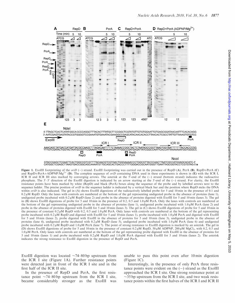
ExoIII digestion was located �74–80 bp upstream fromthe ICR I site (Figure 1A). Further resistance pointswere detected just in front of the ICR I site and in thefirst half of the ICR II site.
In the presence of RepD and PcrA, the first resis-tance point �74–80 bp upstream from the ICR I site,became considerably stronger as the ExoIII was
unable to pass this point even after 10min digestion(Figure 1C).Interestingly, in the presence of only PcrA three resis-
tance points were evident on the (�) strand as the ExoIIIapproached the ICR I site. One strong resistance point at�35 bp upstream from the ICR I site, and two weak resis-tance points within the first halves of the ICR I and ICR II
Figure 1. ExoIII footprinting of the oriD (�) strand. ExoIII footprinting was carried out in the presence of RepD (A), PcrA (B), RepDþPcrA (C)and RepDþPcrAþADPNP/Mg2þ (D). The complete sequence of oriD containing DNA used in these experiments is shown in (E) with the ICR I,ICR II and ICR III sites marked by converging arrows. The asterisk at the 50-end of the (�) strand (bottom strand) indicates the radioactivephosphate. The 30–50 direction of the ExoIII digestion is indicated by an arrow starting at the 30-end of the (�) strand. For clarity, the ExoIIIresistance points have been marked by white (RepD) and black (PcrA) boxes along the sequence of the probe and by labelled arrows next to thesequence ladder. The precise position of oriD in the sequence ladder is indicated by a vertical black bar and the position where RepD nicks the DNAwithin oriD is also indicated. The gel in (A) shows ExoIII digestions of the radioactively labelled probe for 5 and 10min in the presence of 0.1 and0.2 mM RepD. Only the lanes with controls are numbered at the bottom of the gel representing undigested probe in the absence of proteins (lane 1),undigested probe incubated with 0.2 mM RepD (lane 2) and probe in the absence of proteins digested with ExoIII for 5 and 10min (lanes 3). The gelin (B) shows ExoIII digestions of probe for 5 and 10min in the presence of 0.2, 0.5 and 1.0mM PcrA. Only the lanes with controls are numbered atthe bottom of the gel representing undigested probe in the absence of proteins (lane 1), undigested probe incubated with 1.0 mM PcrA (lane 2) andprobe in the absence of proteins digested with ExoIII for 5 and 10min (lanes 3). The gel in (C) shows ExoIII digestions of probe for 5 and 10min inthe presence of constant 0.2 mM RepD with 0.2, 0.5 and 1.0 mM PcrA. Only lanes with controls are numbered at the bottom of the gel representingprobe incubated with 0.2 mM RepD and digested with ExoIII for 5 and 10min (lanes 1), probe incubated with 1.0 mM PcrA and digested with ExoIIIfor 5 and 10min (lanes 2), probe digested with ExoIII in the absence of proteins for 5 and 10min (lane 3), undigested probe in the absence ofproteins (lane 4), undigested probe incubated with 0.2 mM RepD (lane 5), undigested probe incubated with 1.0 mM PcrA (lane 6) and undigestedprobe incubated with 0.2 mM RepD and 1.0 mM PcrA (lane 7). The point of strong resistance to ExoIII digestion is marked by an asterisk. The gel in(D) shows ExoIII digestions of probe for 5 and 10min in the presence of constant 0.2 mM RepD, 50 mM ADPNP, 200 mM MgCl2, with 0.2, 0.5 and1.0 mM PcrA. Only lanes with controls are numbered at the bottom of the gel representing probe digested with ExoIII in the absence of proteins for5 and 10min (lanes 1) and probe incubated with 0.2 mM RepD and 1.0 mM PcrA digested with ExoIII for 5 and 10min (lanes 2). The asteriskindicates the strong resistance to ExoIII digestion in the presence of RepD and PcrA.
Nucleic Acids Research, 2010, Vol. 38, No. 6 1877
at Periodicals D
epartment , H
allward Library, U
niversity of Nottingham
on May 5, 2010
http://nar.oxfordjournals.orgD
ownloaded from
sites (Figure 1B). These data indicate that even in theabsence of RepD, PcrA appears to bind to this DNAfragment, targeting the area upstream from the ICR Isite (see gel shift data later on).In the presence of ADPNP, a non-hydrolysable
analogue of ATP, the ExoIII footprint of the ternarycomplex on the (�) strand changed drastically. As theExoIII approached the ICR I site, the strong resistanceat �74–80 bp upstream from ICR I and the second resis-tance just in front of ICR I disappeared, whereas the resis-tance point within the first half of ICR II strengthenedwith ExoIII being unable to pass this point even after10min digestion (compare the ExoIII digestion patterns
of RepDþPcrA in the absence and presence of ADPNPin Figure 1C and D, respectively).
ExoIII probing at the back of oriD
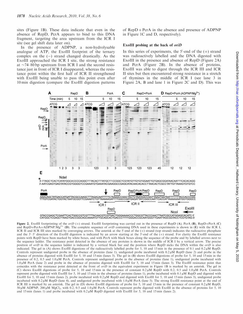
In this series of experiments, the 50-end of the (þ) strandwas radioactively labelled and the DNA digested withExoIII in the presence and absence of RepD (Figure 2A)and PcrA (Figure 2B). In the absence of proteins,ExoIII was able to digest through the ICR III and ICRII sites but then encountered strong resistance in a stretchof thymines in the middle of ICR I (see lane 3 inFigure 2A, B and lane 1 in Figure 2C and D). This was
Figure 2. ExoIII footprinting of the oriD (þ) strand. ExoIII footprinting was carried out in the presence of RepD (A), PcrA (B), RepDþPcrA (C)and RepDþPcrAþADPNP/Mg2þ (D). The complete sequence of oriD containing DNA used in these experiments is shown in (E) with the ICR I,ICR II and ICR III sites marked by converging arrows. The asterisk at the 50-end of the (þ) strand (top strand) indicates the radioactive phosphateand the 30–50 direction of the ExoIII digestion is indicated by an arrow starting at the 30-end of the (þ) strand. For clarity the ExoIII resistancepoints with RepD have been marked by white boxes, and with PcrA with black boxes along the sequence of the probe and by labelled arrows next tothe sequence ladder. The resistance point detected in the absence of any proteins is shown in the middle of ICR I by a vertical arrow. The preciseposition of oriD in the sequence ladder is indicated by a vertical black bar and the position where RepD nicks the DNA within the oriD is alsoindicated. The gel in (A) shows ExoIII digestions of the radioactively labelled probe for 5, 10 and 15min in the presence of 0.1 and 0.2 mM RepD.Controls represent undigested probe in the absence of proteins (lane 1), undigested probe incubated with 0.2 mM RepD (lane 2) and probe in theabsence of proteins digested with ExoIII for 5, 10 and 15min (lanes 3). The gel in (B) shows ExoIII digestions of probe for 5, 10 and 15min in thepresence of 0.2, 0.5 and 1.0 mM PcrA. Controls represent undigested probe in the absence of proteins (lane 1), undigested probe incubated with1.0 mM PcrA (lane 2) and probe in the absence of proteins digested with ExoIII for 5, 10 and 15min (lanes 3). The ExoIII resistance point thatcoincides with the resistance point detected at the front of oriD in the equivalent experiment in Figure 1B is marked by an asterisk. The gel in(C) shows ExoIII digestions of probe for 5, 10 and 15min in the presence of constant 0.2 mM RepD with 0.2, 0.5 and 1.0 mM PcrA. Controlsrepresent probe digested with ExoIII for 5, 10 and 15min in the absence of proteins (lanes 1), probe incubated with 0.1 mM RepD and digested withExoIII for 5, 10 and 15min (lanes 2), probe incubated with 0.2 mM RepD and digested with ExoIII for 5, 10 and 15min (lanes 3), undigested probeincubated with 0.2 mM RepD (lane 4), and undigested probe incubated with 1.0 mM PcrA (lane 5). The strong ExoIII resistance point at the end ofICR III is marked by an asterisk. The gel in (D) shows ExoIII digestions of probe for 5, 10 and 15min in the presence of constant 0.2 mM RepD,50 mM ADPNP, 200 mM MgCl2, with 0.2, 0.5 and 1.0 mM PcrA. Controls represent probe digested with ExoIII in the absence of proteins for 5, 10and 15min (lanes 1) and probe incubated with 0.2 mM RepD digested with ExoIII for 5, 10 and 15min (lanes 2).
1878 Nucleic Acids Research, 2010, Vol. 38, No. 6
at Periodicals D
epartment , H
allward Library, U
niversity of Nottingham
on May 5, 2010
http://nar.oxfordjournals.orgD
ownloaded from
specific for the (þ) strand as it was not observed on the(�) strand when the ExoIII approached from the front ofthe ICR I site (see control lane 3 in Figure 1A–C and lane1 in Figure 1D). The possibility of a secondary DNAstructure inhibiting ExoIII was investigated by creatingmutant oriD fragments with mutations in ICR I (oriDI),ICR II (oriDII) and ICR III (oriDIII), but all mutant oriDfragments exhibited the same resistance point uponExoIII digestion, albeit slightly weaker in the oriDI case(Supplementary Figure 1s). This resistance appears to bespecific to oriD as a non-specific DNA fragment amplifiedfrom the empty pCER19 vector with the same oligo-nucleotides used to amplify oriD did not exhibit resistanceto ExoIII digestion (Supplementary Figure 1s).
In the presence of RepD, protection from ExoIII wasextended �46–50 bp downstream from the ICR III site(Figure 2A). At longer digestions ExoIII was able topass this point but then encountered several more resis-tance points starting at the end of ICR III and extendingall through the ICR II and ICR I sites (compare the 5, 10and 15min digestions in the presence of RepD alone inFigure 2A). With PcrA alone ExoIII digested through theoriD containing DNA fragment but a strong resistancepoint was detected about 30–35 nucleotides past theICR I site (Figure 2B the band marked by an asterisk)which coincides with the same strong resistance pointdetected in the equivalent experiment in Figure 1B.A second minor resistance point was apparent in themiddle of ICR II.
In the presence of RepD and PcrA, the first resistancepoint �46–50 bp downstream from the ICR III site on the(þ) strand, remained the same as in the absence of PcrA.However, the resistance points at the end of ICR IIIbecame considerably stronger in the presence of PcrA.Even after 15min digestion, the ExoIII was halted at theend of ICR III and could not progress into the ICR II andICR I sites (Figure 2C; point marked by an asterisk).
We have already seen ADPNP-induced changes whenwe probed the (�) strand with ExoIII (see above). Similarchanges were also apparent at the other end of the oriD onthe (þ) strand as the ExoIII approached the ICR III site(Figure 2D). In the presence of ADPNP, the first resis-tance point �46–50 bp downstream from the ICR III siteon the (þ) strand became weaker (compare the gels inFigure 2C and D), the resistance points at the end ofICR III also became weaker and the ExoIII was able toprogress beyond ICR III into the ICR II site (compare theExoIII digestion patterns of RepDþPcrA in the presenceand absence of ADPNP in Figure 2C and D).
DNaseI probing of oriD
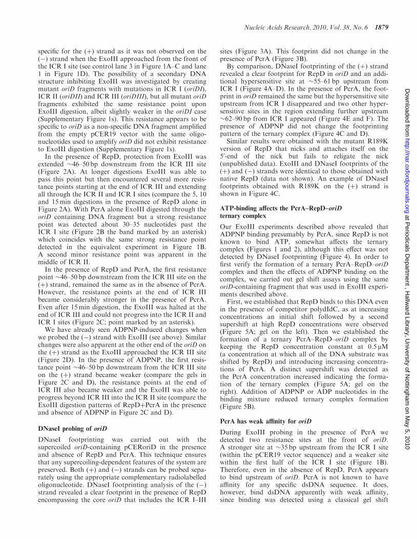
DNaseI footprinting was carried out with thesupercoiled oriD-containing pCERoriD in the presenceand absence of RepD and PcrA. This technique ensuresthat any supercoiling-dependent features of the system arepreserved. Both (þ) and (�) strands can be probed sepa-rately using the appropriate complementary radiolabelledoligonucleotide. DNaseI footprinting analysis of the (�)strand revealed a clear footprint in the presence of RepDencompassing the core oriD that includes the ICR I–III
sites (Figure 3A). This footprint did not change in thepresence of PcrA (Figure 3B).By comparison, DNaseI footprinting of the (þ) strand
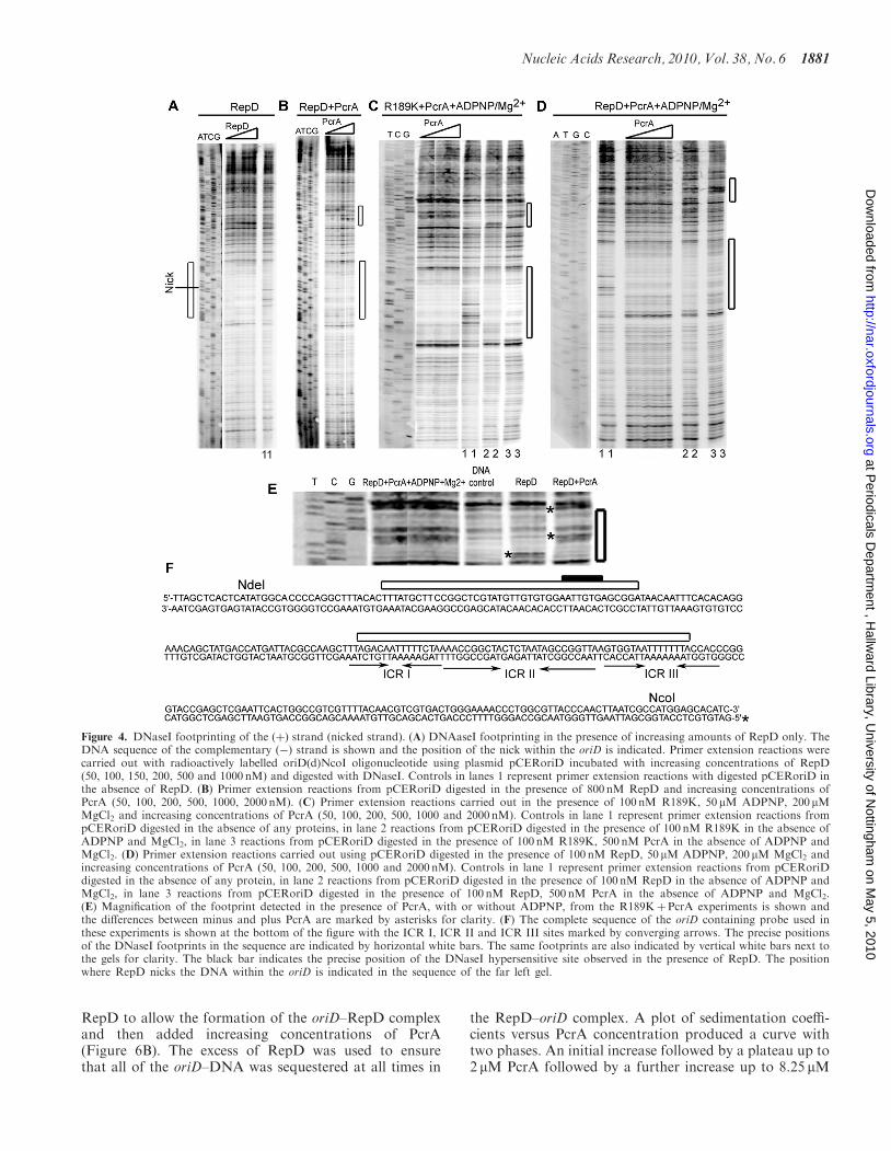
revealed a clear footprint for RepD in oriD and an addi-tional hypersensitive site at �55–61 bp upstream fromICR I (Figure 4A–D). In the presence of PcrA, the foot-print in oriD remained the same but the hypersensitive siteupstream from ICR I disappeared and two other hyper-sensitive sites in the region extending further upstream�62–90 bp from ICR I appeared (Figure 4E and F). Thepresence of ADPNP did not change the footprintingpattern of the ternary complex (Figure 4C and D).Similar results were obtained with the mutant R189K
version of RepD that nicks and attaches itself on the50-end of the nick but fails to religate the nick(unpublished data). ExoIII and DNaseI footprints of the(þ) and (�) strands were identical to those obtained withnative RepD (data not shown). An example of DNaseIfootprints obtained with R189K on the (þ) strand isshown in Figure 4C.
ATP-binding affects the PcrA–RepD–oriDternary complex
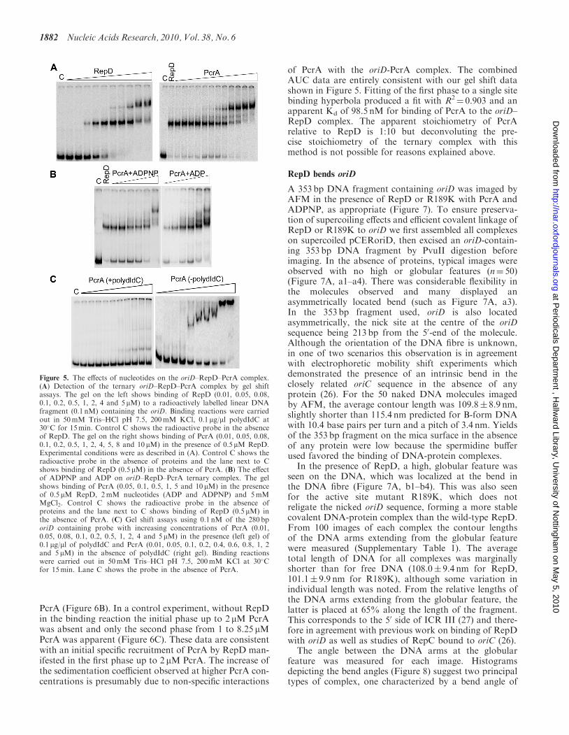
Our ExoIII experiments described above revealed thatADPNP binding presumably by PcrA, since RepD is notknown to bind ATP, somewhat affects the ternarycomplex (Figures 1 and 2), although this effect was notdetected by DNaseI footprinting (Figure 4). In order tofirst verify the formation of a ternary PcrA–RepD–oriDcomplex and then the effects of ADPNP binding on thecomplex, we carried out gel shift assays using the sameoriD-containing fragment that was used in ExoIII experi-ments described above.First, we established that RepD binds to this DNA even
in the presence of competitor polydIdC, as at increasingconcentrations an initial shift followed by a secondsupershift at high RepD concentrations were observed(Figure 5A; gel on the left). Then we established theformation of a ternary PcrA–RepD–oriD complex bykeeping the RepD concentration constant at 0.5 mM(a concentration at which all of the DNA substrate wasshifted by RepD) and introducing increasing concentra-tions of PcrA. A distinct supershift was detected asthe PcrA concentration increased indicating the forma-tion of the ternary complex (Figure 5A; gel on theright). Addition of ADPNP or ADP nucleotides in thebinding mixture reduced ternary complex formation(Figure 5B).
PcrA has weak affinity for oriD
During ExoIII probing in the presence of PcrA wedetected two resistance sites at the front of oriD.A stronger site at �35 bp upstream from the ICR I site(within the pCER19 vector sequence) and a weaker sitewithin the first half of the ICR I site (Figure 1B).Therefore, even in the absence of RepD, PcrA appearsto bind upstream of oriD. PcrA is not known to haveaffinity for any specific dsDNA sequence. It does,however, bind dsDNA apparently with weak affinity,since binding was detected using a classical gel shift
Nucleic Acids Research, 2010, Vol. 38, No. 6 1879
at Periodicals D
epartment , H
allward Library, U
niversity of Nottingham
on May 5, 2010
http://nar.oxfordjournals.orgD
ownloaded from
assay only in the absence of competitor DNA (Figure 5C).There is no higher affinity for oriD because shifts observedwith oriD-containing and a non-specific fragments werecomparable (data not shown).
The stoichiometry of the RepD–oriD complex
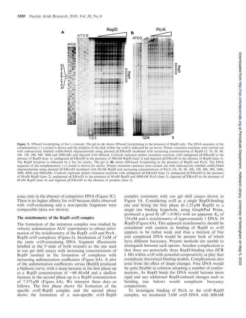
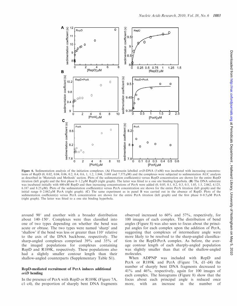
The formation of the initiation complex was studied byvelocity sedimentation AUC experiments to obtain infor-mation of the stoichiometry of the RepD–oriD and PcrA–RepD–oriD complexes (Figure 6). Incubation of 5 nM ofthe same oriD-containing DNA fragment (fluoresceinlabelled at the 50-ends of both strands) as the one usedin our gel shift assays with increasing concentrations ofRepD resulted in the formation of complexes withincreasing sedimentation coefficients (Figure 6A). A plotof the sedimentation coefficients versus [RepD] produceda biphasic curve, with a steep increase in the first phase upto a RepD concentration of �60–80 nM and a shallowincrease in the second phase up to a RepD concentrationof 7.375 mM (Figure 6A). We interpret these data asfollows. The first phase shows the formation of thespecific oriD–RepD complex and the second phaseshows the formation of a non-specific oriD–RepD
complex consistent with our gel shift assays shown inFigure 5A. Considering oriD as a single RepD-bindingsite and fitting the first phase (0–1.25 mM RepD) to asingle site binding hyperbola, using GraphPad Prism,produced a good fit (R2
¼ 0.961) with an apparent Kd of124 nM and a stoichiometry of approximately 1 DNA: 10RepD (Figure 6A). This apparent stoichiometry should beconsidered with caution as binding of RepD to oriDappears to be rather weak and thus a mixture of freeand complexed DNA would be present both of whichhave different buoyancy. Present methods are unable todistinguish between such species. Another complication isthat there are potentially three RepD-binding sites (ICRI–III) within oriD with potential cooperativity at play thatcomplicate theoretical binding models. Complications alsoarise from the effect of shape changes. Free DNA wouldbe quite flexible in solution adopting a number of confor-mations. As RepD binds the DNA would become morerigid and any additional RepD-induced changes such asbending (see below) would complicate buoyancycomparisons.
To investigate binding of PcrA to the oriD–RepDcomplex, we incubated 5 nM oriD–DNA with 600 nM
Figure 3. DNaseI footprinting of the (�) strand. The gel in (A) shows DNaseI footprinting in the presence of RepD only. The DNA sequence of thecomplementary (þ) strand is shown and the position of the nick within the oriD is indicated by an arrow. Primer extension reactions were carried outwith radioactively labelled oriD(r)NdeI oligonucleotide using plasmid pCERoriD incubated with increasing concentrations of RepD (5, 10, 50, 80,100, 150, 200, 500, 1000 and 2000 nM) and digested with DNaseI. Controls represent primer extension reactions with undigested pCERoriD in theabsence of RepD (lane 1), undigested pCERoriD in the presence of 500 nM RepD (lane 2) and digested pCERoriD in the absence of RepD (lane 3).The RepD footprint is indicated by a bar for clarity. The gel in (B) shows DNAaseI footprinting in the presence of RepD and PcrA. The DNAsequence of the complementary (þ) strand is shown for clarity. Primer extension reactions were carried out with radioactively labelled oriD(r)NdeIoligonucleotide using plasmid pCERoriD incubated with 80 nM RepD and increasing concentrations of PcrA (10, 50, 80, 100, 150, 200, 500, 1000,2000, 4000 and 8000 nM). Controls represent primer extension reactions with undigested pCERoriD (lane 1), undigested pCERoriD in the presenceof 80 nM RepD (lane 2), undigested pCERoriD in the presence of 80 nM RepD and 8000 nM PcrA (lane 3), digested pCERoriD in the presence of80 nM RepD (lane 4) and digested pCERoriD in the absence of proteins (lane 5).
1880 Nucleic Acids Research, 2010, Vol. 38, No. 6
at Periodicals D
epartment , H
allward Library, U
niversity of Nottingham
on May 5, 2010
http://nar.oxfordjournals.orgD
ownloaded from
RepD to allow the formation of the oriD–RepD complexand then added increasing concentrations of PcrA(Figure 6B). The excess of RepD was used to ensurethat all of the oriD–DNA was sequestered at all times in
the RepD–oriD complex. A plot of sedimentation coeffi-cients versus PcrA concentration produced a curve withtwo phases. An initial increase followed by a plateau up to2 mM PcrA followed by a further increase up to 8.25 mM
Figure 4. DNaseI footprinting of the (þ) strand (nicked strand). (A) DNAaseI footprinting in the presence of increasing amounts of RepD only. TheDNA sequence of the complementary (�) strand is shown and the position of the nick within the oriD is indicated. Primer extension reactions werecarried out with radioactively labelled oriD(d)NcoI oligonucleotide using plasmid pCERoriD incubated with increasing concentrations of RepD(50, 100, 150, 200, 500 and 1000 nM) and digested with DNaseI. Controls in lanes 1 represent primer extension reactions with digested pCERoriD inthe absence of RepD. (B) Primer extension reactions from pCERoriD digested in the presence of 800 nM RepD and increasing concentrations ofPcrA (50, 100, 200, 500, 1000, 2000 nM). (C) Primer extension reactions carried out in the presence of 100 nM R189K, 50 mM ADPNP, 200mMMgCl2 and increasing concentrations of PcrA (50, 100, 200, 500, 1000 and 2000 nM). Controls in lane 1 represent primer extension reactions frompCERoriD digested in the absence of any proteins, in lane 2 reactions from pCERoriD digested in the presence of 100 nM R189K in the absence ofADPNP and MgCl2, in lane 3 reactions from pCERoriD digested in the presence of 100 nM R189K, 500 nM PcrA in the absence of ADPNP andMgCl2. (D) Primer extension reactions carried out using pCERoriD digested in the presence of 100 nM RepD, 50 mM ADPNP, 200 mM MgCl2 andincreasing concentrations of PcrA (50, 100, 200, 500, 1000 and 2000 nM). Controls in lane 1 represent primer extension reactions from pCERoriDdigested in the absence of any protein, in lane 2 reactions from pCERoriD digested in the presence of 100 nM RepD in the absence of ADPNP andMgCl2, in lane 3 reactions from pCERoriD digested in the presence of 100 nM RepD, 500 nM PcrA in the absence of ADPNP and MgCl2.(E) Magnification of the footprint detected in the presence of PcrA, with or without ADPNP, from the R189KþPcrA experiments is shown andthe differences between minus and plus PcrA are marked by asterisks for clarity. (F) The complete sequence of the oriD containing probe used inthese experiments is shown at the bottom of the figure with the ICR I, ICR II and ICR III sites marked by converging arrows. The precise positionsof the DNaseI footprints in the sequence are indicated by horizontal white bars. The same footprints are also indicated by vertical white bars next tothe gels for clarity. The black bar indicates the precise position of the DNaseI hypersensitive site observed in the presence of RepD. The positionwhere RepD nicks the DNA within the oriD is indicated in the sequence of the far left gel.
Nucleic Acids Research, 2010, Vol. 38, No. 6 1881
at Periodicals D
epartment , H
allward Library, U
niversity of Nottingham
on May 5, 2010
http://nar.oxfordjournals.orgD
ownloaded from
PcrA (Figure 6B). In a control experiment, without RepDin the binding reaction the initial phase up to 2 mM PcrAwas absent and only the second phase from 1 to 8.25 mMPcrA was apparent (Figure 6C). These data are consistentwith an initial specific recruitment of PcrA by RepD man-ifested in the first phase up to 2 mM PcrA. The increase ofthe sedimentation coefficient observed at higher PcrA con-centrations is presumably due to non-specific interactions
of PcrA with the oriD-PcrA complex. The combinedAUC data are entirely consistent with our gel shift datashown in Figure 5. Fitting of the first phase to a single sitebinding hyperbola produced a fit with R2
¼ 0.903 and anapparent Kd of 98.5 nM for binding of PcrA to the oriD–RepD complex. The apparent stoichiometry of PcrArelative to RepD is 1:10 but deconvoluting the pre-cise stoichiometry of the ternary complex with thismethod is not possible for reasons explained above.
RepD bends oriD
A 353 bp DNA fragment containing oriD was imaged byAFM in the presence of RepD or R189K with PcrA andADPNP, as appropriate (Figure 7). To ensure preserva-tion of supercoiling effects and efficient covalent linkage ofRepD or R189K to oriD we first assembled all complexeson supercoiled pCERoriD, then excised an oriD-contain-ing 353 bp DNA fragment by PvuII digestion beforeimaging. In the absence of proteins, typical images wereobserved with no high or globular features (n¼ 50)(Figure 7A, a1–a4). There was considerable flexibility inthe molecules observed and many displayed anasymmetrically located bend (such as Figure 7A, a3).In the 353 bp fragment used, oriD is also locatedasymmetrically, the nick site at the centre of the oriDsequence being 213 bp from the 50-end of the molecule.Although the orientation of the DNA fibre is unknown,in one of two scenarios this observation is in agreementwith electrophoretic mobility shift experiments whichdemonstrated the presence of an intrinsic bend in theclosely related oriC sequence in the absence of anyprotein (26). For the 50 naked DNA molecules imagedby AFM, the average contour length was 109.8� 8.9 nm,slightly shorter than 115.4 nm predicted for B-form DNAwith 10.4 base pairs per turn and a pitch of 3.4 nm. Yieldsof the 353 bp fragment on the mica surface in the absenceof any protein were low because the spermidine bufferused favored the binding of DNA-protein complexes.
In the presence of RepD, a high, globular feature wasseen on the DNA, which was localized at the bend inthe DNA fibre (Figure 7A, b1–b4). This was also seenfor the active site mutant R189K, which does notreligate the nicked oriD sequence, forming a more stablecovalent DNA-protein complex than the wild-type RepD.From 100 images of each complex the contour lengthsof the DNA arms extending from the globular featurewere measured (Supplementary Table 1). The averagetotal length of DNA for all complexes was marginallyshorter than for free DNA (108.0� 9.4 nm for RepD,101.1� 9.9 nm for R189K), although some variation inindividual length was noted. From the relative lengths ofthe DNA arms extending from the globular feature, thelatter is placed at 65% along the length of the fragment.This corresponds to the 50 side of ICR III (27) and there-fore in agreement with previous work on binding of RepDwith oriD as well as studies of RepC bound to oriC (26).
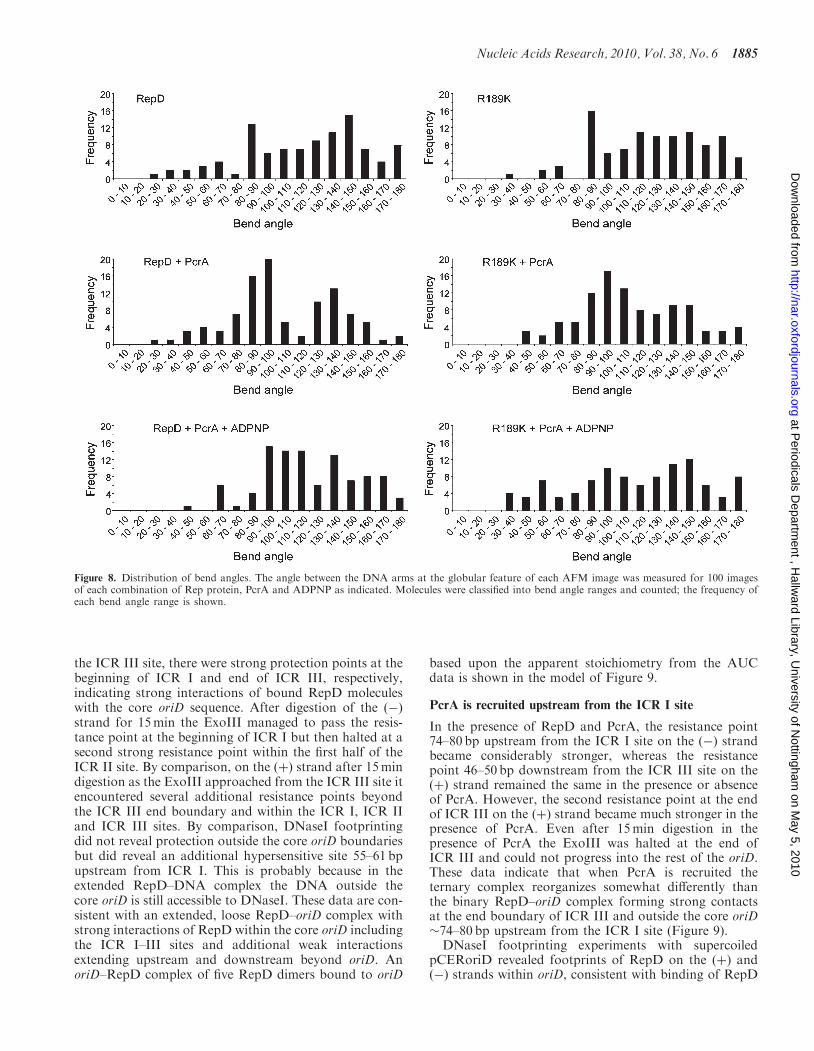
The angle between the DNA arms at the globularfeature was measured for each image. Histogramsdepicting the bend angles (Figure 8) suggest two principaltypes of complex, one characterized by a bend angle of
Figure 5. The effects of nucleotides on the oriD–RepD–PcrA complex.(A) Detection of the ternary oriD–RepD–PcrA complex by gel shiftassays. The gel on the left shows binding of RepD (0.01, 0.05, 0.08,0.1, 0.2, 0.5, 1, 2, 4 and 5 mM) to a radioactively labelled linear DNAfragment (0.1 nM) containing the oriD. Binding reactions were carriedout in 50mM Tris–HCl pH 7.5, 200mM KCl, 0.1 mg/ml polydIdC at30�C for 15min. Control C shows the radioactive probe in the absenceof RepD. The gel on the right shows binding of PcrA (0.01, 0.05, 0.08,0.1, 0.2, 0.5, 1, 2, 4, 5, 8 and 10 mM) in the presence of 0.5 mM RepD.Experimental conditions were as described in (A). Control C shows theradioactive probe in the absence of proteins and the lane next to Cshows binding of RepD (0.5 mM) in the absence of PcrA. (B) The effectof ADPNP and ADP on oriD–RepD–PcrA ternary complex. The gelshows binding of PcrA (0.05, 0.1, 0.5, 1, 5 and 10mM) in the presenceof 0.5 mM RepD, 2mM nucleotides (ADP and ADPNP) and 5mMMgCl2. Control C shows the radioactive probe in the absence ofproteins and the lane next to C shows binding of RepD (0.5 mM) inthe absence of PcrA. (C) Gel shift assays using 0.1 nM of the 280 bporiD containing probe with increasing concentrations of PcrA (0.01,0.05, 0.08, 0.1, 0.2, 0.5, 1, 2, 4 and 5 mM) in the presence (left gel) of0.1 mg/ml of polydIdC and PcrA (0.01, 0.05, 0.1, 0.2, 0.4, 0.6, 0.8, 1, 2and 5 mM) in the absence of polydIdC (right gel). Binding reactionswere carried out in 50mM Tris–HCl pH 7.5, 200mM KCl at 30�Cfor 15min. Lane C shows the probe in the absence of PcrA.
1882 Nucleic Acids Research, 2010, Vol. 38, No. 6
at Periodicals D
epartment , H
allward Library, U
niversity of Nottingham
on May 5, 2010
http://nar.oxfordjournals.orgD
ownloaded from
around 90� and another with a broader distributionabout 140–150�. Complexes were thus classified intoone of two types depending on whether the bend wasacute or obtuse. The two types were named ‘sharp’ and‘shallow’ if the bend was less or greater than 110� relativeto the axis of the DNA backbone, respectively. Thesharp-angled complexes comprised 39% and 35% ofthe imaged populations for complexes containingRepD and R189K, respectively. These populations alsohad a slightly smaller contour length than theirshallow-angled counterparts (Supplementary Table S1).
RepD-mediated recruitment of PcrA induces additionaloriD bending
In the presence of PcrA with RepD or R189K (Figure 7A,c1–c4), the proportion of sharply bent DNA fragments
observed increased to 60% and 57%, respectively, for100 images of each complex. The distribution of bendangles (Figure 8) was also seen to focus about the princi-pal angles for each complex upon the addition of PcrA,suggesting that complexes of intermediate angle weremore likely to be resolved to the sharp-angled classifica-tion in the RepD:PcrA complex. As before, the aver-age contour length of each sharply-angled populationwas slightly smaller than that of the shallow-angledmolecules.When ADPNP was included with RepD and
PcrA or R189K and PcrA (Figure 7A, d1–d4) thenumber of sharply bent DNA fragments decreased to41% and 46%, respectively, again for 100 images ofeach complex. The histograms (Figure 8) show that thefocus about each principal angle is reduced oncemore, with an increase in the number of
Figure 6. Sedimentation analysis of the initiation complexes. (A) Fluorescein labelled oriD-DNA (5 nM) was incubated with increasing concentra-tions of RepD (0, 0.02, 0.04, 0.06, 0.2, 0.4, 0.6, 1, 1.2, 1.844, 3.688 and 7.375 mM) and the complexes were subjected to sedimentation AUC analysisas described in ‘Materials and Methods’ section. Plots of the sedimentation coefficient(s) versus RepD concentration are shown for the entire RepDtitration (left graph) and the first phase 0–1.2 mM RepD (right graph). The latter was fitted to a one site binding hyperbola. (B) The DNA substratewas incubated initially with 600 nM RepD and then increasing concentrations of PcrA were added (0, 0.05, 0.1, 0.2, 0.3, 0.5, 1.03, 1.5, 2.062, 4.125,6.187 and 8.25mM). Plots of the sedimentation coefficient(s) versus PcrA concentration are shown for the entire PcrA titration (left graph) and theinitial range 0–2.062 mM PcrA (right graph). (C) The same experiment as in panel B was carried out in the absence of RepD. Plots of thesedimentation coefficient(s) versus PcrA concentration are shown for the entire PcrA titration (left graph) and the first phase 0–0.5 mM PcrA(right graph). The latter was fitted to a one site binding hyperbola.
Nucleic Acids Research, 2010, Vol. 38, No. 6 1883
at Periodicals D
epartment , H
allward Library, U
niversity of Nottingham
on May 5, 2010
http://nar.oxfordjournals.orgD
ownloaded from
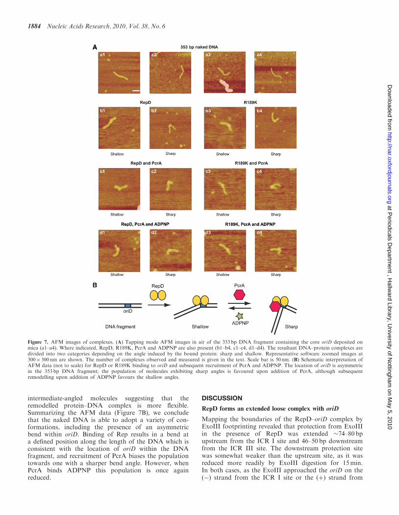
intermediate-angled molecules suggesting that theremodelled protein–DNA complex is more flexible.Summarizing the AFM data (Figure 7B), we concludethat the naked DNA is able to adopt a variety of con-formations, including the presence of an asymmetricbend within oriD. Binding of Rep results in a bend ata defined position along the length of the DNA which isconsistent with the location of oriD within the DNAfragment, and recruitment of PcrA biases the populationtowards one with a sharper bend angle. However, whenPcrA binds ADPNP this population is once againreduced.
DISCUSSION
RepD forms an extended loose complex with oriD
Mapping the boundaries of the RepD–oriD complex byExoIII footprinting revealed that protection from ExoIIIin the presence of RepD was extended �74–80 bpupstream from the ICR I site and 46–50 bp downstreamfrom the ICR III site. The downstream protection sitewas somewhat weaker than the upstream site, as it wasreduced more readily by ExoIII digestion for 15min.In both cases, as the ExoIII approached the oriD on the(�) strand from the ICR I site or the (þ) strand from
Figure 7. AFM images of complexes. (A) Tapping mode AFM images in air of the 353 bp DNA fragment containing the core oriD deposited onmica (a1–a4). Where indicated, RepD, R189K, PcrA and ADPNP are also present (b1–b4, c1–c4, d1–d4). The resultant DNA–protein complexes aredivided into two categories depending on the angle induced by the bound protein: sharp and shallow. Representative software zoomed images at300� 300 nm are shown. The number of complexes observed and measured is given in the text. Scale bar is 50 nm. (B) Schematic interpretation ofAFM data (not to scale) for RepD or R189K binding to oriD and subsequent recruitment of PcrA and ADPNP. The location of oriD is asymmetricin the 353 bp DNA fragment; the population of molecules exhibiting sharp angles is favoured upon addition of PcrA, although subsequentremodelling upon addition of ADPNP favours the shallow angles.
1884 Nucleic Acids Research, 2010, Vol. 38, No. 6
at Periodicals D
epartment , H
allward Library, U
niversity of Nottingham
on May 5, 2010
http://nar.oxfordjournals.orgD
ownloaded from
the ICR III site, there were strong protection points at thebeginning of ICR I and end of ICR III, respectively,indicating strong interactions of bound RepD moleculeswith the core oriD sequence. After digestion of the (�)strand for 15min the ExoIII managed to pass the resis-tance point at the beginning of ICR I but then halted at asecond strong resistance point within the first half of theICR II site. By comparison, on the (þ) strand after 15mindigestion as the ExoIII approached from the ICR III site itencountered several additional resistance points beyondthe ICR III end boundary and within the ICR I, ICR IIand ICR III sites. By comparison, DNaseI footprintingdid not reveal protection outside the core oriD boundariesbut did reveal an additional hypersensitive site 55–61 bpupstream from ICR I. This is probably because in theextended RepD–DNA complex the DNA outside thecore oriD is still accessible to DNaseI. These data are con-sistent with an extended, loose RepD–oriD complex withstrong interactions of RepD within the core oriD includingthe ICR I–III sites and additional weak interactionsextending upstream and downstream beyond oriD. AnoriD–RepD complex of five RepD dimers bound to oriD
based upon the apparent stoichiometry from the AUCdata is shown in the model of Figure 9.
PcrA is recruited upstream from the ICR I site
In the presence of RepD and PcrA, the resistance point74–80 bp upstream from the ICR I site on the (�) strandbecame considerably stronger, whereas the resistancepoint 46–50 bp downstream from the ICR III site on the(þ) strand remained the same in the presence or absenceof PcrA. However, the second resistance point at the endof ICR III on the (þ) strand became much stronger in thepresence of PcrA. Even after 15min digestion in thepresence of PcrA the ExoIII was halted at the end ofICR III and could not progress into the rest of the oriD.These data indicate that when PcrA is recruited theternary complex reorganizes somewhat differently thanthe binary RepD–oriD complex forming strong contactsat the end boundary of ICR III and outside the core oriD�74–80 bp upstream from the ICR I site (Figure 9).DNaseI footprinting experiments with supercoiled
pCERoriD revealed footprints of RepD on the (þ) and(�) strands within oriD, consistent with binding of RepD
Figure 8. Distribution of bend angles. The angle between the DNA arms at the globular feature of each AFM image was measured for 100 imagesof each combination of Rep protein, PcrA and ADPNP as indicated. Molecules were classified into bend angle ranges and counted; the frequency ofeach bend angle range is shown.
Nucleic Acids Research, 2010, Vol. 38, No. 6 1885
at Periodicals D
epartment , H
allward Library, U
niversity of Nottingham
on May 5, 2010
http://nar.oxfordjournals.orgD
ownloaded from
molecules in the ICR I-III sites. However, an additionalDNaseI hypersensitive site appeared to form at 55–61 bpupstream from ICR I site on the (þ) strand when RepDwas bound to oriD. In the presence of RepD and PcrA,this DNaseI hypersensitive site disappeared, whilst twoother hypersensitive sites appeared nearby in the regionextending further upstream 62–90 bp from ICR I. This isconsistent with a reorganization of the binary RepD-DNAcomplex when PcrA is also bound. The area where theseeffects were observed coincided broadly with the ExoIIIresistance point 74–80 bp upstream from the ICR I site.The combined data reinforce the notion that initial RepD
binding to oriD forms an extended loose complex thatrecruits PcrA upstream from the ICR I site (Figure 9).
ATP binding induces re-modelling of thePcrA–RepD–oriD ternary complex
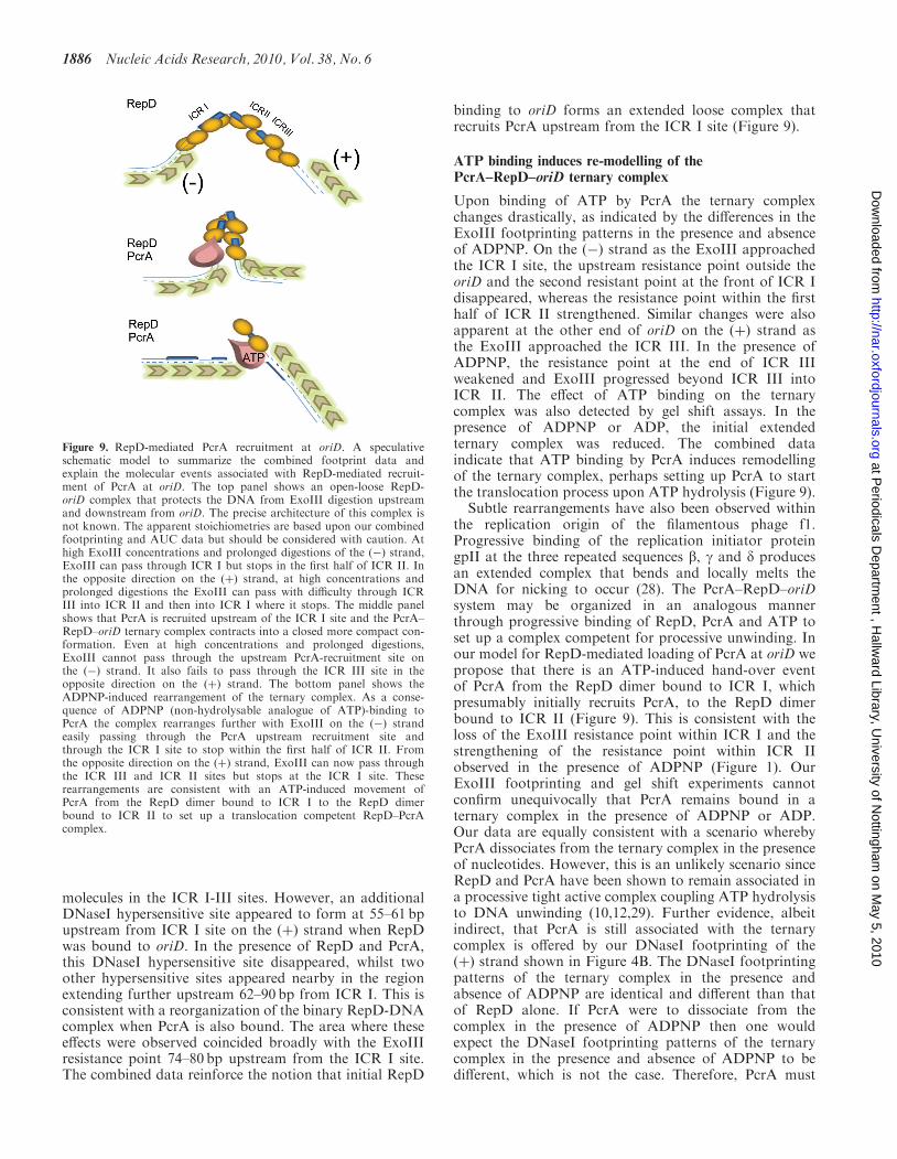
Upon binding of ATP by PcrA the ternary complexchanges drastically, as indicated by the differences in theExoIII footprinting patterns in the presence and absenceof ADPNP. On the (�) strand as the ExoIII approachedthe ICR I site, the upstream resistance point outside theoriD and the second resistant point at the front of ICR Idisappeared, whereas the resistance point within the firsthalf of ICR II strengthened. Similar changes were alsoapparent at the other end of oriD on the (þ) strand asthe ExoIII approached the ICR III. In the presence ofADPNP, the resistance point at the end of ICR IIIweakened and ExoIII progressed beyond ICR III intoICR II. The effect of ATP binding on the ternarycomplex was also detected by gel shift assays. In thepresence of ADPNP or ADP, the initial extendedternary complex was reduced. The combined dataindicate that ATP binding by PcrA induces remodellingof the ternary complex, perhaps setting up PcrA to startthe translocation process upon ATP hydrolysis (Figure 9).
Subtle rearrangements have also been observed withinthe replication origin of the filamentous phage f1.Progressive binding of the replication initiator proteingpII at the three repeated sequences b, g and d producesan extended complex that bends and locally melts theDNA for nicking to occur (28). The PcrA–RepD–oriDsystem may be organized in an analogous mannerthrough progressive binding of RepD, PcrA and ATP toset up a complex competent for processive unwinding. Inour model for RepD-mediated loading of PcrA at oriD wepropose that there is an ATP-induced hand-over eventof PcrA from the RepD dimer bound to ICR I, whichpresumably initially recruits PcrA, to the RepD dimerbound to ICR II (Figure 9). This is consistent with theloss of the ExoIII resistance point within ICR I and thestrengthening of the resistance point within ICR IIobserved in the presence of ADPNP (Figure 1). OurExoIII footprinting and gel shift experiments cannotconfirm unequivocally that PcrA remains bound in aternary complex in the presence of ADPNP or ADP.Our data are equally consistent with a scenario wherebyPcrA dissociates from the ternary complex in the presenceof nucleotides. However, this is an unlikely scenario sinceRepD and PcrA have been shown to remain associated ina processive tight active complex coupling ATP hydrolysisto DNA unwinding (10,12,29). Further evidence, albeitindirect, that PcrA is still associated with the ternarycomplex is offered by our DNaseI footprinting of the(þ) strand shown in Figure 4B. The DNaseI footprintingpatterns of the ternary complex in the presence andabsence of ADPNP are identical and different than thatof RepD alone. If PcrA were to dissociate from thecomplex in the presence of ADPNP then one wouldexpect the DNaseI footprinting patterns of the ternarycomplex in the presence and absence of ADPNP to bedifferent, which is not the case. Therefore, PcrA must
Figure 9. RepD-mediated PcrA recruitment at oriD. A speculativeschematic model to summarize the combined footprint data andexplain the molecular events associated with RepD-mediated recruit-ment of PcrA at oriD. The top panel shows an open-loose RepD-oriD complex that protects the DNA from ExoIII digestion upstreamand downstream from oriD. The precise architecture of this complex isnot known. The apparent stoichiometries are based upon our combinedfootprinting and AUC data but should be considered with caution. Athigh ExoIII concentrations and prolonged digestions of the (�) strand,ExoIII can pass through ICR I but stops in the first half of ICR II. Inthe opposite direction on the (þ) strand, at high concentrations andprolonged digestions the ExoIII can pass with difficulty through ICRIII into ICR II and then into ICR I where it stops. The middle panelshows that PcrA is recruited upstream of the ICR I site and the PcrA–RepD–oriD ternary complex contracts into a closed more compact con-formation. Even at high concentrations and prolonged digestions,ExoIII cannot pass through the upstream PcrA-recruitment site onthe (�) strand. It also fails to pass through the ICR III site in theopposite direction on the (þ) strand. The bottom panel shows theADPNP-induced rearrangement of the ternary complex. As a conse-quence of ADPNP (non-hydrolysable analogue of ATP)-binding toPcrA the complex rearranges further with ExoIII on the (�) strandeasily passing through the PcrA upstream recruitment site andthrough the ICR I site to stop within the first half of ICR II. Fromthe opposite direction on the (þ) strand, ExoIII can now pass throughthe ICR III and ICR II sites but stops at the ICR I site. Theserearrangements are consistent with an ATP-induced movement ofPcrA from the RepD dimer bound to ICR I to the RepD dimerbound to ICR II to set up a translocation competent RepD–PcrAcomplex.
1886 Nucleic Acids Research, 2010, Vol. 38, No. 6
at Periodicals D
epartment , H
allward Library, U
niversity of Nottingham
on May 5, 2010
http://nar.oxfordjournals.orgD
ownloaded from
remain bound in the complex even in the presence ofADPNP or ADP.
Recruitment of PcrA by RepD results in a visible changein DNA conformation
The model proposed above is consistent with our AFMimaging data. AFM images of protein–DNA complexesfell in two categories depending on the severity of theprotein induced bend (sharp or shallow). It is possiblethat wild-type RepD may nick at oriD but religate theDNA during the course of sample preparation, resultingin a mixed population of nicked and religated DNA.For this reason, the RepD mutant R189K was alsostudied, as it is severely compromised with respect toDNA religation and thus yields a population of nickedDNA covalently attached to oriD. As both populationtypes were observed for both Rep proteins, we concludethat they do not correspond to separate populations ofnicked and religated DNA.
When PcrA was incubated with RepD and R189K, theproportion of sharply bent DNA fragments increasedsignificantly in comparison with DNA bound to Repproteins alone, and the contour lengths of those sharplybent fragments were slightly less than their shallow-angledcounterparts. Bending of the closely related RepC–oriCsystem has been documented before (26), however this isthe first time that further induced bending by therecruitment of the PcrA helicase has been shown. Thecontraction in contour length of the sharply bent DNAsuggests a rearrangement of oriD, with possibly a loopingof the DNA around the RepD–PcrA complex. WhenADPNP was added, the sharply bent protein–DNAcomplex was further rearranged, and the increased distri-bution of bend angles suggests a more flexible DNAmolecule in this remodelled complex.
Different functions of the ICR I–III sites
Sequence comparisons of several contiguous plasmidreplication origins revealed that sequence conservation ishighest in ICR II and in ICR I, whilst ICR III exhibitedthe greatest sequence diversity (7). Such sequence conser-vation patterns may be indicative of a functional divisionof labour between the three ICR sites. ICR III confersspecificity on the Rep–ori interaction resulting inplasmid specific replication in vivo (7). ICR II is highlyconserved as it is the site of the cleavage reaction andparticipates in the termination of replication (6,7,30,31).ICR I is also well conserved in terms of sequence and ourdata suggest that its role is to participate in the initialrecruitment of the host helicase at the plasmid ori.Overall we conclude that RepD-mediated recruitment ofPcrA at oriD is a three step process. Initially, an extendedRepD–oriD complex includes a region upstream from thecore oriD. PcrA is recruited to this upstream region andfinally upon ATP-binding PcrA relocates within thecore oriD.
SUPPLEMENTARY DATA
Supplementary Data are available at NAR Online.
FUNDING
Biotechnology Biological Sciences Research Council(BBSRC) grant (BB/E004717/1 to P.S.); DoctoralTraining Award from BBSRC to the Astbury Centre(BB/D526502/1 to G.P.L.); University of Leeds (toN.H.T.); the Aviv fluorescence optics for the analyticalultracentrifuge was provided by a BBSRC equipmentgrant (BBF0111561 to D.J.S.). Funding for open accesscharge: Biotechnology Biological Sciences ResearchCouncil.
Conflict of interest statement. None declared.
REFERENCES
1. Gillespie,M.T. and Skurray,R.A. (1988) Structural relationshipsamong chloramphenicol-resistance plasmids of Staphylococcusaureus. FEMS Microbiol. Lett., 51, 205–210.
2. Daini,O.A. and Akano,S.A. (2009) Plasmid-mediated antibioticresistance in Staphylococcus aureus from patients andnon-patients. Sci. Res. Essay, 4, 346–350.
3. Del Solar,G., Giraldo,R., Ruiz-Echevarria,M.J., Espinosa,M. andDiaz-Orejas,R. (1998) Replication and control of circular bacterialplasmids. Microbiol. Mol. Biol. Rev., 62, 434–464.
4. Khan,S.A. (2000) Plasmid rolling-circle replication: recentdevelopments. Mol. Microbiol., 37, 477–484.
5. Novick,R.P. (1989) Staphylococcal plasmids and their replication.Ann. Rev. Microbiol., 43, 537–565.
6. Koepsel,R.R., Murray,R.W., Rosenblum,W.D. and Khan,S.A.(1985) The replication initiator protein of plasmid pT181 hassequence-specific endonuclease and topoisomerase-like activities.Proc. Natl Acad. Sci. USA, 82, 6845–6849.
7. Thomas,C.D., Balson,D.F. and Shaw,W.V. (1990) In vitro studiesof the initiation of staphylococcal plasmid replication. Specificityof RepD for its origin (oriD), and characterization of the Rep-orityrosyl ester intermediate. J. Biol. Chem., 265, 5519–5530.
8. Zock,J.M., Birch,P. and Khan,S.A. (1990) Specificity of RepCprotein in plasmid pT181 DNA replication. J. Biol. Chem., 265,3484–3488.
9. Khan,S.A. (2005) Plasmid rolling-circle replication: highlights oftwo decades of research. Plasmid, 53, 126–136.
10. Zhang,W., Dillingham,M.S., Thomas,C.D., Allen,S., Roberts,C.J.and Soultanas,P. (2007) Directional loading and stimulationof PcrA helicase by the replication initiator protein RepD.J. Mol. Biol., 371, 336–348.
11. Soultanas,P., Dillingham,M.S., Papadopoulos,F., Phillips,S.E.V.,Thomas,C.D. and Wigley,D.B. (1999) Plasmid replication initiatorprotein RepD increases the processivity of PcrA DNA helicase.Nucleic Acids Res., 27, 1421–1428.
12. Slatter,A.F., Thomas,C.D. and Webb,M.R. (2009) PcrA helicasetightly couples ATP hydrolysis to unwinding double-strandedDNA, modulated by the initiator protein for plasmid replication,RepD. Biochem., 48, 6326–6334.
13. Petit,M.A., Dervyn,E., Rose,M., Entian,K.D., McGovern,S.D.,Ehrlich,S.D. and Bruand,C. (1998) PcrA is an essential DNAhelicase of Bacillus subtilis fulfilling functions both in repair androlling-circle replication. Mol. Microbiol., 29, 261–273.
14. Petit,M.A. and Ehrlich,S.D. (2002) Essential bacterial helicasesthat counteract the toxicity of recombination proteins. EMBO J.,21, 3137–3147.
15. Iordanescu,S. and Bargonetti,J. (1989) Staphylococcus aureuschromosomal mutations that decrease efficiency of Rep utilizationin replication of pT181 and related plasmids. J. Bacteriol., 171,4501–4503.
16. Iordanescu,S. and Basheer,R. (1991) The Staphylococcus aureusmutation pcrA3 leads to the accumulation of pT181 replicationinitiation complexes. J. Mol. Biol., 221, 1183–1189.
17. Iordanescu,S. (1993) Plasmid pT181-linked suppressors of theStaphylococcus aureus pcrA3 chromosomal mutation. J. Bacteriol.,175, 3916–3917.
Nucleic Acids Research, 2010, Vol. 38, No. 6 1887
at Periodicals D
epartment , H
allward Library, U
niversity of Nottingham
on May 5, 2010
http://nar.oxfordjournals.orgD
ownloaded from
18. Iordanescu,S. (1993) Characterization of the Staphylococcusaureus chromosomal gene pcrA, identified by mutations affectingplasmid pT181 replication. Mol. Gen. Genet., 241, 185–192.
19. Chang,T.-L., Naqvi,A., Anand,S.P., Kramer,G., Munshi,R. andKhan,S.A. (2002) Biochemical characterization ofthe Staphylococcus aureus PcrA helicase and its role inplasmid rolling circle replication. J. Biol. Chem., 277,45880–45886.
20. Anand,S.P., Mitra,P., Naqvi,A. and Khan,S.A. (2004) Bacillusanthracis and Bacillus cereus PcrA helicases can support DNAunwinding and in vitro rolling-circle replication of plasmid pT181of Staphylococcus aureus. J. Bacteriol., 186, 2195–2199.
21. Ruiz-Maso,J.A., Anand,S.P., Espinosa,M., Khan,S.A. and delSolar,G. (2006) Genetic and biochemical characterization of theStreptococcus pneumoniae PcrA helicase and its role in plasmidrolling circle replication. J. Bacteriol., 188, 7416–425.
22. Caryl,J.A. and Thomas,C.D. (2006) Investigating the basis ofsubstrate recognition in the pC221 relaxosome. Mol. Microbiol.,60, 1302–1218.
23. Streeter,S.D., Papapanagiotou,I., McGeehan,J.E. and Kneale,G.G.(2004) DNA footprinting and biophysical characterization of thecontroller protein C.Ahdl suggests the basis of a genetic switch.Nucleic Acids Res., 32, 6445–6453.
24. Metzger,W. and Heumann,H. (1994) Footprinting withExonuclease III. Methods Mol. Biol., 30, 11–20.
25. Schuck,P. (2000) Size distribution analysis of macromolecules bysedimentation velocity ultracentrifugation and Lamm equationmodeling. Biophys. J., 78, 1606–1619.
26. Koepsel,R.R. and Khan,S.A. (1986) Static and initiatorprotein-enhanced bending of DNA at a replication origin.Science, 233, 1316–1318.
27. Thomas,C.D., Nikiforov,T.T., Connolly,B.A. and Shaw,W.V.(1995) Determination of sequence specificity between a plasmidreplication initiator protein and the origin of replication.J. Mol. Biol, 254, 381–391.
28. Higashitani,A., Greenstein,D., Hirokawa,H., Asano,S. andHoriuchi,K. (1994) Multiple DNA conformational changesinduced by an initiator protein precede the nicking reaction in arolling circle replication origin. J. Mol. Biol., 237, 388–400.
29. Toseland,C., Martinez-Senac,M.M., Slatter,A.F. and Webb,M.R.(2009) The ATPase cycle of PcrA helicase and its coupling totranslocation on DNA. J. Mol. Biol., 392, 1020–1032.
30. Iordanescu,S. and Projan,S.J. (1988) Replication termination forstaphylococcal plasmids: plasmids pT181 and pC221 cross-react inthe termination process. J. Bacteriol., 170, 3427–3434.
31. Murray,R.W., Koepsel,R.R. and Khan,S.A. (1989) Synthesisof single-stranded plasmid pT181 DNA in vitro: initiationand termination of DNA replication. J. Biol. Chem., 264,1051–1057.
1888 Nucleic Acids Research, 2010, Vol. 38, No. 6
at Periodicals D
epartment , H
allward Library, U
niversity of Nottingham
on May 5, 2010
http://nar.oxfordjournals.orgD
ownloaded from


![PCRA Report II From Intro[1]](https://img.pdfslide.us/doc/110x75/55cf8a9f55034654898c674f/pcra-report-ii-from-intro1.jpg)