Embed Size (px)
Citation preview
Introduction
Even as cells and tissues are injured, events that contain the
damage and prepare the surviving cells to replicate are set into motion.
Repair consists of the replacement of dead cells by viable cells.
These new cells may be either derived from the parenchyma or from
the connective tissue stroma of the injured tissue. During the
evolutionary process, mammals lost the capacity to regenerate total
structures, such as limbs, as many of the simpler aquatic and
amphibious animals can. Indeed, the restorative capacity of humans is
quite limited. Only some of their cells are capable of regeneration.
Repair of destroyed cells because usually involves some connective
tissue proliferation with the formation of a fibrous scar. Although, the
anatomic continuity of the tissue may be restored thereby, such impair
is obviously imperfect, since it replaces functioning parenchymal cells
with non-specialized connective tissue.
Repair begins very early in the process of inflammation and
involves two processes:
I] Regeneration of the injured tissue.
II] Replacement by connective tissue.
1
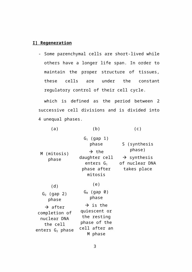
I] Regeneration
- Some parenchymal cells are short-lived while others have a
longer life span. In order to maintain the proper structure of
tissues, these cells are under the constant regulatory control of
their cell cycle.
which is defined as the period between 2 successive cell
divisions and is divided into 4 unequal phases.
(a) (b) (c)
M (mitosis) phase
G1 (gap 1) phase
the daughter cell enters G1 phase after
mitosis
S (synthesis phase)
synthesis of nuclear DNA takes
place
(d)
G2 (gap 2) phase
after completion of nuclear DNA the cell enters G2 phase
(e)
G0 (gap 0) phase
is the quiescent or the resting phase of the cell after an M
phase
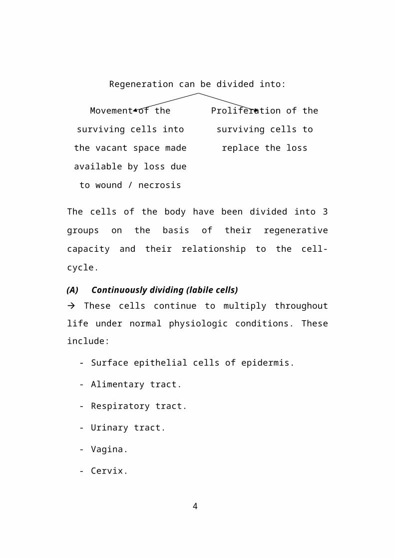
Regeneration can be divided into:
Movement of the surviving cells
into the vacant space made
available by loss due to wound /
necrosis
Proliferation of the surviving cells
to replace the loss
2
The cells of the body have been divided into 3 groups on the basis of
their regenerative capacity and their relationship to the cell-cycle.
(A) Continuously dividing (labile cells)
These cells continue to multiply throughout life under normal
physiologic conditions. These include:
- Surface epithelial cells of epidermis.
- Alimentary tract.
- Respiratory tract.
- Urinary tract.
- Vagina.
- Cervix.
- Uterine endometrium.
- Haemopoietic cells of bone marrow.
- Cells of lymph nodes and spleen.
(B)Quiescent (stable cells)
- These cells decrease or lose their ability to proliferate after
adolescence but retain the capacity to multiply in response to
stimuli throughout life.
The growth capacity of stable cells is best exemplified by the
ability of the liver to regenerate after hepatectomy or following toxic /
viral / chemical injury. These include:
3
- Parenchymal cells of organic like liver, pancreas, kidneys,
adrenal and thyroid.
- Mesenchymal cells like smooth muscle cells
- Fibroblasts.
- Vascular endothelium
- Bone
- Cartilage cells.
(C)Non-dividing (Permanent cells)
- Exited the cell cycle at some point in intra-uterine development
and cannot undergo further mitotic division in post-natal life.
These include:
o Neurons of nervous system.
o Skeletal muscle.
o Cardiac muscle cells.
i.e. why destruction of a neuron whether in central nervous
system / ganglia, represents a permanent loss. The perfection of
parenchymal repair of an injury depends on more than the ability of the
cells to regenerate. Preservation of the stromal architecture of the
injured tissue is also necessary. Thus, the perfection of repair depends
to a considerable extent on the survival of the basic framework of the
4
tissue when this is lost, regeneration may restore mass but not
complete function.
Relationship of parenchymal cells to the cell cycle
a. Labile cells which are continuously dividing cells remain
in the cell cycle from one mitosis to the next.
b. Stable cells are in the resting phase but can be stimulated
to enter the cell cycle.
c. Permanent cells leave the cell cycle and die after injury.
II] Repair
- Repair is the replacement of injured tissue by fibrous tissue.
Two processes are involved.
A. Granulation tissue formation.
B. Contraction of wounds.
A. Granulation tissue formation
Refers / derived from the pink, soft, granular gross
appearance such as that seen beneath the scab of a skin wound.
5
Histologically, each granule corresponds to the proliferation of new
small blood vessels which are slightly lifted on the surface by a thin
covering of fibroblasts and young collagen. There are 3 components
to granulation tissue formation.
i) PHASE OF ACUTE INFLAMMATION
- Following trauma / injury, blood clots at the site of injury.
There is acute inflammatory response with exudation of plasma,
neutrophils and some monocytes within 24 hours.
ii) PHASE OF CLEARANCE
- Combination of proteolytic enzymes liberated from neutrophils,
autolytic enzymes from dead tissue cells, and phagocytic activity
of macrophages clear off the necrotic tissue, debris and red
blood cells.
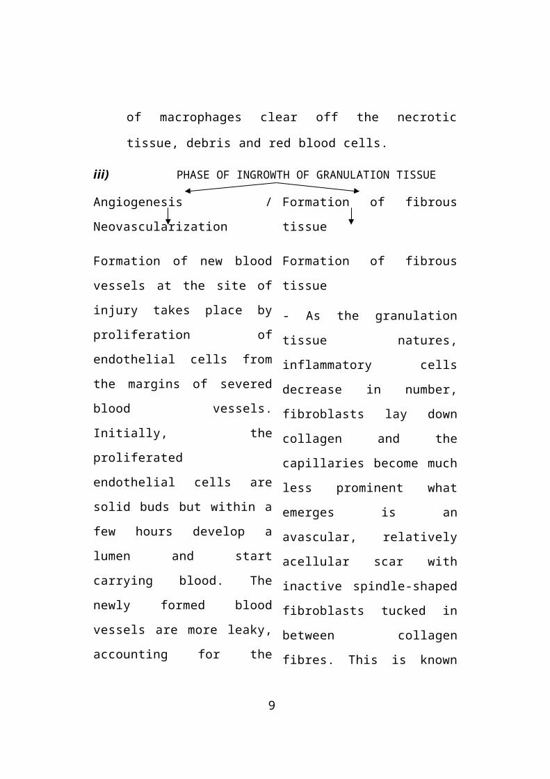
iii) PHASE OF INGROWTH OF GRANULATION TISSUE
Angiogenesis / Neovascularization Formation of fibrous tissue
Formation of new blood vessels at
the site of injury takes place by
proliferation of endothelial cells
from the margins of severed blood
vessels. Initially, the proliferated
endothelial cells are solid buds but
within a few hours develop a
Formation of fibrous tissue
- As the granulation tissue
natures, inflammatory cells
decrease in number, fibroblasts
lay down collagen and the
capillaries become much less
6
lumen and start carrying blood.
The newly formed blood vessels
are more leaky, accounting for the
oedematous appearance of new
granulation tissue.
Summarizing:
- Proteolytic – degeneration
of the parent vessel basement
membrane allowing formation
of a capillary sprout.
- Migration of the endothelial
cells towards the angiogenic
stimulus.
- Proliferation of the
endothelial cells.
- Maturation of the
endothelial cells with
organization.
prominent what emerges is an
avascular, relatively acellular scar
with inactive spindle-shaped
fibroblasts tucked in between
collagen fibres. This is known as
Cicartrisation.
B. Contraction of wounds
The wound starts contracting after 2-3 days and the process is
completed by the 14 th day. During this time, the wound is reduced
to approximately 80% of its original size. Contracted wound results
in rapid healing since lesser surface area of the injured tissue has to
be replaced.
7
In order to explain the mechanism of wound healing, a number
of factors have been proposed. These are:
a) Dehydration as a result of fluid removal by the
drying of the wound but this was not substantiated.
b) Contraction of collagen Was thought to be
responsible for contraction but wound contraction proceeds at
a stage when the collagen content of the granulation tissue is
very small.
c) Discovery of myofibroblasts appearing in active
granulation tissue has resolved the controversy surrounding
the mechanism of wound contraction. These cells have
features intermediate between those of fibroblasts and
smooth muscle cells. Their migration into the wound area and
their active contraction decreases the size of the defect.
With all this background on granulation tissue, we can now
discuss the two forms of repair.
Healing by first intention Healing by second intention
(primary union) (secondary union)
8
The sequence of events are:
1. Initial haemorrhage
Within the first post-operative day, after the wound has been
approximated by surgical sutures, the line of incision promptly fills
with blood clots which seals the wound against dehydration and
infection.
2. Acute inflammatory response
This occurs within 24 hours by appearance of polymorphs from
the margins of the incision. By 3 rd day, polymorphs are replaced by
macrophages.
3. Epithelial changes
The basal cells of epidermis from both the cut margins start
proliferating and migrating towards incisional space in the form of
epithelial spurs. A well approximated wound is covered by a layer of
epithelium in 48 hours. The migrated epidermal cells separate the
underlying viable dermis from the overlying necrotic material and
blood clot, forming a scab which is cast off.
By 5th day a multilayered new epidermis is formed which is
differentiated into superficial and deeper layer.
9
4. Organisation
By 3rd day, fibroblasts also invade the wound area. By 5 th day,
the acute inflammatory response begins to subside and the neutrophils
are replaced by macrophages which debride the wound margins of
destroyed cells and bits and pieces of fibrin.
In 4 weeks, the scar tissue with scanty cellular and vascular
elements, new inflammatory cells and epithelialized surface is formed.
I] Primary union
This is defined as healing of a wound which has the following
characteristics.
- Clean and uninfected.
- Surgically incised.
- Without much loss of cells and tissue, and
- Edges of the wound are approximated by surgical sutures.
10
The incised wound as well as sutures track
on either side are filled with blood clot and
there is inflammatory response from the
margins.
Spurs of epidermal cells migrate along the
incised margin on either side as well as
around the suture track. Formation of
granulation tissue also begins from below.
Removal of suture at around 7 th day results
in scar tissue at the site of incision and
suture track.
Suture tacks
Each suture track is a separate wound and incites the same
phenomenon as in healing of the primary wound.
When sutures are removed around 7 th day, much of the
epithelialized suture tack is avulsed and the remaining epithelial tissue
in the track is absorbed. However, sometimes the suture track gets
infected (Stitch absecess), or the epithelial cells may persist in the
track (implantation / epidermal cysts).
11
II] Secondary union
This is defined as the healing of the wound having, the following
characteristics:
- Open with a large tissue defect; at times infected.
- Having extensive loss of cells and tissue and
- The wound is not approximated by surgical sutures but is left
open.
- The basic events in secondary union are similar to primary union
but differ in having a larger defect which has to be bridged.
Hence, healing takes place from the base upwards, as well as
from the margins inwards. The healing by second intention is
slow and results in a large at times, ugly, scar as compared to
rapid healing and neat scar of primary union.
The open wound is filled with blood clot
and there is inflammatory response at the
junction of viable tissue.
Epithelial spurs from the margins of wound
meet in the middle to cover the gap and
separate the underlying viable tissue from
necrotic tissue at the surface forming.
12
After contraction of the wound, a scar
smaller than the original wound is left.
The sequence of events are:
1. Initial haemorrhage
As a result of injury, the wound space is filled with blood clot
and fibrin clot which dries.
2. Inflammatory phase
There is an initial acute inflammatory phase followed by
appearance of macrophages which clear off the debris as in primary
union.
3. Epithelial changes
As in primary healing, the epidermal cells from both the margins
of wound proliferate and migrate into the wound in the form of
epithelial spurs till they meet in the middle and re-epithelialise the gap
completely. However, the proliferating epithelial cells do not cover the
surface fully till granulation tissue from base has started filling the
wound space. In this way, preexisting viable connective tissue is
separated from necrotic material and clot on the surface, forming seal
which is cast off.
13
4. Granulation tissue
The main bulk of secondary healing is by granulations. The
newly-formed granulation tissue is deep red, granular and very fragile.
With time, this scar on maturation becomes pale and white due to
increase in collagen and decrease in vascularity.
5. Wound contraction
Due to the action of myofibroblasts present in granulation tissue,
the wound contracts to 1/3rd – ¼th of its original size.
6. Presence of infection
Bacterial contamination of an open wound delays the process of
healing due to release of bacterial toxins that provoke necrosis,
suppuration and thrombosis. Surgical removal of dead and necrosed
tissue helps in preventing the bacterial infection of open wounds.
Complications of wound healing:
- Infection (due to entry of bacteria).
- Implantation cyst.
- Pigmentation (healed wounds may at times have rust like colour
due to staining with haemoglobin).
- Deficient scar formation (due to inadequate G.T. formation).
14
- Incisional hermia (a weak scar may be site of bursting open of a
wound) (Wound dehiscence).
Two main aberrations occur in wound healing whether the
process of 1st/2nd intention.
a) The accumulation of excessive amounts of collagen may give
rise to a protruding, tumorous scar known as a keloid thick
interlacing bundles of collagen causing marked swelling at the
site of the wound.
b) The other deviation is the formation of excessive amounts of
granulation tissue which protrudes above the level of the
surrounding skin and in fact blocks re-epithelization. This has
been called as ‘exuberant granulation / pround flesh’.
PATHOLOGIC ASPECTS OF REPAIR / Factors Influencing Repair
In wound healing, normal cell growth and fibrosis may be
altered by a variety of influences, frequently reducing the quality and
adequacy of the reparative process.
These factors may be either extrinsic / intrinsic.
15
Systemic factors: Systemic / Local
1. Hypoxia
Healing is often imperfect in regions with a poor blood
supply e.g., various ulcers in a lower limb made hypoxic by
varicose veins, often fails to heal.
In contrast to this, mild hypoxia helps healing by increasing
the differentiation of fibrocytes, collagen formation and the
cross-linkage of collagen-fibrils.
2. Scurvy
Is a classic cause of defective wound healing though now
rare. Scar that results is weak and in the granulation tissue, new
capillaries are few and ill-formed.
3. Severe and prolonged protein deficiency
Inhibits wound healing. Deficiency of sulfur containing
amino acids like cystine and methionine is particularly
important.
4. Glucocorticoids
Of the adrenal gland interfere with wound healing when
given in large doses.
16
5. Age
Wound healing is rapid in young and slow in aged and
debilitated patients due to poor blood supply to the injured area.
6. Uncontrolled diabetic – are more prone to develop infections
and hence delay in healing.
7. Local factors
a) Movement of the injured part.
b) Fixation of the injured tissue to an unyielding object such
as bone, makes apposition of the wound edges difficult
and hampers healing.
c) Wounds made along a line of mechanical stress in the
tissue tend to fall together and heal easily but wound
across a line of stress tend to gape and heal more slowly.
d) Infection of the wound.
e) Any foreign bodies present.
f) Type, size and location of injury determines whether
healing takes place by resolution / organization.
g) Exposure to u-v light facilitates healing.
17
h) Exposure to ionizing radiation delays granulation tissue
formation.
MECHANISMS INVOLVED IN REPAIR3 processes
Cell-cell interactions.
Cell-matrix interactions.
Stimulatory hormones / growth factors.
1. Cell-cell interactions
As mentioned earlier, re-epithelialization of wound begins
within 24 hours of injury and the gap is usually covered within 48
hours. During regeneration of liver, after partial hepatectomy, the liver
cells burst into mitoses but cease to divide when normal liver substance
is restored. What signals these cells to stop dividing? When certain
normal cells are lightly seeded in Petri dishes to investigate the growth
behaviour of cells, they proliferate migrate and eventually form
confluent monolayers. At this point cells cease to divide – a process
called contact inhibition. It is proposed that cells are inhibited from
proliferation by interchange of signals or substances at contact point.
2. Cell-matrix interactions
Much evidence has accumulated to indicate that the orderly
movement and proliferation of the cells within a healing wound is
18
influenced not only by signals derived from other cells but also from
the extra-cellular matrix, which is an organized complex of collagen,
glycosaminoglycans, proteoglycans and glycoproteins. Of these much
attention is focused on fibronectins which are a family of adhesive
high molecular weight glycoproteins. Experimental evidence suggests
that migration of endothelial cells and their organization into
capillaries is also facilitated by fibronectin.
3. Growth Factors
Thus far, we have discussed the regeneration migration and
organization of cells in the wound. But what triggers these cells to
divide.
Either loss of factors that or release of growth
normally inhibit cell division stimulating factors.
At one time, the concept that proliferation occurred because of
reduced levels of growth inhibition substance called Chalones, was
popular.
But nowadays, the list of growth factors is ever increasing. E.g.,
- Epidermal G.F. (E.G.F.).
- Nerve G.F. (N.G.F.).
- Platelet derived G.F. (P.D.G.F.).
- Fibroblast G.F. (F.G.F.).
All these are polypeptides with hormone like structure.
19
HEALING IN SPECIALIZED TISSUES
a) Internal surfaces – Endothelium
The regeneration of the covering epithelium is very similar to
that of the skin.
b) Bone
Repair of a bone injury is essentially another instance of
connective tissue healing. It differs from soft tissue repair in so far
as formation of the specialized calcified tissues of bone involves the
activity of osteoblasts and osteoclasts.
Repair of a fracture may be taken as a model for the process of
bone healing. However, basic events in healing of any type # are
similar and resemble healing of skin wound to some extent.
Primary Union of fractures occurs occasionally in a few special
situations when the ends of fractures are approximated because bony
union takes place with formation of medullary callus.
Secondary union is the more common process of # healing. It is
described under 3 headings:
- Procallus formation.
- Osseous callus formation.
- Remodelling.
20
1) Procallus formation
a. Haematoma
Forms due to bleeding, filling the area surrounding the #.
b. Local inflammatory response
Occurs at the site of injury with exudation of fibrin, polymorphs
and macrophages. Fragments of necrosed bone are scavenged by
macrophages and osteoclasts.
c. Ingrowth of granulation tissue
Begins with neovascularization. A soft tissue callus is that
formed which joins the ends of # bone without much strength.
d. Callus composed of woven bone and
cartilage
The cells of the inner layer of periosteum have osteogenic
potential and lay down the collagen as well as the osteoid matrix in the
granulation tissue. The osteoid undergoes calcification and is called
woven bone callus. At times, callus is composed of woven bone as well
as cartilage, temporarily immobilizing the bone ends.
This stage is called provisional callus / procallus formation and
is arbitrarily divided into:
- External
21
- Intermediate.
- Internal procallus.
2) Osseous Callus
The procallus acts as scaffolding on which osseous callus
composed of lamellar bone is formed.
3) Remodelling
During the formation of lamellar bone, osteoblastic laying and
osteoclastic removal are taking place, remodeling the united bone ends,
which after sometime, are indistinguishable from normal bone. The
external callus is cleared away, compact bone (cortex) is formed in
place of intermediate callus and the bone marrow cavity develops in
internal callus.
Haematoma formation and local inflammatory
response at the # site.
Ingrowth of granulation tissue with formation of
soft tissue callus.
Formation of procallus composed of woven bone
and cartilage with its characteristic furiform
appearance and having 3 arbitary components
- External.
22
- Intermediate.
- Internal callus
Formation of osseous callus composed of lamellar
bone following clearance of woven bone and
cartilage.
Remodelled bone end; the external callus cleared
away intermediate callus converted into lamellar
bone and internal callus developing bone marrow
cavity.
4) Nervous tissue
Central Nervous System Peripheral Nervous System
Regeneration does not occur. In cases of acute damage, the
initial functional loss often exceeds the loss of actual nerve tissue
because of the reactive changes in the surrounding tissue. As these
changes diminish, functional restoration commences.
Peripheral Nervous System (PNS)
3 types of degenerative processes in the PNS are:
a) Wallerian degeneration
Occurs after transection of the axon which may be as a result
of knife wounds, compression, ischaemia. Then, there is initially
23

accumulation of organelles in the proximal and distal end of
transection sites. Subsequently, the axon and myelin sheath
distal to the transection site undergoes disintegration upto the
next node of Ranvier followed by phagocytosis. The process of
regeneration occurs by sprouting of axons and proliferation of
Schwann cells from the distal end.
b) Axonal degeneration
Degeneration of the axon begins at the peripheral terminal
and proceeds backwards towards the nerve cell body. Changes
similar to those seen in Wallerian degeneration are present but
regenerative reaction is limited / absent.
24
c) Segmental demyelination
Is demyelination of the segment between two consecutive
nodes of Ranvier, leaving a denuded axon segment. The axon,
however remains intact. Schwann cells proliferation also occurs.
This results in re-myelination of the affected axon. Repeated
episodes of demyelination and remyelination are associated with
concentric proliferation of Schwann cells around axons
producing ‘onion bulbs’ found in hypertrophic neuropathy.
Traumatic Neuroma
Normally, the injured axon of a peripheral nerve regenerates
at the rate of approximately 1mm/day. However, if the process
of regeneration is hampered due to an interposed haematoma or
fibrous scar, the axonal sprouts together with Schwann cells and
fibroblasts form a peripheral mass called Traumatic / Stump
Neuroma.
25
d) Muscles
Muscle fibres of all 3 types – Skeletal, Caridac and Visceral
have only limited capacity to regenerate.
- When a mass of muscle tissue is damaged, repair by scarring
occurs.
- If the damage affects individual muscle fibres diffusely and with
varying severity, then regeneration of the specialized fibres is
possible.
Books referred
- Harsh Mohan
- Stanley (Basic Pathology)
- Boyd’s Pathology
- Govan, MacFarlane and Callendar
- Cottran, Robin and Williams
26
REPAIR (IN GENERAL)
CONTENTS
Introduction
Process Involved in Repair Regeneration
Replacement
Wound Healing
Pathologic Aspects of Repair
Mechanisms Involved in Repair
Healing in Specialized tissues
27