Embed Size (px)
Citation preview

Abstracts
Nitric oxide inhibits IKK through S-nitrosation andS-glutathiolation and prevents activation of NF-kB
Nitric oxide (NO) possesses anti-inflammatory effects,which may be exerted via its ability to inhibit the nuclearfactor (NF)-kB via S-nitrosation of cysteine 62 of the p50subunit. In the present study, it was assessed whether NOcould also inhibit NF-kB by preventing the activation of IkBkinase (IKK), the enzyme complex necessary for NF-kBactivation.
Mouse lung epithelial cells (C10 cells) were exposed totumour necrosis factor (TNF) in the presence or absence ofthe S-nitrosothiols S-nitroso-N-acetylpenicillamine (SNAP)or S-nitrosoglutathione (GSNO) for evaluation of IKKenzymatic activity. The addition of GSNO to intact cellsinhibited TNF-induced IKK activity, whereas SNAP inhi-bited IKK only when glutathione (GSH) was depleted. Incontrast, addition of 100 mM–1 mM SNAP or GSNO toisolated active IKK, from cells with a normal GSH content,resulted in a dose-dependent inhibition of enzymatic activity.Assessment of S-nitrosation by biotinylation and Westernblotting revealed that the IKK-b subunit became S-nitrosatedfollowing exposure to SNAP and GSNO. GSH was alsofound to inhibit IKK activity, but was less effective thanGSNO. Immunoprecipitation and Western blotting showedthat GSH and GSNO glutathiolate IKK-b.
When C10 cells were incubated with 1 mM N-monomethyl-L-arginine to inhibit all three NO synthase enzymes IKKactivity was not induced, whereas NF-kB-dependent reporter
gene expression did increase. NFkB DNA binding andreporter gene expression were inhibited following the addi-tion of GSNO, GSH and SNAP, independent of the GSHcontent of the cells. These data demonstrate that oxidativeinactivation of IKK is a mode to downregulate NF-kB inresponse to NO.
N.L. Reynaert*, K. Ckless*, A. van der Vliet*, S. Korn*,N. Vos*, E.F.M. Wouters#, Y. Janssen-Heininger**Dept of Pathology, University of Vermont, Burlington,VT, USA. #Dept of Pulmonology, Maastricht University,Maastricht, The Netherlands.
Modulation of human lung dendritic cell recruitment:role of alveolar epithelium
Chronic obstructive pulmonary disease and asthma bothdisplay a chronic inflammatory component that is thought tobe partly T-cell driven. Studies have shown that dendriticcells (DCs) are elevated in both disorders. It is thought thatthe inflammatory response of T-cells is initiated by antigen-presenting DCs residing within the mucosa and alveolar septae.
The CC chemokine macrophage inflammatory protein(MIP)-3a is a specific chemoattractant for DCs, in particularthe Langerhans9 cell subset that colonise the subepithelia.The authors have shown that primary human alveolar type(HAT)-II cells produce MIP-3a constitutively and that
Repair mechanisms in embryos: wound healing and learning fromhow embryos repair perfectly
P. Martin
Embryos heal wounds very rapidly and efficiently withoutleaving a scar. Studying how they do this can tell a great dealabout the natural morphogenetic movements of embryo-genesis as well as suggesting ways in which adult tissues couldrepair more efficiently. Using live confocal imaging oftransgenic Drosophila embryos expressing green fluorescentprotein-labelled actin in epithelial tissues, the key actinmachineries that drive the paradigm morphogenetic processof dorsal closure, which appear to bear striking analogy withre-epithelialisation of a vertebrate skin wound have beenrevealed. Using embryos expressing mutant forms of thevarious small guanadine triphosphatases, the function of eachof these actin-based elements, the actin cable and dynamicfilopodia and lamellipodia, were tested in both dorsal closureand the repair of laser-generated wound holes in the flyembryo. These experiments conducted in embryonic chicksand mice and in the neonatal PU.1 null mouse, which isgenetically macrophageless, suggest that an inflammatory
response is not essential for healing and may indeed be causalof fibrosis in postembryonic animals. Consequently, amicroarray approach was used with this mouse in order toidentify a portfolio of candidate inflammation/fibrosis genes.Finally, by taking advantage of the translucency of thezebrafish larval tail, the author9s group has begun to makedifferential interference contrast movies of the inflammatoryresponse and to dissect the genetics of this process byscreening for mutants that fail to recruit leukocytes to thewound site and by morpholino knockdown of candidate"inflammation" genes. The hope is that these basic cell andmolecular studies in genetically tractable organisms willsupply the clues needed to design the new repair andregeneration medicines of the future.
Correspondence: P. Martin, Dept of Anatomy and DevelopmentBiology, University College London, Gower Street, London WC1E6BT, UK. Fax: 44 2076797349. E-mail: [email protected]
Eur Respir J 2003; 22: Suppl. 44, 39sDOI: 10.1183/09031936.03.00000003mPrinted in UK – all rights reserved
Copyright #ERS Journals Ltd 2003European Respiratory Journal
ISSN 0904-1850

release is increased in response to lipopolysaccharide (LPS) ina time- and dose-dependent manner (fig. 1). Using immuno-cytochemistry it was also shown that the majority ofmonocytes in the lung interstitium are CD1az DCs. Mono-cytes found in washings from the airspace are predominantlyCD68z i.e. macrophages; very few are CD1az.
The effect of MIP-3a on DC migration was investigated byculturing immature DCs derived from peripheral bloodmonocytes. DCs were added to the upper part of an invasionchamber and varying concentrations of recombinant MIP-3ato the lower. Migration increased in a dose-dependent mannerwith a three-fold increase at 6,000 pg?mL-1 (pv0.0001). Inaddition the chemotactic capacity of conditioned media fromLPS-stimulated HAT II cells (n=3) was assessed. Conditionedmedia was collected 24 h post-LPS and therefore LPS-free. DCmigration towards the conditioned media correlated directlywith the amount of MIP-3a (r2
=0.94) but not with other chemo-kines detected in the media (interleukin-8, monocyte chemo-tactic protein-1 and growth-related protein-a). Furthermore,
using selective chemokine antibodies, blockade of MIP-3a wasshown to cause the greatest inhibition in DC migration (fig. 2).
To conclude, HAT II cells play an important role inregulation of DC recruitment in the lung, most likely via MIP-3a production.
A.J. Thorley*, P. Goldstraw*, A. Young#, T.D. Tetley**National Heart and Lung Institute, Imperial College,London, and #AstraZeneca, Loughborough, UK.
Effects of anti-IL-5 on bone marrow CD34z eosinophilsafter allergen exposure: influence on airway eosinophilia
Airway allergen exposure induces enhanced eosinophil(eos) production, and increases the number of circulatingCD34z cells. Interleukin (IL)-5 is an especially importantmediator in orchestrating this eosinophilic inflammatoryresponse. The aim was to elucidate the onset of effect of amonoclonal antibody to IL-5 (TRFK5) in reducing eosino-philia induced by airway allergen exposure.
Ovalbumin (OVA)-sensitised Balb/c mice were exposed toOVA (100 mg) intranasally on 10 days with 2 days of restbetween exposure days 5 and 6. TRFK5 (50 mg?animal-1) orits isotype control was given once intraperitoneally beforethe last 5 days of allergen exposure. Bronchoalveolar lavage(BAL), peripheral blood (PB) and bone marrow (BM) cellswere collected at different days after the treatment. Newlyproduced eos were pulse-labelled with bromodeoxyuridine(BrdU). BrdU-labelled eos and CD34z eos numbers wereexamined by immunocytochemistry.
BM eos were significantly reduced on the third day afterTRFK5 administration versus vehicle-treated mice, due to thedecrease of immature eos (3.05¡0.50 versus 6.08¡0.82% oftotal cells, pv0.05). This effect was further enhanced on the fifthday, as well as on BM BrdU-labelled eos, and CD34z eos(1.10¡0.22 versus 4.13¡0.91% of total cells, pv0.05). Similareffects were observed on PB eos counts. However, a significanteffect on BAL fluid eos was found only on the fifth day afterTRFK5 administration (3.14¡0.99 versus 62.20¡13.786104?mL-1, pv0.05). At this time point, there was also a significantinhibitory effect on BrdU-labelled eos (0.58¡0.39 versus15.54¡6.556104?mL-1) as well as on CD34zeos in BAL.
A single dose of anti-IL-5 extensively reduces BM CD34zeos, immature BM eos and PB eos, as well as BAL BrdU-positive eos and BAL CD34z eos numbers. Together thesedata argue that anti-IL-5, in vivo, is important to an extent toregulate eosinophilia by action within the BM, probably byinhibition of the early maturation of eos from CD34zprogenitor cells.
B. Sitkauskiene*, M. Sjostrand#, A-K. Johansson#, J. Lotvall#
*Clinic of Pulmonology and Immunology, Kaunas Universityof Medicine, Lithuania. #Lung Pharmacology Group,Dept of Respiratory Medicine and Allergology, GothenburgUniversity, Sweden.
Attenuation of IgE-receptor signalling in mast cells as amolecular basis for the antiallergic action of glucocorticoids
Glucocorticoids exhibit anti-inflammatory, immune sup-pressive and antiallergic activities. They inhibit the release ofallergic mediators and the expression of proinflammatorycytokines, processes induced by activation of the receptorwith high affinity for immunoglobulin E (FceRI) upon
����
����
����
���
�
������� ����
� �� ��� ���
Fig. 1. – Time- and dose-dependent release of macrophage inflamma-tory protein (MIP)-3a from primary human alveolar type-II cells inresponse to lipopolysaccharide (LPS) stimulation (n=6). &: control;q: 1 ng?mL-1 LPS; : 10 ng?mL-1 LPS; u: 100 ng?mL-1 LPS; h:1000 ng?mL-1 LPS.
��
�
��
��
�
����� �������
�� �� ���� ��� ��� ����� �� ��!�
"
Fig. 2. – Inhibition of dendritic cell migration by antibodies (Ab) tochemokines present in conditioned media from lipopolysaccharide-stimulated human alveolar type-II cells (n=3). UC: unstimulated con-trol; SC: stimulated control; IL: interleukin; GRO: growth-related protein;MCP: monocyte chemotactic protein; MIP: macrophage inflamma-tory protein; All–M: all antibodies except MIP-a.
40s TAORMINA MEETING
antigen trigger in mast cells. The mode(s) of action ofglucocorticoids in inhibiting FceRI signalling were analysed.Glucocorticoids suppress the expression of FceRI a-chaingene at the promoter level. The downregulation of the FceRIa-chain gene expression requires new protein synthesis andregulatory elements at the FceRI a-chain promoter andcorrelates with a reduced surface expression of the FceRI.This downregulation would possibly suppress signal trans-duction originating from the FceRI and ending up with theactivation of the downstream targets, extracellular signal-related kinase (ERK)1/2.
In addition, glucocorticoids enhance the expression of themitogen-activated protein kinase phosphatase MKP-1, whichinhibits the activation of ERK1/2, at the promoter level. Thisregulation requires the glucocorticoid receptor dimerisationfunction and the presence of discrete elements on the promoterproximal sequence. The role of MKP-1 in glucocorticoid-mediated repression of ERK1/2 phosphorylation was confirmedin primary bone marrow-derived mast cells from MKP-1-deficient mice, where ERK1/2 are no longer inhibited byglucocorticoids, while this is the case in cells from wildtypemice. Instead, ERK1/2 activity in other cell types, such asthymocytes and splenocytes of MKP-1-deficient mice, couldstill be inhibited by glucocorticoids, demonstrating thatrepression of ERK1/2 through MKP-1 is a cell-type specificprocess. Glucocorticoid-mediated inhibition of expression ofFceRIa gene and increased expression of MKP-1 may possiblyfunction together in the attenuation of FceRI signalling byglucocorticoids.
A. Sancono*, O. Kassel*, J. Maier*, C. Hesslinger#,A.C.B. Cato**Forschungszentrum Karlsruhe, Institute of Toxicology andGenetics, and #Pharmazentrum Frankfurt, Institute forGeneral Pharmacology, Klinikum of the J. W. GoetheUniversity, Frankfurt am Main, Germany.
The presence of quorum-sensing signal molecules inclinically stable lung
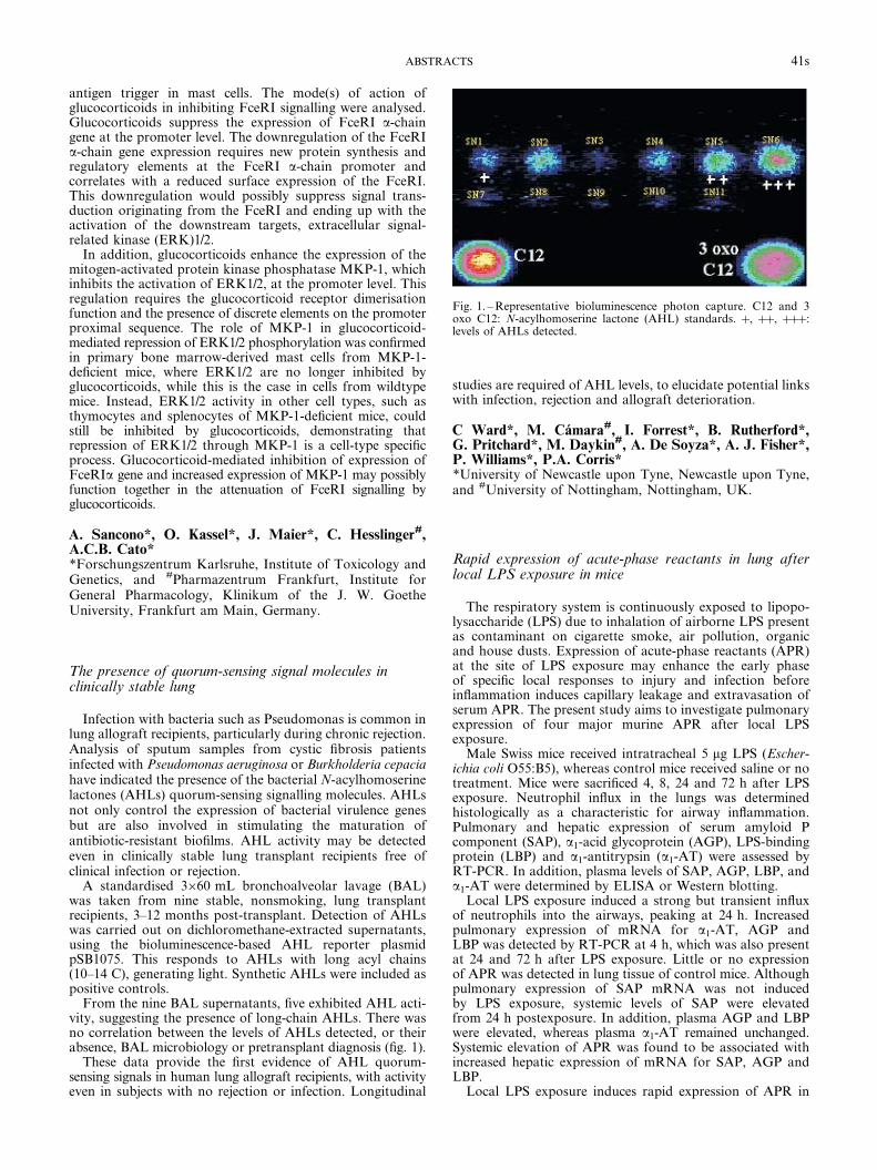
Infection with bacteria such as Pseudomonas is common inlung allograft recipients, particularly during chronic rejection.Analysis of sputum samples from cystic fibrosis patientsinfected with Pseudomonas aeruginosa or Burkholderia cepaciahave indicated the presence of the bacterial N-acylhomoserinelactones (AHLs) quorum-sensing signalling molecules. AHLsnot only control the expression of bacterial virulence genesbut are also involved in stimulating the maturation ofantibiotic-resistant biofilms. AHL activity may be detectedeven in clinically stable lung transplant recipients free ofclinical infection or rejection.
A standardised 3660 mL bronchoalveolar lavage (BAL)was taken from nine stable, nonsmoking, lung transplantrecipients, 3–12 months post-transplant. Detection of AHLswas carried out on dichloromethane-extracted supernatants,using the bioluminescence-based AHL reporter plasmidpSB1075. This responds to AHLs with long acyl chains(10–14 C), generating light. Synthetic AHLs were included aspositive controls.
From the nine BAL supernatants, five exhibited AHL acti-vity, suggesting the presence of long-chain AHLs. There wasno correlation between the levels of AHLs detected, or theirabsence, BAL microbiology or pretransplant diagnosis (fig. 1).
These data provide the first evidence of AHL quorum-sensing signals in human lung allograft recipients, with activityeven in subjects with no rejection or infection. Longitudinal
studies are required of AHL levels, to elucidate potential linkswith infection, rejection and allograft deterioration.
C Ward*, M. Camara#, I. Forrest*, B. Rutherford*,G. Pritchard*, M. Daykin#, A. De Soyza*, A. J. Fisher*,P. Williams*, P.A. Corris**University of Newcastle upon Tyne, Newcastle upon Tyne,and #University of Nottingham, Nottingham, UK.
Rapid expression of acute-phase reactants in lung afterlocal LPS exposure in mice
The respiratory system is continuously exposed to lipopo-lysaccharide (LPS) due to inhalation of airborne LPS presentas contaminant on cigarette smoke, air pollution, organicand house dusts. Expression of acute-phase reactants (APR)at the site of LPS exposure may enhance the early phaseof specific local responses to injury and infection beforeinflammation induces capillary leakage and extravasation ofserum APR. The present study aims to investigate pulmonaryexpression of four major murine APR after local LPSexposure.
Male Swiss mice received intratracheal 5 mg LPS (Escher-ichia coli O55:B5), whereas control mice received saline or notreatment. Mice were sacrificed 4, 8, 24 and 72 h after LPSexposure. Neutrophil influx in the lungs was determinedhistologically as a characteristic for airway inflammation.Pulmonary and hepatic expression of serum amyloid Pcomponent (SAP), a1-acid glycoprotein (AGP), LPS-bindingprotein (LBP) and a1-antitrypsin (a1-AT) were assessed byRT-PCR. In addition, plasma levels of SAP, AGP, LBP, anda1-AT were determined by ELISA or Western blotting.
Local LPS exposure induced a strong but transient influxof neutrophils into the airways, peaking at 24 h. Increasedpulmonary expression of mRNA for a1-AT, AGP andLBP was detected by RT-PCR at 4 h, which was also presentat 24 and 72 h after LPS exposure. Little or no expressionof APR was detected in lung tissue of control mice. Althoughpulmonary expression of SAP mRNA was not inducedby LPS exposure, systemic levels of SAP were elevatedfrom 24 h postexposure. In addition, plasma AGP and LBPwere elevated, whereas plasma a1-AT remained unchanged.Systemic elevation of APR was found to be associated withincreased hepatic expression of mRNA for SAP, AGP andLBP.
Local LPS exposure induces rapid expression of APR in
Fig. 1. – Representative bioluminescence photon capture. C12 and 3oxo C12: N-acylhomoserine lactone (AHL) standards. z, zz, zzz:levels of AHLs detected.
41sABSTRACTS

the lung, which clearly precedes systemic elevation of APRassociated with hepatic APR expression.
J.H.J. Vernooy, N. Reynaert, T.G. Wolfs, B. de Vries,M.A. Dentener, W.A. Buurman, E.F.M. WoutersNutrition and Toxicology Research Institute Maastricht(NUTRIM), Depts of Pulmonology and General Surgery,University Hospital Maastricht, Maastricht, The Netherlands.
De novo synthesis of IL-17 in human airways followingexposure to organic dust
The cytokine interleukin (IL)-17 is produced by activatedT-lymphocytes and may contribute to recruitment and acti-vation of neutrophils in the airways. The aim of this studywas to determine whether IL-17 is synthesised de novo duringairway inflammation induced by organic dust.
Four healthy nonsmoking volunteers were exposed toorganic dust for 3 h while working in a swine confinement.Bronchoalveolar lavage (BAL) fluid was gathered 2 weeksbefore and 24 h after the exposure and IL-17 mRNA wasmeasured using RT-PCR-ELISA. Total and cell differentialcounts were also performed in BAL fluid.
The exposure to organic dust caused an 11.1-fold increaseof IL-17 mRNA levels (% HPRT; median (range)) from 7.45(0–21.9) to 82.7 (0.35–118.2), (n=4; pv0.05). In BAL fluid,there was a corresponding increase (before versus aftermedian million cells?L-1) in lymphocytes (3.7 versus 13.15)and neutrophils (2.25 versus 15.87).
In conclusion, IL-17 can be synthesised de novo duringairway inflammation induced by organic dust. The synthesisof IL-17 is associated with the recruitment of lymphocytesand neutrophils. Further investigations are needed to deter-mine whether IL-17 constitutes a potential target for pharma-cotherapy of airway disease characterised by an exaggeratedmobilisation of neutrophils.
S. Ivanov*, O. Prause*, L. Palmberg#, K. Larsson#,A. Linden**Lung Pharmacology Group, Dept of Respiratory Medicineand Allergology, Gothenburg University, Gothenburg, and#Programme for Respiratory Health and Climate, NationalInstitute of Environmental Medicine, Karolinska Institute,Stockholm, Sweden.
Lack of the transcription factor CEBP-a and differentintracellular signalling in asthmatic bronchial smoothmuscle cells
The pathogenesis of asthma is characterised by increasedproliferation of bronchial smooth muscle cells (BSMCs) andenhanced production of proinflammatory cytokines. How-ever, the molecular basis of this disease remains unclear. Theauthors have previously shown that b-receptor agonists arecapable of activating the glucocorticoid receptor (GR) inBSMCs and in peripheral blood leukocytes. The transcriptionfactor CEBP-a is centrally involved in the steroid activationpathway forming a complex when activated.
The expression and activation of the GR and the trans-cription factor CEBP-a, and the secretion of interleukin (IL)-6in BSMCs cultured from asthmatics and nonasthmatic con-trols were studied. GR and CEBP-a were assessed by electro-phoretic mobility shift assay (EMSA) and Western blot. IL-6
was determined by ELISA. In asthmatic BSMCs, the GRwas activated by steroids. The glucocorticoid-dependentinhibition of cell proliferation was not significant, while theanti-inflammatory effect shown as downregulation of IL-6secretion was functioning. In contrast, the antiproliferativeeffect of b2-agonists was observed in asthmatic and nonasth-matic BSMCs, as well as an induction of IL-6 secretion. UsingWestern blot and EMSA, it was shown that asthmaticBSMCs lack CEBP-a. When pretreated with CEBP-a anti-sense oligonucleotides or with the steroid antagonist RU486,the antiproliferative action of steroids and the downregula-tion of IL-6 was counterbalanced.
In summary, there is evidence that the lack of the anti-proliferative action of steroids in asthmatic BSMCs is basedon a lack of the transcription factor CEBP-a; this lack canbe bypassed by b2-agonists. The antiproliferative and anti-inflammatory signalling pathway of both drug types involvesthe GR, but splits up subsequently.
M. Roth*, P.R.A. Johnson*, P. Borger*, G. G. King#, Q. Ge#,J. Burgess#, J. Black#, M. Tamm**Pulmonary Cell Research University Hospital Basel, Swit-zerland. #Dept of Pharmacology and Woolcock Institute,University of Sydney, Australia.
Involvement of IKK signalsome complex in b-cateninsignalling pathway regulation in human bronchial epithelialcells
In asthma, the epithelium is frequently damaged. b-catenin(b-cat) is an intracellular protein essential for E-cadherin-mediated cell adhesion, but can also act as a transcriptionfactor. The authors have shown that dexamethasone (Dex)induces the formation of b-cat/Tcf-4 complex and itstranslocation in the nucleus where it can activate targetgene transcription. The fate of human bronchial epithelialcells (HBEC) including cohesion and repair may be relatedto these processes. b-cat, as a transcription factor, has beenshown to induce proliferation and inhibit apoptosis of HBEC.In the present study, the molecular process implicated inthe Dex-induced b-cat signalling pathway in HBEC wasinvestigated.
After Dex stimulation of HBEC the b-cat/Tcf-4 complexfunctionality was investigated by transfection and luciferaseassay. The implications of the regulatory molecules GSK-3b,E-cadherin, IKK-a and IKK-b, in the Dex-induced b-catsignalling pathway, were assessed by immunoprecipitationand immunofluorescence.
Luciferase assay showed that b-cat/Tcf-4 complex is func-tional (pv0.05). Immunoprecipitation and immunofluorescenceanalysis showed that the Dex-induced b-cat transcriptionalactivity was mediated by the IKK signalsome and tightlyregulated by GSK-3b and E-cadherin-mediated intercellularadhesion complex.
To conclude, steroids regulated the b-cat transcriptionalactivity outside of the canonical Wnt-regulated pathway, bythe IKK signalsome complex. This mechanism may beessential in promoting the injury-repair cycle in damagedepithelium of asthmatics.
N. Carayol*, R. Gagliardo#, I. Vachier*, A. M Vignola#,P. Godard*, J. Bousquet*, P. Chanez**INSERM U454, CHU Montpellier, France. #IBIM, CNRPalermo, Italy.
42s TAORMINA MEETING

Role of oxidative stress in the regulation of iNOS andarginase in rat alveolar macrophages
L-Arginine is a substrate of nitric oxide synthase (NOS)and arginase, pathways of particular importance in macro-phages. Arginase can limit the L-arginine supply for NOS andmay be involved in the development of airway hyperreactiv-ity. A role for oxidative stress, known to be associated withacute inflammatory reactions, in the regulation of arginaseand inducible (i)NOS in alveolar macrophages (AM) wasstudied.
Rat AM were cultured for 1–20 h in the absence or pre-sence of 1 mg?mL-1 lipopolysaccharide (LPS) and/or apocynin(NADPH oxidase inhibitor). Nitrite accumulation in cul-ture media and arginase activity at the end of the cultureperiods were determined or RNA was isolated for use inRT-PCR.
Arginase activity in AM cultured in the absence of LPS was31¡4 mU?106 cells-1 and presence of LPS caused an increaseof 115¡11%. Apocynin (500 mM) reduced basal arginaseactivity by 54¡9% and largely attenuated the LPS-mediatedincrease. After 5 and 20 h of exposure to LPS, arginase ImRNA was clearly increased and apocynin, which slightlyreduced basal arginase I mRNA, inhibited the LPS-inducedincrease. Apocynin also attenuated the LPS-induced increaseof iNOS mRNA, but had no clear effect on the expression ofarginase II mRNA. Exposure to hydrogen peroxide (H2O2)caused an increase in arginase I mRNA. This effect, startedafter 1 h, was maximal after 2 h, and was lost after 5 h ofexposure to H2O2. With a similar time course, exposure toH2O2 also caused a transient increase in iNOS mRNA (allno4).
To conclude, in rat AM the expression of arginase ImRNA, like that of iNOS mRNA, is highly sensitive tooxidative stress. The inhibitory effect of apocynin on LPS-induced increase in arginase I and iNOS expression sug-gests that oxygen radicals may play a role as intracellularsignals.
K. Racke, D. Lindemann, F. WenzelInstitute of Pharmacology and Toxicology, University ofBonn, Bonn, Germany.
Novel roles for elafin in modulating LPS-mediatedinflammation
Elafin is an elastase inhibitor that plays a role in thelung9s defence against tissue damage mediated by humanneutrophil elastase. The authors have recently demonstratedthat, in addition, elafin may contribute to host defencemechanisms both as a "defensin-like" antimicrobial peptide andas a neutrophil chemoattractant. Here, the lipopolysaccharide(LPS)-binding properties of elafin and the effects of thisinteraction on the inflammatory response of host cells to LPS aredescribed.
Direct binding of elafin to both smooth-form and rough-form LPS serotypes was demonstrated using native acidic(pH 4.5) polyacrylamide gel electrophoresis techniques. Further,an ELISA assay was used to demonstrate that elafin in theconcentration range 10–160 nM can inhibit the interaction ofLPS with the acute-phase serum component LPS-binding protein,an important step in the LPS-mediated activation of macro-phages; maximal inhibition observed was 42% at 160 nM elafin.
To determine the effects of elafin9s LPS-binding propertieson cellular responses to LPS, the murine macrophage cell lineRAW 264.7 was used. Cells were stimulated with 50 ng?mL-1
LPS of Escherichia coli serotype O55:B5 for 4 h in thepresence or absence of 0–100 nM elafin, and secretion oftumour necrosis factor (TNF)-a was measured by ELISA.Cell stimulations were carried out in culture medium eithercontaining or lacking in serum. In medium containing 0.2%serum, elafin was shown to inhibit LPS-mediated TNF-arelease by 35–40% at 10–100 nM. However, in serum-freeconditions, elafin increased LPS-induced TNF-a releasesix-fold at these concentrations indicating an enhancementof activation of cells by LPS.
These findings suggest that the effects of elafin may bedependent upon the site of LPS stimulation in vivo; forexample elafin may act to downregulate LPS activity insystemic serum-containing milieu, but may enhance theinflammatory response to LPS in sites where serum isabsent, such as the airways. This may have physiologicalrelevance in the control of local lung inflammation whiledampening potentially deleterious systemic responses.
J.W. McMichael, J-M. SallenaveRayne Laboratory, Centre for Inflammation Research,University of Edinburgh, UK.
Pulmonary inflammation and promotion of peripheralvascular thrombosis by particulate pollutants
Pollution by particulates has been associated with cardio-pulmonary morbidity and mortality, but biological plaus-ibility for this association is lacking. Ultrafine particles(v100 nm) are believed to play an important role. Their rolewas studied in an in vivo hamster model of peripheral vascularthrombosis induced by free radical-mediated endothelialinjury, using intravenous Rose Bengal and local illumination.Pulmonary inflammation was assessed by bronchoalveolarlavage (BAL).
First, the acute (1 h) effects of intratracheally instilled poly-styrene particles with differing surface charges was studied.Unmodified (60 nm) and negatively charged (60 nm) particlesdid not affect venous thrombosis or BAL indices. Positivelycharged (60 nm) particles increased thrombosis at 500 and50 mg?animal-1, but not at 5 mg?animal-1. Neutrophils, lactatedehydrogenase and histamine were increased in BAL at allthese doses. Positive 400 nm particles (500 mg?animal-1) didnot affect thrombosis, although they led to an increase inneutrophils, proteins and histamine in BAL.
Using the platelet function analyser, the platelets ofhamsters were shown to be activated by the in vitro additionof positive 60 nm and 400 nm particles to blood. Subsequentexperiments using intratracheally instilled diesel exhaustparticles also showed a dose-dependent (5, 50, 500 mg?animal-1)enhancement of venous thrombosis, with evidence of plateletactivation and pulmonary inflammation, 1 h after instillation.The prothrombotic effect was also observed for arterialthrombosis. These effects persisted at 6 and 24 h afterinstillation (50 mg?animal-1). Preliminary data indicate thatthese effects can be mitigated by pretreatment with an H1-histamine-receptor antagonist (diphenhydramine).
These results provide plausible mechanistic explanationsfor the epidemiologically established link between air pollu-tion and acute cardiopulmonary effects.
A. Nemmar*, M.F. Hoylaerts#, P.H.M. Hoet*,B. Nemery**Laboratory of Pneumology, Unit of Lung Toxicology, and#Centre for Molecular and Vascular Biology, K.U. Leuven,Belgium.
43sABSTRACTS

Cigarette smoke and oxidative stress alter histoneacetylation and deacetylation in alveolar epithelial cells:potential mechanism in inflammatory gene transcription
Cigarette smoke contains w1015 free radicals per puff andmany of these are relatively long-lived such as tar-semiquinone,which can generate hydrogen peroxide (H2O2). Cigarette smokeinduces an abnormal inflammatory response by upregulationof proinflammatory genes in the lungs of susceptible smokers.Chromatin remodelling by histone acetylation:deacetylationregulates gene transcription by modulating transcriptionfactor accessibility to promoters on genes.
In this study, the effects of cigarette smoke condensate (CSC)and H2O2 on histone acetylation (histone 4):deacetylation(HDAC 2), nuclear factor (NF)-kB transactivation and theexpression of interleukin (IL)-8 in alveolar epithelial cells(A549) were determined.
Treatment with CSC (1, 5, 10%) and H2O2 (100 mM)significantly increased acetylation of H4 (210, 385, 590 and410%, respectively; pv0.001), compared with control values(100%) at 1 h as detected by immunocytochemistry. CSC(1, 5, 10%) and H2O2 (100 mM) also significantly increasedhistone acetyltransferase activity (HAT) and protein levelsassessed by [3H] acetate incorporation assay and Western blot-ting respectively, compared with the controls. H2O2 and theHDAC inhibitor, trichostatin A (100 ng?mL-1) increased NF-kB activity (210 and 190%, respectively, versus control 100%) asmeasured by luciferase reporter assay. This was associatedwith increased coactivator CBP binding with NF-kB. CSCand H2O2 treatment also resulted in a decrease in HDAC 2levels (CSC 60%, H2O2 52% versus control 100%; pv0.01) inA549 cells. H2O2 exposure also significantly increased IL-8release (H2O2 1.7¡0.29 versus controls 0.44¡0.11 ng?mL-1,n=4; pv0.001), and IL-8 gene expression in A549 cells.
Thus, cigarette smoke-derived oxidants modulate intrinsicHAT activity, activate NF-kB and inhibit HDAC 2 levelsleading to increased histone acetylation. This suggests thatcigarette smoke induces proinflammatory effects via histoneacetylation:deacetylation in epithelial cells.
I. Rahman, F.M. Moodie, J.A. Wickenden, J.A. Marwick,L.A. Jimenez, W. MacNeeELEGI/Colt Laboratory, University of Edinburgh MedicalSchool, UK.
IL-6 in exhaled breath condensate as an inflammatorymarker in pulmonary diseases
Airway inflammation plays a key role in the pathogenesisof several respiratory diseases. The discovery of noninvasiveinflammatory markers may therefore be useful in the dia-gnosis and in monitoring of these diseases. Interleukin (IL)-6is a proinflammatory cytokine involved in the resolution ofacute and chronic inflammation. The aim of this study wasto investigate the presence of the IL-6 in exhaled breathcondensate of patients with cystic fibrosis (CF), chronicobstructive pulmonary disease (COPD), bronchiectasis andasthma and to assess its usefulness as an inflammatorymarker.
Twenty patients with CF (13 males, 28¡9 yrs), 20 withCOPD (17 males, 56¡8 yrs), 15 with mild asthma (sevenmales, 30¡4 yrs), 15 with bronchiectasis (eight males,43¡6 yrs) and 15 healthy controls (seven males, 33¡4 yrs)were recruited. IL-6 concentrations were measured in theirbreath condensate by a specific enzyme immunoassay kit.
Higher exhaled IL-6 concentrations were found in patients
with CF (6.4¡0.1 pg?mL-1), COPD (6.2¡0.1 pg?mL-1), asthma(8.0¡0.1 pg?mL-1), bronchiectasis (7.9¡0.2 pg?mL-1) com-pared with control subjects (2.6¡0.1 pg?mL-1). A furtherincrease in IL-6 levels during exacerbations of these diseaseswas also observed.
These results suggest that the measurement of exhaled IL-6may therefore be of clinical value in diagnosis and monitoringof airway inflammation in these diseases.
G.E. Carpagnano*, E. Bucchioni#, S.A. Kharitonov#,P.J. Barnes#
*Institute of Respiratory Diseases, University of Bari, Italy.#Dept of Thoracic Medicine, National Heart and LungInstitute, Imperial College, London, UK.
STAT4 overexpression in bronchial biopsies from smokerswith COPD
The expression of the transcription factor signal trans-ducer and activators of transcription (STAT)4 is critical forthe differentiation of Th1/Tc1 cells and the production ofinterferon (IFN)-c. Phosphorylation of STAT4 on both tyro-sine and serine residues is important in promoting STAT4activation.
The expression and localisation of STAT4, phospho(Y693)-STAT4 and IFN-c were investigated in the bronchial mucosaof patients with chronic obstructive pulmonay disease (COPD),and the relationship between their expression and disease statuswas examined. Bronchial biopsies were obtained from 12smokers with COPD (59¡16 FEV1 % predicted), 14 smokerswith normal lung function (106¡12 FEV1 % pred) and 12 non-smokers with normal lung function (111¡14 FEV1 % pred).
The number of STAT4, phospho(Y693)-STAT4 and IFN-c(z) cells were quantified by immunohistochemistry in bron-chial biopsies from the three groups. Results are expressedas median (range). COPD patients had increased numbersof phospho(Y693)-STAT4z cells in the submucosa (240(22–406) versus 125 (0–492) versus 29 (0–511) cells?mm-2,respectively; pv0.05) in comparison with both control groups.In all smokers the number of submucosal phospho(Y693)-STAT4zcells correlated with the degree of airflow limitation(r=-0.46, p=0.022) and with the number of submucosal IFN-cz cells (R=0.45, p=0.041).
To conclude, bronchial biopsies in smokers with mild/moder-ate COPD show increased activation of STAT4 protein in thesubmucosa suggesting an increased presence of Th1/Tc1 cells.
S.E. D9Anna*, A. Capelli*, M. Lusuardi*, B. Balbi#,P. Balbo#, C. F. Donner*, A. Di Stefano**S. Maugeri Foundation, IRCCS, Division of PulmonaryDisease, Verona and #Pavia, Pneumology Unit, Novara, Italy.
Terbutaline improves ischaemia/reperfusion injury afterleft-sided orthotopic rat lung transplantation
b2-Agonists have been shown to increase alveolar fluidresorption, and at least part of their effect depends on activesodium transport from the alveolus into the epithelial cell bythe amiloride-sensitive epithelial sodium channel. Few dataexist on their effect in the inflamed lung. Therefore the effectof intrabronchially administered terbutaline was investigatedone day after experimental transplantation of donor lungswith very severe injury due to prolonged ischaemia.
Orthotopic single left-sided lung isotransplantation was
44s TAORMINA MEETING
performed in female rats (Wistar to Wistar, 234¡13 g(mean¡SD)) after a total ischaemic time of 20 h.
Graft arterial oxygen tension (Pa,O2)/inspiratory oxygen frac-tion (FI,O2) in six recipients treated with 100 mM terbutalinein 500 mL NaCl 0.9%, instilled into the left lung immediatelybefore reimplantation, was superior 24 h after transplantationwith a Pa,O2 of 329¡111 mmHg versus five controls with44¡15 mmHg (p=0.004). The coadministration of terbutalineand 10-4 M of the sodium channel blocker amiloride in threerecipients abrogated graft Pa,O2/FI,O2 of control level with71¡34 mmHg.
Terbutaline at a high dose significantly improved the trans-planted rat lung function at 24 h after transplantation. As theeffect could be blocked by amiloride, part of the mode of actionmay be due to increased epithelial sodium transport, thus aneffect of oedema resorption was shown in this acute lunginjury model.
J. Hamacher, R. Lucas, A. WendelBiochemical Pharmacology, University of Konstanz,Konstanz, Germany.
The role of GM-CSF for the development of pulmonaryemphysema in SP-D-deficient mice
Surfactant protein (SP)-D has important functions in themodulation of the inflammatory response. At least someof the changes in SP-D-knockout mice may therefore bedue to an uninhibited inflammatory process. In additionto an increased production of matrix metalloproteinases andgranulocyte/macrophage colony-stimulating factor (GM-CSF),these lungs are characterised by an enhanced number ofenlarged macrophages and pulmonary emphysema. Thisraises the question of whether the changes in lung morpho-logy are due to a secondary upregulation of GM-CSF.
The aim of this study was to quantify the pulmonaryemphysema in SP-D and GM-CSF single-knockout micecompared with SP-D/GM-CSF double-knockout mice bymeans of unbiased stereological methods. Besides classicalestimators a new method was applied, based on the so-calledEuler number estimation to determine the number of alveoliand to calculate alveolar size. The authors were also interestedin how the number and size of alveolar macrophages andtype-II pneumocytes coincided with the degree of pulmonaryemphysema. Therefore, they made use of the physical disectorand rotator method.
Both the GM-CSF- and the SP-D-knockout mice wereafflicted with an emphysema of a similar degree. Whereas thenumber of alveolar macrophages in GM-CSF-knockout miceseemed to be normal, the SP-D-knockout mice showed anincreased number and size. Regarding the double-knockoutmice, a significantly higher degree of pulmonary emphysemawas found although the number of alveolar macrophages was,as opposed to the size, not increased.
To conclude, these data suggest that SP-D deficiency doesnot lead to pulmonary emphysema via an upregulation ofGM-CSF. Instead GM-CSF seems to be necessary forproliferation and hypertrophy of both alveolar macrophagesand type-II pneumocytes in SP-D-knockout mice. Moreover,SP-D deficiency and GM-CSF deficiency seem to haveadditive effects on emphysema development.
L. Knudsen*, M. Ochs*, S. Hawgood#
*Dept of Anatomy, University of Goettingen, Germany.#Cardiovascular Research Institute, University of California,San Francisco, CA, USA.
Resolution of airway eosinophilic inflammation in vivoinvolves egression of granulocytes into the airway lumenrather than apoptosis of airway tissue eosinophils
Based on in vitro data it is believed that apoptosis ofgranulocytes, especially steroid-induced eosinophil apoptosis,followed by ingestion by macrophages may resolve airwayinflammation. This hypothesis was previously tested byexamination of thousands of individual eosinophils in tissuespecimens obtained from patients with asthma and rhinitiswithout detecting any apoptotic eosinophil, inside or outsidemacrophages. The present study examined the effects onresolution of established eosinophilic inflammation in mouselungs by local treatment with either anti-Fas monoclonalantibody (mAb) or airway steroids.
Immunised and ovalbumin-challenged mice with an estab-lished eosinophilia were treated with either anti-Fas mAb(30 mg intranasally, once) or budesonide (1 mg?kg-1 intraper-itoneally for 4 days). Bronchoalveolar lavage fluid (BALF)and lung tissues were obtained 8, 24 and 96 h after thetreatments. Luminal entry was determined as BALF eosino-phils. Apoptosis in the airway lumen was analysed morpho-logically on cytospin slides and apoptosis in the tissue wasassessed by transmission electron microscopy (TEM) andTUNEL-staining.
Prolonged steroid treatment (96 h) permitted luminalentry of eosinophils (BALF eosinophilia was not reduced)and reduced the tissue eosinophilia without any signs ofapoptosis. Anti-Fas treatment induced apoptosis of lungtissue eosinophils (5% after 24 h) as confirmed by TEM butthe eosinophilia remained. Neighbouring macrophages leftthe apoptotic eosinophils unengulfed leading to secondarynecrosis of these cells and a general aggravation of lunginflammation with upregulation of CC-chemokines, increasedmucus-exudate plugs, eosinophil cytolysis and cell debris(pv0.05).
To conclude, eosinophil apoptosis is a rare event in vivoin airway tissues even at steroid-mediated resolution. Withenforced eosinophil apoptosis, secondary necrosis and aggra-vated inflammation resulted rather than efficient engulfmentof the apoptotic cells. Noninjurious elimination of airwaytissue eosinophils occurs through egression into the airwaylumen rather than through apoptosis.
L. Uller*, C.G.A. Persson#, J.S. Erjefalt**Dept of Physiological Sciences and #Dept of ClinicalPharmacology, Lund University, Lund, Sweden.
IL-4 enhances wound rate closure in lung epithelialcells that is EGFR-dependent and induces activationof ERK1/2
Goblet cell hyperplasia and mucus hypersecretion areimportant features in the pathogenesis of asthma. Studiesusing cultured airway epithelial cells as well as animal studieshave indicated that epithelial mucin production is, at leastin part, regulated by the epidermal growth factor receptor(EGFR). The EGFR is also a crucial mediator in epithelialremodelling and repair processes. Increasing evidenceobtained from in vivo and cell culture studies points to arole of the T-helper cell (Th) type-2 cytokines interleukin(IL)-4, IL-9 and IL-13 in goblet cell hyperplasia and mucushypersecretion. However, the effect of Th2 cytokines onepithelial repair is unclear. Therefore, the aim of this studywas to examine the effect of IL-4 on airway epithelial woundclosure and the involvement of the EGFR.
45sABSTRACTS

Using H292 and 16HBE bronchial epithelial cells it wasobserved that IL-4 induced a time- and dose-dependentenhancement of the wound closure. The stimulatory effectsof IL-4 were observed at concentrations of 10 ng?mL-1 andhigher. A significant difference in wound closure was alreadyobserved after 24 h, but was most prominent between48–72 h where the closure rate in the presence of IL-4 was1.5–2-fold higher compared with control-treated cells. In thepresence of an antibody against the EGFR these effectswere completely abolished. Following activation of theEGFR, stimulation of various signalling pathways occurs,including activation of extracellular signal-related kinase(ERK)1/2. Therefore, the effect of IL-4 on ERK1/2 activationwas also studied. IL-4 induced ERK1/2 activation within5 min that persisted up to 20 min and was prevented by theEGFR inhibitor AG1478 and the mitogen-activated proteinkinase kinase (MEK) inhibitor U0126, which prevent activa-tion of the MEK pathway. These results indicate that theTh2 cytokine IL-4 promotes epithelial restitution thatinvolves activation of the EGFR and downstream signallingpathways.
S. van Wetering, K.F. Rabe, P.S. HiemstraDept of Pulmonology, LUMC, Leiden, The Netherlands.
Cellular infiltrates and injury evaluation in a rat modelof warm ischaemia/reperfusion in lung tissue
Besides lung transplantation, thoracic surgery and pulmo-nary embolism result in serious pulmonary ischaemia/reperfusion injury. Therefore, a model of warm ischaemia/reperfusion injury was developed to differentiate cellularinfiltrates and to quantify tissue damage.
Fifty rats were randomised into eight groups. Five groupsunderwent warm ischaemia during 60 min followed by30 min, 1, 2, 3 and 4 h of warm reperfusion, respectively(n=7 each). An additional group was flushed with bufferedstarch during 4 min by using isolated lung perfusion (ILuP)after 4 h of reperfusion (n=7). One of two sham groups wasalso flushed with buffered starch using ILuP (n=4 each).Samples processed with haematoxylin and eosin were usedin order to visualise neutrophils and oedema. Immuno-histochemistry with ED-1 and 1F4 was applied to visualisemacrophages and T-cells, respectively. Apoptotic cells andbodies were stained by the TUNEL method. Statisticalsignificance was accepted at pv0.05.
Neutrophils were increased after 30 min until 4 h ofreperfusion, as well as after flushing. Doubling of macro-phages and a four-fold increase of T-cells were observed after30 min until 1 and 2 h of reperfusion, respectively. Apoptosiswith important oedema in the absence of necrosis was seenduring the whole study period.
After warm ischaemia/reperfusion, an important increasein infiltration of neutrophils, a four-fold increase of T-cellsand doubling of macrophages were observed in this study.Warm ischaemia/reperfusion followed by flushing alsoresulted in a significant increase in infiltration of neutro-phils. Finally, this study showed apoptosis with seriousoedema in the absence of necrosis after all periods ofreperfusion.
B.P. van Putte, V.P. Persy, J.M.H. Hendriks, V. van derMeiren, M.E. De Broe, P.E.Y. van SchilDepts of Thoracic and Vascular Surgery and Nephrology,University Hospital Antwerp, Edegem, Belgium.
Cytotoxic effects of infection with rhinovirus on asthmaticprimary bronchial epithelial cells
Rhinovirus (RV) is an important trigger of acute asthmaand in vitro studies have shown that infection of epithelialcells leads to the release of proinflammatory mediators.Although cytopathic effects of RV have been reported, theunderlying mechanisms of cell death have not been eluci-dated. The aim of this study was to analyse cell deathfollowing infection and to relate this to virus production andthe inflammatory response of the epithelial cells.
Primary bronchial epithelial cells (PBEC) were obtainedby bronchial brushings and grown to confluence, thenserum starved for 12 h. Cells were treated with RV-16 andmeasurements taken between 8–48 h. Cells were photo-graphed under time-lapse phase contrast microscopy; theywere then analysed by flow cytometry using Annexin-V and7AAD to detect apoptosis or necrosis. Supernatants wereremoved and assayed for lactate dehydrogenase (LDH)activity or ELISAs performed for measurement of interleukin(IL)-8 and tumour necrosis factor-a.
As previously reported, RV-infected cells demonstrated asignificant increase in IL-8 release that was evident at 24 hand maximal by 48 h. However, in these cultures, the cyto-pathic effect of RV was evident 2 h after infection, with cellsincreasing in size and detaching. By 8 h, there was a higherproportion of necrotic cells (41%) compared with controls(14%, pv0.001), but no significant difference in apoptoticcells. A significant rise in LDH activity was not seen at 8 hbut was evident 48 h postinfection (83% of total cellular LDHreleased, pv0.001).
Infection of PBEC with RV leads to significant cell necrosisevident by 8 h that precedes the release of IL-8 from cells.The extent of cell death appears to be in excess of that whichcould be accounted for by the initial RV infection, and mayresult from secondary release of virions or cytotoxic factorsfrom affected cells.
P.A.B. Wark*, F. Bucchieri*, S.M. Puddicombe*,A.L. Andrews*, S.L. Johnston#, D.E. Davies*,S.T. Holgate**Brooke Laboratories, Southampton General Hospital,Southampton, and #Dept of Respiratory Medicine, NationalHeart and Lung Institute at St Mary9s Imperial College,School of Medicine, London, UK.
Specific modulation of CaM activity induces a dramaticproduction of superoxide by alveolar macrophages
Airway inflammation is a characteristic feature in airwaydiseases such as asthma and chronic obstructive pulmonarydisease. Oxidative stress, caused by the excessive produc-tion of reactive oxygen species (ROS) by inflammatory cellslike macrophages, eosinophils and neutrophils, is thoughtto be important in the complex pathogenesis of such airwaydiseases. The calcium-sensing regulatory protein calmodulin(CaM) binds and regulates different target enzymes andproteins, including calcium channels. In the present study, itwas investigated whether CaM, via the modulation of calciumchannel function, influences intracellular calcium concentrationin pulmonary inflammatory cells, and consequently, modu-lates the production of ROS by these cells. This was testedwith a peptide termed calcium-like peptide 2 (CALP2),which was previously shown to regulate such channels.Specifically, radical production by purified bronchoalveolarlavage cells from guinea-pigs in response to CALP2 was
46s TAORMINA MEETING

measured. CALP2 was a strong activator of alveolarmacrophages. In contrast, CALP2 was only a mild activatorof neutrophils and did not induce radical production byeosinophils. The CALP2-induced radical production was mainlyintracellular, and was completely blocked by the reducednicotinamide adenine dinucleotide phosphate-oxidase inhibi-tor DPI, the superoxide inhibitor superoxide dismutase, andby the CaM antagonist W7. Furthermore, the calcium channelblocker lanthanum partly inhibited the cellular activationby CALP2. It was concluded that alveolar macrophages, butnot neutrophils or eosinophils, can produce extremely highamounts of ROS when stimulated via the calcium/CaMpathway. These results may contribute to new therapeuticstrategies against oxidative stress in airway diseases.
R. Ten Broeke, A. Leusink-Muis, R. Hilberdink, I. van Ark,M. Villain, F. De Clerck, J.E. Blalock, F.P. Nijkamp,G. FolkertsDept of Pharmacology and Pathophysiology, Utrecht Insti-tute for Pharmaceutical Sciences, Utrecht University, TheNetherlands.
The myeloid-related protein-8/14 heterodimer is thepredominant stimulator of epithelial IL-8 in airwaysecretions
The myeloid-related protein (MRP)-8/14 heterodimer(S100A8/A9; calprotectin) constitutes 60% of neutrophiliccytoplasmic protein and is a member of the S100 family ofcalcium-binding proteins. It is elevated in a number ofinflammatory disorders such as inflammatory bowel disease,rheumatoid arthritis and cystic fibrosis. Although it has beenshown to exhibit antimicrobial properties and may inhibittumour invasiveness, the functions of MRP-8/14 remainlargely unexplored and unlike its contemporary S100 mole-cules, it is not directly chemotactic.
It was found that MRP-8/14 is present in sputum solphase from patients with chronic obstructive pulmonarydisease and its concentration is associated (Spearman9srank correlations) with sputum concentrations of myeloper-oxidase (r=0.811, pv0.01; n=28), neutrophil elastase (r=0.894,pv0.01; n=16), interleukin (IL)-8 (r=0.585, p=0.01; n=28)and sputum colour number (r=0.87, pv0.001; n=28). Further-more, size exclusion chromatography of sputum sol phasesuggests that MRP-8/14 is a key stimulator of IL-8 secretionfrom airway epithelial cells in vitro (898¡16 pg IL-8 6106
stimulated cells versus 374¡19 pg IL-8 6106 control cells,pv0.001).
Since IL-8 is a major neutrophil chemoattractant, this alludesto a potentially pivotal role of MRP-8/14 in the propagationof neutrophil-mediated inflammation in bronchial disease.
A. Ahmad, D. Bayley, N. Carrabino, R. StockleyUniversity of Birmingham, Birmingham, UK.
Inflammatory cells within the airway smooth muscle inCOPD
A mast cell infiltration of airway smooth muscle hasbeen reported in patients with asthma. To determine if thisinfiltration is a specific feature of asthma or a generalcharacteristic of obstructive airway diseases, the localisationof inflammatory cells within the airway smooth muscle of
smokers with chronic ostructive pulmonary disease (COPD)was investigated.
Using immunohistochemical methods, the number of mastcells, neutrophils and macrophages infiltrating the smoothmuscle of peripheral airways were quantified. Surgical speci-mens were obtained from three groups of subjects undergoingthoracotomy for localised pulmonary lesions: 10 smokers withsymptoms of chronic bronchitis and fixed airflow limitation(FEV1 66¡3% predicted), six asymptomatic smokers withnormal lung function (FEV1 102¡4% pred) and nineasymptomatic nonsmoking controls with normal lung func-tion (FEV1 106¡6% pred).
The number of neutrophils was significantly increased inboth smokers with COPD (54 (9–93) cells?mm-2) and smokerswith normal lung function (33 (11–77) cells?mm-2) comparedwith nonsmokers (6 (0–38) cells?mm-2), p=0.0014 and p=0.017,respectively. The number of mast cells and macrophages wasnot different in the three groups examined. When all subjectswere considered together, the number of neutrophils withinairway smooth muscle showed a negative correlation withboth the values of FEV1 % pred (r=-0.53, p=0.009) and FEV1/FVC % (r=-0.47, p=0.022).
To conclude, at variance with asthma, mast cell infiltrationof airway smooth muscle is not a feature characteristic ofCOPD. By contrast, in this disease the neutrophil appears tobe the predominant cell infiltrating the airway smoothmuscle. The correlation observed between the neutrophilnumber and the degree of airway obstruction suggests apossible role for these cells in the remodelling of peripheralairways that characterises COPD.
S. Baraldo*, G. Turato*, C. Badin*, A. Papi#,GL. Casoni#, B. Beghe*, R. Zuin*, P. Maestrelli*,L.M. Fabbri}, M. Saetta**Universities of Padova, #Ferrara and }Modena and ReggioEmilia, Italy.
Decreased TIMP-1 and TGF-b in cultured alveolarmacrophages from patients with COPD
Alveolar macrophages (AM) are key cells in the develop-ment of the inflammatory process. There is increasing evidencethat AM play a role in the pathogenesis of chronic obstructivepulmonary disease (COPD). In this study, the concentrationof several inflammatory mediators (tissue inhibitor of metallo-proteinase (TIMP)-1, matrix metalloproteinase (MMP)-9,
Table 1. – Tissue inhibitor of metalloproteinase (TIMP)-1 andtransforming growth factor (TGF)-b levels in chronicobstructive pulmonary disease (COPD)
TIMP-1 ng?mL-1 TGF-b pg?mL-1
Baseline LPS Baseline LPS
COPD4 h 1.7¡0.4 2.4¡0.8 ND ND
24 h 4.2¡2.1 6.9¡4.9 17¡5 67¡13Smokers
4 h 5.7¡2 7.4¡2.6 ND ND
24 h 28.3¡10.9 33.5¡12.2 25.9¡6.6 109.6¡10.4Never-smokers
4 h 10¡3 15.2¡4.7 ND ND
24 h 69.5¡17.6 73¡24.2 13.9¡5.1 135¡54.5
Data are presented as mean¡SEM. LPS: lipopolysaccharide; ND: notdetectable.
47sABSTRACTS

transforming growth factor (TGF)-b and leukotriene (LT)B4)released by cultured AM (at 4 and 24 h, with and withoutlipopolysaccharide stimulation) obtained from bronchoalveolarlavage samples in 17 COPD patients (65¡2 yrs, 59¡5 pack-yrs, FEV1 56¡4% (mean¡SEM) predicted), 17 smokers withnormal lung function (55¡2 yrs, 42¡4 pack-yrs, FEV1 97¡4%pred) and seven never-smokers (67¡7 yrs, FEV1 94¡4% pred)were determined by ELISA. Results show that: 1) TIMP-1and TGF-b levels were lower in COPD patients (pv0.05) thanin the other two groups (table 1); and 2) MMP-9 and LTB4
levels were similar in all groups (data not shown).These results show that AM harvested from patients with
COPD release low antielastolytic and anti-inflammatorymediators than smokers and never-smokers with normallung function.
J. Sauleda, A.R. Pons, J. Pons, A. Noguera, B. Barcelo,A. Fuster, A.G.N. AgustıServei de Pneumologia, Servei d9Analisis Clınics and UnitatdInvestigacio, Hospital Universitari Son Dureta, Palma deMallorca, Spain.
The Th1/Th2 paradigm in bronchoalveolar lavage fromchildren with asthma
There is increasing evidence that T-helper cell (Th) type-2cytokines play a pivotal role in the pathogenesis of asthma.However, no published studies have investigated the cyto-kine production at the single cell level in paediatric broncho-alveolar lavage fluid (BALF). The aim of this study wasto simultaneously detect surface markers and intracellularproduction of cytokines in T-cells from the airways ofchildren with and without asthma.
BALF was obtained by a nonbronchoscopic lavage tech-nique immediately prior to elective surgery. A total of 60subjects were included in this study (39 male, medianage 7.79 yrs, range 2.08–13.92), which included 18 atopicasthmatics, 14 nonasthmatic atopic subjects and 29 normalcontrol subjects. Cells were stimulated with phorbol myristateacetate and ionomycin, and intracytoplasmic cytokine reten-tion was achieved using monensin. Cells were stained with therelevant antibodies and analysed flow cytometrically.
Unstimulated cells did not express detectable levels ofintracellular cytokines, even in the presence of monensin. Nostatistical difference was observed in the percentage of CD3zcells that produced interleukin (IL)-2 or -4 between atopicasthmatics, atopic nonasthmatic subjects and normal con-trols. However, it was noted that the use of inhaledcorticosteroids was associated with a significant reduction inthe percentage of IL-4z/CD3z cells (p=0.008, n=5). Thepercentage of interferon (IFN)-cz T-cells was significantlyincreased in atopic asthmatics (median 71.3%, interquartilerange 65.1–82.2, n=13) compared with both atopic nonasth-matic subjects (51.9%, 37.2–70.3, n=12; pv0.05) and normalcontrols (58.1%, 36.1–66.1, n=23; pv0.01).
These findings indicate that IFN-c-producing T-cells aremore abundant in the airways of children with atopic asthmacompared with atopic nonasthmatic subjects and controls.The proinflammatory activities of IFN-c may play animportant role in the pathogenesis of childhood asthma andmay suggest that asthma is not simply a Th2-driven response.
V. Brown, T.J. Warke, M.D. Shields, M. EnnisDepts of Clinical Biochemistry and Child Health, Queen9sUniversity Belfast, Belfast, UK.
The effect of mometasone furoate on gene expression inprimary human lung fibroblasts
Inhaled steroids are important modifiers of airwayinflammation and remodelling. However, the exact mechan-isms are still largely unknown. The effect of mome-tasone furoate (MF), a potent synthetic glucocorticoid,was investigated on gene expression in primary lungfibroblasts.
Purified mRNA from human lung fibroblasts treated witheither MF, formoterol or MF/RU486 was reverse-transcribedand hybridised onto oligonucleotide microarrays (HG-U133a;Affymetrix, Santa Clara, CA, USA). Gene expression wasmeasured at seven different time points up to 6 h aftertreatment with MF or formoterol. Group differences wereassessed after standardisation with the Kruskal-Wallis test.Significantly dysregulated genes were further analysed bycluster analysis.
Of the 44,760 total gene sequences, 9,898 (22.1%) wereexpressed. One hundred and five (1.1%) genes were signifi-cantly dysregulated. Genes significantly transactivated by MF(n=53) included WNT1-inducible signalling pathway protein1 and insulin-like growth factor-binding protein 2. Severalgenes involved in inflammation and remodelling, such asinterleukin (IL)-6, IL-8, leukaemia inhibitory factor, matrixmetalloproteinase-1, and hyaluronan synthase 2, were trans-repressed w50% by MF (n=52). This effect was completelyreversed by simultaneous treatment with the glucocorticoid-antagonist RU486. The expression of collagen genes was notinfluenced by MF.
These data suggest that MF modulates expression ofrelevant genes involved in airway inflammation. The reduc-tion of hyaluronan synthase transcripts may indicate aregulatory effect in airway remodelling.
L. Joos, E. Eryuksel, J.J. Rudiger, M. Hermann,K. Laule-Kilian, A.P. Perruchoud, M. Tamm,M.H. BrutschePulmonary Cell Research Laboratory, University HospitalBasel, Basel, Switzerland.
The role of toll-like receptors in the regulation ofneutrophilic lung inflammation
Neutrophils express toll-like receptors (TLR)2 and TLR4,and respond to lipopolysaccharide (LPS) with prolongationof lifespan that is in part monocyte-dependent. CommercialLPS can stimulate both TLR2 and TLR4, and there isincreasing evidence that these receptors can mediate independ-ent responses.
The proinflammatory responses activated in highlypurified neutrophils by selective TLR2 and TLR4 agonists,and the signalling pathways mediating these responseswere investigated. Activation of either receptor inducedchanges in adhesion molecule expression with shedding ofL-selectin and upregulation of CD11b expression, cytokinegeneration with significant upregulation of interleukin-8protein generation, modulation of chemokine receptorexpression with loss of cell surface CXCR2, and respiratoryburst.
Prolongation of neutrophil lifespan measured by flowcytometry and cellular morphology was a marked featureof TLR4 stimulation, but was much less evident witha TLR2 ligand. Inhibitors of pathways regulating genetranscription exerted differential effects on TLR4-induced
48s TAORMINA MEETING

cytokine generation versus TLR4-induced neutrophilsurvival.
I. Sabroe, L.R. Prince, E.C. Jones, S.K. Dower, M.K.B.WhyteAcademic Unit of Respiratory Medicine, Section of Func-tional Genomics, Division of Genomic Medicine, Universityof Sheffield, Royal Hallamshire Hospital, Sheffield, UK.
The effect of Pseudomonas aeruginosa and pyocyaninon neutrophil apoptosis in vivo
Pseudomonas aeruginosa, a human opportunistic pathogen,colonises the lungs of patients with cystic fibrosis (CF) and isa major cause of CF pulmonary damage and mortality.Polymorphonuclear neutrophils (PMN), recruited to the lungsby chemotactic signals, cytokines and cell adhesion molecules,function to phagocytose the invading bacteria.
Pyocyanin, a phenazine pigment produced by P. aerugi-nosa, induces apoptosis of peripheral blood PMN. This mayfavour bacterial evasion of host defences but could also pro-tect the host against tissue damage associated with persistentaccumulation of inflammatory cells. The effect of wildtype(PA14) and phenazine-deficient (DeltaphnAB) strains of P.aeruginosa upon neutrophil accumulation, apoptosis andbacterial clearance in C57BL/6 mice were therefore compared.
Bacterial colony-forming units (cfu) 6107 were instilledintratracheally and, at time points up to 72 h, bronchialalveolar lavage (BAL) was performed or whole lungsremoved. Total neutrophil numbers in BAL fluid wereassessed and apoptotic neutrophils were counted by morpho-logy on Diff-Quick-stained cytospins. Serially dilute homo-genised lung tissue was cultured overnight and cfu counted.Total BAL neutrophil numbers in DeltaphnAB-infectedmice were significantly higher at 48 h (6.52¡0.876106)compared with PA14-infected mice (4.26¡1.966105,pv0.001). Phenazine-producing P. aeruginosa strains showedsignificantly enhanced neutrophil apoptosis (20¡7 %), at 72 hcompared with phenazine-deficient strains (7¡2 %, pv0.01)and significantly reduced bacterial clearance (4.43¡0.55versus 1.18¡0.53 log10 cfu, pv0.01) at 48 h.
To conclude, phenazine-producing strains showed enhancedneutrophil apoptosis and reduced bacterial clearance whencompared with infection by a nonphenazine-producing strainof P. aeruginosa. This provides in vivo evidence that pyo-cyanin production contributes to P. aeruginosa evasion ofneutrophil host defences.
L. Allen, D. Dockrell, P. Hellewell, M. WhyteUniversity of Sheffield, Sheffield, UK.
Mutation analysis of the human IL-18 intron-1 promoterin sarcoidosis: a possible role for single nucleotidepolymorphisms in expression regulation
Sarcoidosis is a multisystemic disease of unknown aetio-logy, characterised by a granulomatous inflammatory process.The primary manifestation of the disorder is an accumulationof mononuclear inflammatory cells, mostly activated CD4zT-helper cell type-1 T-lymphocytes that produce interleukin(IL)-2. IL-18, a proinflammatory cytokine, is important inthe pathogenesis of sarcoidosis, via its activator protein-1and nuclear factor-kB-mediated regulation of IL-2 gene trans-cription and protein production. In two of three sarcoidosis
patients, spontaneous remission occurs and the remainingpatients develop a chronic/progressive form of the disease.Resolution of sarcoidosis correlates with high levels of IL-18 in the lung. This study sought to determine whethermutations in the regulatory regions of the IL-18 geneinfluence physiological levels of this cytokine and thuscontribute to disease phenotype. Mutation detection wasperformed using single strand conformation polymorphismand restriction fragment length polymorphism analyses andsequencing. Single nucleotide polymorphisms (SNPs) wereidentified and screened in 80 sarcoid patients and 80 healthycontrols. The functional consequences of these mutationswere assessed using reporter gene assays. Nine sequencevariations were detected in this case-control study. Using theGenomatix Matinspector program, it was found that fourSNPs are positioned at important transcription factor-binding sites including sequences specific for the GATA-binding proteins. Results indicated that the most commonallele (T1336) in the IL-18 intron-1 promoter region wassignificantly associated with sarcoidosis. Predominant pro-moter haplotypes were identified in the Irish population.Functional consequences of these haplotypes on transcrip-tional control were assessed. All IL-18 promoter haplotypeswere found to have promoter activity. Regulation of IL-18promoter activation was elucidated in vitro by exposingtransfected cells to various stimuli. Polymorphisms in theIL-18 intron-1 promoter region may play a role in sarcoidosisby upregulating IL-18 expression. The IL-18 1366T/C pro-moter polymorphism showed an association with sarcoido-sis patients, due primarily to an increased frequency of theT-allele.
D. Kelly, P. Gallagher, C. Greene, C. Taggart,G. Meachery, S. O9Neill, N.G. McElvaneyDept of Respiratory Research, Royal College of SurgeonsERC, Beaumont Hospital, Dublin, Ireland.
Pulmonary eosinophils and T-lymphocytes possessdistinct roles in the extension of bleomycin-induced lunginjury and fibrosis
Pulmonary eosinophil accumulation is a common charac-teristic of lung injury and fibrosis both in human and inanimal models. In this study, the role of eosinophils in thecellular and molecular mechanisms of bleomycin (blm)-inducedtissue repair and fibrosis were investigated by studying, in parti-cular, their intimately associated cytokine interleukin (IL)-5.
Overexpression of IL-5 by using transgenic mice (IL-5TG)or adenoviral constructs (adIL-5) was associated with amore marked lung fibrosis as well as a massive eosinophilinfiltration in comparison with appropriate controls. Surpri-singly, blm-treated IL-5-deficient mice (IL-5-/-) developed amore pronounced pulmonary fibrosis accompanied by a greaterinfiltration of T-lymphocytes compared with wildtype mice,despite the relative absence of lung eosinophilia. In culture,purified lung eosinophils from blm-treated IL-5TG mice directlystimulated a-smooth muscle actin (a-SMA) and type-I collagenexpression in lung fibroblasts isolated from the wildtype mice,without affecting cellular proliferation rate.
Lung T-lymphocytes purified from blm-treated IL-5-/- micewere able to stimulate fibroblast proliferation, but, in con-trast, unable to induce a-SMA and type-I collagen expression.Instillation of purified eosinophils into the lungs of naive andblm-treated wildtype mice resulted in a significant increase inlung hydroxylproline content. Administration of anti-CD3antibodies inhibited lung fibrosis suggesting a role for
49sABSTRACTS

T-lymphocytes. Pulmonary fibrosis in blm-treated IL-5TG
mice was preferentially associated with production oftransforming growth factor (TGF)-b as well as type-2cytokines, such as IL-4 and IL-13, whereas fibrotic lesionsin IL-5-/- animals were accompanied preferentially by TGF-band proinflammatory cytokines, such as tumour necrosisfactor-a, IL-1b and interferon-c.
To conclude, eosinophils and T-cells contribute to thedevelopment of blm-induced lung fibrosis by directly stimu-lating specific functions of fibroblasts potentially via theproduction of different cytokine patterns.
F. Huaux*, T. Liu#, B. McGarry#, M. Ullenbruch#,Z. Xing}, S.H. Phan#
*Unit of Industrial Toxicology, Universite Catholique deLouvain, Brussels, Belgium. #Dept of Pathology, Universityof Michigan, USA. }McMaster University, Canada.
sVEGF-R1: does this explain the paradox of lowepithelial lining fluid VEGF in ARDS?
Vascular endothelial growth factor (VEGF) has beenreported to be decreased in the epithelial lining fluid (ELF)of patients with acute respiratory distress synbdrome (ARDS).The mechanism of this reduction is unknown, but is not dueto differential inflammatory cell production. Recovery fromlung injury is associated with an eight-fold increase in ELFVEGF levels, supporting a role in alveolar repair. The aimof this study was to see if a biological inhibitor of VEGF,soluble VEGF-receptor 1 (sVEGF-R1) is present withinARDS lungs and whether this influences VEGF levels.
Bronchoalveolar lavage (BAL) was performed on 36patients at the onset of ARDS and nine patients at risk ofARDS and 10 normal subjects. sVEGF-R1 and VEGF weremeasured by ELISA in plasma and BAL fluid. ELF VEGFlevels were calculated using urea dilution.
sVEGF-R1 was detectable in the BAL fluid of 10 of 36ARDS patients while none was detected in at-risk ornormal controls (p=0.048). Median concentration of VEGF(ELF) was lower in patients with detectable sVEGF-R1(1,033 pg?mL-1) than those without detectable levels(4,929 pg?mL-1, p=0.0031). Patients without detectable levelshad similar levels as at-risk patients (3,744 pg?mL-1) andnormals (5,261 pg?mL-1, p=0.35). sVEGF-R1 was undetect-able in normal serum (n=5) and in only two of 16 ARDSpatients9 serum suggesting its origin may be pulmonary.
This is the first study to report the presence of sVEGR-R1in BAL fluid of patients with ARDS. As it was detectedin only two patients9 plasma, this suggests that sVEGF-R1 isproduced locally within the lung in ARDS. This naturalinhibitory role of of VEGF in protecting the lung in patientswith ARDS requires further evaluation.
G.D. Perkins, F. GaoBirmingham Heartlands Hospital, Birmingham, UK.
Cigarette smoke condensate inhibits apoptosis andpromotes necrosis
Cigarette smoking is the major aetiological factor in thepathogenesis of emphysema, an inflammatory disease, whichis characterised by destruction of alveolar walls thought tobe as a result of alveolar epithelial and endothelial cellapoptosis. However, necrosis and not apoptosis was observed
in response to cigarette smoke condensate (CSC) in alveolarepithelial cells (mean¡SEM, control 0¡0%, CSC 63.65¡18.1%,n=3), human umbilical vein endothelial cells (control 0¡0%,CSC 53.5¡4.14%, n=3) and Jurkat cells (control 0.17¡0.23%,CSC 63.95¡9.67%, n=3). In addition, CSC treatment ofJurkat T-cells resulted in inhibition of apoptosis (control0.1¡0.12%, SS 96¡1.38%, CSC 0.22¡0.26%, SS/CSC0.27¡0.25%, n=3), which could be prevented by addition ofextracellular glutathione and dithiothreitol, but not mannitolor vitamin E. Mechanistic analysis of the cell death pathway,by reconstitution of an active apoptosome in cytoplasmicextracts from Jurkat cells, showed that CSC treatmentinhibited caspase-9 activation and thereby prevented acti-vation of caspase-3, detected by Western blot analysis. Pre-incubation with LY294002, PD98059 or SB203580 indicatedthat caspase-9 inhibition was not dependent on phosphoino-sitide 3-kinase, mitogen-activated protein kinase kinase-1 orp38 activation. Thus, it is proposed that necrosis, notapoptosis, may be responsible for the inflammation and lossof alveolar tissue mass observed in emphysema.
J. A. Wickenden*, M.C.H. Clarke#, K. Donaldson*,W. MacNee**ELEGI/Colt Research Laboratories and #PhagocyteLaboratories, Medical Research Council Centre for Inflam-mation Research, University of Edinburgh Medical School,Edinburgh, UK.
Reduced expression of COX-2 results in limited inductionof PGE2 and an enhanced fibrotic response followingbleomycin-induced lung injury
Prostaglandin (PG)E2 is a potent inhibitor of fibroblastproliferation and collagen production, however, levels ofPGE2 are reduced in bronchoalveolar lavage fluid (BALF),and lung fibroblasts from patients with pulmonary fibrosis(PF). Limited induction of PGE2 in fibroblasts has beenshown to be due to a failure to upregulate cyclooxygenase(COX)-2, the rate-limiting enzyme in PGE2 biosynthesis. Tofurther investigate the roles of COX-2 and PGE2 in the develop-ment of PF, the bleomycin model in mice heterozygous forCOX-2 (COX-2z/-) was used compared with wildtype (WT)animals and BALF PGE2 (pg?mL-1) and collagen content(mg) measured, or lung tissue for histology processed. Levelsof BALF PGE2 are shown in table 1.
Seven days following bleomycin instillation, synthesis ofPGE2 is significantly reduced in COX-2z/- mice comparedwith WT animals, pv0.05. BALF PGE2 production peaks at14 days in both genotypes and is still significantly upregulatedin WT mice 28 days following bleomycin. However, PGE2
synthesis returns to saline levels in COX-2z/- mice at day 28.Reduced levels of PGE2 in COX-2z/- mice correlated with
Table 1. - Levels of bronchoalveolar lavage fluidprostaglandin E2
Days# Saline Bleomycin
WT COX-2z/- WT COX-2z/-
3 89.0¡29.8 43.9¡2.0 283.3¡104.0 344.6¡121.67 59.6¡11.2 61.8¡10.7 1234.0¡407.2 321.8¡72.214 233.3¡64.6 58.9¡8.6 1530.5¡387.8 612.3¡193.828 177.2¡70.3 165.9¡59.0 535.1¡166.6 221.4¡49.2
Data are presented as mean¡SEM pg?mL-1. WT: wildtype; COX:cyclooxygenase. #: following instillation.
50s TAORMINA MEETING
an approximate 44% increase in lung collagen content com-pared with WT mice after 28 days (WT 2.6¡0.22, COX-2z/-3.77¡0.12; pv0.001). Histological analysis also showed greaterstaining for matrix proteins in COX-2z/- mice compared withWT mice. Together, these data suggest that reduced expres-sion of COX-2 leads to limited production of PGE2 and anenhanced fibrotic response to lung injury. Therefore, upregu-lation of COX-2 expression in the lungs of patients with PFmay represent a novel therapeutic strategy for this condition.
R.J. Hodges, R.G. Jenkins, S.E. Bottoms, G.J. Laurent,R.J. McAnultyCentre for Respiratory Research, University College,London, UK.
Early growth response genes are preferentiallytransactivated in stage II/III sarcoidosis
Sarcoidosis is a disease with an unknown pathogenetic mecha-nism and growth factors are thought to play a major role inits pathogenesis. They may mediate differences betweenphenotypes, i.e. self-limited disease versus progressive fibro-proliferative lung disease. The aim of the present study was toinvestigate the expression pattern of growth factors in differentsarcoidosis phenotypes using gene expression array technology.
A comprehensive genomic analysis was performed applyinghigh density human GeneChip1 probe arrays (U95A; Affyme-trix, Santa Clara, CA, USA) to blood of 12 patients withsarcoidosis (stage I n=7, stage II/III n=5) and 12 matchedhealthy controls. Two hundred and sixty growth-related geneswere identified and analysed. Target validation was performedusing immunohisto- and cytochemistry in tissue microarrays.
Of the 260 growth genes, 193 (74%) were expressed and 39(15%) were dysregulated in sarcoidosis. Of these genes, 19were up- and 20 genes were downregulated. In patients withprogressive fibroproliferative lung involvement, early growthresponse (EGR) genes 1, 2, 3, 4, and a were preferentiallyupregulated. Immunohistochemistry using tissue microarraysof normal human organs demonstrated EGR expressionuniquely in thymus (cortex) and lymph nodes (germinalcentre). Lung biopsy specimens of sarcoidosis patientsconfirmed EGR positivity in granulomas, some (myo)fibro-blasts, lymphocytes and macrophages.
In conclusion, patients with sarcoidosis stage II/III showan upregulation of EGR genes. These data suggest that EGRgenes play an important role in the pathogenesis of fibro-proliferation in sarcoidosis. Gene Chip1 technology mayhelp to stratify sarcoidosis patients according to prognosis.
M.H. Brutsche, K. Laule-Kilian, E. Eryuksel, R.Rutherford, M. Bihl, L. Joos, J. Kehren, F. Staedtler,M. Tamm, L. BubendorfPulmonology Dept, Internal Medicine, University HospitalBasel, and Novartis Pharma AG, Preclinical Safety, Toxico-logy/Pathology, Pharmacogenomics, Basel, Switzerland. Deptof Respiratory Medicine, University Hospital Galway,Galway, Ireland.
Neutrophils contribute to the phagocytosis of apoptoticcells and cell debris during resolution of a lungneutrophilic inflammation
Cell apoptosis and engulfment of apoptotic cells by macro-phages is a key mechanism for silent removal of peripheral
lung neutrophils. However, if the phagocytic capacity isoverwhelmed apoptotic cells may die violently throughsecondary necrosis. Hence, in this situation it is possiblethat nonmacrophage cells may also assist in the engulfment ofapoptotic cells and cell debris. This study explores the fate ofneutrophils in tissue areas where the phagocytotic capacityof macrophages is insufficient.
Balb/C mice were given lipopolysaccharide (LPS; Escher-ichia coli 10 mg) by intranasal administration. The experimentwas terminated at 4, 12, 24, 36, 48, 60 and 72 h post-LPSexposure when bronchoalveolar lavage (BAL) and tissuesamples were collected.
BAL neutrophils increased significantly by 4 h, peaked at36 h, and returned to control levels by 72 h (pv0.001–0.05).In lung sections, TUNEL-positive cells, which were mainlydistributed in the air spaces, were significantly increased 12 hand onwards (pv0.001–0.01). BAL macrophages containingDNA-positive (Hoechst 33342) phagosomes were significantlyincreased from 36 h and onwards (pv0.01–0.05). At thesetime points TUNEL-positive cell debris was evident inneutrophil-rich lung areas. Detailed transmission electronmicroscopic analysis confirmed the rich occurrence of apo-ptotic neutrophils and their engulfment by macrophages. Inaddition, neutrophils with phagosomes containing condensedcell nuclei and cell debris were present in areas rich insecondary necrosis and extracellular cell debris.
This study suggests that high numbers of apoptotic cellsmay exceed the phagocytic capacity of macrophages resultingin secondary necrosis and local areas with aggravated inflam-mation. Interestingly, in such areas viable neutrophils mayalso significantly contribute to the engulfment of apoptoticcells and cell debris. The data further indicate that resolutionof high numbers of inflammatory cells involves a delicatebalance between apoptosis and potentially pathogenic shapesof death. Hence, caution is required for treatment strategiesinvolving pro-apoptotic drugs.
J.S. Erjefalt, L. Uller, K. Rydell, C.G.A. PerssonDept of Physiological Sciences, Lund University Hospital,Lund, Sweden.
Retinoic acid depletion downregulates sonic hedgehog indifferentiated human bronchial epithelial cells in vitro
The retinoic acid (RA) system is a key pathway in patternformation in the mammalian lung. Recent studies havesuggested that RA may induce realveolarisation in damagedrodent airways. In addition, patients with chronic obstructivepulmonary disease show decreased levels of RA in their serum.The authors studied how RA and cigarette smoke extract(CSE) modulate the expression of three pattern-related genes,sonic hedgehog (SHH), hepatocyte nuclear factor-3/Forkheadhomologue-4 (HFH-4) and 10 kD Clara cell secreted protein(CCSP), in a model system for redifferentiation of normalhuman bronchial epithelial (NHBE) cells.
Air-liquid interface (ALI) cultures of NHBE cells wereallowed to differentiate for up to 60 days. Cultures of 21 dayswere maintained for 54 h in minimal medium (1% ITS/0.1%bovine serum albumin in BEBM) with or without 15 ng?mL-1
RA. Separate cultures were exposed to apical CSE (5–40%)during the last 30 h in minimal medium. Gene expression wasmonitored by Taqman analysis.
While undifferentiated NHBE cells expressed very lowlevels of mRNA for all genes, differentiation induced expres-sion of all genes. This was accompanied by the appearance ofciliated cells and mucus-producing goblet cells. Depletion of
51sABSTRACTS

RA downregulated SHH significantly (pv0.001), while theexpression of HFH-4 and CCSP remained unchanged. Inaddition CSE exposure downregulated all genes in a dose-dependent fashion up to a concentration of 20% CSE.
Differentiation of NHBE cells in ALI cultures inducesgenes involved in developmental signalling systems. While allgenes are affected by CSE, only SHH expression seems to beregulated by RA, which is in line with data from rat lungexplants. The main source of RA in vivo are the subepithe-lial fibroblasts, highlighting the regulatory function of theepithelial mesenchymal trophic unit, while the downregula-tion of the investigated genes by CSE may suggest animpaired interaction in smoke-related disease.
S. Moller, C. Zackrisson, K. Kristensson, E. WieslanderDiscovery Biosciences, AstraZeneca R&D Lund, Lund, Sweden.
Critical levels of GSH are needed for differentiationin skeletal muscle cells C2C12
Human skeletal muscle atrophy or cachexia are associatedwith a variety of serious clinical conditions including someinflammatory disorders, sepsis and chronic obstructivepulmonary disease (COPD). Many studies have implicatedthe induction of oxidative stress in muscle in these largelycatabolic events, and this phenomenon is associated with theloss of glutathione (GSH).
An abnormal GSH regulation in muscle of COPD patientscorrelating with high levels of tumour necrosis factor (TNF)-a plasma levels was previously demonstrated. A recent studyshows a decrease in GSH during myogenic differentiation ofC2C12 cells elicited by TNF-a. Using myoblasts and myo-tubes from C2C12 cells, the role of GSH during differentia-tion process was studied. Myoblasts at confluence (day 0)formed myotubes after serum restriction, with an 80% ofmyotubes at day 7.
Total GSH levels during differentiation were similar in myo-blasts and myotubes from days 4–7 (25¡6 nmol?mg protein-1),while during the first 2 days these levels significantly increased(40¡3 nmol?mg protein-1, pv0.05), accompanied with an induc-tion of the c-glutamylcysteine synthetase activity (3.9¡0.1 and2.6¡0.01 nmol?min-1?mg protein-1, days 1 and 0, respectively;pv0.05). As these findings suggested that GSH inductioncontributes to differentiation, GSH levels were first depletedwith diethyl maleate (D) and buthionine sulfoximine (B) for24 h, 1 day after serum restriction. Seven days later, GSH levelswere only 33% of the control levels (15.5¡3 and 4.9¡4 nmol?mgprotein-1, control and treated, respectively, pv0.05). This treat-ment did not cause toxicity as assessed by lactate dehydro-genase release, but resulted in a 20% reduction in total proteincontent of myosin and in creatine kinase (CK) activity (1.15¡0.02 and 0.86¡0.09 U?mg protein-1, control and treated, res-pectively; pv0.05). To restore GSH levels, cells were incubatedwith GSH ethyl ester for 24 or 48 h, 1 day after the D/B treat-ment. At day 7, GSH levels were completely restored to controllevels and accompanied with a normalisation of CK activity andthe content of major histocompatibilty complex.
These results indicate that an optimal GSH content isnecessary for differentiation of myoblasts into myotubes,which can be of relevance as a strategy to restore muscledamage in catabolic events associated with COPD.
E. Ardite*, R. Rabinovich*, J.A. Barbera*, J. Roca*, J.C.Fernandez-Checa#
*Depts of Pulmonary Medicine and #Liver Unit, HospitalClinic, University of Barcelona, Barcelona, Spain.
Hypersensitivity of bronchopulmonary C-neurones inducedby airway mucosal inflammation
Stimulation of vagal bronchopulmonary C-fibres inducesbronchoconstriction and hypersecretion of mucus, andenhances the sensitivity of cough reflex. Recent studies inthe laboratory have shown that the excitability of theseafferents is elevated in several experimental conditionsinvolving acute injury and inflammation of airway mucosa.Although the underlying mechanisms are not yet fullyunderstood, endogenous cyclooxygenase metabolites arebelieved to play a part because the C-fibre hypersensitivitywas attenuated by pretreatment with indomethacin in someof these experiments. Indeed, recently it was found thatprostaglandin (PG)E2 alone can generate a pronouncedsensitising effect on pulmonary C-fibres in anaesthetised rats.
To investigate the cellular mechanisms involved in thePGE2-induced hypersensitivity of these afferents, the mem-brane excitability of capsaicin-sensitive nodose and jugularganglion neurones in primary culture were measured using thewhole-cell perforated patch-clamp recording technique; theneurones innervating the lungs were identified by retrogradelabelling with a fluorescent dye (DiI) in young Sprague-Dawley rats. PGE2 (10-6 or 10-7 M) perfusion markedly andreversibly increased both the magnitude of inward current(in voltage-clamp mode) and the number of action potentials(in current-clamp mode) evoked by capsaicin (10-7 M, 4 s) inthe same neurones.
The current/voltage curves obtained by applying depolaris-ing voltage steps also showed that PGE2 perfusionsignificantly increased the magnitude of peak inward current,did not change the reversal potential, and shifted theactivation curve to the left (hyperpolarisation). Furthermore,the PGE2-induced hypersensitivity of these neurones could beclosely mimicked by perfusion with butaprost (2610-7 M), aselective agonist of the EP2 prostanoid receptor. In addition,results obtained in a parallel study suggest that the PGE2-induced potentiation of calcium influxes evoked by variouschemical stimuli in cultured pulmonary C-neurones is mediatedthrough the cyclic adenosine monophosphate/protein kinaseA transduction cascade initiated by activation of the EP2
receptor.To conclude, PGE2 enhances the sensitivity of pulmonary
nodose and jugular capsaicin-sensitive neurones to bothchemical and electrical stimulations, and activation of theEP2 receptor is probably involved.
L-Y. Lee, Q. Gu, K. KwongDept of Physiology, University of Kentucky Medical Center,Lexington, USA.
Airway neutrophilic inflammation decreases after exercise-based rehabilitation in COPD patients
Exercise-based rehabilitation in chronic obstructive pulmo-nary disease (COPD) patients is known to improve functionalcapacity and decrease dyspnoea, but the possible contributionof airway inflammatory cells to these changes is unknown.Airway cells were studied in induced sputum samplescollected under resting conditions in seven COPD patientsbefore and after a 4-week rehabilitation programme based onendurance training.
The mean¡SD age of the sample was 66.4¡4.5 yrs, FEV1
was 1.61¡0.28 L (55¡13% predicted), FVC was 3.22¡0.39 L(90¡13% pred), resting arterial oxygen tension and arterialcarbon dioxide tension were 9.6¡1.5 and 5.3¡0.4 kPa,
52s TAORMINA MEETING
respectively (72¡11 and 40¡3 mmHg, respectively). Allpatients were on inhaled bronchodilator plus steroid treat-ment during the entire duration of the study. Afterrehabilitation, peak oxygen uptake per kg increased from16.9¡3.6 to 20.3¡4.3 mL?min-1, peak minute ventilationincreased from 46¡7 to 56¡9 L?min-1, while dyspnoea index(Borg scale) decreased from 7.4¡2.6 to 5.0¡3.2.
Total corrected cell counts in induced sputum tended todecrease (prerehabilitation 2.296¡2.0646106 cells?mL-1, postre-habilitation 1.303¡1.6876106 cells?mL-1), with significantlydecreased neutrophil (pre 76¡9%, post 67¡8%; pv0.05) andincreased macrophage (pre 19¡7%, post 25¡8%; pv0.05paired t-test) differential counts. All other cell types wereunaffected.
These preliminary data suggest a positive effect of exercise-based rehabilitation on airway inflammatory cells in COPD,supporting their possible involvement in modulating thedyspnoea sensation in these patients.
M. R. Bonsignore*, A. Satta#, P. Palange}, B. Chizzoni#,A.M. Vignola *, M. Neri#, V. Bellia*, G. Bonsignorez
*Institute of Medicine and Pneumology, University ofPalermo, #Fondazione S. Maugeri IRCCS, Tradate (VA),}University La Sapienza, Rome, and zIBIM-CNR, Palermo,Italy.
Cigarette smoke extract inhibits angiogenesis ofpulmonary artery endothelial cells: role of calpainand eNOS
Angiogenesis is an integral part of both the pulmonaryinflammatory response to chronic exposure to cigarettesmoke and the vascular remodelling associated with cigarettesmoke-induced chronic obstructive lung disease (COLD).
In the present study, the effect of cigarette smoke extract(CSE) on angiogenesis of pulmonary artery endothelial cells(PAEC) was evaluated. The role of calpain and endothelialnitric oxide synthase (eNOS) were focused upon because CSEhas previously been reported to decrease eNOS expressionand eNOS activity is regulated, in part, by the calcium-dependent, neutral endopeptidase calpain. PAEC wereincubated with and without (control) CSE (7.5%) in thepresence and absence of calpain inhibitor I, antisense ODNof calpastatin, the major endogenous calpain inhibitor, N-nitro-L-arginine methyl ester HCI (L-NAME), an eNOSinhibitor, and S-nitroso-N-acetyl-D,L-penicillamine (SNAP),an NO donor, after which wound repair, tube formation onmatrigel, cell migration in a Boyden chamber, and cellproliferation were assayed.
Incubation of PAEC with CSE for 16 h resulted in25–35% decreases in wound repair (pv0.05), tube formation(pv0.05), and proliferation (pv0.05). However, in the pre-sence of calpain inhibitor I (10 mM) or L-NAME (5 mM),incubation of PAEC with 7.5% CSE caused decreases of50–70% in wound repair, tube formation, cell migration,and proliferation (pv0.05 for all versus no inhibitor). Incontrast, in the presence of antisense ODN of calpastatin(0.16 ng?mL-1), incubation of PAEC with 7.5% CSE causeddecreases of only 8–12% in wound repair, tube formation,migration and proliferation. Moreover, incubation of PAECwith SNAP (10 mM) prevented CSE-induced inhibition ofwound repair and tube formation.
These results suggest that CSE attenuates angiogenesisof PAEC and that the mechanism involves inhibition ofcalpain and eNOS. Impaired angiogenesis may impede theinflammatory response in the lungs of cigarette smokers
and contribute to the altered vascular remodelling observedin the lungs of patients with cigarette-related COLD.
W. Cao, Y. Su, E. BlockUniversity of Florida and Gainesville, VA Medical Center,Gainesville, FL, USA.
TNF-a converting enzyme: altered regulation of a keyregulator of TNF-a activity in inflammatory lung disease
Tumour necrosis factor (TNF)-a converting enzyme(TACE) cleaves membrane-associated TNF-a (mTNF) atthe Ala-76–Val-77 site to yield the 26 kD soluble product.Consequently, TACE activity is a key step in the regulation ofTNF-a biological activity. Regulation of cleavage by TACEin human cells is poorly understood and, in the context oflung inflammation, there have been no studies relating to thealveolar macrophage (AM).
AMs were isolated from normal subjects and subjects withidiopathic pulmonary fibrosis (IPF) or pulmonary sarcoidosisby bronchoalveolar lavage in order to evaluate: 1) basal levelsof TACE protein expression; 2) constitutive catalytic activityof TACE; and 3) modification of TACE expression by theinflammatory mediators. The relationship between TACEexpression and activity relative to mTNF surface expressionwas also examined. Using flow cytometry, constitutive TACEexpression was detected on the surface of all AMs, but thisexpression was higher in IPF and sarcoidosis subjects relativeto normal subjects (median fluorescent intensities 3.50, 5.15and 0.30, respectively; p=0.03 and 0.006).
TACE catalytic activity as assessed by cleavage ofglutathione-S-transferase-proTNF was also significantly ele-vated in IPF and sarcoidosis subjects relative to normalsubjects (p=0.002 and p=0.05, respectively). Catalytic activitypositively correlated with TACE protein expression (p=0.04)and negatively correlated with mTNF expression (p=0.05)suggesting that TACE detectable by flow cytometry reflectsfunctional activity. Addition of inflammatory mediators toAMs from normal subjects revealed that TACE wassignificantly upregulated by lipopolysaccharide (p=0.02) andinterferon-c (p=0.02) and downregulated by interleukin-10(p=0.02). By contrast, TACE expression on AMs fromsubjects with IPF or sarcoidosis was not significantly alteredby any stimuli. These data indicate that regulation of TACEis abnormal in IPF and sarcoidosis and may contribute tothe increased levels of TNF-a activity associated withinflammatory lung disease.
L. Armstrong, S.I.H. Godinho, K.M. Uppington,A.B. MillarLung Research Group, University of Bristol, Division ofMedicine, Bristol, UK.
Proliferation and inflammation after allergen exposure inbronchial epithelium of atopic asthmatics
The mechanisms that regulate epithelial integrity and repairin asthma are poorly understood. It was hypothesised thatallergen exposure could alter epithelial inflammation, damageand proliferation in atopic asthma. Epithelial cell infiltration,shedding, expression of the proliferation marker Ki-67 andthe epithelial cell-cell adhesion molecules Ep-CAM and E-cadherin were studied in bronchial biopsies of 10 atopicmild asthmatics 48 h after experimental diluent (D) and
53sABSTRACTS

allergen (A) challenge in a crossover design. Ki-67z, eosino-phil/EG-2z, CD4zand CD8z cells were quantified by imageanalysis in bronchial epithelium, and adhesion molecules wereanalysed semiquantitatively.
Epithelial shedding (per cent not intact epithelium) wasnot altered by A (D 88.1¡3.1% versus A 89.2¡3.7%; p=0.63).The numbers of Ki-67z epithelial (D 10.2¡0.2 versus A19.9¡0.3 cells?mm-3; p=0.03), EG-2z (D 4.3¡0.5 versus A27¡0.3 cells?mm-3; p=0.04) and CD4zcells (D 1.7¡1.2 versusA 12.3¡0.6 cells?mm-3; p=0.04) were significantly increasedafter A, whilst CD8znumbers were not significantly changed(pw0.05). E-cadherin and Ep-CAM epithelial staining showeda similar intensity after D and A (pw0.05).
A positive correlation between EG-2z and Ki-67z cells inepithelium (r 0.63, p=0.02) was found. This study indicatesthat allergen challenge increases epithelial proliferation inconjunction with inflammation at 2 days after exposure.
F.L.M. Ricciardolo*,#, A. Di Stefano}, J.H.J.M. vanKriekenz, J.K. Sont§, A. van Schadewijk*, K.F. Rabe*,C.F. Donner}, P.S. Hiemstra*, P.J. Sterk*, T. Mauad**Dept of Pulmonology and §Dept of Medical Decision-Making, Leiden University Medical Centre, and zDept ofPathology, University Medical Center St Radboud, Nijme-gen, The Netherlands. #Division of Pulmonology, OspedaliRiuniti, Bergamo, and }S. Maugeri Foundation, Verona,Italy.
Neutrophil apoptosis and sputum microbiology in CF
Chronic endobronchial inflammation and bacterial infec-tion are the main causes of morbidity and mortality in cysticfibrosis (CF). Inflammation is characterised by excessiveneutrophil influx. Pseudomonas aeruginosa infection is asso-ciated with an exaggerated inflammatory response andcolonisation with Burkholderia cepacia is often accompaniedby progressive pulmonary deterioration. Apoptosis of inflam-matory cells is essential for the resolution of inflammation. Itwas hypothesised that the number of neutrophils undergoingapoptosis/necrosis would alter with the causative infectiousagent in CF lungs. The study aimed to assess the relationshipbetween levels of neutrophil cell death and sputum micro-biology in matched clinically stable CF patients.
In total 34 patients were recruited; eight (three males) withno Gram-negative infection, 10 (four male) colonised withP. aeruginosa, 10 (four male) with B. cepacia infection and five(three male) with other Gram-negative infections such asStenotrophomonas maltophilia. Sputum was induced asdescribed previously. Apoptosis was investigated by stainingsputum cells with propidium iodide (PI) and annexin V (AV),and subsequent flow cytometric analysis. Nonparametricstatistical analyses were used.
The percentage of necrotic granulocytes (AV-PIz) wassignificantly higher in the P. aeruginosa group (17.1¡2.5%,p=0.03) and the B. cepacia group (13.9¡1.2%, p=0.002)compared with those with no Gram-negative infection(7.1¡1.4%). B. cepacia patients also had a significantlyhigher percentage secondary necrotic cells (AVzPIz) thanthose with no Gram-negative infection (12.5¡2.3%,5.8¡1.5%, p=0.04), whilst those with no Gram-negativeinfection had a significantly higher percentage of viable cells(AV-PI- 79.2¡4.3%) than the B. cepacia and P. aeruginosagroups (60.2¡4.3%, p=0.006; 62¡5.6%, p=0.03, respectively).
These results indicate that neutrophil cell death isassociated with the type of organism colonising the CFlung. The greater number of granulocytes in necrotic pathways
from patients infected with P. aeruginosa or B. cepaciasuggests that this may be a mechanism of greater inflamma-tion and subsequent lung injury.
A.P. Watt*, J. Courtney*, J. Moore#, J.S. Elborn*,M. Ennis**Respiratory Research Group, Centre for Infection, Inflam-mation and Repair, The Queen9s University of Belfast and#Dept of Bacteriology, Belfast City Hospital, Belfast, UK.
Assessment of ventilation defects in emphysematous ratlung using hyperpolarised 3He-MRI
The measurement of regional pulmonary ventilation is ofmajor importance in studies of lung physiology and investi-gations of lung disease. By utilising 3He-magnetic resonanceimaging (MRI), the authors recently developed a method forquantitative measurement of regional pulmonary ventilationwith high spatial and temporal resolution in small animals.The method exploits the build-up of the signal intensity aftera variable number of 3He breaths (usually 1–4). The regionalventilation value is calculated on a voxel-by-voxel basis and isexpressed as the fraction of gas exchanged per breath. Theaim of this study was to measure regional ventilation in amodel of early stage pulmonary emphysema in rats. Fourweeks prior to imaging, the rats were pretreated with porcineelastase (75 U per 100 g body weight) or saline (controls)intratracheally into the left lung. On the day of experiment,the rats were anaesthetised, tracheotomised, paralysed andplaced in supine position in the MR scanner. Air or 3He weredelivered to the rats via an MR-compatible ventilator.Coronal 3He ventilation maps of the control lungs showedventilation values close to 1.0 in the trachea and the majorairways. In the lung parenchyma, values of 0.6–0.8 wereobserved in regions adjacent to the hilum, whereas lowervalues (0.2–0.5) was measured in peripheral regions. Noventilation differences were found between left and rightlungs of the control animals. In the elastase-treated animals,however, the mean ventilation values of both peripheral andcentral regions of the left lung were significantly decreasedcompared with the right lung (15.2% and 13.5% reduction,respectively; pv 0.01). 3He-MRI may provide new opportu-nities to determine ventilation defects in experimental modelsof emphysema. The method may also open novel possibilitiesfor detailed evaluation of regional ventilation after drugtreatment.
G. Pettersson*, G. Hansson*, P. Magnusson*, J.S.Erjefalt#, I. Erjefalt*, S. Mansson}
*Amersham Health R&D AB, and }Dept of ExperimentalResearch, Malmo University Hospital, Malmo, and #Dept ofPhysiological Sciences, Lund University, Lund, Sweden.
Asthma-like symptoms during work in swine confinementbuildings are associated with a reduced acute immuneresponse
Respiratory diseases due to exposure to organic dust area major occupational health problem. Eight persons (repre-senting 2.4% of the studied population) who had in anearlier study developed asthma-like work-related respiratorysymptoms (persistent cough, wheeze, tightness in the chest)in swine confinement units, and eight persons who toleratedthis environment (controls) were selected in order to study
54s TAORMINA MEETING

the differentiated immune response in the two groups.One-half of each group smoked. Cases and matched controlswere studied with bronchoscopy, lung function measure-ments and blood samples at baseline, and after a 3-h exposurein a representative swine confinement building (SCB).Compared with controls, cases had a significantly reduceddiffusion capacity 24 h past exposure start (10%, p=0.001)without appreciable changes in total lung or vital capacity,lowered serum interleukin (IL)-1b at baseline (0.082 versus0.181 pg?mL-1, p=0.047) and during exposure (p=0.048),reduced bronchoalveolar lavage IL-1b after exposure(p=0.010), increased serum IL-8 (p=0.014), upregulation ofCD106 on blood monocytes (p=0.046) and plasma hyalur-onan levels (p=0.010) during the 24 h following exposure. Theinability of cases to mount a sufficient acute immune responsemay in the long term lead to development of asthma-likesymptoms.
H.J. Hoffmann, M. Iversen, T. Sigsgaard, Ø. Omland,H. Takai, L. Bjermer, E. Bonefeldt-Jørgensen, C. Oxvig,J. Seedorf, R. DahlDept of Pulmonary Medicine, and Institute of EnvironmentalMedicine, University of Aarhus, Aarhus, Denmark.
Recombinant SP-D reduces lung inflammation bypromoting clearance of apoptotic alveolar macrophagesin mice developing pulmonary emphysema
Surfactant protein (SP)-D deficiency in mice causes chroniclung inflammation characterised by an excess of alveolarmacrophages (AM), hyperplastic type-II cells, an accumula-tion of surfactant phospholipids and the development ofpulmonary emphysema. The development of emphysema hasbeen linked to high numbers of apoptotic AM in the bron-choalveolar lavage (BAL) of smokers. It was hypothesisedthat delayed clearance of apoptotic cells in SP-D-deficientmice contributed to the accumulation of AM and chroniclung inflammation. Using Annexin V and propidium iodideas markers for apoptosis and necrosis, respectively,SP-D-deficient mice have a 5–10-fold increase in the numberof apoptotic and necrotic AM in the lungs. These findingswere confirmed on TUNEL assay by incorporation ofdeoxyuridine triphosphate-fluoroscein isothiocyanate anddirect visualisation by confocal microscopy. Administrationof recombinant SP-D to SP-D-knockout mice reducedthe number of apoptotic and necrotic AM and partiallycorrected the accumulation of surfactant lipids in the alveolarspace. The overall number of AM recovered after BALin recombinant SP-D-treated knockout mice was 50%lower compared with controls (pv0.05, n=4–6 mice in eachgroup) after 3 weeks of treatment. Surfactant phospholipidlevels were significantly lower after 6 weeks of exogenousrecombinant SP-D replacement (pv0.05). Using a ribonu-clease protection assay for chemokines expressed in totallung RNA, monocyte chemoattractant protein-1 and macro-phage inflammatory protein-1a mRNAs were significantlyraised in SP-D-knockout mice (pv0.01) compared withwildtype, but reduced after treatment with recombinantSP-D (pv0.05), consistent with decreased influx of AMinto the lungs. The same recombinant SP-D binds pre-ferentially to apoptotic and necrotic AM in vitro, consistentwith a role for SP-D in controlling lung inflammationby targeting apoptotic cells for clearance from the lungsand by decreasing AM recruitment into the alveolarspace. Therapeutic application of recombinant SP-D may
offer a novel strategy in macrophage-mediated lunginflammation.
H.W. Clark*, N. Palaniyar*, P. Strong*, J. Edmondson#,S. Hawgood#, K.B.M. Reid*MRC Immunochemistry Unit, Dept of Biochemistry, Uni-versity of Oxford, Oxford, UK. #Cardiovascular ResearchInstitute, University of California, San Francisco, CA, USA.
Potential role for CD1d restricted T-cells in theimmunopathology of sarcoidosis
CD1d-restricted NKT cells (cNKT) are a distinctive subsetof T-lymphocytes that, upon activation by a lipid ligand,produce large amounts of both T-helper cell (Th) type-1 andTh2 cytokines. They are thought to have an immunoregula-tory role. This study investigated whether these cells could beinvolved in the immunopathology of granulomatous inflam-matory lung disease using sarcoidosis as the disease model.
CD1d-lipid-tetramers and fluorescence-activated cell sorterstaining of the invariant Va24Vb11 T-cell receptors weregenerated and utilised to specifically examine the frequency ofcNKT in the lungs and peripheral blood of sarcoid patients.This showed a distinct reduction of cNKT in the peripheralblood of all 21 sarcoid patients compared with normalcontrols, regardless of disease activity (median frequency asper cent of lymphocytes 0.02 versus 0.19; pv0.001). This wasnot due to reduced CD1d expression on dendritic cells ormonocytes. In the lungs (bronchoalveolar lavage), two of sixsarcoid patients showed increased staining for cNKT, in thepresence of extensive interferon-c production by CD4 T-cells.
In order to examine the effect of these cells on otherlymphocytes, cNKT clones were derived from two normaldonors and the proliferation of peripheral blood mononuclearcells co-cultured with these clones was examined. Specificactivation of these clones causes an early increase in pro-liferation of the co-cultured peripheral blood mononuclearcells and enhances the recall response of T-cells. To determineif the ligand for cNKT is found in Kveim reagent, glycolipidswere purified from Kveim reagent and examined if theyactivated the cNKT clones. This was negative, suggesting thatactivation of cNKT is likely to be independent of the initialtrigger of this disease.
To conclude, the study shows that cNKT is deficient in theperipheral blood of sarcoidosis, possibly due to homing ofthese cells to the lungs after the initial trigger of the disease.One consequence of its presence in the lungs could beaugmentation of the inflammatory response at this site.
L.P. Ho, G.S. Besra, V. Cerundolo, A.J. McMichaelMedical Research Council Human Immunology Unit, Insti-tute of Molecular Medicine, and Centre for RespiratoryMedicine, Churchill Hospital, Oxford, UK.
Exon 22 A3650G, intron 27 T520C and exon 33 C5507Gpolymorphisms of complement receptor 1 gene areassociated with IPF
Erythrocyte (E) complement receptor 1 (CR1) is a mem-brane protein mediating the transport of immune complexes(ICs) to phagocytes. Three polymorphic sites of the CR1 gene(exon 22 A3650G, intron 27 T520C and exon 33C5507G) arerelated to E surface density of CR1, in turn related to therate of IC clearance. The authors previously demonstrated
55sABSTRACTS

an association between sarcoidosis and CR1 genotypes codingfor reduced CR1/E ratio. Since idiopathic pulmonary fibrosis(IPF) shares with sarcoidosis the hypothesis of putativetriggering exogenous factor(s), it was hypothesised that a lateclearance of ICs may also be involved in the pathogenesisof IPF. To this aim, the three CR1 polymorphisms wereanalysed by polymerase chain reaction-restriction fragmentlength polymorphism and direct sequencing in a group of73 patients affected by IPF, as well as in 166 sex-, age-, andethnicity-matched controls was performed. Evidence wasfound that the intron 27 polymorphism (previously referredto as intron 27 HindIII RFLP) is a TRC substitution nt520 (GeneBank access number AY 158532). The threepolymorphisms were in strong linkage disequilibrium. TheGG genotype for the C5507G (Pro1827Arg) polymorphism,related to an E surface low density of CR1 molecules, wassignificantly associated with IPF in comparison with con-trols (Chi-squared=8.7496, odds ratio 6.86, 95% confidenceinterval 2.00–14.93). These findings are consistent with anovel, possible susceptibility gene factor for IPF. Moreover,it allows intriguing speculations about a susceptibilityfactor shared by two interstitial lung diseases, sarcoidosisand IPF.
I. Ferrarotti, M. Luisetti, R. Scabini, R. Trisolini,V. Poletti, M. Patelli, M. Novo, L. Lazzari Agli, M.Cuccia, M. Martinetti, E. Pozzi, M. ZorzettoClinica Malattie Apparato Respiratorio, IRCCS PoliclinicoSan Matteo, Universita di Pavia, Italy.
Zinc depletion causes a reduction in the ciliary beatfrequency of human airway epithelial cells
Zinc (Zn) is an important biometal, found in all cellsthroughout the body. It is potentially important in protectingthe airway epithelium against oxidant stress caused byinflammation as well as in the repair of injured epithelium.In human airway epithelial cells (HAEC) there is anabundance of Zn in the apical cytoplasm, especially in andaround the basal bodies. In chronic inflammation, this Znmay be depleted by shedding of the epithelial cells. The aim ofthis study was to investigate whether Zn has an importantfunction in maintaining ciliary beat frequency (CBF).
HAEC were obtained from patients undergoing broncho-scopy, by brushing the nasal and bronchial mucosa. The HAECwere then treated with N,N,N,N-tetrakis(2-pyridylmethyl)-ethylene diamine (TPEN), a membrane-permeable Zn chelator,to functionally deplete labile intracellular Zn. The CBF wasmeasured at 33uC by a photometric technique using a micro-scope with heated stage and a photodiode.
Zn depletion significantly reduced the CBF of HAEC(table 1). This effect was small but present for both nasal andbronchial cells with Zn deficiency affecting bronchial cells to
a greater degree than nasal cells. Zn deficiency appears toreduce bronchial cell CBF to a greater degree (22.7%) thannasal cells (14.0%)
These studies show that CBF is reduced by Zn depletion.Understanding the role of Zn in protecting HAEC mayprovide a new focus in measuring and manipulating Zn levels.
D. Grosser, P. Zalewski, R. RuffinDept of Medicine, Basil Hetzel Institute, and University ofAdelaide, The Queen Elizabeth Hospital, Woodville, Australia.
TNF-a is increased during respiratory syncytial virusinfection and correlates with virus load
It remains an enigma why respiratory syncytial virus(RSV) infection may progress from sparse symptoms tosevere bronchiolitis. Recently, researchers have targetedimmune-mediated reactions. Much effort has been put intoinvestigating murine models and in vitro production ofcytokines both of which, however, have only limited usewhen applied to humans.
To test whether an in vivo predominance of the pro-inflammatory cytokine, tumour necrosis factor (TNF)-a, innasopharyngeal aspirations (NPA) from RSV-infected child-ren, was related to severity of disease and the need fortreatment, children aged 0–36 months with acute respiratorytract symptoms were recruited during RSV epidemics in2001–2002. NPAs were obtained in a highly standardisedway. RSV was detected by direct immunofluorescence andscored according to virus load. TNF-a was analysed byELISA (CLB).
Of 235 children (male:female 55:45%), mean age 6.7 (range0–36) months were recruited. Children with RSV weresignificantly longer hospitalised than children without RSV(pv0.00001; fig. 1). TNF-a was significantly higher in RSVzchildren than in RSV- ones (pv0.05), the highest level wasfound in the most infected patients (pv0.05; fig. 2). If childrenwere febrile (rectal temperature w38uC), TNF-a was signifi-cantly higher (pv0.01). TNF-a did not correlate with oxygensaturation, respiratory rate, C-reactive protein, capillary pHor carbon dioxide tension.
In vivo secretion of TNF-a in airway secretions fromrespiratory ill children predominates in RSVz children,especially in those with fever and high-level virus load.Characterisation of cytokine imbalances may pin-point infants at
Table 1. - Ciliary beat frequency in normal and zinc-depletednasal and bronchial human airway epithelial cells
Location Zn-deficient Control
Nasal 8.0¡3.2** 9.3¡2.9Bronchial 6.8¡3.0** 8.8¡3.2Combined 7.4¡3.1** 9.0¡3.0
Data are presented as mean¡SD. n are between 82–180. **: pv0.01between control and N,N,N,N-tetrakis(2-pyridylmethyl)ethylenediamine-treated cells, t-test.
�
�
�
�
�
��
��#� ��#$
%&���'(��'()�()�
Fig. 1. – Respiratory syncytial virus (RSV) versus length of stay.RSV-: without RSV, n=79; RSVz: with RSV, n=156.
56s TAORMINA MEETING

risk of severe respiratory illness, and facilitate the devel-opment of new treatment modalities.
M. Breindahl*,#, K. Bendtzen#, K. Muller#,}
*Dept of Paediatrics, Copenhagen University Hospital,Hilleroed, #Institute for Inflammation Research, and}Paediatric Clinic II, the National University Hospital(Rigshospitalet), Copenhagen, Denmark.
Molecular mechanism of the damage of the alveolar-capillary barrier in response to inflammatorystimuli
The damage of the alveolar-capillary barrier plays animportant role in a large number of chronic lung diseases,such as bronchial asthma. In this process, inflammatorymediators may have a significant role. The aim of this studywas to elucidate the molecular mechanisms of the barrierdamage elicited by inflammation, which has been shown to bea key element in the pathogenesis of bronchial asthma. Theinterepithelial junctional proteins (occludin, cadherin, cate-nins), which form the molecular basis of the alveolar-capillarybarrier, have been investigated by using dexamethasone.Reduction of the possible barrier damage caused by inflam-matory cytokines was attempted.
Experiments were performed on the A549 alveolar epithelialcell line, which has been largely used as a model system. Thecells were treated with different concentrations of tumournecrosis factor (TNF)-a. The expression and localisation ofjunctional proteins have been studied by Western blot ana-lysis and immunofluorescence, respectively. The interactionof junctional proteins was investigated by coimmunoprecipi-tation. The possible activation of proteolytic mechanisms wasstudied by zymography.
Treatment with TNF-a of these cells caused no significantchange in the expression of occludin, a transmembrane com-ponent of the tight junction, however, immunofluorescentstudies showed a redistribution of this protein. Moreover,coimmunoprecipitation studies revealed a disruption of thecadherin-catenin complex that could lead to damage of thebarrier. TNF-a was found to induce a strong activation ofthe 72 kD matrix metalloproteinase-2 that is released in theculture medium. This effect could be blocked by dexamethasone.
These results indicate that the inflammatory processes ofthe airways induce an increased proteolytic activity of theepithelial cells, and by disrupting the junctional complex lead
to the damage of the alveolar-capillary barrier, which isaccompanied by increased permeability and antigen invasion.
H. Szabo*, H. Bauer#, Z. Novak}, E. Szatmari}, I.A. Krizbai#
*Institute for Molecular Biology, Salzburg, and #Dept ofPaediatrics, University of Szeged, Faculty of Medicine, AlbertSzent-Gyorgyi Medical and Pharmaceutical Centre, Austria.}Hungarian Academy of Sciences, Biological ResearchCentre, Szeged, Hungary.
Induction of apoptosis in asthmatic epithelial cells inresponse to decreased SOD activity
Superoxide dismutase (SOD) is an essential enzyme thateliminates superoxide and protects cells from damagingeffects of oxidant stress. Asthma is associated with an increasein superoxide production, and the authors have previouslyreported that SOD activity is decreased in asthmatic lungs. Itwas hypothesised that increased superoxide production andlow SOD activity in asthmatic lung contribute to epithelialcell injury, a common manifestation of asthma, in part throughactivation of apoptotic pathways. Endobronchial biopsies ofasthmatic individuals (n=4) had increased TUNEL-positivecells (28¡3%) versus controls (0.4¡0.2%, n=5; pv0.05). Further-more, activation of caspase 3 and 9 and poly (adenosinediphosphate)-ribose polymerase (PARP) was noted in asth-matic bronchial epithelial cells. To test whether loss of SODactivation mediates apoptosis, the effect of 2-methoxyoestra-diol, a SOD inhibitor, was studied on human airway epithelialcells. Targeting SOD activity led to cleavage/activation ofcaspase 3 and 9 and PARP, concomitant with increasedoxidative stress as shown by increased numbers of nitratedprotein. Upregulation of BAX and lack of caspase 8 cleavagesuggests the involvement of a mitochondria-dependent path-way of apoptosis. These findings indicate that the loss of SODalong with increased oxidative and nitrosative stress inasthma causes apoptosis of epithelial cells, manifested as inepithelial shedding in asthma.
S.A.A. Comhair, W. Xu, H.M. Markgraaff, S.C. ErzurumThe Cleveland Clinic Foundation, Lerner Research Institute,Cleveland, OH, USA.
IL-4 concentrations in the lungs of infants with severerespiratory syncytial virus bronchiolitis decline over thecourse of the illness
Bronchiolitis caused by respiratory syncytial virus (RSV)infection is an important cause of lung disease in infantsand increasing evidence suggests that it is immunologicallymediated. The authors have shown previously that infantswith severe RSV bronchiolitis demonstrate cytokine expres-sion skewed to either type-1 or type-2 responses, but it wasunclear whether these responses changed during recoveryfrom the illness. The aim of this study was to characterisethe T-helper cell response in infants ventilated with RSVbronchiolitis throughout the course of the illness.
Nonbronchoscopic bronchoalveolar lavage (BAL) wasperformed on infants ventilated for RSV bronchiolitis overthe first 5 days following intubation. BAL samples wereanalysed for interleukin (IL)-4, interferon (IFN)-c and IL-4receptor mRNA expression using RNase protection assays.BAL IL-4 and IFN-c protein concentration was quantifiedusing commercially available ELISAs.
�
���
���
���
���
����
����
����*+,���� ����
+&-� � �!� .�
Fig. 2. – Tumour necrosis factor (TNF)-a versus viral load. n valuesfrom 32–79.
57sABSTRACTS

BAL samples (n=143) were collected from 47 infants(median age 11 weeks, median ventilation time 136 h). Thecellular composition of BAL predominantly comprisedneutrophils (83%). IL-4 and IFN-c mRNA expression wasnot detected in BAL from any infant. IL-4 protein wasdetected in 81 of 143 and IFN-c protein in 92 of 143 BALsamples. IL-4 showed a sharp decline over the first 5 days ofventilation (fig. 1). In contrast, IFN-c protein stayed rela-tively constant over this period (fig. 2). IL-4 receptor mRNAexpression was detected in 26 of 30 infants.
In these infants with severe RSV bronchiolitis, the presenceof IFN-c and IL-4 protein in the BAL in the absence ofmRNA suggests that production of these cytokines isoccurring within the lung parenchyma. The decline in IL-4concentration during the period of ventilation, suggests thatthis may contribute to recovery from RSV disease.
P.S. McNamara, B.F. Flanagan, C.A. Hart, R.L. SmythUniversity of Liverpool, Liverpool, UK.
Granulocyte apoptosis in the airways of normalvolunteers and COPD patients
Chronic obstructive pulmonary disease (COPD) is aneutrophilic airway inflammatory disorder. Neutrophils die
constitutively by undergoing apoptosis. This mechanismis poorly understood in vivo. It was postulated that granulo-cyte apoptosis is affected in the airways of patients withCOPD and inflammatory mediators may play a role. Thisstudy examined bronchoalveolar lavage (BAL) and inducedsputum (IS) from normal volunteers and patients withCOPD.
Ten normal volunteers (mean¡SEM age 56¡4 yrs, FEV1
92.6¡2.1%) and 10 patients with COPD (age 65¡2 yrs, FEV1
46.8¡4%) underwent BAL followed at least 1 week later byIS. BAL and IS were processed using standard methods. Cellcytospins were prepared and apoptotic neutrophils (NA %)were assessed by morphology. Soluble interleukin (IL)-8, IL-10, leukotriene (LT)B4 and neutrophil elastase (NE) wereassayed by immunoassay. Granulocytes were assessed by flowcytometry using annexin V (AV) and propidium iodide (PI)and categorised as positive (z) or negative (-) for each marker.
In COPD BAL, a higher percentage of granulocytes are inthe fully active form (AV-PI-) compared with normal BAL(69.5% (65.3–83.8) versus 32.2% (20.0–45.7); pv0.05) and alesser percentage of granulocytes are in the late necrotic phase(AV-PIz) compared with normal BAL (9.9% (3.6–23.4) versus28.1% (19.5–39.5); pv0.05). There is a similar (nonsignificant)trend in COPD IS. In COPD IS, lymphocyte percentagecorrelates negatively with AV-PI- (r=-0.7, p=0.04). In COPDBAL, there is a positive correlation with AV-PI- (r=0.72,p=0.02). LTB4 also correlates with COPD BAL AV-PI-(r=0.74, p=0.01).
There is evidence of abnormal granulocyte apoptosis inBAL in COPD. Lymphocytes and LTB4 may have a role.Different effects are noted in IS compared with BAL, sup-porting the concept of compartmentalisation of the airways.
M.G. Kelly, V. Brown, J.S. Elborn, M. EnnisRespiratory Research Group, Centre for Inflammation,Infection and Repair, Queen9s University, Belfast, UK.
Cigarette smoke-induced inflammatory changes as amechanism of pulmonary vascular impairment in COPD
Structural abnormalities and endothelial dysfunction inpulmonary arteries can be observed in both smokers withnormal lung function and nonhypoxaemic patients with mildchronic obstructive pulmonary disease (COPD). These observa-tions suggest that mechanisms other than hypoxaemia couldbe at the origin of pulmonary hypertension in COPD. Theaetiopathogenic mechanisms of these early vascular abnor-malities may be related to an inflammatory process promotedby cigarette smoke components.
Accordingly, the number of CD8z cells infiltrating theadventitia, the endothelium-dependent relaxing response, andthe expression of endothelial nitric oxide (eNOS) in pulmo-nary arteries (PA) of smokers were counted. Patients (n=29)who underwent lung resection were divided according to theirsmoking habit. To rule out chronic lung disease or hypo-xaemia as potential causes of pulmonary vascular changes,patients with an arterial oxygen tension of v75 mmHg and,those who had moderate-to-severe airflow obstruction (FEV1
v65% predicted) were excluded.Lung paraffin-embedded tissue sections were immunostained
with antibodies for CD4 and CD8 T-cells and endothelialnitric oxide synthase (eNOS). Positive cells were counted inthe adventitia of 10 PA and reported as cells?mm-2. Positivearteries for eNOS were counted and expressed as per cent ofthe total number of arteries. Maximal relaxation of PA was
� � � �*����/&��-'0"('�&-�()�
�1�
��1�
��1�
2�1�
���1������/&'��-�� ����
�
Fig. 1. – Interleukin (IL)-4 protein in the first 5 days of ventilation. *: pv0.05.
�����1�
��1�
���1�
���1�
*����/&��-'0"('�&-�()�
�,+�
��/&'��-�� ����
Fig. 2. – Interferon (IFN)-c protein in the first 5 days of ventilation.
58s TAORMINA MEETING

evaluated in vitro using adenosine diphosphate (ADP), anendothelium-dependent vasodilator (table 1).
These results show that smokers had a lower numberof arteries with positive immunoreactivity for eNOS andgreater inflammatory infiltrate, compared with nonsmokers.The number of CD8z cells was inversely correlated with theimmunoreactivity to eNOS (r=-0.52, pv0.01) and with themaximal relaxation to ADP (r=-0.39, pv0.05). It is concludedthat the reduction of eNOS expression in smokers is asso-ciated with greater inflammatory infiltrate in the arterial wall,largely constituted by CD8z T-lymphocytes, suggesting thatpulmonary vascular impairment in COPD may be initiated bycigarette smoking.
V.I. Peinado, J.A. BarberaServei de Pneumologia, Hospital Clınic, Universitat deBarcelona, Spain.
A functional COX-2 promoter variant, -765GwC,associates with poorer outcome in sarcoidosis
In normal lung fibroblasts upregulation of cyclooxygenase(COX)-2 results in the synthesis of antifibrotic prostaglandin(PG)E2. Patients with idiopathic pulmonary fibrosis (IPF)have low levels of lung PGE2 despite the presence ofmediators capable of stimulating its synthesis. This is thoughtto be due to a reduced capacity to upregulate COX-2. Thisphenomenon may in part be due to sequence variants presentin the regulatory regions of the gene.
The authors have previously reported a novel promotervariant, -765GwC, that is carried by 25% of a UK healthypopulation. The -765C allele decreases COX-2 promoteractivity by 30% in reporter gene assays in human lungfibroblasts, under both basal and stimulated conditions. Itwas hypothesised that the -765C allele would associate withlung fibrosis. The -765GwC variant was genotyped in healthyUK subjects, IPF and sarcoidosis patients (table 1). There
was no difference in allele or genotype frequencies overallbetween the healthy subjects and either disease group.
However, within the sarcoidosis group carriage of the rare–765C allele was associated with the risk of poorer outcome(odds ratio (OR) 2.4, 95% confidence interval (CI) 1.01–5.92;p=0.04). The genotype distribution of -765GwC in sarcoidosispatients with nonpersistent disease 2 yrs from diagnosisappeared similar to that of the healthy subjects. Whenpatients with progressive disease were compared with thehealthy subjects, carriage of the rare allele gave an OR of 2.6,a 95% CI of 1.29–5.31, and p=0.006. In conclusion, theseresults suggest that the -765GwC variant predicts poorer outcomein sarcoidosis.
M. HillCentre for Respiratory Research, University College,London, UK.
Stimulation of lung epithelial wound repair by humanneutrophil defensins
Human airways are frequently exposed to potentiallyharmful agents that cause tissue injury. Upon such injury, arepair process is initiated that comprises cell migration,proliferation and differentiation. The authors have previouslyshown that human neutrophil defensins (HNP)1–3, smallcationic antimicrobial peptides, induce lung epithelial cellproliferation. Here, the effect of HNP1–3 on airway epithelialwound closure, the role of cell migration and proliferationin this process, and the signalling pathways involved wereinvestigated. Using mechanically wounded NCI-H292 air-way epithelial cell monolayers, it was shown that HNP1–3induced a dose- and time-dependent increase of woundclosure. HNP1–3 also significantly increased cell migrationas determined in modified Boyden chamber experiments.Furthermore, bromodeoxyuridine-incorporation experimentsin wounded NCI-H292 cell monolayers showed that HNP1–3caused a localised increase in cell proliferation within thewound area in the time period 24–48 h after stimulation, butnot in the first 24 h. The effects of HNP1–3 on wound closurewere blocked by specific inhibitors of the mitogen-activatedprotein kinase (MAPK) kinase MEK (U0126 and PD98059),epidermal growth factor (EGF) receptor tyrosine kinase(AG1478), phosphatidylinositol 3-kinase (LY294002) andSrc (PP1). Finally, HNP1–3 induced activation of theMAPKs extracellular signal-related kinase 1/2, that wereinhibited by U0126 and PD98059. These results indicate thatneutrophil defensins increase epithelial wound repair, whichinvolves migration and proliferation. Neutrophil defensin-induced wound repair appears to require EGF receptoractivation and downstream signalling pathways.
J. Aarbiou, R.M. Verhoosel, S. van Wetering, K.F. Rabe,P.S. HiemstraDept of Pulmonology, Leiden University Medical Centre,Leiden, The Netherlands.
SP-C gene mutations and lung fibrosis: a novel form ofconformational disease
Human surfactant protein (hSP)-C is synthesised as a 197amino acid proprotein and proteolytically processed to the3.7 kD alveolar form ("mature SP-C"). Recently a number ofmutations in the SP-C proprotein have been associated with
Table 1. - Pulmonary artery abnormalities in smokers
Nonsmokers Smokers p-value
Maximal relaxation to ADP % 82¡14 73¡14 0.009eNOS % total arteries 87¡9 64¡19 0.007Intimal area % total area 21¡5 29¡6 0.002CD8z cells?mm-2 82¡32 210¡112 0.007CD4z cells?mm-2 175¡65 185¡122 0.836
ADP: adenosine diphosphate; eNOS: endothelial nitric oxide synthase.p-value from unpaired t-test.
Table 1. - Genotype distribution and rare allele frequency(RAF) of -765GwC in UK healthy subjects, idiopathicpulmonary fibrosis (IPF) and sarcoidosis patients
Healthy subjects IPF Sarcoidosis
n 454 78 94RAF %95% CI
0.130.11–0.16
0.140.08–0.19
0.190.13–0.24
p-value 0.99 0.16GD % 339¡74.7 59¡75.6 62¡66.0
109¡24.0 17¡21.8 29¡30.86¡1.3 2¡2.6 3¡3.2
p-value 0.66 0.14
CI: confidence interval; GD: genotype distribution.
59sABSTRACTS

an absence of mature SP-C and the development of chroniclung disease in humans.
It was hypothesised that mutations in the SP-C gene,associated with chronic lung disease, lead to disruption ofintracellular trafficking of pro-SP-C. Heterologous fusion pro-teins consisting of epidermal growth factor (EGF) attached tothe NH2 terminus of wildtype human SP-C were comparedwith fusions containing one of several novel mutationsidentified in children with chronic lung disease. The fate ofthe proteins expressed in A549 cells after transfection with theconstructs was investigated. Whereas the wildtype proteinfused to EGFP was expressed in a punctate pattern withinCD-63-positive, EEA1-negative vesicles, the expression pat-tern of the mutated proteins differed greatly. The previouslypublished DExon 4 mutation in the COOH propeptide(hSP-CDExon4) formed ubiquitinated perinuclear aggregates.Aggresome formation was also observed following transfec-tion of mutant SP-C lacking two conserved Cys residuesencoded by Exon 4 (EGFP/hSP-CC120/121G). The expressed
protein of a mutation (E66K) found in a newborn withnonspecific interstitial pneumonia and alveolar proteinosiswas located at the plasma membrane and in small EEA1-positive cytoplasmic vesicles with a variable degree ofaggregation.
From these and other experiments it is concluded thatmutations in the coding regions of the SP-C gene can leadto intracellular trafficking defects of the SP-C protein.The toxic effects of these defects lead to clinically relevantlung pathology. Depending on the location of the mutationin the SP-C protein-coding gene sequence, different patternsof transport defects and different lung pathology occur.
P.A. Stevens, H. Witt, M.F. BeersUniversity Children9s Hospital Charite, Humboldt Univer-sity, Berlin, Germany. Pulmonary and Critical Care Division,Dept of Medicine, University of Pennsylvania School ofMedicine, Philadelphia, PA, USA.
60s TAORMINA MEETING