Embed Size (px)
Citation preview

JOURNAL OF VIROLOGY,0022-538X/99/$04.0010
Apr. 1999, p. 2963–2973 Vol. 73, No. 4
Copyright © 1999, American Society for Microbiology. All Rights Reserved.
Reovirus Virion-Like Particles Obtained by Recoating InfectiousSubvirion Particles with Baculovirus-Expressed s3 Protein:
an Approach for Analyzing s3 Functions during Virus EntryJUDIT JANE-VALBUENA,1,2 MAX L. NIBERT,1,2* STEPHAN M. SPENCER,1,2 STEPHEN B. WALKER,3
TIMOTHY S. BAKER,3 YA CHEN,4 VICTORIA E. CENTONZE,4 AND LESLIE A. SCHIFF5
Department of Biochemistry, College of Agricultural and Life Sciences,1 and Institute for Molecular Virology2 andIntegrated Microscopy Resource,4 The Graduate School, University of Wisconsin—Madison, Madison, Wisconsin53706; Department of Biological Sciences, Purdue University, West Lafayette, Indiana 479073; and Department of
Microbiology, University of Minnesota Medical School, Minneapolis, Minnesota 554555
Received 20 August 1998/Accepted 8 December 1998
Structure-function studies with mammalian reoviruses have been limited by the lack of a reverse-geneticsystem for engineering mutations into the viral genome. To circumvent this limitation in a partial way for themajor outer-capsid protein s3, we obtained in vitro assembly of large numbers of virion-like particles bybinding baculovirus-expressed s3 protein to infectious subvirion particles (ISVPs) that lack s3. A level of s3binding approaching 100% of that in native virions was routinely achieved. The s3 coat in these recoated ISVPs(rcISVPs) appeared very similar to that in virions by electron microscopy and three-dimensional imagereconstruction. rcISVPs retained full infectivity in murine L cells, allowing their use to study s3 functions invirus entry. Upon infection, rcISVPs behaved identically to virions in showing an extended lag phase prior toexponential growth and in being inhibited from entering cells by either the weak base NH4Cl or the cysteineproteinase inhibitor E-64. rcISVPs also mimicked virions in being incapable of in vitro activation to mediatelysis of erythrocytes and transcription of the viral mRNAs. Last, rcISVPs behaved like virions in showing minorloss of infectivity at 52°C. Since rcISVPs contain virion-like levels of s3 but contain outer-capsid proteinm1/m1C mostly cleaved at the d-f junction as in ISVPs, the fact that rcISVPs behaved like virions (and notISVPs) in all of the assays that we performed suggests that s3, and not the d-f cleavage of m1/m1C, determinesthe observed differences in behavior between virions and ISVPs. To demonstrate the applicability of rcISVPsfor genetic studies of protein functions in reovirus entry (an approach that we call recoating genetics), we usedchimeric s3 proteins to localize the primary determinants of a strain-dependent difference in s3 cleavage rateto a carboxy-terminal region of the ISVP-bound protein.
Mammalian orthoreoviruses (reoviruses) serve as usefulmodels to study the viral and cellular determinants that enablenonenveloped viruses to enter cells and initiate infection. Themature reovirus virion comprises two concentric icosahedralcapsids, which in turn surround the segmented double-stranded RNA genome. Outer-capsid proteins s1, s3, andm1/m1C play critical roles in virus entry. The first step in entry,binding to cell surface receptors, is mediated by the s1 trimerlocated at each fivefold axis in virions (24, 26, 49). Followingreceptor binding, reovirus virions are delivered into endocyticcompartments, where they undergo partial uncoating. By thisprocess the major outer-capsid proteins s3 and m1/m1C,present in 600 copies each, are proteolytically cleaved, yieldingsubvirion particles (16, 48) with similarities to the infectioussubvirion particles (ISVPs) that can be generated by in vitroproteolysis (7, 30, 45). Notable features of these subvirionparticles include loss of s3 and cleavage of m1/m1C within adefined region near its C terminus to generate particle-boundfragments m1d/d and f (40). Subsequent to the required cleav-ages of s3 and/or m1/m1C, the m1/m1C protein is thought toundergo a change in conformation analogous to those by thefusion proteins of enveloped viruses, giving it the capacity toperturb the integrity of the adjacent membrane bilayer (9, 14,
15, 27, 35, 39, 52). This interaction provides access to thecytoplasm for the resulting subvirion particle in which theparticle-associated enzymes for transcription of the viralmRNAs are activated from their latent state in virions (8, 10,14, 19, 22, 30, 45).
Although proteolysis of outer-capsid proteins is essential forproductive infections (see below), the molecular basis for thisrequirement remains to be fully characterized. In nature, reo-viruses infect via the enteric and respiratory tracts. Studies withproteinase inhibitors have shown that in the intestinal tract,proteolysis of outer-capsid proteins by pancreatic serine pro-teinases, generating ISVP-like subvirion particles, is requiredfor at least some reovirus strains to adhere to M cells and infectintestinal target tissues (1, 6). In contrast, when reovirusesinfect via the respiratory tract, where the concentration ofextracellular proteinases is low, proteolysis more likely occursafter uptake of virions into the acidic endocytotic compart-ments of target cells. Studies with cultured cell lines haveclearly demonstrated the need for intracellular proteolysis dur-ing infections with intact reovirus virions. In culture, infectionswith virions, but not ISVPs, can be blocked by treating cellswith weak bases like NH4
1 (e.g., from NH4Cl) that raise pH inacidic compartments in cells, including endosomes and lyso-somes (3, 12, 50). Similar results are obtained with E-64, aninhibitor of papain family cysteine proteinases, including sev-eral that reside in mammalian lysosomes (3, 15). Treatmentwith pepstatin A, an inhibitor of aspartic proteinases includingcathepsin D in mammalian lysosomes, however, has no effect
* Corresponding author. Mailing address: Institute for MolecularVirology, University of Wisconsin—Madison, 1525 Linden Dr., Mad-ison, WI 53706. Phone: (608) 262-4536. Fax: (608) 262-7414. E-mail:[email protected].
2963
on reovirus infections (32). Together, these data indicate thatspecific cellular proteinases participate in cleaving s3 and/orm1/m1C during entry into cells. Recent evidence indicates thatthe effects of E-64 and NH4Cl on reovirus infections can beovercome by infecting cells either with ISVPs (3, 50) or withdpSVPs, distinct subvirion particles which are like ISVPs inlacking s3 but like virions in having very few m1/m1C moleculescleaved at the d-f junction (15). The latter finding suggeststhat cleavage of m1/m1C at the d-f junction during reovirusentry is dispensable for infection and that only cleavages of s3are required. Many molecular details of reovirus entry, includ-ing which cleavages of s3 are needed to activate the mem-brane-disrupting potential of the underlying m1/m1C proteinand which host proteinases effect these cleavages, remain to bedetermined.
To understand the molecular basis of reovirus entry ingreater detail, we sought to develop a system in which the s3protein could be altered (e.g., by site-directed mutagenesis)and assembled into virion-like particles, after which the con-sequences of these mutations on virion structure and entry intocells could be analyzed. Chang and Zweerink (16) provided thefirst evidence that s3 protein derived from reovirus-infected Lcells can bind to ISVP-like subvirion particles generated duringreovirus infection. Subsequent studies by Astell et al. (2)showed that the reassembled particles obtained in this mannerare similar to native virions in density, appearance in electronmicrographs, and insensitivity to transcriptase activation. Re-cent work in one of our laboratories showed that s3 generatedby in vitro transcription-translation, using a s3-encoding S4cDNA (31), can bind to purified ISVPs and remain boundthrough purification in a CsCl gradient (46). Moreover, theconformation of the s3 molecules bound to ISVPs appearedidentical to that bound to virions, based on an identical patternof s3 cleavage fragments following limited in vitro digestionwith proteinase K (46).
Although in vitro transcription-translation generates suffi-cient amounts of s3 for many types of analytical studies withrecoated particles (46), that approach is not conducive to ob-taining large numbers of virion-like particles. Here, we dem-onstrate that this limitation can be overcome by using s3 pro-tein from lysates of insect cells that are infected with arecombinant baculovirus providing high levels of s3 expres-sion. Using baculovirus-expressed s3 protein, we approxi-mated stoichiometric recoating of large numbers of ISVPs andperformed studies which indicate that these recoated ISVPs(rcISVPs) mimic both the appearance and the behavior ofvirions in several respects. We conclude from these studies thats3, and not the d-f cleavage of m1/m1C, is the primary deter-minant of structural and functional differences between virionsand ISVPs. Since virion-like particles can be similarly recon-stituted with s3 mutants (29), we can now explore the roles ofs3 in reovirus entry by using an approach that we call recoatinggenetics. To demonstrate this approach, we used chimeric s3proteins to localize the primary determinants of a strain-de-pendent difference in s3 cleavage rate to a C-terminal regionof the ISVP-bound protein.
MATERIALS AND METHODS
Cells. Spinner-adapted L cells were grown in Joklik’s modified minimal es-sential medium (Irvine Scientific Co., Irvine, Calif.) supplemented to contain 2%fetal bovine serum, 2% neonatal bovine serum (HyClone Laboratories, Logan,Utah), 2 mM glutamine, 100 U of penicillin per ml, and 100 mg of streptomycinper ml (Irvine Scientific). Spodoptera frugiperda clone 21 (Sf21) insect cells(Invitrogen, Carlsbad, Calif.) were grown in TC-100 medium (Gibco BRL,Gaithersburg, Md.) supplemented to contain 10% heat-inactivated fetal bovineserum.
Recombinant baculovirus containing the reovirus T3D S4 gene. A cDNA copyof the reovirus type 3 Dearing (T3D) S4 gene (31) was subcloned into the EcoRIsite of the transfer plasmid pEV/35K/ polybsmcr (34) under transcriptionalcontrol of the baculovirus polyhedrin promoter. Recombinant baculovirusescontaining the polyhedrin-S4 construct (termed S4D-baculoviruses) were thenobtained and amplified as described for the reovirus S3 gene (25). Expression ofthe T3D s3 protein upon infection of Sf21 cells with S4D-baculoviruses wasconfirmed by sodium dodecyl sulfate (SDS)-polyacrylamide gel electrophoresis(PAGE) and immunoblotting.
Expression of s3 in insect cells. Sf21 cells (5 3 106) were infected withS4D-baculovirus at 10 PFU/cell. Cells were harvested at 48 h postinfection,washed with phosphate-buffered saline (PBS: 137 mM NaCl, 8.1 mM Na2HPO4,2.7 mM KCl, 1.5 mM KH2PO4 [pH 7.5]), and lysed with 800 ml of lysis buffer (20mM Tris, 5 mM MgCl2, 1% Triton X-100, 0.1 M NaCl, 5 mg of leupeptin per ml,1 mM phenylmethylsulfonyl fluoride [pH 7.4]) by incubation on ice for 30 min.Immediately after incubation, the lysed cells were pelleted by centrifugation at500 3 g for 10 min at 4°C. The soluble (cytoplasmic) fraction was removed, andthe pellet was resuspended in 800 ml of PBS adjusted to contain 500 mM NaCl.After centrifugation at 22,000 3 g for 10 min, the new soluble fraction (nuclearlysate), which contained most of the s3 protein, was harvested.
Virions and ISVPs. Virions of reovirus type 1 Lang (T1L) or T3D wereobtained by the standard protocol (24) and stored in virion buffer (150 mM NaCl,10 mM MgCl2, 10 mM Tris [pH 7.5]). To obtain T1L ISVPs, the same protocolwas followed except that after the second freon extraction, virions were dilutedin virion buffer and pelleted by centrifugation at 5°C in an SW28 rotor (BeckmanInstruments, Palo Alto, Calif.) spun at 25,000 rpm for 2 h. The pelleted virionswere resuspended in virion buffer at a concentration lower than 1013 particles/mland treated with 200 mg of Na-p-tosyl-L-lysine chloromethyl ketone (TLCK)-treated a-chymotrypsin (CHT) (Sigma Chemical Co., St. Louis, Mo.) per ml for50 min at 37°C. ISVPs were purified by centrifugation in a preformed CsClgradient (24) and stored in virion buffer after dialysis. All particle concentrationswere determined from A260 (17).
rcISVPs. Purified T1L ISVPs were incubated at room temperature for 30 minwith nuclear lysate derived from Sf21 cells infected with S4D-baculovirus. Stoi-chiometric recoating was routinely achieved by adding lysate from 5 3 106
infected cells to 2.5 3 1012 ISVPs. Virion buffer was added to decrease the NaClconcentration of the final mixture to 250 to 350 mM. Immediately after incuba-tion, the sample was layered on a preformed CsCl density gradient (1.27 to 1.46g/cm3) and subjected to centrifugation at 5°C in an SW41 rotor (Beckman) spunat 35,000 rpm for at least 4 h. The particle band was harvested and dialyzed intovirion buffer. Particle concentration was determined from A260 values, using theconversion factor corresponding to virions (17).
SDS-PAGE and immunoblotting. Samples to be analyzed by SDS-PAGE werediluted 2:1 with 33 sample buffer (375 mM Tris, 30% sucrose, 3% SDS, 6%b-mercaptoethanol, 0.03% bromophenol blue [pH 8.0]) and boiled for 1 to 2min. Ten percent acrylamide gels were used, and proteins were visualized bystaining with Coomassie brilliant blue R-250 (Sigma). For immunoblots, proteinsamples were subjected to SDS-PAGE and transferred to nitrocellulose at 4°Covernight at 30 V in transfer buffer (25 mM Tris, 192 mM glycine, 20% methanol[pH 8.3]). Mouse-derived, s3-specific monoclonal antibody 4F2 (53) was used ata 1/2,000 dilution of a 1-mg/ml stock. Detection of this antibody was achieved byusing 1/3,000 alkaline phosphatase-coupled goat anti-mouse immunoglobulin(Bio-Rad Laboratories, Hercules, Calif.) with 300 mg of p-nitroblue tetrazoliumchloride per ml and 150 mg of 5-bromo-4-chloro-3-indolyl phosphate p-toluidineper ml (Bio-Rad) in substrate buffer (100 mM Tris, 0.5 mM MgCl2 [pH 9.5]).
Buoyant densities. Particles (2 3 1011) were layered on a 10-ml 1.30- to1.46-g/cm3 preformed CsCl gradient and subjected to centrifugation at 5°C in anSW41 rotor (Beckman) spun at 25,000 rpm for 12 to 16 h. Gradients werefractionated into 300-ml aliquots by collection from bottom to top, using aperistaltic pump (Rainin Instrument Co., Woburn, Mass.). Fractions containingvirus particles were located by measuring A260, and buoyant densities of the peakfractions were determined by measuring refractive indices with a digital refrac-tometer (Bausch & Lomb, Rochester, N.Y.).
Electron microscopy and 3-D image reconstruction. Fresh rcISVPs sampleswere prepared for conventional transmission electron microscopy (TEM) asdescribed previously (13). Samples of freshly generated rcISVPs were preparedfor low-temperature, high-resolution scanning electron microscopy (cryo-SEM)and viewed as described previously (13). Images were acquired in digital formatdirectly from the microscope by using Digital Micrograph (Gatan), and theirbrightness and contrast were optimized by using Photoshop (Adobe Systems,Mountain View, Calif.). For low-temperature, high-resolution TEM (cryo-TEM), purified rcISVPs were embedded in vitreous ice, and micrographs wererecorded at a nominal magnification of 338,000, using standard low-dose cryo-TEM procedures on a Philips CM200 microscope (5). Ninety-one particles wereselected from three micrographs (defocus values of 2.1, 2.6, and 3.3 mm) andanalyzed with image processing techniques for icosahedral particles (4, 23). Theparticle orientations were evenly distributed throughout the asymmetric unit, asevidenced by all inverse eigenvalues being ,1.0 and at least 99% being ,0.1 (23).The final three-dimensional (3-D) reconstruction was calculated at 33-Å resolu-tion.
Plaque assays. Plaque assays to determine particle/PFU ratios of reoviruspreparations were done as described previously (24). To determine infectious
2964 JANE-VALBUENA ET AL. J. VIROL.
titers in other experiments, a modified procedure was used. Prior to addition ofvirus, the monolayers were washed to remove serum with PBS plus 2 mM MgCl2.After a 1-h attachment period, the monolayers were covered with 2 ml of 1%Bacto Agar and serum-free medium 199 containing 10 mg of trypsin per ml(Sigma). Plaques were counted 2 days later.
Infectivity experiments. Single-step growth curves were determined as de-scribed elsewhere (15). Endpoint experiments with NH4Cl were performed as forsingle-step growth curves except that only 0- and 24-h time point samples wereharvested and 20 mM NH4Cl was included in the culture medium. Endpointexperiments with E-64 (Sigma) and pepstatin A (Sigma) were performed asfollows. L cells at 4 3 105 cells/ml were dispensed in 2-dram vials and pretreatedfor 2 h at 37°C with 300 mM E-64 or 30 mM pepstatin A (each dissolved indimethyl sulfoxide), or with dimethyl sulfoxide alone (for samples without inhib-itor). After pretreatment, cells were chilled at 4°C for $15 min, the growthmedium was removed, and virus particles were added at 3 PFU/cell. Adsorptionproceeded for 1 h at 4°C, after which the inoculum was removed, and the originalmedium (with or without inhibitor) was added back to each vial. Vials were thentransferred to a 37°C incubator, and 0- and 24-h time point samples wereharvested by freezing.
Hemolysis and transcription. The capacity of reovirus particles to lyse eryth-rocytes was measured by a spectrophotometric assay for hemoglobin release (15,39). The capacity of reovirus particles to mediate transcription of the viralmRNAs in vitro was measured by a radiometric assay for incorporation of[a-32P]GTP into acid-precipitable material (36).
Heat inactivation. Viral particles at a concentration of 4 3 107 particles/ml in500 ml of virion buffer were incubated at 52°C for 1 h or were kept on ice as acontrol. Heat treatment was ended by transferring the 52°C samples onto ice, andinfectious titers were determined for all samples by plaque assay. The decreasein infectivity due to heat inactivation was expressed as the difference between thelog10 titers of each heat-treated sample and the corresponding control sample.
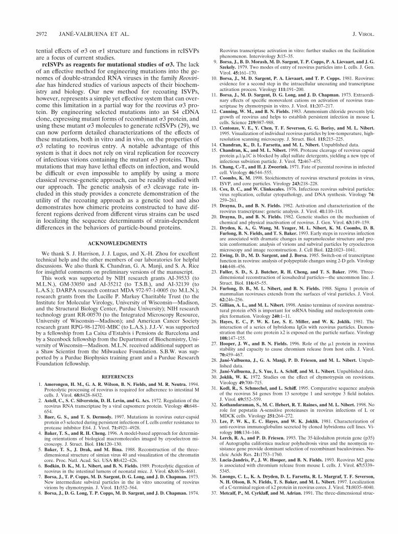
Construction of T1L and T3D s3 chimeras. DNA clones of the T1L and T3DS4 genes, inserted into pBluescript KS1 (Stratagene, La Jolla, Calif.) at theEcoRI site, were cleaved with BsaI and StyI (both within the S4 gene) or with StyIand EcoRV (the latter within the vector beyond the 39 end of the S4 plus strand)to obtain middle (S4 nucleotides 552 to 791, corresponding to s3 amino acids 186to 265) or C-terminal (S4 nucleotides 792 to 1095, corresponding to s3 aminoacids 266 to 365) fragments of S4, respectively. These fragments were exchangedbetween the T1L and T3D S4 constructs to generate chimeras as shown in Fig.10A. The chimeric S4 genes were subcloned into the pEV/35K/polybsmcr plas-mid (34), and the resulting constructs were used to express each s3 protein ininsect cells by using a novel baculovirus system (28). Protein was labeled at 40 hpostinfection with 25 mCi of [35S]methionine/cysteine per ml, and 8 h later cellswere harvested as described above.
RESULTS
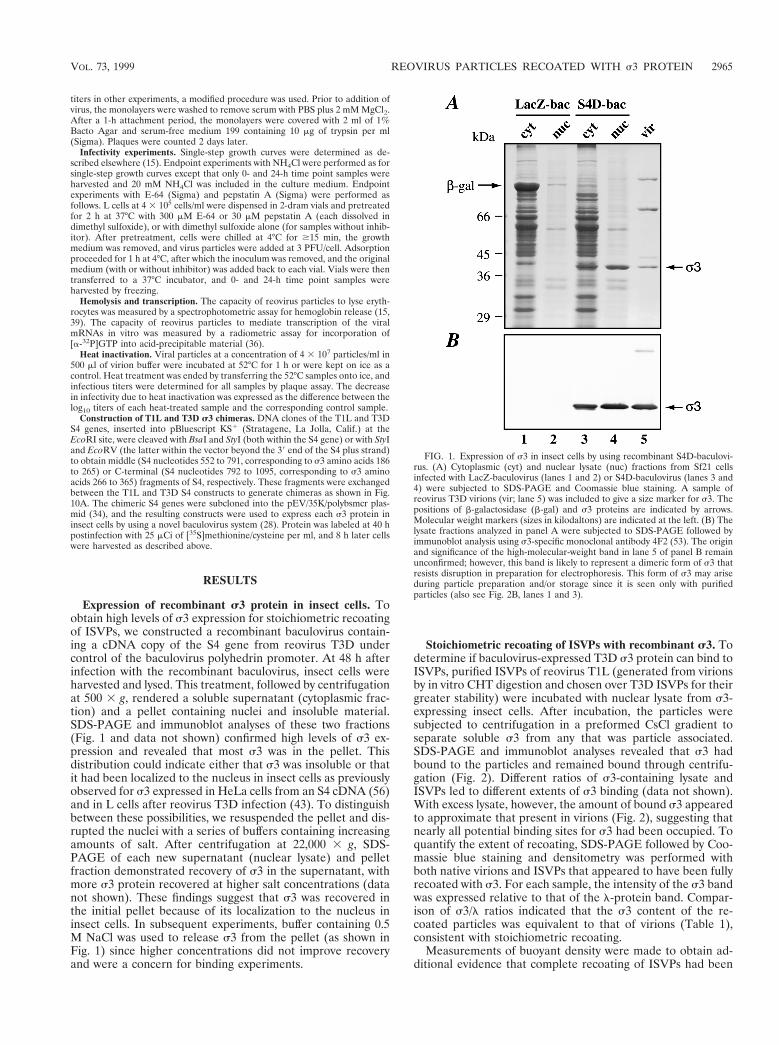
Expression of recombinant s3 protein in insect cells. Toobtain high levels of s3 expression for stoichiometric recoatingof ISVPs, we constructed a recombinant baculovirus contain-ing a cDNA copy of the S4 gene from reovirus T3D undercontrol of the baculovirus polyhedrin promoter. At 48 h afterinfection with the recombinant baculovirus, insect cells wereharvested and lysed. This treatment, followed by centrifugationat 500 3 g, rendered a soluble supernatant (cytoplasmic frac-tion) and a pellet containing nuclei and insoluble material.SDS-PAGE and immunoblot analyses of these two fractions(Fig. 1 and data not shown) confirmed high levels of s3 ex-pression and revealed that most s3 was in the pellet. Thisdistribution could indicate either that s3 was insoluble or thatit had been localized to the nucleus in insect cells as previouslyobserved for s3 expressed in HeLa cells from an S4 cDNA (56)and in L cells after reovirus T3D infection (43). To distinguishbetween these possibilities, we resuspended the pellet and dis-rupted the nuclei with a series of buffers containing increasingamounts of salt. After centrifugation at 22,000 3 g, SDS-PAGE of each new supernatant (nuclear lysate) and pelletfraction demonstrated recovery of s3 in the supernatant, withmore s3 protein recovered at higher salt concentrations (datanot shown). These findings suggest that s3 was recovered inthe initial pellet because of its localization to the nucleus ininsect cells. In subsequent experiments, buffer containing 0.5M NaCl was used to release s3 from the pellet (as shown inFig. 1) since higher concentrations did not improve recoveryand were a concern for binding experiments.
Stoichiometric recoating of ISVPs with recombinant s3. Todetermine if baculovirus-expressed T3D s3 protein can bind toISVPs, purified ISVPs of reovirus T1L (generated from virionsby in vitro CHT digestion and chosen over T3D ISVPs for theirgreater stability) were incubated with nuclear lysate from s3-expressing insect cells. After incubation, the particles weresubjected to centrifugation in a preformed CsCl gradient toseparate soluble s3 from any that was particle associated.SDS-PAGE and immunoblot analyses revealed that s3 hadbound to the particles and remained bound through centrifu-gation (Fig. 2). Different ratios of s3-containing lysate andISVPs led to different extents of s3 binding (data not shown).With excess lysate, however, the amount of bound s3 appearedto approximate that present in virions (Fig. 2), suggesting thatnearly all potential binding sites for s3 had been occupied. Toquantify the extent of recoating, SDS-PAGE followed by Coo-massie blue staining and densitometry was performed withboth native virions and ISVPs that appeared to have been fullyrecoated with s3. For each sample, the intensity of the s3 bandwas expressed relative to that of the l-protein band. Compar-ison of s3/l ratios indicated that the s3 content of the re-coated particles was equivalent to that of virions (Table 1),consistent with stoichiometric recoating.
Measurements of buoyant density were made to obtain ad-ditional evidence that complete recoating of ISVPs had been
FIG. 1. Expression of s3 in insect cells by using recombinant S4D-baculovi-rus. (A) Cytoplasmic (cyt) and nuclear lysate (nuc) fractions from Sf21 cellsinfected with LacZ-baculovirus (lanes 1 and 2) or S4D-baculovirus (lanes 3 and4) were subjected to SDS-PAGE and Coomassie blue staining. A sample ofreovirus T3D virions (vir; lane 5) was included to give a size marker for s3. Thepositions of b-galactosidase (b-gal) and s3 proteins are indicated by arrows.Molecular weight markers (sizes in kilodaltons) are indicated at the left. (B) Thelysate fractions analyzed in panel A were subjected to SDS-PAGE followed byimmunoblot analysis using s3-specific monoclonal antibody 4F2 (53). The originand significance of the high-molecular-weight band in lane 5 of panel B remainunconfirmed; however, this band is likely to represent a dimeric form of s3 thatresists disruption in preparation for electrophoresis. This form of s3 may ariseduring particle preparation and/or storage since it is seen only with purifiedparticles (also see Fig. 2B, lanes 1 and 3).
VOL. 73, 1999 REOVIRUS PARTICLES RECOATED WITH s3 PROTEIN 2965
approximated with baculovirus-expressed s3. Virions and IS-VPs contain the same complement of nucleic acids, but virionshave a higher protein content due to the presence of s3 andthus exhibit a buoyant density in CsCl (1.36 g/cm3) lower thanthat of ISVPs (1.38 g/cm3) (24, 30). Recoated particles mi-grated at a buoyant density of 1.361 g/cm3 (60.003, n 5 3),equivalent to that of native virions, suggesting that stoichio-metric or near-stoichiometric recoating had been achieved.
Henceforth, we use the term rcISVPs to refer to ISVPs thathave been recoated with approximately stoichiometric levels ofbaculovirus-expressed s3 protein. It is important to note thatrcISVPs are similar to virions in that they contain a full com-plement of s3 but similar to ISVPs in that they contain m1/m1Cmostly cleaved at the d-f junction. The implications of these
features for the structure and function of rcISVPs are ad-dressed below.
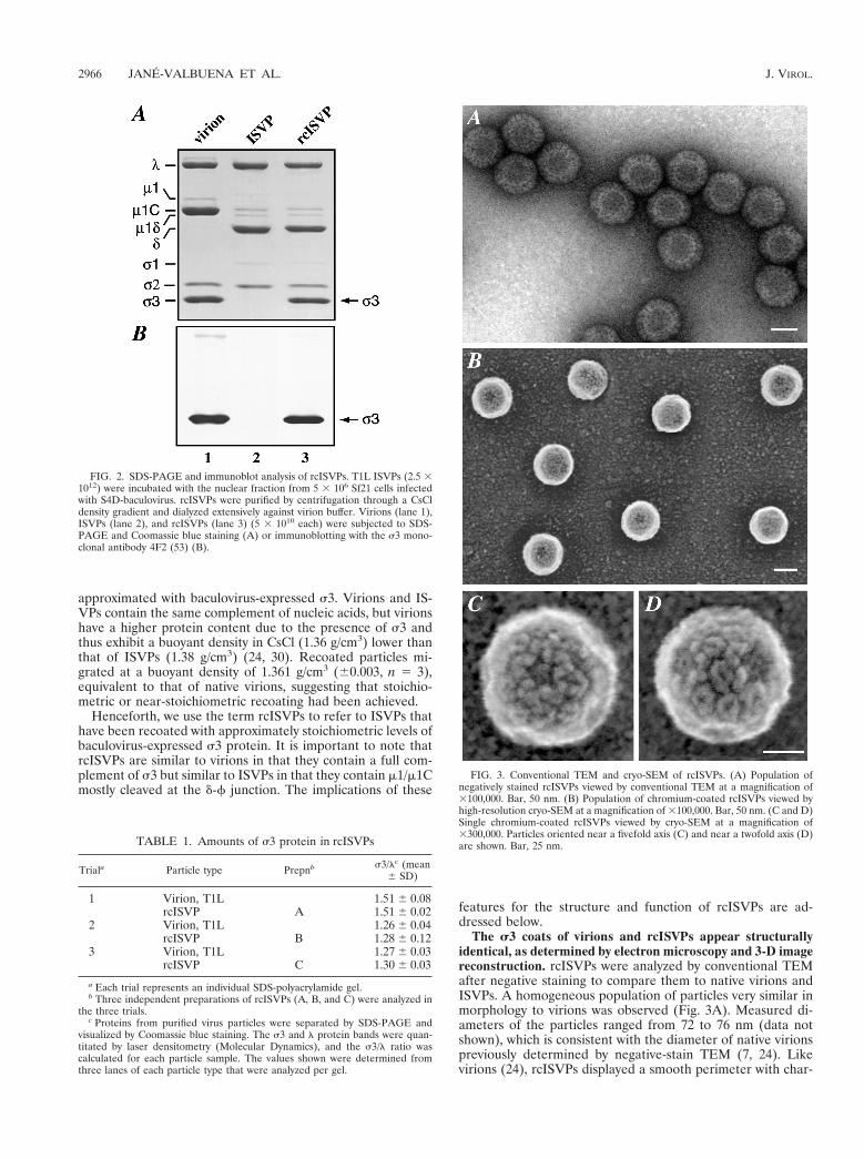
The s3 coats of virions and rcISVPs appear structurallyidentical, as determined by electron microscopy and 3-D imagereconstruction. rcISVPs were analyzed by conventional TEMafter negative staining to compare them to native virions andISVPs. A homogeneous population of particles very similar inmorphology to virions was observed (Fig. 3A). Measured di-ameters of the particles ranged from 72 to 76 nm (data notshown), which is consistent with the diameter of native virionspreviously determined by negative-stain TEM (7, 24). Likevirions (24), rcISVPs displayed a smooth perimeter with char-
FIG. 2. SDS-PAGE and immunoblot analysis of rcISVPs. T1L ISVPs (2.5 31012) were incubated with the nuclear fraction from 5 3 106 Sf21 cells infectedwith S4D-baculovirus. rcISVPs were purified by centrifugation through a CsCldensity gradient and dialyzed extensively against virion buffer. Virions (lane 1),ISVPs (lane 2), and rcISVPs (lane 3) (5 3 1010 each) were subjected to SDS-PAGE and Coomassie blue staining (A) or immunoblotting with the s3 mono-clonal antibody 4F2 (53) (B).
TABLE 1. Amounts of s3 protein in rcISVPs
Triala Particle type Prepnb s3/lc (mean6 SD)
1 Virion, T1L 1.51 6 0.08rcISVP A 1.51 6 0.02
2 Virion, T1L 1.26 6 0.04rcISVP B 1.28 6 0.12
3 Virion, T1L 1.27 6 0.03rcISVP C 1.30 6 0.03
a Each trial represents an individual SDS-polyacrylamide gel.b Three independent preparations of rcISVPs (A, B, and C) were analyzed in
the three trials.c Proteins from purified virus particles were separated by SDS-PAGE and
visualized by Coomassie blue staining. The s3 and l protein bands were quan-titated by laser densitometry (Molecular Dynamics), and the s3/l ratio wascalculated for each particle sample. The values shown were determined fromthree lanes of each particle type that were analyzed per gel.
FIG. 3. Conventional TEM and cryo-SEM of rcISVPs. (A) Population ofnegatively stained rcISVPs viewed by conventional TEM at a magnification of3100,000. Bar, 50 nm. (B) Population of chromium-coated rcISVPs viewed byhigh-resolution cryo-SEM at a magnification of 3100,000. Bar, 50 nm. (C and D)Single chromium-coated rcISVPs viewed by cryo-SEM at a magnification of3300,000. Particles oriented near a fivefold axis (C) and near a twofold axis (D)are shown. Bar, 25 nm.
2966 JANE-VALBUENA ET AL. J. VIROL.
acteristic flattened edges that arise at fivefold axes where theml-s3 lattice is interrupted by l2 (21, 37).
We used cryo-SEM to compare the organization of s3 sub-units on individual rcISVPs and virions. This technique showsindividual proteins on the surfaces of reovirus particles at aresolution of 2 to 4 nm (13). rcISVPs exhibited characteristicrings of protein distributed over much of their surface andincomplete rings surrounding the fivefold axes (Fig. 3B to D).These features correspond to complete and partial hexamersof s3 (see below), consistent with the results of the previouscryo-SEM study of reovirus virions (13). As no clear differ-ences distinguish the cryo-SEM images of rcISVPs and nativevirions (13), these findings suggest that a native s3 coat wasreconstituted in rcISVPs.
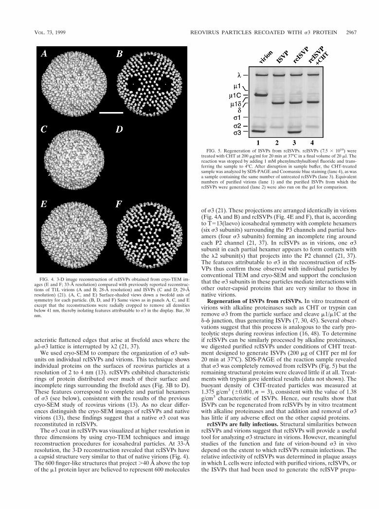
The s3 coat in rcISVPs was visualized at higher resolution inthree dimensions by using cryo-TEM techniques and imagereconstruction procedures for icosahedral particles. At 33-Åresolution, the 3-D reconstruction revealed that rcISVPs havea capsid structure very similar to that of native virions (Fig. 4).The 600 finger-like structures that project .40 Å above the topof the m1 protein layer are believed to represent 600 molecules
of s3 (21). These projections are arranged identically in virions(Fig. 4A and B) and rcISVPs (Fig. 4E and F), that is, accordingto T513(laevo) icosahedral symmetry with complete hexamers(six s3 subunits) surrounding the P3 channels and partial hex-amers (four s3 subunits) forming an incomplete ring aroundeach P2 channel (21, 37). In rcISVPs as in virions, one s3subunit in each partial hexamer appears to form contacts withthe l2 subunit(s) that projects into the P2 channel (21, 37).The features attributable to s3 in the reconstruction of rcIS-VPs thus confirm those observed with individual particles byconventional TEM and cryo-SEM and support the conclusionthat the s3 subunits in these particles mediate interactions withother outer-capsid proteins that are very similar to those innative virions.
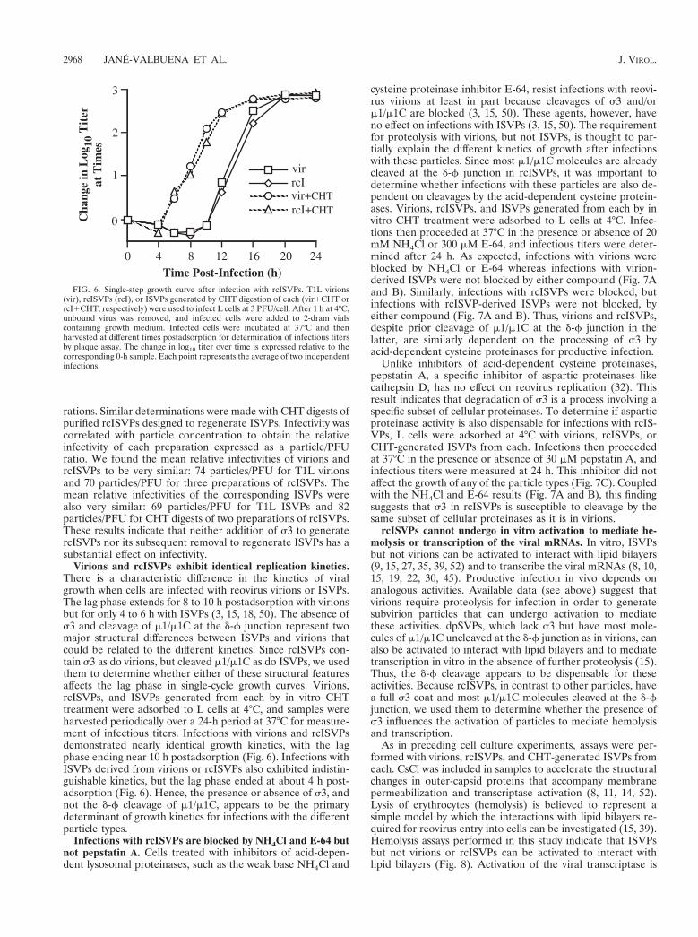
Regeneration of ISVPs from rcISVPs. In vitro treatment ofvirions with alkaline proteinases such as CHT or trypsin canremove s3 from the particle surface and cleave m1/m1C at thed-f junction, thus generating ISVPs (7, 30, 45). Several obser-vations suggest that this process is analogous to the early pro-teolytic steps during reovirus infection (16, 48). To determineif rcISVPs can be similarly processed by alkaline proteinases,we digested purified rcISVPs under conditions of CHT treat-ment designed to generate ISVPs (200 mg of CHT per ml for20 min at 37°C). SDS-PAGE of the reaction sample revealedthat s3 was completely removed from rcISVPs (Fig. 5) but theremaining structural proteins were cleaved little if at all. Treat-ments with trypsin gave identical results (data not shown). Thebuoyant density of CHT-treated particles was measured at1.375 g/cm3 (60.001, n 5 3), consistent with the value of 1.38g/cm3 characteristic of ISVPs. Hence, our results show thatISVPs can be regenerated from rcISVPs by in vitro treatmentwith alkaline proteinases and that addition and removal of s3has little if any adverse effect on the other capsid proteins.
rcISVPs are fully infectious. Structural similarities betweenrcISVPs and virions suggest that rcISVPs will provide a usefultool for analyzing s3 structure in virions. However, meaningfulstudies of the function and fate of virion-bound s3 in vivodepend on the extent to which rcISVPs remain infectious. Therelative infectivity of rcISVPs was determined in plaque assaysin which L cells were infected with purified virions, rcISVPs, orthe ISVPs that had been used to generate the rcISVP prepa-
FIG. 4. 3-D image reconstruction of rcISVPs obtained from cryo-TEM im-ages (E and F; 33-Å resolution) compared with previously reported reconstruc-tions of T1L virions (A and B; 28-Å resolution) and ISVPs (C and D; 29-Åresolution) (21). (A, C, and E) Surface-shaded views down a twofold axis ofsymmetry for each particle. (B, D, and F) Same views as in panels A, C, and Eexcept that the reconstructions were radially cropped to remove all densitiesbelow 41 nm, thereby isolating features attributable to s3 in the display. Bar, 30nm.
FIG. 5. Regeneration of ISVPs from rcISVPs. rcISVPs (7.5 3 1010) weretreated with CHT at 200 mg/ml for 20 min at 37°C in a final volume of 20 ml. Thereaction was stopped by adding 1 mM phenylmethylsulfonyl fluoride and trans-ferring the sample to 4°C. After disruption in sample buffer, the CHT-treatedsample was analyzed by SDS-PAGE and Coomassie blue staining (lane 4), as wasa sample containing the same number of untreated rcISVPs (lane 3). Equivalentnumbers of purified virions (lane 1) and the purified ISVPs from which thercISVPs were generated (lane 2) were also run on the gel for comparison.
VOL. 73, 1999 REOVIRUS PARTICLES RECOATED WITH s3 PROTEIN 2967
rations. Similar determinations were made with CHT digests ofpurified rcISVPs designed to regenerate ISVPs. Infectivity wascorrelated with particle concentration to obtain the relativeinfectivity of each preparation expressed as a particle/PFUratio. We found the mean relative infectivities of virions andrcISVPs to be very similar: 74 particles/PFU for T1L virionsand 70 particles/PFU for three preparations of rcISVPs. Themean relative infectivities of the corresponding ISVPs werealso very similar: 69 particles/PFU for T1L ISVPs and 82particles/PFU for CHT digests of two preparations of rcISVPs.These results indicate that neither addition of s3 to generatercISVPs nor its subsequent removal to regenerate ISVPs has asubstantial effect on infectivity.
Virions and rcISVPs exhibit identical replication kinetics.There is a characteristic difference in the kinetics of viralgrowth when cells are infected with reovirus virions or ISVPs.The lag phase extends for 8 to 10 h postadsorption with virionsbut for only 4 to 6 h with ISVPs (3, 15, 18, 50). The absence ofs3 and cleavage of m1/m1C at the d-f junction represent twomajor structural differences between ISVPs and virions thatcould be related to the different kinetics. Since rcISVPs con-tain s3 as do virions, but cleaved m1/m1C as do ISVPs, we usedthem to determine whether either of these structural featuresaffects the lag phase in single-cycle growth curves. Virions,rcISVPs, and ISVPs generated from each by in vitro CHTtreatment were adsorbed to L cells at 4°C, and samples wereharvested periodically over a 24-h period at 37°C for measure-ment of infectious titers. Infections with virions and rcISVPsdemonstrated nearly identical growth kinetics, with the lagphase ending near 10 h postadsorption (Fig. 6). Infections withISVPs derived from virions or rcISVPs also exhibited indistin-guishable kinetics, but the lag phase ended at about 4 h post-adsorption (Fig. 6). Hence, the presence or absence of s3, andnot the d-f cleavage of m1/m1C, appears to be the primarydeterminant of growth kinetics for infections with the differentparticle types.
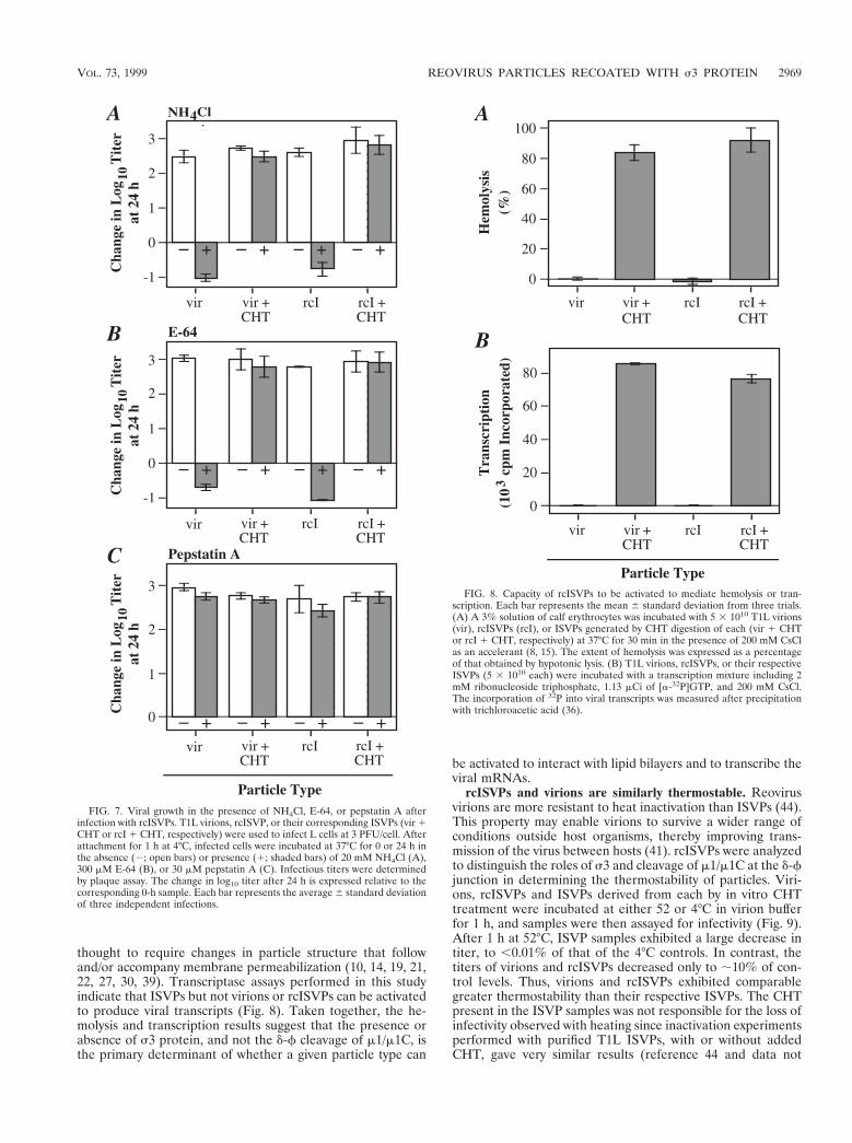
Infections with rcISVPs are blocked by NH4Cl and E-64 butnot pepstatin A. Cells treated with inhibitors of acid-depen-dent lysosomal proteinases, such as the weak base NH4Cl and
cysteine proteinase inhibitor E-64, resist infections with reovi-rus virions at least in part because cleavages of s3 and/orm1/m1C are blocked (3, 15, 50). These agents, however, haveno effect on infections with ISVPs (3, 15, 50). The requirementfor proteolysis with virions, but not ISVPs, is thought to par-tially explain the different kinetics of growth after infectionswith these particles. Since most m1/m1C molecules are alreadycleaved at the d-f junction in rcISVPs, it was important todetermine whether infections with these particles are also de-pendent on cleavages by the acid-dependent cysteine protein-ases. Virions, rcISVPs, and ISVPs generated from each by invitro CHT treatment were adsorbed to L cells at 4°C. Infec-tions then proceeded at 37°C in the presence or absence of 20mM NH4Cl or 300 mM E-64, and infectious titers were deter-mined after 24 h. As expected, infections with virions wereblocked by NH4Cl or E-64 whereas infections with virion-derived ISVPs were not blocked by either compound (Fig. 7Aand B). Similarly, infections with rcISVPs were blocked, butinfections with rcISVP-derived ISVPs were not blocked, byeither compound (Fig. 7A and B). Thus, virions and rcISVPs,despite prior cleavage of m1/m1C at the d-f junction in thelatter, are similarly dependent on the processing of s3 byacid-dependent cysteine proteinases for productive infection.
Unlike inhibitors of acid-dependent cysteine proteinases,pepstatin A, a specific inhibitor of aspartic proteinases likecathepsin D, has no effect on reovirus replication (32). Thisresult indicates that degradation of s3 is a process involving aspecific subset of cellular proteinases. To determine if asparticproteinase activity is also dispensable for infections with rcIS-VPs, L cells were adsorbed at 4°C with virions, rcISVPs, orCHT-generated ISVPs from each. Infections then proceededat 37°C in the presence or absence of 30 mM pepstatin A, andinfectious titers were measured at 24 h. This inhibitor did notaffect the growth of any of the particle types (Fig. 7C). Coupledwith the NH4Cl and E-64 results (Fig. 7A and B), this findingsuggests that s3 in rcISVPs is susceptible to cleavage by thesame subset of cellular proteinases as it is in virions.
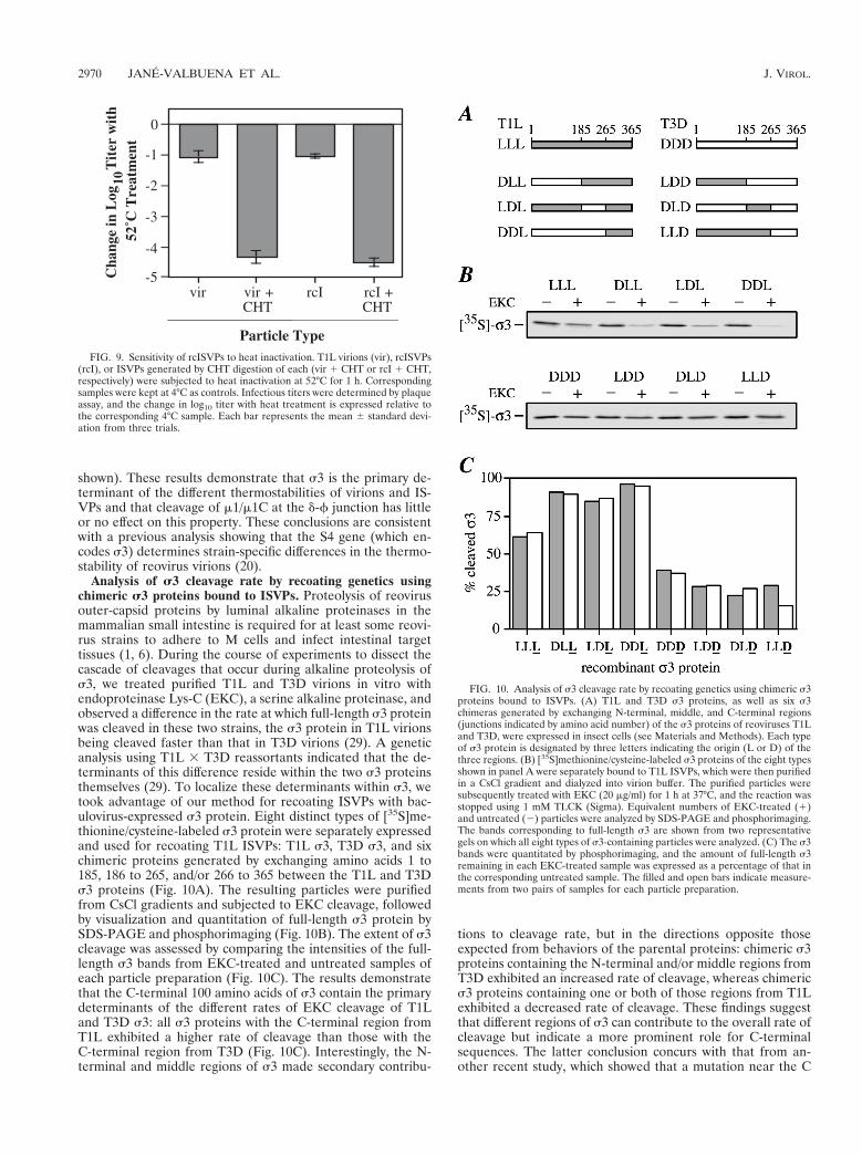
rcISVPs cannot undergo in vitro activation to mediate he-molysis or transcription of the viral mRNAs. In vitro, ISVPsbut not virions can be activated to interact with lipid bilayers(9, 15, 27, 35, 39, 52) and to transcribe the viral mRNAs (8, 10,15, 19, 22, 30, 45). Productive infection in vivo depends onanalogous activities. Available data (see above) suggest thatvirions require proteolysis for infection in order to generatesubvirion particles that can undergo activation to mediatethese activities. dpSVPs, which lack s3 but have most mole-cules of m1/m1C uncleaved at the d-f junction as in virions, canalso be activated to interact with lipid bilayers and to mediatetranscription in vitro in the absence of further proteolysis (15).Thus, the d-f cleavage appears to be dispensable for theseactivities. Because rcISVPs, in contrast to other particles, havea full s3 coat and most m1/m1C molecules cleaved at the d-fjunction, we used them to determine whether the presence ofs3 influences the activation of particles to mediate hemolysisand transcription.
As in preceding cell culture experiments, assays were per-formed with virions, rcISVPs, and CHT-generated ISVPs fromeach. CsCl was included in samples to accelerate the structuralchanges in outer-capsid proteins that accompany membranepermeabilization and transcriptase activation (8, 11, 14, 52).Lysis of erythrocytes (hemolysis) is believed to represent asimple model by which the interactions with lipid bilayers re-quired for reovirus entry into cells can be investigated (15, 39).Hemolysis assays performed in this study indicate that ISVPsbut not virions or rcISVPs can be activated to interact withlipid bilayers (Fig. 8). Activation of the viral transcriptase is
FIG. 6. Single-step growth curve after infection with rcISVPs. T1L virions(vir), rcISVPs (rcI), or ISVPs generated by CHT digestion of each (vir1CHT orrcI1CHT, respectively) were used to infect L cells at 3 PFU/cell. After 1 h at 4°C,unbound virus was removed, and infected cells were added to 2-dram vialscontaining growth medium. Infected cells were incubated at 37°C and thenharvested at different times postadsorption for determination of infectious titersby plaque assay. The change in log10 titer over time is expressed relative to thecorresponding 0-h sample. Each point represents the average of two independentinfections.
2968 JANE-VALBUENA ET AL. J. VIROL.
thought to require changes in particle structure that followand/or accompany membrane permeabilization (10, 14, 19, 21,22, 27, 30, 39). Transcriptase assays performed in this studyindicate that ISVPs but not virions or rcISVPs can be activatedto produce viral transcripts (Fig. 8). Taken together, the he-molysis and transcription results suggest that the presence orabsence of s3 protein, and not the d-f cleavage of m1/m1C, isthe primary determinant of whether a given particle type can
be activated to interact with lipid bilayers and to transcribe theviral mRNAs.
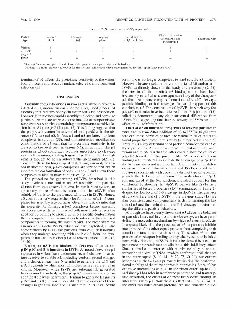
rcISVPs and virions are similarly thermostable. Reovirusvirions are more resistant to heat inactivation than ISVPs (44).This property may enable virions to survive a wider range ofconditions outside host organisms, thereby improving trans-mission of the virus between hosts (41). rcISVPs were analyzedto distinguish the roles of s3 and cleavage of m1/m1C at the d-fjunction in determining the thermostability of particles. Viri-ons, rcISVPs and ISVPs derived from each by in vitro CHTtreatment were incubated at either 52 or 4°C in virion bufferfor 1 h, and samples were then assayed for infectivity (Fig. 9).After 1 h at 52°C, ISVP samples exhibited a large decrease intiter, to ,0.01% of that of the 4°C controls. In contrast, thetiters of virions and rcISVPs decreased only to ;10% of con-trol levels. Thus, virions and rcISVPs exhibited comparablegreater thermostability than their respective ISVPs. The CHTpresent in the ISVP samples was not responsible for the loss ofinfectivity observed with heating since inactivation experimentsperformed with purified T1L ISVPs, with or without addedCHT, gave very similar results (reference 44 and data not
FIG. 7. Viral growth in the presence of NH4Cl, E-64, or pepstatin A afterinfection with rcISVPs. T1L virions, rcISVP, or their corresponding ISVPs (vir 1CHT or rcI 1 CHT, respectively) were used to infect L cells at 3 PFU/cell. Afterattachment for 1 h at 4°C, infected cells were incubated at 37°C for 0 or 24 h inthe absence (2; open bars) or presence (1; shaded bars) of 20 mM NH4Cl (A),300 mM E-64 (B), or 30 mM pepstatin A (C). Infectious titers were determinedby plaque assay. The change in log10 titer after 24 h is expressed relative to thecorresponding 0-h sample. Each bar represents the average 6 standard deviationof three independent infections.
FIG. 8. Capacity of rcISVPs to be activated to mediate hemolysis or tran-scription. Each bar represents the mean 6 standard deviation from three trials.(A) A 3% solution of calf erythrocytes was incubated with 5 3 1010 T1L virions(vir), rcISVPs (rcI), or ISVPs generated by CHT digestion of each (vir 1 CHTor rcI 1 CHT, respectively) at 37°C for 30 min in the presence of 200 mM CsClas an accelerant (8, 15). The extent of hemolysis was expressed as a percentageof that obtained by hypotonic lysis. (B) T1L virions, rcISVPs, or their respectiveISVPs (5 3 1010 each) were incubated with a transcription mixture including 2mM ribonucleoside triphosphate, 1.13 mCi of [a-32P]GTP, and 200 mM CsCl.The incorporation of 32P into viral transcripts was measured after precipitationwith trichloroacetic acid (36).
VOL. 73, 1999 REOVIRUS PARTICLES RECOATED WITH s3 PROTEIN 2969
shown). These results demonstrate that s3 is the primary de-terminant of the different thermostabilities of virions and IS-VPs and that cleavage of m1/m1C at the d-f junction has littleor no effect on this property. These conclusions are consistentwith a previous analysis showing that the S4 gene (which en-codes s3) determines strain-specific differences in the thermo-stability of reovirus virions (20).
Analysis of s3 cleavage rate by recoating genetics usingchimeric s3 proteins bound to ISVPs. Proteolysis of reovirusouter-capsid proteins by luminal alkaline proteinases in themammalian small intestine is required for at least some reovi-rus strains to adhere to M cells and infect intestinal targettissues (1, 6). During the course of experiments to dissect thecascade of cleavages that occur during alkaline proteolysis ofs3, we treated purified T1L and T3D virions in vitro withendoproteinase Lys-C (EKC), a serine alkaline proteinase, andobserved a difference in the rate at which full-length s3 proteinwas cleaved in these two strains, the s3 protein in T1L virionsbeing cleaved faster than that in T3D virions (29). A geneticanalysis using T1L 3 T3D reassortants indicated that the de-terminants of this difference reside within the two s3 proteinsthemselves (29). To localize these determinants within s3, wetook advantage of our method for recoating ISVPs with bac-ulovirus-expressed s3 protein. Eight distinct types of [35S]me-thionine/cysteine-labeled s3 protein were separately expressedand used for recoating T1L ISVPs: T1L s3, T3D s3, and sixchimeric proteins generated by exchanging amino acids 1 to185, 186 to 265, and/or 266 to 365 between the T1L and T3Ds3 proteins (Fig. 10A). The resulting particles were purifiedfrom CsCl gradients and subjected to EKC cleavage, followedby visualization and quantitation of full-length s3 protein bySDS-PAGE and phosphorimaging (Fig. 10B). The extent of s3cleavage was assessed by comparing the intensities of the full-length s3 bands from EKC-treated and untreated samples ofeach particle preparation (Fig. 10C). The results demonstratethat the C-terminal 100 amino acids of s3 contain the primarydeterminants of the different rates of EKC cleavage of T1Land T3D s3: all s3 proteins with the C-terminal region fromT1L exhibited a higher rate of cleavage than those with theC-terminal region from T3D (Fig. 10C). Interestingly, the N-terminal and middle regions of s3 made secondary contribu-
tions to cleavage rate, but in the directions opposite thoseexpected from behaviors of the parental proteins: chimeric s3proteins containing the N-terminal and/or middle regions fromT3D exhibited an increased rate of cleavage, whereas chimerics3 proteins containing one or both of those regions from T1Lexhibited a decreased rate of cleavage. These findings suggestthat different regions of s3 can contribute to the overall rate ofcleavage but indicate a more prominent role for C-terminalsequences. The latter conclusion concurs with that from an-other recent study, which showed that a mutation near the C
FIG. 9. Sensitivity of rcISVPs to heat inactivation. T1L virions (vir), rcISVPs(rcI), or ISVPs generated by CHT digestion of each (vir 1 CHT or rcI 1 CHT,respectively) were subjected to heat inactivation at 52°C for 1 h. Correspondingsamples were kept at 4°C as controls. Infectious titers were determined by plaqueassay, and the change in log10 titer with heat treatment is expressed relative tothe corresponding 4°C sample. Each bar represents the mean 6 standard devi-ation from three trials.
FIG. 10. Analysis of s3 cleavage rate by recoating genetics using chimeric s3proteins bound to ISVPs. (A) T1L and T3D s3 proteins, as well as six s3chimeras generated by exchanging N-terminal, middle, and C-terminal regions(junctions indicated by amino acid number) of the s3 proteins of reoviruses T1Land T3D, were expressed in insect cells (see Materials and Methods). Each typeof s3 protein is designated by three letters indicating the origin (L or D) of thethree regions. (B) [35S]methionine/cysteine-labeled s3 proteins of the eight typesshown in panel A were separately bound to T1L ISVPs, which were then purifiedin a CsCl gradient and dialyzed into virion buffer. The purified particles weresubsequently treated with EKC (20 mg/ml) for 1 h at 37°C, and the reaction wasstopped using 1 mM TLCK (Sigma). Equivalent numbers of EKC-treated (1)and untreated (2) particles were analyzed by SDS-PAGE and phosphorimaging.The bands corresponding to full-length s3 are shown from two representativegels on which all eight types of s3-containing particles were analyzed. (C) The s3bands were quantitated by phosphorimaging, and the amount of full-length s3remaining in each EKC-treated sample was expressed as a percentage of that inthe corresponding untreated sample. The filled and open bars indicate measure-ments from two pairs of samples for each particle preparation.
2970 JANE-VALBUENA ET AL. J. VIROL.
terminus of s3 affects the proteinase sensitivity of the virion-bound protein in a reovirus mutant selected during persistentinfection (55).
DISCUSSION
Assembly of s3 into virions in vivo and in vitro. In reovirus-infected cells, mature virions undergo a regulated process ofassembly that remains poorly characterized. One observation,however, is that outer-capsid assembly is blocked and core-likeparticles accumulate when cells are infected at nonpermissivetemperatures with virus containing a temperature-sensitive le-sion in the S4 gene (tsG453) (38, 47). This finding suggests thatthe m1 protein cannot be assembled into particles in the ab-sence of functional s3. In fact, m1 and s3 are known to formcomplexes in solution (33), and this interaction modifies theconformation of s3 such that its proteinase sensitivity is in-creased to the level seen in virions (46). In addition, the m1protein in m1-s3 complexes becomes susceptible to cleavagenear its N terminus, yielding fragments m1N and m1C throughwhat is thought to be an autocatalytic mechanism (42, 51).Together, these findings suggest that during assembly of viri-ons in infected cells, m1-s3 complexes are formed first, whichmodifies the conformation of both m1 and s3 and allows thesecomplexes to bind to nascent particles (38, 47).
The procedure for generating rcISVPs described in thisstudy involves a mechanism for s3 assembly into particlesdistinct from that observed in vivo. In our in vitro system, anapparently native s3 coat is reconstituted in rcISVPs aftersoluble s3 binds to the preformed m1 lattice in ISVPs. Hence,s3 does not strictly require the prior formation of m1-s3 com-plexes for assembly into particles. Given this fact, we infer thatthe necessity for forming m1-s3 complexes before assemblyonto core-like particles in infected cells most likely reflects theneed for s3 binding to induce m1 into a specific conformationthat is competent to self-associate or to interact with other viralcomponents in forming the outer capsid. The mechanism forassembling s3 onto ISVPs, which we have exploited, is alsodemonstrated by ISVP-like particles from cellular lysosomeswhen they undergo recoating with soluble s3 from the cyto-plasm or nucleus upon disruption of reovirus-infected cells (2,16, 50).
Binding to s3 is not blocked by cleavages of m1 at them1N-m1C and d-f junctions in ISVPs. As noted above, the m1molecules in virions have undergone several changes in struc-ture relative to soluble m1, including conformational changesand a cleavage near their N termini to generate the m1N andm1C fragments by which most m1 molecules are represented invirions. Moreover, when ISVPs are subsequently generatedfrom virions by proteolysis, the m1/m1C molecules undergo anadditional cleavage near their C termini to generate fragmentsm1d/d and f (40). It was conceivable that one or more of thesechanges might have modified m1 such that, in its ISVP-bound
form, it was no longer competent to bind soluble s3 protein.However, because soluble s3 can bind to m1d/d and/or f inISVPs, as directly shown in this study and previously (2, 46),the sites in m1 that mediate s3 binding cannot have beenirreversibly modified as a consequence of any of the changes inm1 that accompany complex formation, m1N-m1C cleavage,particle binding, or d-f cleavage. In partial support of thisconclusion, a 3-D reconstruction of dpSVPs, in which very fewm1/m1C molecules have been cleaved at the d-f junction (15),failed to demonstrate any clear structural differences fromISVPs (54), suggesting that the d-f cleavage in ISVPs has littleeffect on m1 conformation.
Effect of s3 on functional properties of reovirus particles invitro and in vivo. After addition of s3 to ISVPs, to generatercISVPs, these particles behave like virions in all of the func-tional properties tested in this study (summarized in Table 2).Thus, s3 is a key determinant of particle behavior for each ofthese properties. An important structural distinction betweenvirions and rcISVPs is that the latter contain most molecules ofm1/m1C cleaved at the d-f junction, like ISVPs. As a result, ourfindings with rcISVPs also indicate that cleavage of m1/m1C atthe d-f junction is not an important determinant of the differ-ences in behavior between virions and ISVPs studied here.Previous experiments with dpSVPs, a distinct type of subvirionparticle that lacks s3 but contains most molecules of m1/m1Cstill uncleaved at the d-f junction, provided support for thisconclusion by showing that dpSVPs behave like ISVPs in asimilar set of tested properties (15) (summarized in Table 2),despite the low level of d-f cleavage in dpSVPs. The analysesof rcISVPs here and of dpSVPs in the previous study (15) arethus consistent and complementary in demonstrating the keyrole of s3 and the negligible role of d-f cleavage in determin-ing the different particle behaviors.
Although we have clearly shown that s3 affects the behaviorof particles in several in vitro and in vivo assays, we have yet todefine the molecular mechanisms by which s3 exerts its effects.It appears likely that the presence of s3 in particles inhibitsone or more of the other capsid proteins from completing theirfunction or functions in reovirus entry. Thus, when s3 remainspresent after receptor binding and uptake by cells, as in infec-tions with virions and rcISVPs, it must be cleaved by a cellularproteinase or proteinases to eliminate this inhibitory effect.Since activation to interact with membrane bilayers and totranscribe the viral mRNAs involves conformational changesin the outer capsid (8, 10, 14, 19, 22, 27, 30, 39), our currenthypothesis is that s3 acts primarily by limiting the conforma-tional mobility of the relevant protein or proteins. Since s3 hasextensive interactions with m1 in the virion outer capsid (21),and since m1 has roles in membrane penetration and transcrip-tase activation, the effects of s3 most likely occur through itsinteractions with m1. Nonetheless, effects of s3 on l2 or s1,the other two outer capsid proteins, are also conceivable. Po-
TABLE 2. Summary of rcISVP propertiesa
Particletype
Presenceof s3
Cleavageat d-f
Long lagphase
Sensitivity toNH4Cl and E-64
Block to activationof hemolysis and
transcriptionThermostability
Virion 1 2 1 1 1 1rcISVP 1 1 1 1 1 1dpSVPb 2 2 2 2 2 2ISVP 2 1 2 2 2 2
a See text for more complete descriptions of the particle types, properties, and behaviors.b Findings are from reference 15 except for the thermostability data, which were generated for this report (data not shown).
VOL. 73, 1999 REOVIRUS PARTICLES RECOATED WITH s3 PROTEIN 2971
tential effects of s3 on s1 structure and functions in rcISVPsare a focus of current studies.
rcISVPs as reagents for mutational studies of s3. The lackof an effective method for engineering mutations into the ge-nomes of double-stranded RNA viruses in the family Reoviri-dae has hindered studies of various aspects of their biochem-istry and biology. Our new method for recoating ISVPs,however, represents a simple yet effective system that can over-come this limitation in a partial way for the reovirus s3 pro-tein. By engineering selected mutations into an S4 cDNAclone, expressing mutant forms of recombinant s3 protein, andusing these mutant s3 molecules to generate rcISVPs (29), wecan now perform detailed characterizations of the effects ofthese mutations, both in vitro and in vivo, on the properties ofs3 relating to reovirus entry. A notable advantage of thissystem is that it does not rely on viral replication for recoveryof infectious virions containing the mutant s3 proteins. Thus,mutations that may have lethal effects on infection, and wouldbe difficult or even impossible to amplify by using a moreclassical reverse-genetic approach, can be readily studied withour approach. The genetic analysis of s3 cleavage rate in-cluded in this study provides a concrete demonstration of theutility of the recoating approach as a genetic tool and alsodemonstrates how chimeric proteins constructed to have dif-ferent regions derived from different virus strains can be usedin localizing the sequence determinants of strain-dependentdifferences in the behaviors of particle-bound proteins.
ACKNOWLEDGMENTS
We thank S. J. Harrison, J. J. Lugus, and X.-H. Zhou for excellenttechnical help and the other members of our laboratories for helpfuldiscussions. We also thank K. Chandran, G. A. Manji, and S. A. Ricefor insightful comments on preliminary versions of the manuscript.
This work was supported by NIH research grants AI-39533 (toM.L.N.), GM-33050 and AI-35212 (to T.S.B.), and AI-32139 (toL.A.S.); DARPA research contract MDA 972-97-1-0005 (to M.L.N.);research grants from the Lucille P. Markey Charitable Trust (to theInstitute for Molecular Virology, University of Wisconsin—Madison,and the Structural Biology Center, Purdue University); NIH researchtechnology grant RR-00570 (to the Integrated Microscopy Resource,University of Wisconsin—Madison); and American Cancer Societyresearch grant RPG-98-12701-MBC (to L.A.S.). J.J.-V. was supportedby a fellowship from La Caixa d’Estalvis i Pensions de Barcelona andby a Steenbock fellowship from the Department of Biochemistry, Uni-versity of Wisconsin—Madison. M.L.N. received additional support asa Shaw Scientist from the Milwaukee Foundation. S.B.W. was sup-ported by a Purdue Biophysics training grant and a Purdue ResearchFoundation fellowship.
REFERENCES
1. Amerongen, H. M., G. A. R. Wilson, B. N. Fields, and M. R. Neutra. 1994.Proteolytic processing of reovirus is required for adherence to intestinal Mcells. J. Virol. 68:8428–8432.
2. Astell, C., S. C. Silverstein, D. H. Levin, and G. Acs. 1972. Regulation of thereovirus RNA transcriptase by a viral capsomere protein. Virology 48:648–654.
3. Baer, G. S., and T. S. Dermody. 1997. Mutations in reovirus outer-capsidprotein s3 selected during persistent infections of L cells confer resistance toprotease inhibitor E64. J. Virol. 71:4921–4928.
4. Baker, T. S., and R. H. Cheng. 1996. A model-based approach for determin-ing orientations of biological macromolecules imaged by cryoelectron mi-croscopy. J. Struct. Biol. 116:120–130.
5. Baker, T. S., J. Drak, and M. Bina. 1988. Reconstruction of the three-dimensional structure of simian virus 40 and visualization of the chromatincore. Proc. Natl. Acad. Sci. USA 85:422–426.
6. Bodkin, D. K., M. L. Nibert, and B. N. Fields. 1989. Proteolytic digestion ofreovirus in the intestinal lumens of neonatal mice. J. Virol. 63:4676–4681.
7. Borsa, J., T. P. Copps, M. D. Sargent, D. G. Long, and J. D. Chapman. 1973.New intermediate subviral particles in the in vitro uncoating of reovirusvirions by chymotrypsin. J. Virol. 11:552–564.
8. Borsa, J., D. G. Long, T. P. Copps, M. D. Sargent, and J. D. Chapman. 1974.
Reovirus transcriptase activation in vitro: further studies on the facilitationphenomenon. Intervirology 3:15–35.
9. Borsa, J., B. D. Morash, M. D. Sargent, T. P. Copps, P. A. Lievaart, and J. G.Szekely. 1979. Two modes of entry of reovirus particles into L cells. J. Gen.Virol. 45:161–170.
10. Borsa, J., M. D. Sargent, P. A. Lievaart, and T. P. Copps. 1981. Reovirus:evidence for a second step in the intracellular uncoating and transcriptaseactivation process. Virology 111:191–200.
11. Borsa, J., M. D. Sargent, D. G. Long, and J. D. Chapman. 1973. Extraordi-nary effects of specific monovalent cations on activation of reovirus tran-scriptase by chymotrypsin in vitro. J. Virol. 11:207–217.
12. Canning, W. M., and B. N. Fields. 1983. Ammonium chloride prevents lyticgrowth of reovirus and helps to establish persistent infection in mouse Lcells. Science 219:987–988.
13. Centonze, V. E., Y. Chen, T. F. Severson, G. G. Borisy, and M. L. Nibert.1995. Visualization of individual reovirus particles by low-temperature, high-resolution scanning microscopy. J. Struct. Biol. 115:215–225.
14. Chandran, K., D. L. Farsetta, and M. L. Nibert. Unpublished data.15. Chandran, K., and M. L. Nibert. 1998. Protease cleavage of reovirus capsid
protein m1/m1C is blocked by alkyl sulfate detergents, yielding a new type ofinfectious subvirion particle. J. Virol. 72:467–475.
16. Chang, C.-T., and H. J. Zweerink. 1971. Fate of parental reovirus in infectedcell. Virology 46:544–555.
17. Coombs, K. M. 1998. Stoichiometry of reovirus structural proteins in virus,ISVP, and core particles. Virology 243:218–228.
18. Cox, D. C., and W. Clinkscales. 1976. Infectious reovirus subviral particles:virus replication, cellular cytopathology, and DNA synthesis. Virology 74:259–261.
19. Drayna, D., and B. N. Fields. 1982. Activation and characterization of thereovirus transcriptase: genetic analysis. J. Virol. 41:110–118.
20. Drayna, D., and B. N. Fields. 1982. Genetic studies on the mechanism ofchemical and physical inactivation of reovirus. J. Gen. Virol. 63:149–159.
21. Dryden, K. A., G. Wang, M. Yeager, M. L. Nibert, K. M. Coombs, D. B.Furlong, B. N. Fields, and T. S. Baker. 1993. Early steps in reovirus infectionare associated with dramatic changes in supramolecular structure and pro-tein conformation: analysis of virions and subviral particles by cryoelectronmicroscopy and image reconstruction. J. Cell Biol. 122:1023–1041.
22. Ewing, D. D., M. D. Sargent, and J. Borsa. 1985. Switch-on of transcriptasefunction in reovirus: analysis of polypeptide changes using 2-D gels. Virology144:448–456.
23. Fuller, S. D., S. J. Butcher, R. H. Cheng, and T. S. Baker. 1996. Three-dimensional reconstruction of icosahedral particles—the uncommon line. J.Struct. Biol. 116:45–55.
24. Furlong, D. B., M. L. Nibert, and B. N. Fields. 1988. Sigma 1 protein ofmammalian reoviruses extends from the surfaces of viral particles. J. Virol.62:246–256.
25. Gillian, A. L., and M. L. Nibert. 1998. Amino terminus of reovirus nonstruc-tural protein sNS is important for ssRNA binding and nucleoprotein com-plex formation. Virology 240:1–11.
26. Hayes, E. C., P. W. K. Lee, S. E. Miller, and W. K. Joklik. 1981. Theinteraction of a series of hybridoma IgGs with reovirus particles. Demon-stration that the core protein l2 is exposed on the particle surface. Virology108:147–155.
27. Hooper, J. W., and B. N. Fields. 1996. Role of the m1 protein in reovirusstability and capacity to cause chromium release from host cells. J. Virol.70:459–467.
28. Jane-Valbuena, J., G. A. Manji, P. D. Friesen, and M. L. Nibert. Unpub-lished data.
29. Jane-Valbuena, J., S. Yue, L. A. Schiff, and M. L. Nibert. Unpublished data.30. Joklik, W. K. 1972. Studies on the effect of chymotrypsin on reovirions.
Virology 49:700–715.31. Kedl, R., S. Schmechel, and L. Schiff. 1995. Comparative sequence analysis
of the reovirus S4 genes from 13 serotype 1 and serotype 3 field isolates.J. Virol. 69:552–559.
32. Kothandaraman, S., M. C. Hebert, R. T. Raines, and M. L. Nibert. 1998. Norole for pepstatin A-sensitive proteinases in reovirus infections of L orMDCK cells. Virology 251:264–272.
33. Lee, P. W. K., E. C. Hayes, and W. K. Joklik. 1981. Characterization ofanti-reovirus immunoglobulins secreted by cloned hybridoma cell lines. Vi-rology 108:134–146.
34. Lerch, R. A., and P. D. Friesen. 1993. The 35-kilodalton protein gene (p35)of Autographa californica nuclear polyhedrosis virus and the neomycin re-sistance gene provide dominant selection of recombinant baculoviruses. Nu-cleic Acids Res. 21:1753–1760.
35. Lucia-Jandris, P., J. W. Hooper, and B. N. Fields. 1993. Reovirus M2 geneis associated with chromium release from mouse L cells. J. Virol. 67:5339–5345.
36. Luongo, C. L., K. A. Dryden, D. L. Farsetta, R. L. Margraf, T. F. Severson,N. H. Olson, B. N. Fields, T. S. Baker, and M. L. Nibert. 1997. Localizationof a C-terminal region of l2 protein in reovirus cores. J. Virol. 71:8035–8040.
37. Metcalf, P., M. Cyrklaff, and M. Adrian. 1991. The three-dimensional struc-
2972 JANE-VALBUENA ET AL. J. VIROL.
ture of reovirus obtained by cryo-electron microscopy. EMBO J. 10:3129–3136.
38. Morgan, E. M., and H. J. Zweerink. 1974. Reovirus morphogenesis. Corelikeparticles in cells infected at 39° with wild-type reovirus and temperature-sensitive mutants of groups B and G. Virology 59:556–565.
39. Nibert, M. L. 1993. Structure and function of reovirus outer-capsid proteinsas they relate to early steps in infection. Ph.D. thesis. Harvard University,Cambridge, Mass.
40. Nibert, M. L., and B. N. Fields. 1992. A carboxy-terminal fragment of proteinm1/m1C is present in infectious subvirion particles of mammalian reovirusesand is proposed to have a role in penetration. J. Virol. 66:6408–6418.
41. Nibert, M. L., D. B. Furlong, and B. N. Fields. 1991. Mechanisms of viralpathogenesis. Distinct forms of reoviruses and their roles during replicationin cells and host. J. Clin. Investig. 88:727–734.
42. Nibert, M. L., L. A. Schiff, and B. N. Fields. 1991. Mammalian reovirusescontain a myristoylated structural protein. J. Virol. 65:1960–1967.
43. Schmechel, S., M. Chute, P. Skinner, R. Anderson, and L. Schiff. 1997.Preferential translation of reovirus mRNA by a s3-dependent mechanism.Virology 232:62–73.
44. Severson, T. F., K. Chandran, A. L. Gillian, and M. L. Nibert. Unpublisheddata.
45. Shatkin, A. J., and A. J. LaFiandra. 1972. Transcription by infectious sub-viral particles of reovirus. J. Virol. 10:698–706.
46. Shepard, D. A., J. G. Ehnstrom, and L. A. Schiff. 1995. Association ofreovirus outer capsid proteins s3 and m1 causes a conformational changethat renders s3 protease sensitive. J. Virol. 69:8180–8184.
47. Shing, M., and K. M. Coombs. 1996. Assembly of the reovirus outer capsidrequires m1/s3 interactions which are prevented by misfolded s3 protein intemperature-sensitive mutant tsG453. Virus Res. 46:19–29.
48. Silverstein, S. C., M. Schonberg, D. H. Levin, and G. Acs. 1970. The reovirusreplicative cycle: conservation of parental RNA and protein. Proc. Natl.Acad. Sci. USA 67:275–281.
49. Strong, J. E., G. Leone, R. Duncan, R. K. Sharma, and P. W. K. Lee. 1991.Biochemical and biophysical characterization of the reovirus cell attachmentprotein s1: evidence that it is a homotrimer. Virology 184:23–32.
50. Sturzenbecker, L. J., M. Nibert, D. Furlong, and B. N. Fields. 1987. Intra-cellular digestion of reovirus particles requires a low pH and is an essentialstep in the viral infectious cycle. J. Virol. 61:2351–2361.
51. Tillotson, L., and A. J. Shatkin. 1992. Reovirus polypeptide s3 and N-terminal myristoylation of polypeptide m1 are required for site-specific cleav-age to m1C in transfected cells. J. Virol. 66:2180–2186.
52. Tosteson, M. T., M. L. Nibert, and B. N. Fields. 1993. Ion channels inducedin lipid bilayers by subvirion particles of the nonenveloped mammalianreoviruses. Proc. Natl. Acad. Sci. USA 90:10549–10552.
53. Virgin, H. W., IV, M. A. Mann, B. N. Fields, and K. L. Tyler. 1991. Mono-clonal antibodies to reovirus reveal structure/function relationships betweencapsid proteins and genetics of susceptibility to antibody action. J. Virol.65:6772–6781.
54. Walker, S. B., K. Chandran, M. L. Nibert, and T. S. Baker. Unpublisheddata.
55. Wetzel, J. D., G. J. Wilson, G. S. Baer, L. R. Dunnigan, J. P. Wright, D. S. H.Tang, and T. S. Dermody. 1997. Reovirus variants selected during persistentinfections of L cells contain mutations in the viral S1 and S4 genes and arealtered in viral disassembly. J. Virol. 71:1362–1369.
56. Yue, Z., and A. J. Shatkin. 1996. Regulated, stable expression and nuclearpresence of reovirus double-stranded RNA-binding protein s3 in HeLa cells.J. Virol. 70:3497–3501.
VOL. 73, 1999 REOVIRUS PARTICLES RECOATED WITH s3 PROTEIN 2973