Embed Size (px)
Citation preview

Kidney International, VoL 46 (1994), pp. 639—646
Renal vasoconstriction during inhibition of NO synthase: Effectsof dietary salt
XIA0uN DENG, WILLIAM J. WELCH, and CHRISTOPHER S. WILcox
Division of Nephrology, Hypertension and Transplantation and Hypertension Center, University of Florida, College of Medicine and Veteran'sAdministration Medical Center, Gainesville, Florida, USA
Renal vasoconstriction during inhibition of NO synthase: Effects ofdietary salt. Since dietary salt loading enhances nitric oxide (NO)generation in the kidney, we investigated the hypothesis that changes insalt intake have specific effects on vascular resistance in the kidneymediated by the L-arginine-NO pathway. We contrasted changes in renaland hindquarter vascular resistances (RVR and HQVR) in anesthetizedrats during intravenous infusions of graded doses of the NO synthaseinhibitor N°-nitro-L-arginine methyl ester (L-NAME). Groups (N = 8 to10) of rats were maintained on a high salt (HS) or low salt (LS) diet fortwo weeks. Compared to those on LS, rats on HS had a greater increasein mean arterial pressure (AMAP; +32 4 vs. +22 3%; P 0.05) andRVR (+ 160 17 vs. +83 10%; P < 0.005) and a greater fall in renalblood flow (zRBF; —47 3 vs. —32 4%; P < 0.01); changes in HQVRwere similar in the two groups. The enhanced RVR response to L-NAMEin HS rats could not be ascribed to the higher renal perfusion pressure(RPP) since it persisted in rats whose RPP was controlled by adjustmentof a suprarenal aortic clamp. Changes in RVR with an NO donor (SIN-i)were similar in HS and LS rats. L-NAME reduced plasma renin activity inboth HS and LS rats. After inhibition of ACE with captopril, or ofangiotensin II type I (AT1) receptor with losartan, the increase in RVRwith L-NAME remained greater in HS than LS rats. In conclusion, anincrease in dietary salt potentiates the renal vascular response to L-NAME. This effect is specific for the kidney and cannot be ascribed tochanges in NO responsiveness or RPP or to effects of Ang II generationor action on AT1 receptors.
Generation of nitric oxide (NO) from L-arginine by nitric oxidesynthase (NOS) leads to vasorelaxation of renal and systemicblood vessels [1—4]. Micropuncture and microperfusion studies invivo have disclosed a functional role for NO as a signallingmolecule between the macula densa (MD) and the afferentarteriole [5]. This MD-derived NO vasodilates the afferent arte-riole and counteracts vasoconstrictor stimuli generated by thetubuloglomerular feedback (TGF) response [5]. Dietary salt load-ing enhances the renal production of NO, as indexed by theexcretion of NO2 and NO3 and cyclic guanosine monophosphate(cGMP) [6]. Since TGF is a specific renal response, the first aimof these studies was to examine the hypothesis that there is aspecific renal vasodilator pathway mediated by L-arginine-derivedNO that is activated by dietary salt loading.
Some investigators have shown that increases in renal vascular
Received for publication December 6, 1993and revised form April 13, 1994Accepted for publication April 14, 1994
© 1994 by the International Society of Nephrology
resistance (RVR) during low-dose infusions of N°-nitro-L-argi-nine methyl ester (L-NAME) are blunted or prevented by ma-neuvers that decrease the plasma renin activity (PRA) or blockangiotensin II (Ang II) generation or action [3, 7—9]. Indeed, sincea three day infusion of L-NAME into conscious dogs at sub-pressor doses increases the PRA, it has been suggested that theincreases in renal vascular resistance (RVR) induced by inhibitionof NOS can be mediated by increases in renin release and Ang IIgeneration [10]. However, short-term infusions of L-NAME pro-duce variable effects on PRA or renal tissue Ang II concentration,depending upon changes in renal perfusion pressure or renalnerve activity [3, 11]. Moreover, some studies have failed to detectany blunting of the RVR response to inhibition of NOS byblockade of the renin-angiotensin system [12—14]. Indeed, theconclusion that Ang II mediates or promotes the response to NOSinhibition is hard to reconcile with the finding that a high salt dietwhich suppresses the renin-angiotensin system increases renal NOgeneration [6]. Therefore, the second aim of these studies was toinvestigate the role of Ang II generation and action on type I(AT1) receptors in the regulation of RVR by the L-arginine-NOpathway in rats adapted to high or low levels of dietary salt intake.
A preliminary account of this work was presented at theAmerican Society of Nephrology meeting in Boston and waspublished (J Am Soc Nephrol 4:547, 1993).
Methods
Animal preparation
Male Sprague-Dawley rats (250 to 350 g) were maintained onstandard rat chow (Rodent Laboratory Chow 5001, Ralston-Purina Co., St. Louis, Missouri, USA; Na content 0.3 g 100 g')until 14 to 17 days prior to study when they received a specialchow with a low salt content (LS; Na content 0.03 g 100 g1;Tekiad Inc., Madison, Wisconsin, USA) or the same chow with ahigh salt content (HS; Na content 6 g 100 g'). On the day ofstudy, they were anesthetized with an intraperitoneal injection ofmactin (100 mg kg 1; BYK Gulden, Konstanz, Germany) andmaintained at 37°C on a servo-controlled heated rodent operatingtable. Both jugular veins were cannulated; one cannula was usedfor infusion of albumin (6g. dl; Sigma Chemical Co., St. Louis,Missouri, USA) dissolved in 0.154 M NaCI solution• (for HS rats)or 5 g 100 g' dextrose solution (for LS rats). The other was usedto infuse drugs dissolved in 0.154 M NaC1 that were infusedcontinuously at 2 ml h . The left carotid artery was cannulatedfor blood sampling and recording of mean arterial pressure
639

640 Deng et al: RVR duringNOS inhibition
(MAP) from the electrically damped output of a pressure trans-ducer (Statham model P23, Gould, Oxnard, California, USA).The abdomen was opened via a midline incision. The left renalartery was cleaned and a transit time blood flow probe (1RB)placed around it and connected to a two channel transit timeblood flow meter (Transonic Systems Inc., Ithaca, New York,USA). The terminal aorta was cleaned and a second blood flowprobe (2SB) was placed around it and connected to the secondchannel of the transit time blood flow meter.
After 30 minutes for equilibration, baseline measurements ofMAP, renal blood flow (RBF) and hindquarter blood flow(HQBF) were made at five minute intervals for 15 minutes. Thesevalues were averaged for the basal data, Thereafter, rats receivedan intravenous infusion of either vehicle (Group la) or N°-nitro-L-arginine methyl ester (L-NAME) (Sigma Chemical Co.) (re-maining groups) in graded doses (3.7, 37 and 370nmol kg' min1) or the maximally effective dose of 11.11mol kg1 min. Each infusion or dose was given for 45minutes. After 20 minutes of infusion, the MAP, RVR andHQVR had reached maximal values; therefore, data for analysiswere taken at the 20 minute period of L-NAME or vehicleinfusion.
After the maximal dose of L-NAME, rats of group lb receivedgraded intravenous bolus injections of the NO donor compound3-morpholino-sydnonimine hydrochloride (SIN-i; Cassella AG.,Frankfurt, Germany) at 9.7, 97, 970, 9700, and 97,000 nmol kg—1;measurements of MAP, RBF and HQBF were made at fiveminute intervals after each bolus injection. Ten minutes afterSIN-i injections, the MAP, RVR and HQVR had reachedminimal values; therefore, data for analysis were taken at the 10minute period.
At the completion of the study, the animals were euthanizedand the kidneys removed and weighed.
Measurements of renal and hindquarter blood flow rates
The transit time blood flow technique used in this study wasevaluated in our laboratory. The probes used to measure RBF andHQBF were calibrated in vitro using isolated vessels. The transittime measurement of blood flow was quite accurate across a widerange of flows and blood hematocrit values. An in vivo validationwas undertaken by comparing the transit time blood flow mea-surements of RBF with simultaneous measurements of RBF fromthe clearance and renal extraction of [14C]-paraminohippurate.There was a good correlation between the two methods (r = 0.84;N = 74; P < 0.0001) without a systematic deviation [15]. Bloodflow probes were calibrated at monthly intervals. Zero blood flowwas determined during periods of occluded flow (—0.2 0.2ml min') and after death of the animal (—0.3 0.2 ml min)and did not differ significantly from zero. RBF was expressed asml min g of kidney weight and hindquarter blood flow asml min 100 g' of body weight. Vascular resistances werecalculated by dividing the MAP by the simultaneous measure-ments of blood flow to the kidneys or hindquarters.
Experimental protocolsEach experiment followed a similar plan. Groups of rats were
equilibrated to HS or LS diet, received a pretreatment and werestudied during a basal period, and during graded or maximalinfusions of L-NAME or vehicle. Rats of group lb were alsostudied during graded intravenous infusions of SIN-i.
Group 1. The aim was to contrast the effects of dietary salt onthe dose-response relationships for systemic and renal hemody-namic responses to graded intravenous doses of vehicle (group la)or L-NAME (group ib). Preliminary studies were undertaken todetermine the threshold response and maximal doses for L-NAME. During these studies, it was apparent that the MAPbecame unstable during prolonged incremental infusions of L-NAME. Therefore, two groups of HS and LS rats were infusedwith L-NAME. The first group were studied during a basal periodand during infusion of L-NAME at 3.7, 37, and 370 nmol kgmin'. The second group was studied during a basal period andduring infusion of L-NAME at 11.11 mol kg' min. Sincebaseline values were not different between the two HS and the twoLS groups, they were pooled for analysis. Separate groups of HSand LS rats received vehicle infusions.
Group 2. Since L-NAME infusion appeared to increase theMAP more in HS than LS rats, these studies were designed toassess the effects of salt intake on the renal and hindquarter bloodflow responses to L-NAME independent of changes in perfusionpressure. During surgery, a clamp [16] was placed around theaorta above the origin of the renal arteries. After the basal period,L-NAME was infused intravenously at 111, 370, and 11,100nmol kg' .min for 30 minute periods. The femoral arterialpressure was regulated each five minutes at a level equivalent tothat at the end of the basal period by adjustment of the aorticclamp. The RBF and HQBF were recorded every five minutes.Minimal values were obtained after 20 minutes of infusion.Therefore, data were analyzed at this time period.
Group 3. The aim was to determine the effects of dietary salt onthe dose-response relationship for changes in PRA during gradedintravenous infusions of L-NAME without control of the renalperfusion pressure. These animals were prepared as in group 1with a laparotomy and dissection of the renal artery and terminalaorta, except that RBF and HQBF were not measured. Arterialblood samples (200 pA) were taken at the end of the basal periodand at the end of each dose of L-NAME for measurement ofPRA; samples were replaced with an equivalent volume ofalbumin-in-saline solution (3 g' 100 m11). Using this replace-ment protocol, the arterial blood hematocrit (Hct) at the end ofthe study was 44.8 0.7% (N = 15). This is similar to the Hctvalues of HS or LS rats prepared similarly for micropuncture [17],but not subjected to blood sampling with albumin replacementand L-NAME infusion. Blood samples were taken into EDTA-containing tubes, centrifuged immediately and the plasma sepa-rated and stored at —70°C for subsequent analysis. For this group,rats received the full range of infusion of L-NAME (3.7, 37, 370,and 11,110 nmol kg' .min).
Group 4. The aim was to determine whether differences in AngII generation in HS and LS rats governed the effects of dietary salton the renal and hindquarter hemodynamic responses to L-NAME. Groups of rats received the angiotensin convertingenzyme (ACE) inhibitor captopril in the drinking water (25mg 100 ml1) for three days prior to study. This resulted in adaily captopril dose of 20 to 50mg kg. On the day of study, ratsreceived an additional bolus dose of captopril of 10 mg .kg1given intravenously after anesthesia. Preliminary studies in ratsgiven bolus graded intravenous doses of Ang I demonstrated thatthe expected dose of Ang Ito increase MAP by 30 mm Hg (ED30)was 0.97 0.13 g kg (N = 7) in normal rats and increased

Deng et a!: RVR during NOS inhibition 641
fivefold (P < 0.05) in rats with this schedule of captopril admin-istration in the drinking water to 4.9 1.6 ig kg (N = 4). Forthis and subsequent groups, L-NAME was infused at the maximaldose of 11 11 tmol kg min only.
Group 5. Since ACEI administration might modulate theresponse to L-NAME infusion independent of its action to reduceAng II generation the aim of this study was to block Ang II by useof a drug with a different mode of action. Group 1 of HS and LSrats that received the Ang II type 1 (AT1) receptor antagonistlosartan in the drinking water (100 mg 100 ml) for three daysprior to study. This resulted in a losartan dose of 60 to 150mg kg1 24 hr. On the day of the study, rats received anadditional bolus dose of intravenous losartan of 10 mg kg' withsurgery. Preliminary studies in rats given bolus i.v. doses of Ang IIshowed that this schedule of losartan administration in thedrinking water significantly (P < 0.01) increased the ED30 from0.4 0.1 (N = 11) in normal rats to 10.0 0.5 tg kg (N = 3)in rats given losartan.
Chemical methods
The PRA was analyzed from the rate of generation of Ang Iover a 90 minute incubation period at 37°C using methodsdescribed previously [18].
DrugsNGnitroLarginine methyl ester was obtained from Sigma
Chemical Co., and dissolved in 0.154 M NaC1 solution. Captoprilwas obtained from Bristol-Myers-Squibb Pharmaceuticals Inc.(Princeton, New Jersey, USA) and was dissolved in drinkingwater. Losartan was obtained from DuPont-Merck Pharmaceuti-cals Inc., (Wilmington, Delaware, USA) and was dissolved in0.154 M NaC1 solution. SIN-i was obtained from Cassella Ag(Frankfurt/Main, Germany) and dissolved in 0.154 M NaCl solu-tion. SIN-i was protected from exposure to light.
Statistical methods
Data are presented as mean SEM values. Some of theperturbations were found to have altered the baseline data. Theabsolute changes in RVR with L-NAME were dependent on thebaseline values of RVR (r = 0.43; N = 47; P < 0.005) but thefractional changes were not significantly dependent (r 0.22; N =47; NS). Therefore, we elected to analyze fractional changes inresponse to L-NAME [i9J. Within group analyses were per-formed using an unpaired Student's t-test and between groupanalyses using an analysis of variance (ANOVA). Results areconsidered significant if P < 0.05.
Results
Group 1
In the basal state, there were no significant differences betweenthe MAP, RBF, or HQBF of the HS and LS groups of rats.Vehicle-infused rats showed no significant changes in any param-eters. L-NAME induced graded pressor responses (Fig. iA) andreduced the RBF and HQBF (Table i); this resulted in sharpincreases in RVR (Fig. 1B) and HQVR (Fig. 1C). At the highestdose of L-NAME, there were greater fractional rises in MAP andfalls in RBF in HS than LS rats. Salt intake had consistent effectson the changes in RVR with L-NAME; HS rats had a 70% greaterincrease in RVR than LS rats during infusion of L-NAME at the
highest dose (Fig. 1B). In contrast, the increases in HQVR weresimilar in both groups (Fig. 1C; Table 1).
Infusions of SIN-i into rats that had received L-NAME led tograded and complete reversal of the increases in MAP, RVR andHQVR. SIN-i caused comparable changes in RVR (Fig. 2A) orHQVR (Fig. 2B) in HS and LS rats. There were no differencesbetween HS and LS rats in the MAP responses to SIN-i (data notshown).
Group 2Rats prepared with a suprarenal aortic clamp had basal values
for MAP, RBF and HQVR that were not significantly differentfrom those in group 1. The clamp was adjusted successfully ineach group to obviate any significant changes in perfusion pres-sure during graded infusions of L-NAME. Both RBF and HQBFappeared to be more responsive to L-NAME during control ofperfusion pressure than in the uncontrolled state. The gradedreductions in RBF with increasing doses of L-NAME were greaterin HS than LS rats at each dose (Fig. 3A), whereas the reductionsin HQBF were not different between the two groups at any doseof L-NAME (Fig. 3B).
Group 3In the basal state, the PRA was greater (P < 0.OOi) in LS than
HS rats (LS; 29.5 4.7 vs. HS; 8.07 1.82 ng Ang I ml1 hr1).Infusion of L-NAME led to graded reductions in PRA thatparalleled the increases in MAP (Fig. 4A). Since the baselinevalues of PRA were dependent on the salt intake, Figure 4Bdepicts the fractional changes in PRA during L-NAME. It isapparent that L-NAME led to equivalent fractional reductions inPRA in HS and LS rats. In contrast, there were no significantchanges in the PRA of a group of vehicle-infused LS control rats(APRA: —5.0 3.5 ng Ang I ml hr'; N = 7, NS).
Group 4After captopril, the baseline values for MAP and RVR were
significantly lower in LS than HS rats (Table 1). However, as inthe rats that had not received captopril, the increment in RVRproduced by L-NAME was greater in HS than LS rats. Aftercaptopril, the baseline values for HQVR were not significantlyaltered and there were no differences in the HQVR response toL-NAME between HS and LS rats.
Group 5To determine whether the effects of captopril could be ascribed
to inhibition of Ang II generation, studies were undertaken ingroups of HS and LS rats given losartan in a similar dosingschedule to captopril in group 4. As with captopril, losartan alsoreduced the basal RVR but did not significantly modify thefractional increases in RVR with L-NAME in either HS or LSrats. After losartan, the fractional decrease in RBF and increasein RVR with L-NAME remained greater in HS than LS rats,whereas there were no effects of losartan on the fractionalchanges in HQBF or HQVR with L-NAME. Thus, the effects oflosartan pretreatment on the response to L-NAME were qualita-tively similar to the effects of captopril pretreatment.
Discussion
Infusion of maximal doses of NGmonomethylLarginine (L-NMA) into anesthetized Sprague-Dawley rats causes a greater

642 Deng et al: RVR during NOS inhibition
AEffect of L-NAME: NS NS P<O.O1 P<O.OO1
BEffect of L-NAME: NS NS P<O.001 P<o.oo1
Effect of salt onresponse to L-NAME NS NS NS
Effect of salt onNS response to L-NAME: NS
C
L-NAME (pmol/kg/min) or equivalent vehicle L-NAME (pmol/kg/min) or equivalent vehicle
Effect of L-NAME: NS NS NS P<O.OO1
0
Fig. 1. Mean SEM values for fractional changes in mean arterial pressure(A), renal vascular resistance (B) and hindquarter vascular resistance (C)during graded intravenous doses of vehicle (group la; square symbol and
___________________________________ broken lines) orL-NAME (group ib; round symbols and continuous lines) in' rats equilibrated to high salt (solid symbols) or low salt (open symbols) diets.
0.01 0.1 1 1 0 100 Data were analyzed by analysis of variance and results are presented forthe effects of L-NAME compared to vehicle and the effects of dietaty salt
L-NAME (pmol/kg/min) or equivalent vehicle on the response to L-NAME.
pressor response in those adapted to a high salt diet [201. Ourresults confirm the rather greater pressor response in HS than LSrats during maximal doses of L-NAME. Schultz and Tolins [61demonstrated that salt-loaded rats had enhanced renal hemody-namic responses to intrarenal L-NMA, compared to rats on anormal salt intake. These results were confirmed in the presentstudy in which NOS was inhibited with intravenous L-NAME. Thepresent study demonstrated further that the effects of dietary salton the vascular responsiveness to NOS inhibition were notuniform. Thus, HS led to a marked potentiation of the renalvasoconstrictor response to L-NAME without modifiing thehindquarter response. Renal autoregulation remains intact after
NOS inhibition in the rat [31. However, these effects of dietary salton the vasoconstrictor responses were not simply autoregulatoryadjustments to the rather greater increase in renal perfusionpressure in HS rats, since these rats had a greater maximalreduction in RBF than LS rats, despite the greater increase inblood pressure. To fully dissociate any effects of changes inperfusion pressure on salt-induced changes in vascular resistance,separate groups of rats were studied while the perfusion pressureto the kidneys and hindquarters was maintained with a suprarenalaortic clamp. In these rats, the reductions in RBF with L-NAMEwere greater in HS than LS rats whereas the reductions in HQBFremained similar in each group. We conclude that a high salt
0
CU)a)
C(U-CC.)
+40
+30
+20
+10
0
—10
NS P<O.05 P<O.02
>CU)ci)
CCU
0
+200
÷150
+100•
+50
0
—50
—1000.01 0.1 1 10 100 0.01 0.1 1 10 100
Effect of salt onresponse to L-NIAME: NS NS NS NS
+200
÷150
+100•aIC_ ÷50U)a)0)CCU
0—50
—100

Deng et a!: RVR during NOS inhibition 643
Table 1. Body weight, mean arterial pressure, renal blood flow, renal vascular resistance, hind-quarter blood flow and hind-quarter vascularresistance in groups of rats equilibrated to high salt and low salt diets and fractional changes produced by L-NAME infusion
MAP RBF RVR HQBFHQVR
BasalNo. Body with Basal with Basal with Basal with mm Hg! with
Group of wt Basal L-NAME mi/mm! L-NAME mm Hg! L-NAME mi/mm! L-NAME mi/mm! L-NAME(Pretreatment) rats g mm Hg % g % milminig % 100 g % 100 g %
No pretreatmentHS 10 304±10 123±5 +32±4 8.1±0.6 —47±3 15.8±1.1 +159±18 4.2±0.3 —46±4 29.8±1.9 +151±12LS 9 294±7 125±5 +22±3 7.5±0.7 —32±4 17.5±1.2 +83±10 3.9±0.4 —51±4 33.8±2.8 +161±19HS vs. LS NS P = 0.05 NS P < 0.01 NS P < 0.005 NS NS NS NS
CaptoprilHS 8 302±12 107±5 +47±5 9.6±0.9 —40±2 11.6± 1.0 +150±13 3.3±0.4 —35±7 34.7±2.9 +141±23LS 7 268±12 94±5 +42±4 10.7±1.0 —23±5 9.0±1.0 +87±8 3.3±0.4 —43±2 31.1±5.0 +150±12HS vs. LS NS NS NS P < 0.005 P < 0.05 P < 0.005 NS NS NS NS
LosartanHS 7 290±5 99±4 +43±3 9.4±0.7 —44±4 10.8±0.7 +164±19 5.7±0.4 —36±4 17.8±1.3 +131±16LS 6 281±12 93±4 +40±3 9.5±0.6 —33±4 10.1±1.0 +112±12 4.5±0.4 —31±4 21.1±1.7 +104±9HS vs. LS NS NS NS P < 0.05 NS P < 0.05 NS NS NS NS
Mean SEM values for basal data and fractional changes produced by infusion of L-NAME at 11.11 j.g kg1 min1.
intake enhances the vascular reactivity to L-NAME specifically inthe kidneys. This effect is independent of changes in perfusionpressure.
The pressor response to L-NAME in conscious rats is accom-panied by a decrease in renal and hindquarter blood flow and adecrease in cardiac output [21]. The present studies confirm thepresence of intense renal and hindquarter vasoconstriction inanesthetized rats infused with L-NAME and show further that thiscan be reversed in full by the NO donor, SIN-i. SIN-i causedsimilar hemodynamic changes in HS and LS rats. This suggeststhat the enhanced renal vasoconstrictor response to inhibition ofNOS in HS rats was not due to an enhanced responsiveness of therenal circulation to NO and therefore more likely entails anenhanced NO generation. This is consistent with the finding thatdietary salt loading enhances the excretion of the NO metabolites,NO2 and NO3 and of cGMP which is an index of the action of NOon target tissues [6]. The present findings indicate that theseeffects of dietary salt on NO generation are specific for thekidneys since the hindquarter vascular response to NOS inhibitionwas independent of dietary salt intake. Our preliminary studieshave disclosed that an HS diet promotes a NO signalling pathwaybetween the macula densa and the afferent arteriole [22]. Thissalt-sensitive vasodilator pathway might contribute to salt-sensi-tive generation of NO by the kidney. However, the presentfindings do not identify the location of the NOS that is responsiblefor relating whole-kidney hemodynamics to changes in dietarysalt.
The renin-angiotensin system mediates many of the renaladaptive responses to changes in dietary salt. Moreover, there arestrong interactions between Ang II and NO in the kidneys. Thus,inhibition of NOS potentiates and prolongs the contractile re-sponse to Ang II on the isolated rabbit afferent arteriole [2]whereas afferent and efferent arterioles harvested from ratspretreated with Ang II blockers exhibit less vasoconstriction withN'3-nitro-L-arginine (L-NA) [4]. Therefore, we designed studiesto investigate the role of Ang II in mediating the effects of dietarysalt on the renal vascular responsiveness to NOS inhibition. Sincewe found that L-NAME reduced the PRA of HS and LS rats, itseemed unlikely that changes in renin release could explain the
effects of salt intake on the renal vascular response to L-NAME inour study. However, NOS inhibition has complicated effects onrenin secretion. In an isolated JGA cell system, NO donors causean abrupt reduction in renin release into the supernatant followedby a sustained increase. In whole animal studies, short-termintravenous infusions of L-NAME at pressor doses normallysuppresses PRA [11], as in the present study. However, aftercontrol of the renal perfusion pressure and blockade of the effectsof renal nerve activity by a 13-receptor antagonist, L-NAMEstimulates PRA [11]. Likewise, renal tissue Ang II concentrationsare reduced during infusion of L-NA at pressor doses but areincreased during L-NA infusion with control of the RPP [3].Studies of renal tissue Ang II levels have shown that they may bedissociated from changes in PRA during NOS blockade [3].Therefore, we elected to study the role of Ang II in mediatingsalt-dependent changes in RVR with L-NAME more directly byblockade of Ang II generation with three days of captopriladministration or of AT1 receptors with three days of losartanadministration. The dosing schedules for captopril and losartanwere found to blunt the pressor responses to Ang I and Ang II,respectively. Moreover, both of these drugs reduced the BP andRVR; these effects were most prominent in LS rats, whichindicates that they were effective in blocking Ang II generation oraction within the kidneys. After captopril or losartan, the reduc-tion in RBF and the increase in RVR with L-NAME remainedgreater in HS than LS rats whereas there was no effect of dietarysalt on the changes in HQBF or HQVR. These data indicate thatthe specific renal effects of dietary salt on the response toL-NAME are not dependent on changes in Ang II generation orAT1 receptor activation.
Short-term administration of ACEI's does not alter release ofendothelium-derived relaxing factor from isolated vessels [23], butmay have effects in vivo mediated by changes in blood flow thatalters the vessel wall shear stress [24]. Sigmon et al [7, 8] foundthat blockade of AT1 receptors with losartan or of ACE withenalaprilat prevented the increase in RVR during L-NAMEinfusion. Tolins and Raij [9] and Takenaka, Mitchell and Navar[31 used a peptide Ang II antagonist or losartàn respectively andfound a blunting of the renal vasoconstrictor response to L-NMA
644 Deng et al: RVR during NOS inhibition
Effect of salt: NS NS NS NS NS
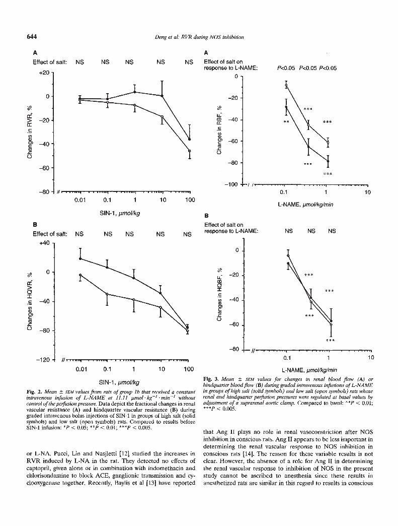
SIN-i, pmol/kgFig. 2. Mean SEM values from rats of group lb that received a constantintravenous infusion of L-NAME at 11.11 .unol kg1 min' withoutcontrol of the perfusion pressure. Data depict the fractional changes in renalvascular resistance (A) and hindquarter vascular resistance (B) duringgraded intravenous bolus injections of SIN-i in groups of high salt (solidsymbols) and low salt (open symbols) rats. Compared to results beforeSIN-i infusion: *P < 0.05; ** < 0.01; 4"P < 0.005.
0.1 1 10
Fig. 3. Mean SEM values for changes in renal blood flow (A) orhindquarter blood flow (B) during graded intravenous infusions of L-NAMEin groups of high salt (solid symbols) and low salt (open symbols) rats whoserenal and hindquarter perfusion pressures were regulated at basal values byadjustment of a suprarenal aortic clamp. Compared to basal: ** < 0.01;***p < 0.005.
P<0.05 P<0.05 P<0.05
**>CCl)a)CCe
0
>0ICCl)
C)C(a
0
0.01 0.1 1 10 100
A AEffect of salt: NS NS NS NS NS Effect of salt on
response to L-NAME:+20
0
0 —20
U-
j::
: ::
B Effect of salt onresponse to L-NAME:
0
—20
0ICU)a)0)C
0-60
—80
SIN-1,pmol/kg B
100.1
L-NAME, pmol/kg/min
NS NS NS
+40,
0.
—40 -
//
0.01 0.1 1 10 100 L-NAME, jimol/kg/mm
or L-NA. Pucci, Lin and Nasjletti [12] studied the increases inRVR induced by L-NA in the rat. They detected no effects ofcaptopril, given alone or in combination with indomethacin andchiorisondamine to block ACE, ganglionic transmission and cy-clooxygenase together. Recently, Baylis et al [13] have reported
that Ang II plays no role in renal vasoconstriction after NOSinhibition in conscious rats. Ang II appears to be less important indetermining the renal vascular response to NOS inhibition inconscious rats [14]. The reason for these variable results is notclear. However, the absence of a role for Ang II in determiningthe renal vascular response to inhibition of NOS in the presentstudy cannot be ascribed to anesthesia since these results inanesthetized rats are similar in this regard to results in conscious

Deng et al: RVR duringNOS inhibition 645
NS NS NS
Fig. 5. Mean SEM values for fractional changes in renal vascular resistance(panel A) or hindquarter vascular resistance (B) in groups of rats adapted tohigh salt () or low salt (E') diets in response to intravenous infusion ofL-NAME at 11.11 p.inol kg1 mm without control of perfusion pressure.Abbreviations are: Control, no pretreatment; Cap, 3 days of captopriladministration in the drinking water and bolus intravenous captopril withsurgely on the study day (group 4); LOS, 3 days of losartan administrationin the drinking water and bolus IV losartan with surgery on the study day(group 5).
L-NAME, pmol/kg/min
Fig. 4. Mean SEM values for plasma renin activity (PRA; A) or fractionalchanges (B) during intravenous infusions of L-NAME (group 2) in groups ofhigh salt (solid symbols) and low salt (open symbols) rats whose renalperfusion pressure was not controlled. Compared to baseline: *P < 0.05;***p < 0.005.
rats [121. Nor are the results readily explained by differences in thebaseline values for PRAsince captopril or losartan did not modifythe RVR response to L-NAME in HS rats whose basal value forPRA were less than 30% of the value of LS rats. One possible
explanation for the different conclusion from the present study isthe experimental condition. In this study, the drugs were given forthree days prior to testing.
In conclusion, dietary salt intake modulates the renal, but notthe hindquarter, vascular response to inhibition of NOS. Aselective increase in NO generation in the kidneys during a highsalt intake may be important for homeostasis by diverting bloodflow to the kidneys, maintaining or increasing the GFR, bluntingthe tubuloglomerular feedback response [5], and inhibiting NaCIreabsorption by the thick ascending limbs of the loop of Henle[25]. A high salt intake appears to increase renal NO generationlargely independent of the effects of Ang II generation.
ANo
pretreatment
P<0.005
Captoprilpretreatment
P<O.005
A
B
+200
+150
Losartanpretreatment
P<0.05
+100
+50
>m
U)a)
(5
* **
>0IU)
C)CS
0
.q:
a-
a-CU)a)C)CCS
0
I —, 1 IBasal 0.01 0.1 1 10
40
30
20
10
0
+20
0
—20
—40
—60
—80
0
L-NAME, pmol/kg/min
BNo
+250 pretreatment
— —---Captopril
pretreatmentLosartan
pretreatment
+200
+150
+100
+50
0-
I0.01 0.1 1 10

646 Deng et al: RVR during NOS inhibition
Acknowledgments
We are grateful to Harold Snellen, B.Sc. for performing the PRA assayand Ms. Michelle Saunier for typing the manuscript. The work wassupported by grants to C.S.W. from the Department of Veteran's Affairs,Washington, D.C. and from the National Institutes of Health (RO1-DK-46995-01). The 3-morpholino-sydnonimine hydrochloride was a gift fromDr. Henning of Cassella AG, Pharmaforschung Galenik Hanauer, Land-strasse 526, 6000 Frankfurt, Germany and the losartan from Ron Smith,Ph.D. (DuPont-Merck Pharmaceuticals, Inc., Wilmington, Delaware,USA).
Reprint requests to Christopher S. Wilcox, M.D., Ph.D., Division ofNephrology and Hypertension, Georgetown University Medical Center, 3800Reservoir Road NW, PHC F6003, Washington, D.C. 20007, USA.
References
1. 1Mb JD, ROMAN RJ: Nitric oxide modulates vascular tone in preglo-merular arterioles. Hypertens 19:770—774, 1992
2. ITo S, JoHNSoN CS, CARRETrERO OA: Modulation of angiotensin11-induced vasoconstriction by endothelium-derived relaxing factor inthe isolated micro-perfused rabbit afferent arteriole. (abstract) J ClinInvest 87:1656—1663, 1993
3. TAKENAKA T, MITCHELL KD, NAVAR LG: Contribution of Angioten-sin II to renal hemodynamic and excretory responses to nitric oxidesynthesis inhibition in the rat. JAm Soc Nephrol 4:1046—1053, 1993
4. OHISHI K, CARMINES PK, INscHo EW, NA VAR LG: EDRF-angiotensinII interactions in rat juxtamedullary afferent and efferent arterioles.Am J Physiol 263:F900—F906, 1992
5. Wii.cox CS, WELCH WJ, MURAD F, GRoss SS, TAYLOR G, LEVI R,SCHMIDT HH: Nitric oxide synthase in macula densa regulates gb-merular capillary pressure. Proc Nati Acad Sd USA 89:11993—11997,1992
6. SHULTZ PJ, TouNs JP: Adaptation to increased dietary salt intake inthe rat: Role of endogenous nitric oxide. J Gun Invest 91:642—650,1993
7. SIGMON DH, CARRETERO OA, BEIERWALTES WH: Plasma reninactivity and the renal response to nitric oxide synthesis inhibition. JAm Soc Nephrol 3:1288—1294, 1992
8. SIGM0N DH, CARRETERO OA, BEIERWALTES WH: Angiotensin de-pendence of endothelium-mediated renal hemodynamics. Hypertens20:643—650, 1992
9. TOLINS JP, RAIJ L: Effects of amino acid infusion on renal hemody-namics: Role of endothelium-derived relaxing factor. Hypertens 17:1045—1051, 1991
10. SALAzAR FJ, PINILLA JM, LOPEZ F, ROMERO JC, QUESADA T: Renal
effects of prolonged synthesis inhibition of endothelium-derived nitricoxide. Hypertens 20:113—117, 1992
11. SIGMON DH, CARRETERO OA, BEIERWALTES WH: Endothelium-derived relaxing factor regulates renin release in vivo. Am J Physiol256(Renal, Fluid, and Electrol Physiol 32) F256—F261, 1992
12. PUCCI ML, LIN L, NASJLETFI A: Pressor and renal vasoconstrictoreffects of NG-nitro-L-arginine as affected by blockade of pressormechanisms mediated by the sympathetic nervous system, angioten-sin, prostanoids and vasopressin. JPharmacol Exp Ther 261:240—245,1992
13. BAYLIS C, ENGLES K, SAMSELL L, HARTON P: Renal effects of acuteendothelium-derived relaxing factor blockade are not mediated byangiotensin II. Am J Physiol 264:F74—F78, 1993
14. SIGMON DH, BEIERWALTES WH: Angiotensin H: Nitric oxide interac-tion and the distribution of blood flow. Am J Physiol 265:R1276—R1283, 1993
15. DENG X, SNELLEN H, WILCOX CS, WELCH WJ: The ultrasonic flowmeter: An accurate and reliable measurement of renal blood flow.(abstract) JAm Soc Nephrol 3:528, 1992, 1993
16. WILCOX CS, WELCH WJ, SNELLEN H: Thromboxane mediates renalhemodynamic response to infused angiotensin II. Kidney Int 40:1090—1097, 1991
17. WELCH WJ, On CE, LORENZ JN, KOTCHEN TA: Control of reninrelease by dietary NaCI in the rat. Am J Physiol 253 (Renal FluidElectrol Physiol 22) F1051—F1057, 1987
18. WELCH WJ, WiLcox CS, DUNEAR KR: Modulation of renin bythromboxane: studies with thromboxane synthase inhibitor, receptorantagonists, and mimetic. Am J Physiol 257 (Renal, Fluid ElectrolPhysiol) 25:F554—F560, 1989
19. KAISER L: Adjusting for baseline: Change or percentage change?Statistics in Med 8:1183—1190, 1989
20. CHEN PY, SANDERS PW: L-arginine abrogates salt-sensitive hyperten-sion in Dahl/Rapp rats. J Cliii Invest 88:1559—1567, 1991
21. GARDINER SM, COMPTON AM, KEMP PA, BENNETr T: Regional andcardiac haemodynamics effects of N°-nitro-L-arginine methyl ester inconscious, Long Evans rats. Br J Pharmacol 101:625—631, 1990
22. WELCH WJ, WILcox CS: Independent effects of salt intake andangiotensin II on the macula densa-nitric oxide signalling pathway(abstract). JAm Soc Nephrol 4:572, 1993
23. VANHOUETTE PM, AUCH-SCI-IWELK W, BIONDI ML, LORENZ RR,SCHINI VB, VIDAL Mi: Why are converting enzyme inhibitors vasodi-lators. Br J Cliii Pharmacol 28:95S—104S, 1989
24. HOLTZ J, BUSSE R, SOMMER 0, BASSENGE E: Dilation of epicardialarteries in conscious dogs induced by angiotensin-converting enzymeinhibition with enalaprilat. J Cardiovasc Pharmacol 9:348—355, 1987
25. BAILLY C, NEANT F, AMIEL C: Cyclic GMP mediates the inhibition byplatelet-activating factor of Cl reabsorption in the cTAL. (abstract) JAm Soc Nephrol 4:450, 1992