Embed Size (px)
Citation preview
Renal TumorsMB3 Lecture series 2015
June 2015 Dr. Andrew Odhiambo MBChB, MMed (Int. Med)
Assistant Lecturer (Hematology/Oncology)Department of Clinical Medicine
Anatomy of Kidney • pair of organs located in the
abdominal cavity on either side of the spine in a retroperitoneal position.
• Adrenal glands rest on top of each kidney
• Approx. at vertebral level T12 to L3, right kidney being slightly lower than the left.
• Long axis of kidney is directed downward and laterally
• Approx. 11–14 cm in length, 6 cm wide and 4 cm thick
• weighs around 150 gm in males & 135 gm in females.
• Mobile Organ, move vertically within retroperitoneum 0.9 cm to 1.3 cm., as much as 4 cm during normal respiration
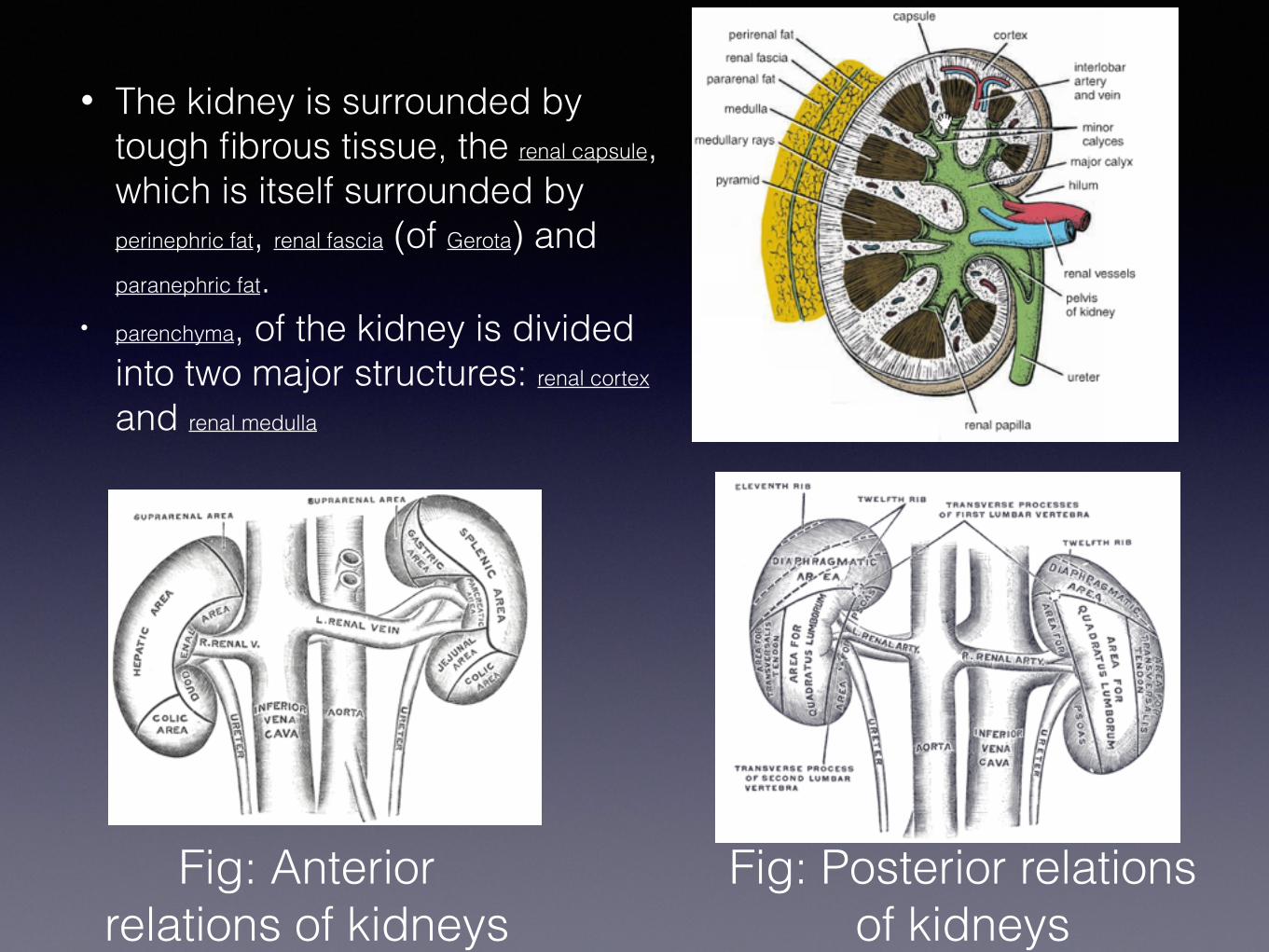
• The kidney is surrounded by tough fibrous tissue, the renal capsule, which is itself surrounded by perinephric fat, renal fascia (of Gerota) and paranephric fat.
• parenchyma, of the kidney is divided into two major structures: renal cortex and renal medulla
Fig: Anterior relations of kidneys
Fig: Posterior relations of kidneys
Anat contd• Blood Supply
• Approx. 20% of the cardiac output . • From renal arteries, left and right, which branch directly
from the abdominal aorta. • renal vein emerges from hilum and drains into the inferior vena cava.
• Lymph Drainage : to the lateral aortic lymph nodes around the origin of the renal artery.
• Nerve Supply: • Through renal sympathetic plexus (T10 – L1) fibres, mainly vasomotor. • Afferent nerves T10 to T12 thoracic nerves.
• Functions • Excretion of wastes, • Acid-base homeostasis, • Osmolality regulation, Blood pressure regulation and Hormone secretion
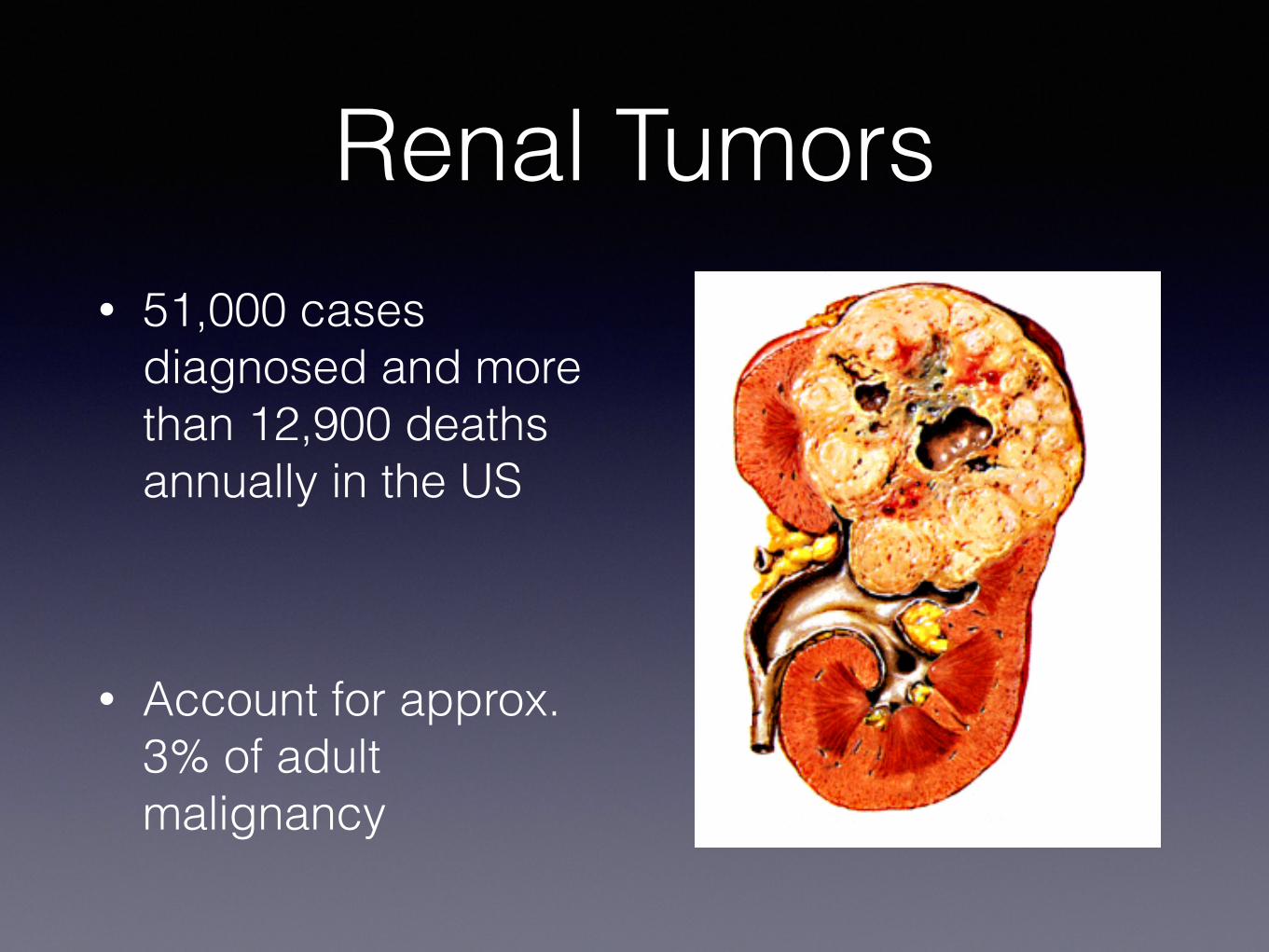
Renal Tumors• 51,000 cases
diagnosed and more than 12,900 deaths annually in the US
• Account for approx. 3% of adult malignancy
Renal tumors…
• Benign • Oncocytoma • Papillary adenoma • Angiomyolipoma
• Malignant• Renal Cell Carcinoma (Adenocarcinoma of
Kidney)
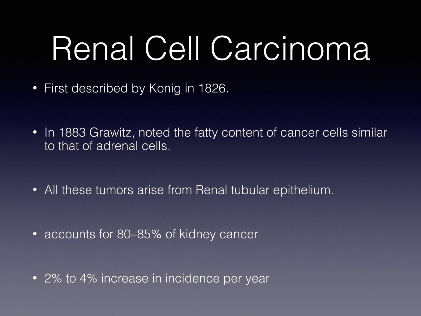
Renal Cell Carcinoma• First described by Konig in 1826.
• In 1883 Grawitz, noted the fatty content of cancer cells similar to that of adrenal cells.
• All these tumors arise from Renal tubular epithelium.
• accounts for 80–85% of kidney cancer
• 2% to 4% increase in incidence per year
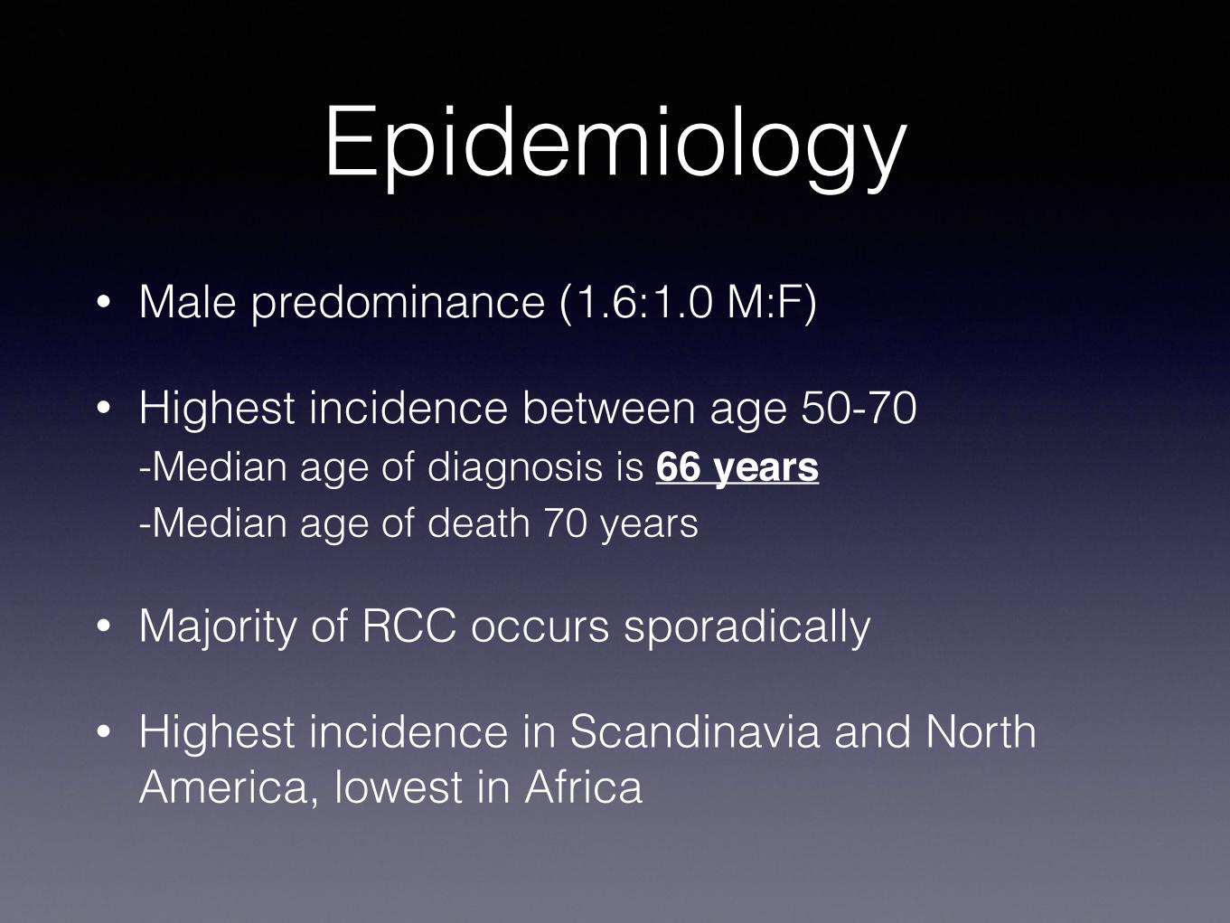
Epidemiology• Male predominance (1.6:1.0 M:F)
• Highest incidence between age 50-70 -Median age of diagnosis is 66 years-Median age of death 70 years
• Majority of RCC occurs sporadically
• Highest incidence in Scandinavia and North America, lowest in Africa
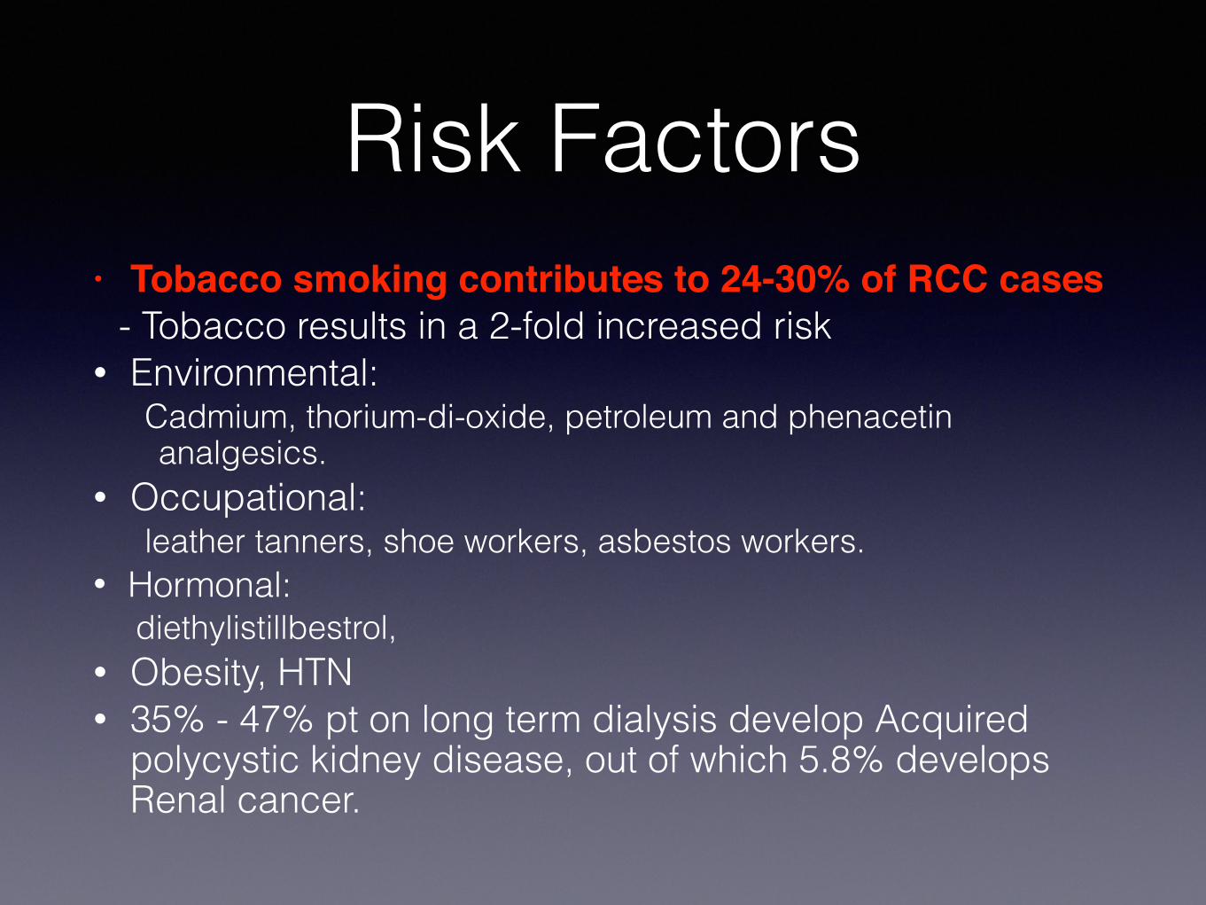
Risk Factors • Tobacco smoking contributes to 24-30% of RCC cases - Tobacco results in a 2-fold increased risk • Environmental:
Cadmium, thorium-di-oxide, petroleum and phenacetin analgesics.
• Occupational: leather tanners, shoe workers, asbestos workers.
• Hormonal: diethylistillbestrol,
• Obesity, HTN • 35% - 47% pt on long term dialysis develop Acquired
polycystic kidney disease, out of which 5.8% develops Renal cancer.
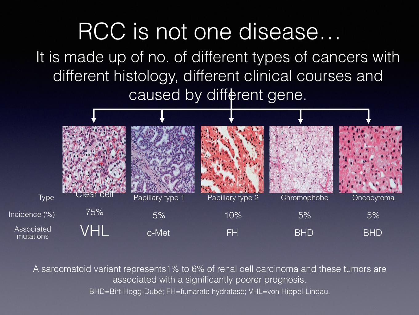
A sarcomatoid variant represents1% to 6% of renal cell carcinoma and these tumors are associated with a significantly poorer prognosis.
BHD=Birt-Hogg-Dubé; FH=fumarate hydratase; VHL=von Hippel-Lindau.
RCC is not one disease…
Clear cell
75% Type
Incidence (%)
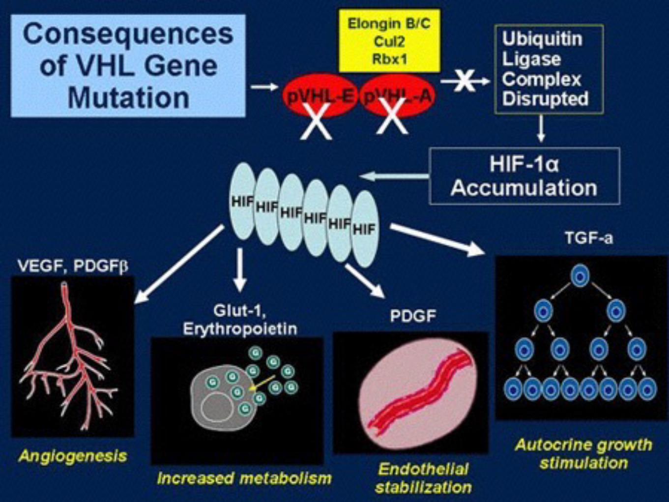
Associated mutations VHL
Papillary type 1
5%
c-Met
Papillary type 2
10%
FH
Chromophobe
5%
BHD
Oncocytoma
5%
BHD
It is made up of no. of different types of cancers with different histology, different clinical courses and
caused by different gene.
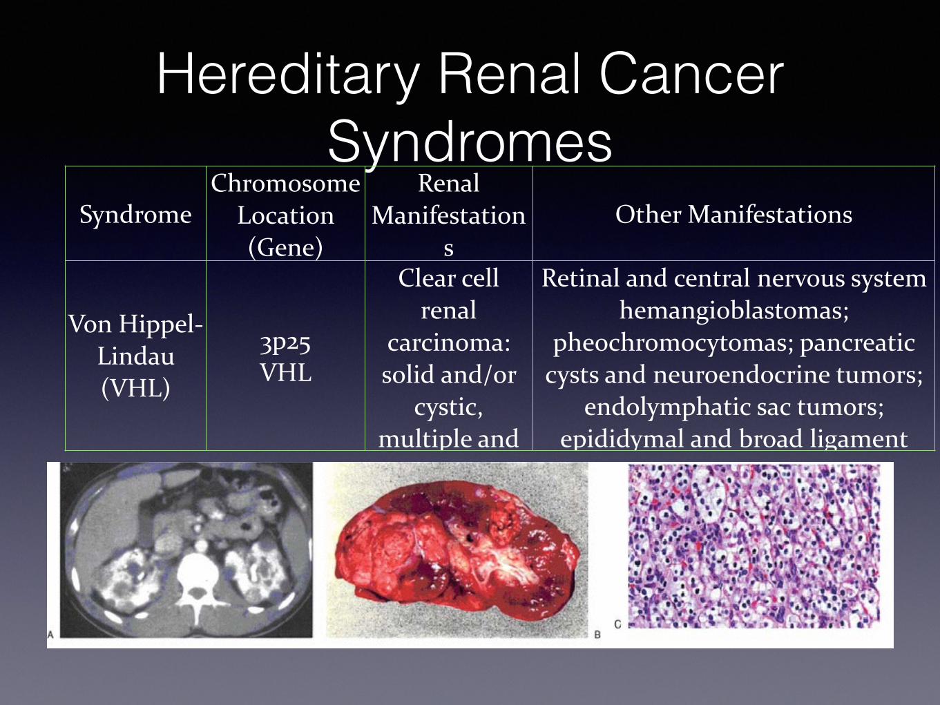
Hereditary Renal Cancer Syndromes
SyndromeChromosome
Location (Gene)
Renal Manifestation
sOther Manifestations
Von Hippel-‐Lindau (VHL)
3p25 VHL
Clear cell renal
carcinoma: solid and/or
cystic, multiple and
Retinal and central nervous system hemangioblastomas;
pheochromocytomas; pancreatic cysts and neuroendocrine tumors;
endolymphatic sac tumors; epididymal and broad ligament
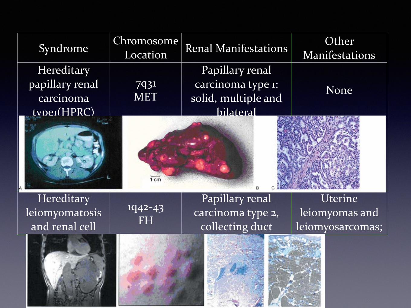
SyndromeChromosome
Location (Gene)
Renal ManifestationsOther
ManifestationsHereditary
papillary renal carcinoma
type1(HPRC)
7q31 MET
Papillary renal carcinoma type 1: solid, multiple and
bilateral
None
Hereditary leiomyomatosis and renal cell carcinoma
1q42-‐43 FH
Papillary renal carcinoma type 2, collecting duct
carcinoma: solitary,
Uterine leiomyomas and leiomyosarcomas;
cutaneous
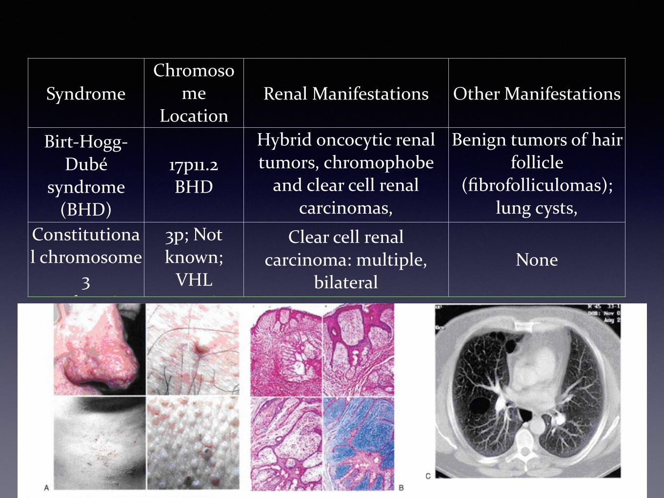
SyndromeChromoso
me Location (Gene)
Renal Manifestations Other Manifestations
Birt-‐Hogg-‐Dubé
syndrome (BHD)
17p11.2 BHD
Hybrid oncocytic renal tumors, chromophobe and clear cell renal
carcinomas, oncocytomas: multiple,
Benign tumors of hair follicle
(fibrofolliculomas); lung cysts, spontaneous Constitutiona
l chromosome 3
translocation
3p; Not known; VHL
somatic
Clear cell renal carcinoma: multiple,
bilateralNone
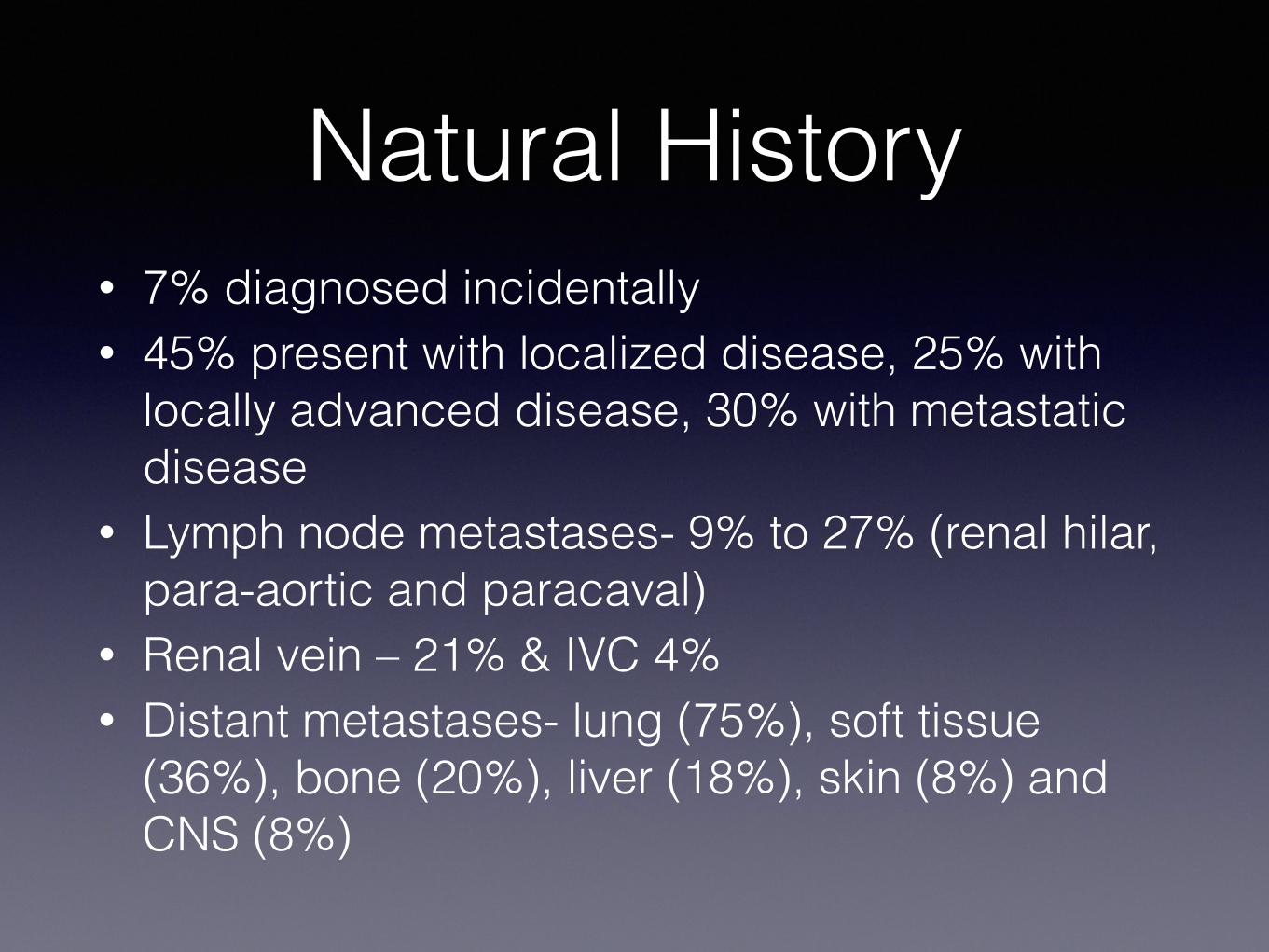
Natural History• 7% diagnosed incidentally • 45% present with localized disease, 25% with
locally advanced disease, 30% with metastatic disease
• Lymph node metastases- 9% to 27% (renal hilar, para-aortic and paracaval)
• Renal vein – 21% & IVC 4% • Distant metastases- lung (75%), soft tissue
(36%), bone (20%), liver (18%), skin (8%) and CNS (8%)
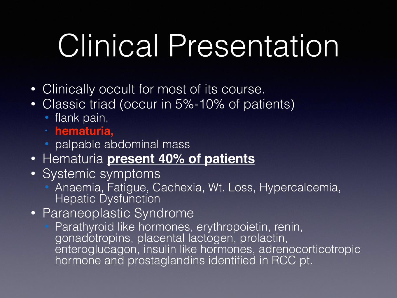
Clinical Presentation• Clinically occult for most of its course. • Classic triad (occur in 5%-10% of patients)
• flank pain, • hematuria, • palpable abdominal mass
• Hematuria present 40% of patients• Systemic symptoms
• Anaemia, Fatigue, Cachexia, Wt. Loss, Hypercalcemia, Hepatic Dysfunction
• Paraneoplastic Syndrome • Parathyroid like hormones, erythropoietin, renin,
gonadotropins, placental lactogen, prolactin, enteroglucagon, insulin like hormones, adrenocorticotropic hormone and prostaglandins identified in RCC pt.
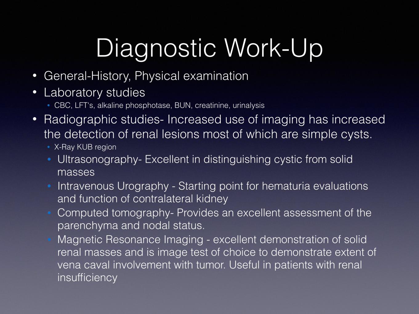
Diagnostic Work-Up • General-History, Physical examination • Laboratory studies
• CBC, LFT's, alkaline phosphotase, BUN, creatinine, urinalysis
• Radiographic studies- Increased use of imaging has increased the detection of renal lesions most of which are simple cysts.
• X-Ray KUB region • Ultrasonography- Excellent in distinguishing cystic from solid
masses • Intravenous Urography - Starting point for hematuria evaluations
and function of contralateral kidney • Computed tomography- Provides an excellent assessment of the
parenchyma and nodal status. • Magnetic Resonance Imaging - excellent demonstration of solid
renal masses and is image test of choice to demonstrate extent of vena caval involvement with tumor. Useful in patients with renal insufficiency
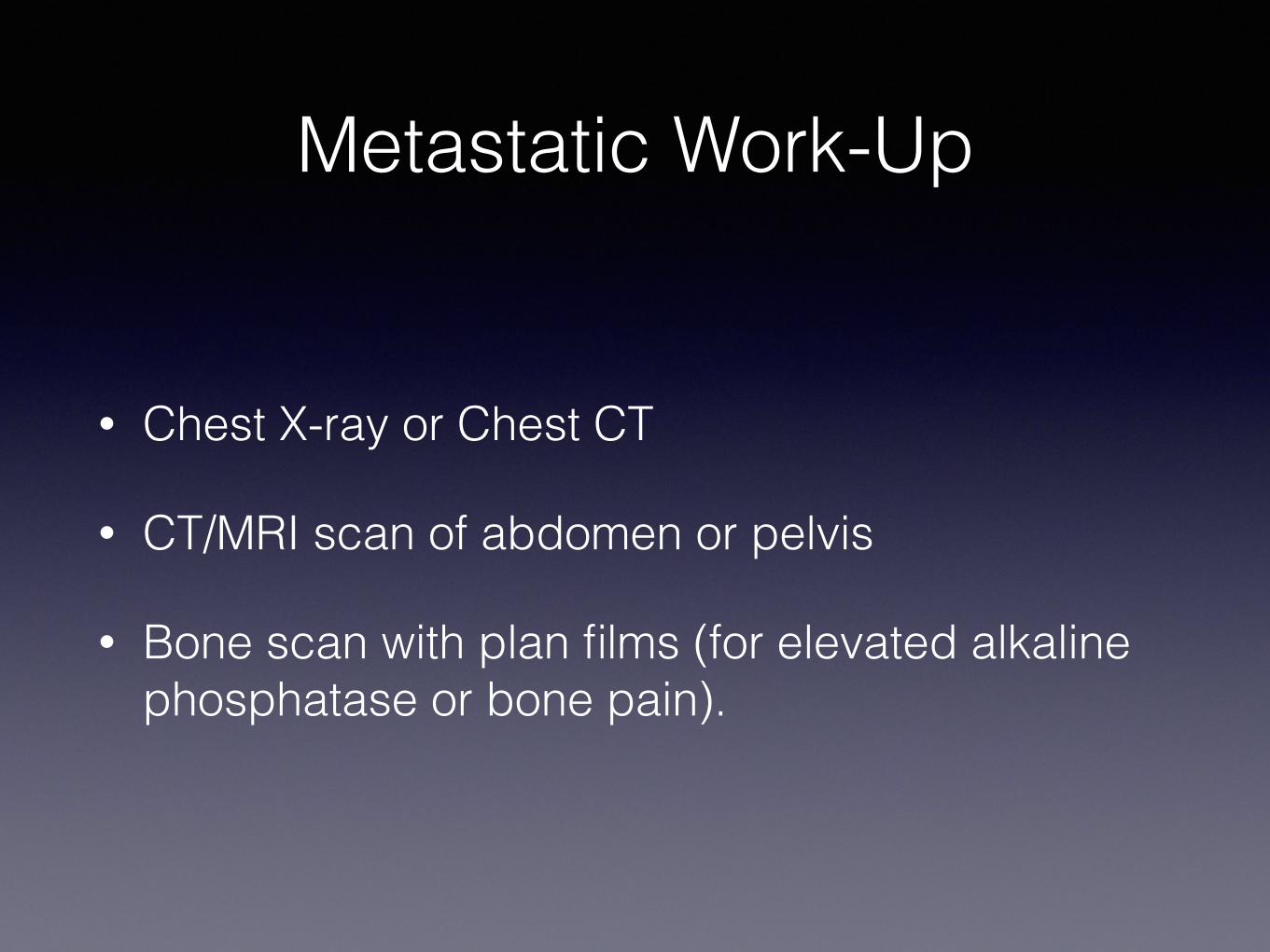
Metastatic Work-Up
• Chest X-ray or Chest CT
• CT/MRI scan of abdomen or pelvis
• Bone scan with plan films (for elevated alkaline phosphatase or bone pain).
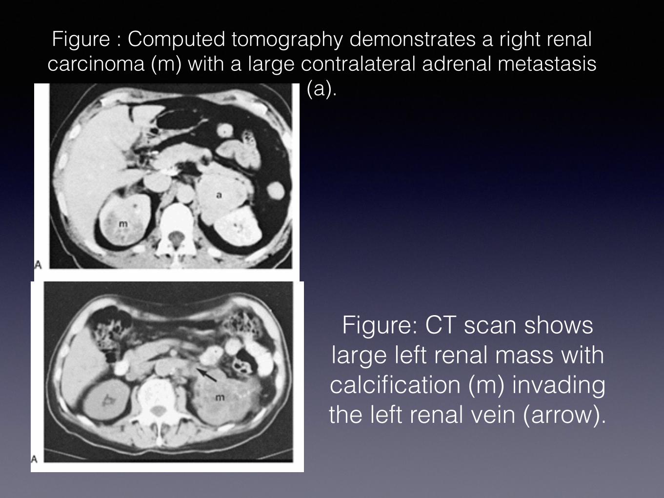
Figure : Computed tomography demonstrates a right renal carcinoma (m) with a large contralateral adrenal metastasis
(a).
Figure: CT scan shows large left renal mass with calcification (m) invading the left renal vein (arrow).
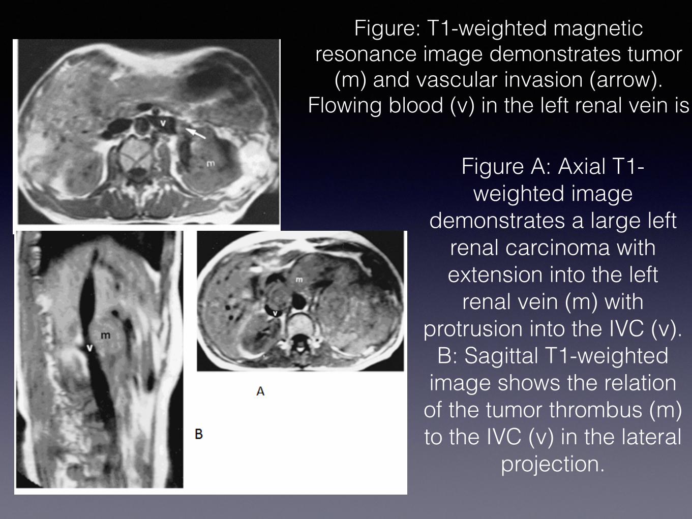
Figure: T1-weighted magnetic resonance image demonstrates tumor
(m) and vascular invasion (arrow). Flowing blood (v) in the left renal vein is
Figure A: Axial T1-weighted image
demonstrates a large left renal carcinoma with extension into the left
renal vein (m) with protrusion into the IVC (v).
B: Sagittal T1-weighted image shows the relation of the tumor thrombus (m) to the IVC (v) in the lateral
projection.
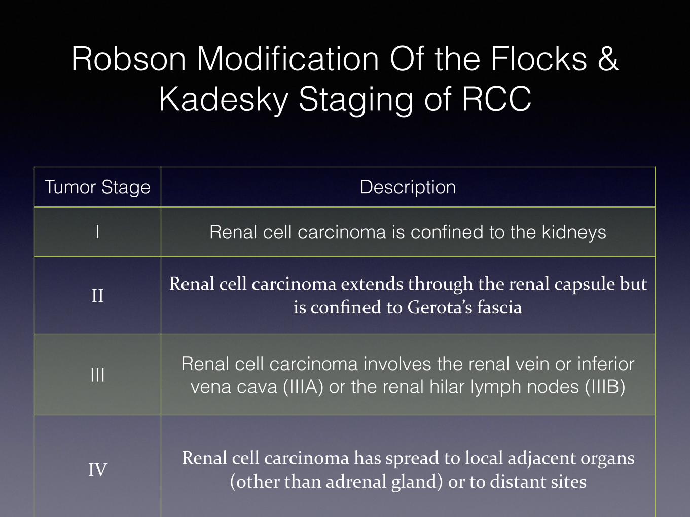
Robson Modification Of the Flocks & Kadesky Staging of RCC
Tumor Stage Description
I Renal cell carcinoma is confined to the kidneys
IIRenal cell carcinoma extends through the renal capsule but
is confined to Gerota’s fascia
III Renal cell carcinoma involves the renal vein or inferior vena cava (IIIA) or the renal hilar lymph nodes (IIIB)
IVRenal cell carcinoma has spread to local adjacent organs
(other than adrenal gland) or to distant sites
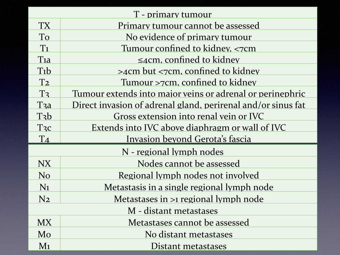
TNM stagingT -‐ primary tumourTX Primary tumour cannot be assessedT0 No evidence of primary tumourT1 Tumour confined to kidney, <7cmT1a ≤4cm, confined to kidneyT1b >4cm but <7cm, confined to kidneyT2 Tumour >7cm, confined to kidneyT3 Tumour extends into major veins or adrenal or perinephric T3a Direct invasion of adrenal gland, perirenal and/or sinus fatT3b Gross extension into renal vein or IVCT3c Extends into IVC above diaphragm or wall of IVCT4 Invasion beyond Gerota’s fascia
N -‐ regional lymph nodesNX Nodes cannot be assessedN0 Regional lymph nodes not involvedN1 Metastasis in a single regional lymph nodeN2 Metastases in >1 regional lymph node
M -‐ distant metastasesMX Metastases cannot be assessedM0 No distant metastasesM1 Distant metastases
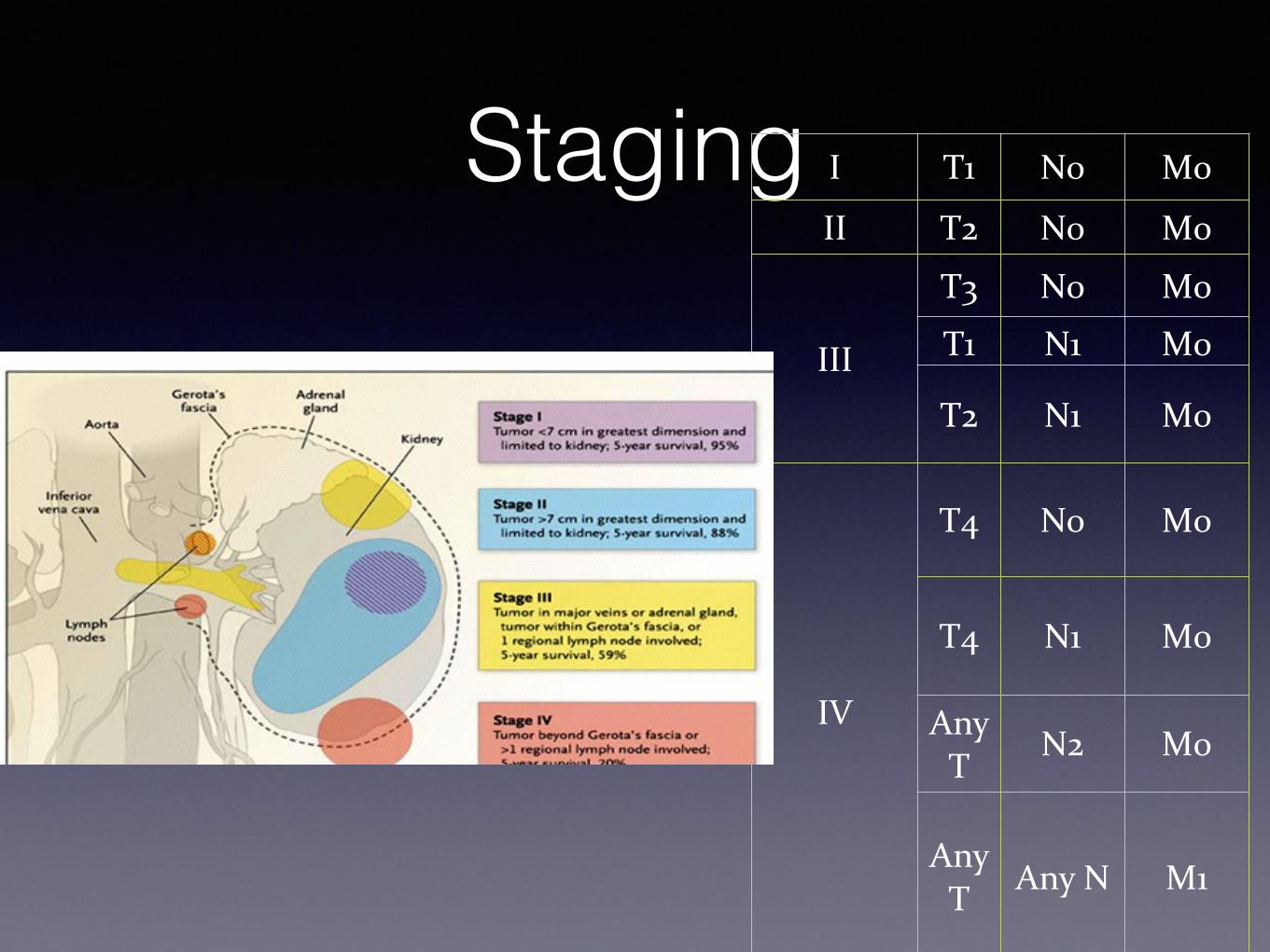
Staging
- Stage I-III: Localized disease - Stage IV: Advanced, metastatic disease
I T1 N0 M0
II T2 N0 M0
III
T3 N0 M0
T1 N1 M0
T2 N1 M0
IV
T4 N0 M0
T4 N1 M0
Any T
N2 M0
Any T Any N M1
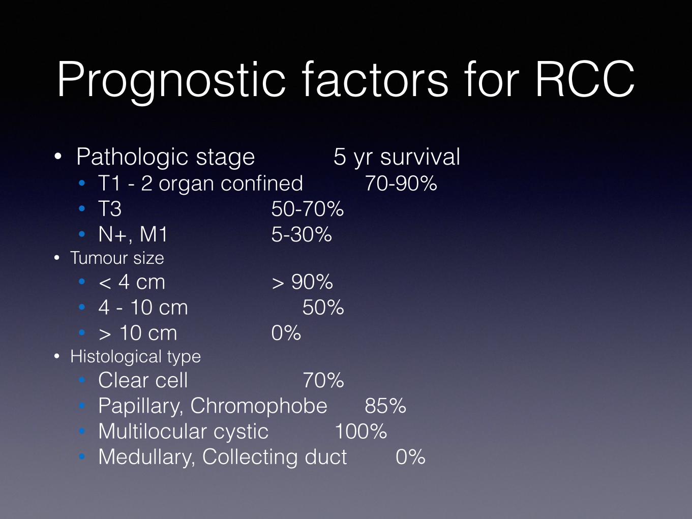
Prognostic factors for RCC• Pathologic stage 5 yr survival
• T1 - 2 organ confined 70-90% • T3 50-70% • N+, M1 5-30%
• Tumour size • < 4 cm > 90% • 4 - 10 cm 50% • > 10 cm 0%
• Histological type • Clear cell 70% • Papillary, Chromophobe 85% • Multilocular cystic 100% • Medullary, Collecting duct 0%
Management
• Localized disease • Metastatic disease
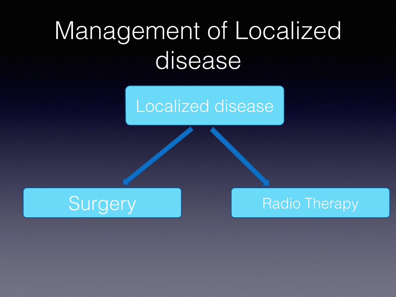
Management of Localized disease
Localized disease
Surgery Radio Therapy
Surgery- Radical nephrectomy
Gold standard treatment for localized RCC with contralateral normal kidney, adequate surgical margin.
Principles of Surgery- Early ligation of renal artery and vein , removal of kidney including Gerota’s fascia, removal of ipsilateral adrenal gland, regional lymphadenectomy from crus of diaphragm to aortic bifurcation.
Indications 1. Bilateral RCC 2. RCC in a solitary functioning kidney 3. Unilateral RCC with contralateral kidney under threat of its future function (Renal artery stenosis, Chronic pyelonephritis , Hydronephrosis, Ureteral reflux, Calculus disease, Systemic disease such as diabetes )4. Tumor less than 4cms with normal opposite kidney.
5. Five year survival rate 75% to 85% 6. Local tumor recurrence of 10% is reported.
Other Approaches 1. Radio frequency ablations 2. Cryo ablation
Nephron Sparing Surgery
Radiotherapy
• Radiosensitivity of RCC is variable • Animal experiments suggest a theoretical benefit
to preoperative RT (? Reduce intra-operative seeding)
• Historically several series suggested clinical benefit to adjuvant (post-op) RT • Limited applicability because of long time span,
improvements in staging, surgery, changing RT technology
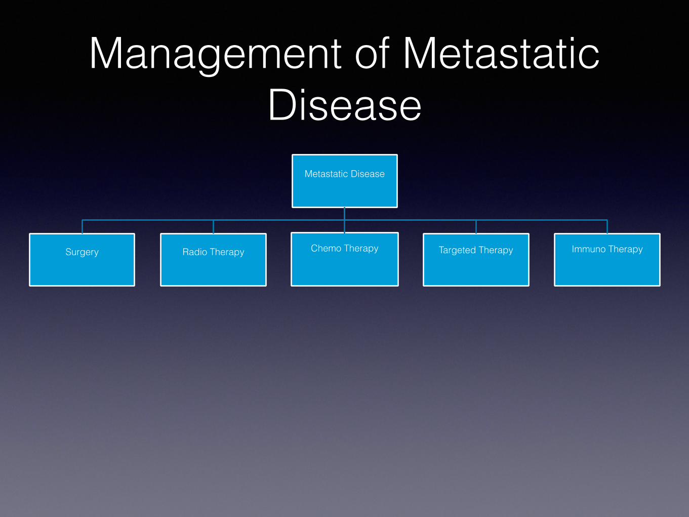
Management of Metastatic Disease
Metastatic Disease
Surgery Radio Therapy Chemo Therapy Targeted Therapy Immuno Therapy
Surgery• Palliative Nephrectomy – Indicated in patients with
• Severe hemorrhage, • Severe pain, • Paraneoplastic syndrome • or compression of adjacent viscera • Solitary metastasis can be resected and may show some survival
advantage • Therapeutic:
• Not curative but produce some long-term survivors. • The possibility of disease-free survival increases after resection of
primary tumor and isolated metastasis excision. • to decrease tumor burden in preparation for subsequent therapy
Surgery…
• Resection of met’s • in pt. not relieved from palliative RT • In solitary mets.
• Spontaneous regression of met’s • < 1 % of cases • only 4 (0.8%) of 474 patients in 9 series who
underwent nephrectomy experienced regression of metastatic foci
Radio Therapy
• Palliation • Used for local or symptomatic metastatic disease,
such as painful osseous lesions or brain metastasis. • Treatment field encompasses metastatic deposit (or
local recurrence) with 2-3cm margins • Higher doses (up to 35-40Gy) may be required to
overcome radioresistance • Symptomatic relief in 64-84% of patients
Chemotherapy• RCC is a chemo resistant tumor. Phenomenon due to
presence of multi drug resistant glycoprotein (MDR) in tumor cell - causes extrusion of the drug
• Conventional therapy has little to offer
• 5-FU alone has a response rate of 10%,
• On-going clinical trials of combination chemotherapy including Gemcitabine and 5-FU
• Limited data reveals some response in non-clear cell RCC to Carboplatin, Cisplatin plus Gemcitabine
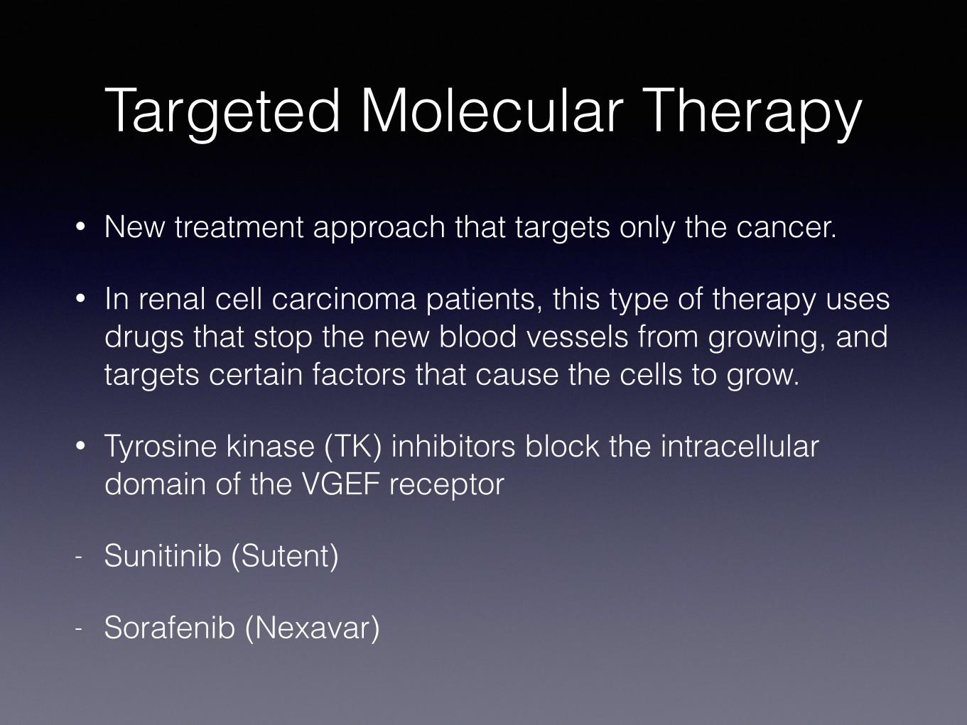
Targeted Molecular Therapy • New treatment approach that targets only the cancer.
• In renal cell carcinoma patients, this type of therapy uses drugs that stop the new blood vessels from growing, and targets certain factors that cause the cells to grow.
• Tyrosine kinase (TK) inhibitors block the intracellular domain of the VGEF receptor
- Sunitinib (Sutent)
- Sorafenib (Nexavar)
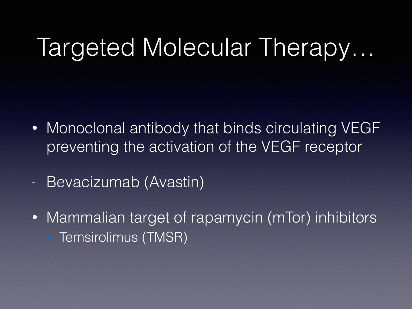
Targeted Molecular Therapy…
• Monoclonal antibody that binds circulating VEGF preventing the activation of the VEGF receptor
- Bevacizumab (Avastin)
• Mammalian target of rapamycin (mTor) inhibitors • Temsirolimus (TMSR)
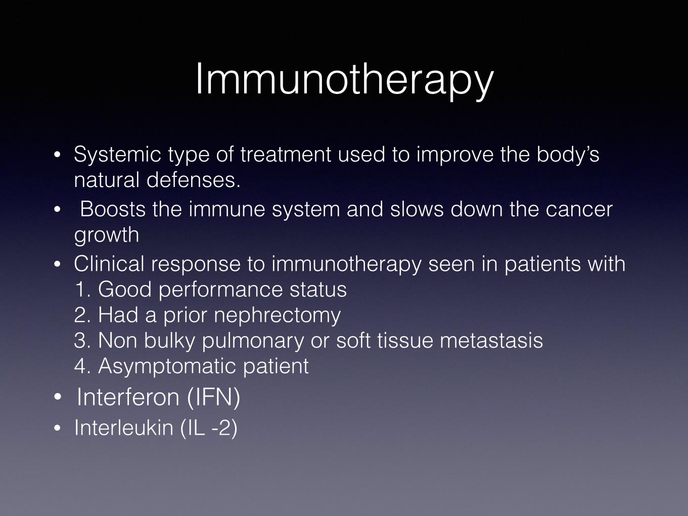
Immunotherapy• Systemic type of treatment used to improve the body’s
natural defenses. • Boosts the immune system and slows down the cancer
growth • Clinical response to immunotherapy seen in patients with
1. Good performance status 2. Had a prior nephrectomy 3. Non bulky pulmonary or soft tissue metastasis 4. Asymptomatic patient
• Interferon (IFN) • Interleukin (IL -2)
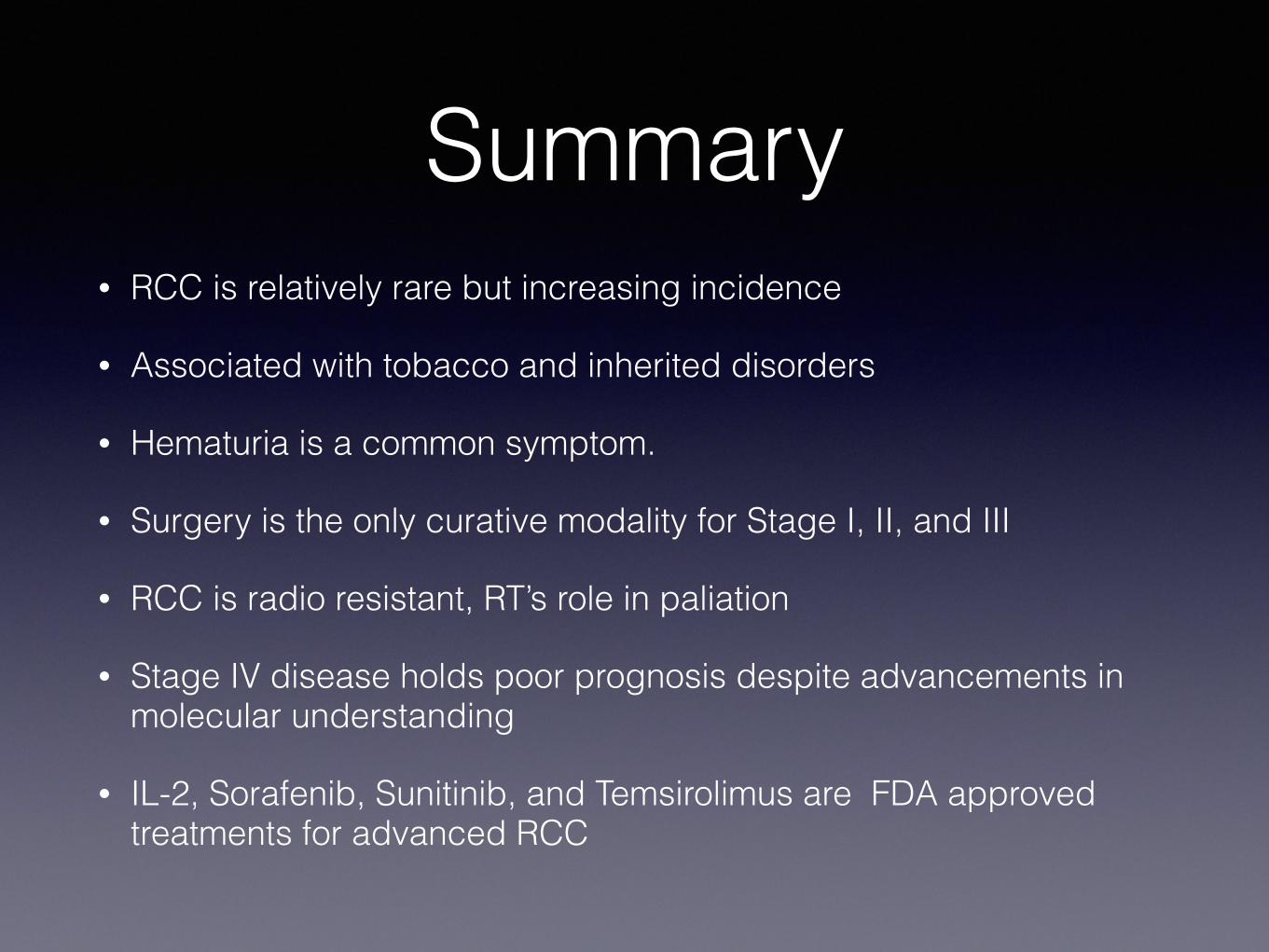
Summary• RCC is relatively rare but increasing incidence
• Associated with tobacco and inherited disorders
• Hematuria is a common symptom.
• Surgery is the only curative modality for Stage I, II, and III
• RCC is radio resistant, RT’s role in paliation
• Stage IV disease holds poor prognosis despite advancements in molecular understanding
• IL-2, Sorafenib, Sunitinib, and Temsirolimus are FDA approved treatments for advanced RCC
Thank you !
• Questions

![4- GU Onc351[1] Semester/Surgery/22- Common...Adrenal Tumors. Renal Tumors. Renal Tumors Benign tumours of the kidney are rare All renal neoplasms should be regarded as potentially](https://img.pdfslide.us/doc/110x75/5f0983b17e708231d4273048/4-gu-onc3511-semestersurgery22-common-adrenal-tumors-renal-tumors-renal.jpg)