Embed Size (px)
Citation preview

REMOVAL OF HEAVY METAL IONS FROM AQUEOUS SOLUTIONS
ONTO BIOMASS OF ASPERGILLUS NIGER.
CHEONG HUI MIN
A project report submitted in partial fulfilment of the
requirements for the award of the degree of
Bachelor (Hons.) of Chemical Engineering
Faculty of Engineering and Science
Universiti Tunku Abdul Rahman
April 2011

DECLARATION
I hereby declare that this project report is based on my original work except for
citations and quotations which have been duly acknowledged. I also declare that it
has not been previously and concurrently submitted for any other degree or award at
UTAR or other institutions.
Signature : _________________________
Name : Cheong Hui Min n
ID No. : 07UEB06774 2
Date : _________________________

ii
APPROVAL FOR SUBMISSION
I certify that this project report entitled “REMOVAL OF HEAVY METAL IONS
FROM AQUEOUS SOLUTIONS ONTO BIOMASS OF ASPERGILLUS
NIGER” was prepared by CHEONG HUI MIN has met the required standard for
submission in partial fulfilment of the requirements for the award of Bachelor of
ENGINEERING (Hons.) CHEMICAL ENGINEERING at Universiti Tunku Abdul
Rahman.
Approved by,
Signature : _________________________
Supervisor : Dr. Gulnaziya Issabayeva
Date : _________________________

iii
The copyright of this report belongs to the author under the terms of the
copyright Act 1987 as qualified by Intellectual Property Policy of University Tunku
Abdul Rahman. Due acknowledgement shall always be made of the use of any
material contained in, or derived from, this report.
© 2011, Cheong Hui Min. All right reserved.

iv
Specially dedicated to
my beloved family.

v
ACKNOWLEDGEMENTS
I would like to thank everyone who had contributed to the successful completion of
this project. First and foremost, I would like to express my gratitude to my project
supervisor, Dr. Gulnaziya Issabayeva for her invaluable advices, guidance and her
enormous patience throughout the development of the research. I thank her for
providing us useful information, materials and techniques required to complete our
research. Besides, million thanks to Ms Chen Siew Kim for her utmost assistance in
providing useful information regarding the project.
In addition, I would also like to express my gratitude to my course-mate, Lau
Miew Cheong who has been working together with me and discussing the problems
faced, and also to my loving parents who had being supportive and encouraging.
Last but not least, I would like to express my appreciation to Universiti
Tunku Abdul Rahman (UTAR) for providing me a platform to gain extra knowledge
and develop problem solving skills throughout the experimental process and report
writing.

vi
REMOVAL OF HEAVY METAL IONS FROM AQUEOUS SOLUTIONS
USING BIOMASS OF ASPERGILLUS NIGER.
ABSTRACT
There is a need to develop cost-effective technologies that can remove toxic heavy
metals ions such as lead from wastewater. In this study, the biosorption characteristic
of Aspergillus niger (A.niger) fungal biomass on lead, Pb(II) removal was examined
as a function of initial pH and metal ion concentrations. Dried biomass of A.niger
was used in the study. The equilibrium data obtained were analysed using both
Langmuir and Freundlich isotherm models and examined using non-linear regression
analysis. The results showed that the equilibrium data fitted well to both Langmuir
and Freundlich models except at pH 2 and pH 3, respectively. The pH of the metal
ion solutions strongly affected the degree of heavy metal ions on the dried biomass.
Biosorption of metal ions was inhibited at pH values below 3 whereas the adsorption
capacity increased sharply when the pH values increased from 5 to 6. The maximum
adsorption uptake was found to be 66.23 mg/g at pH 6. The percentage removal of
lead ions by A.niger increased when the initial concentration of metal ions solution
increased from 5 to 200ppm. The kinetic study of biosorption of heavy metal ions by
A.niger presents the Ho and McKay’ pseudo-second order adsorption with a
correlation coefficient of 0.99 which also described a chemisorption happened during
the metal ion uptake mechanism. The results of this study indicated that A.niger is a
suitable biosorbent for the removal of Pb(II) ions from aqueous solution.

vii
TABLE OF CONTENTS
DECLARATION i
APPROVAL FOR SUBMISSION ii
ACKNOWLEDGEMENTS v
ABSTRACT vi
TABLE OF CONTENTS vii
LIST OF TABLES ix
LIST OF FIGURES x
LIST OF SYMBOLS / ABBREVIATIONS xii
CHAPTER
1 INTRODUCTION 1
1.1 Biosorption prospects 1
1.1.1 The achievement over the past decades 2
1.2 Objectives 3
2 LITERATURE REVIEW 4
2.1 Presence of heavy metals in environment 4
2.2 Conventional technologies in metal ion removal 6
2.3 Biosorption technology 7
2.4 Biosorption versus bioaccumulation 10
2.5 Mechanism of metal uptake 12
2.6 Fungi 15
2.6.1 Classification and general characteristics 15
2.6.2 Cell wall and its main composite 17

viii
2.6.3 Fungal biomass as biosorbent 17
2.6.4 Aspergillus niger 18
3 METHODOLOGY 21
3.1 Preparation of adsorbent 21
3.2 Preparation of metal ion solutions 22
3.3 Preparation of blank solution 22
3.4 Metal ions solution 22
3.5 Batch biosorption studies 23
3.6 Equipments 24
3.7 Metal uptake capacity 25
3.8 Biosorption isotherms 26
3.8.1 Langmuir isotherm model 27
3.8.2 Freundlich isotherm model 29
3.9 Biosorption kinetic studies 29
3.9.1 Pseudo-first order kinetics model 30
3.9.2 Pseudo-second order kinetics model 31
4 RESULTS AND DISCUSSIONS 32
4.1 Batch isotherm studies 32
4.2 Batch kinetic studies 37
4.3 Effect of pH on the metal uptake 39
4.4 Effect of initial concentration on metal uptake 41
5 CONCLUSION AND RECOMMENDATIONS 45
5.1 Conclusion 45
5.2 Recommendations 46
5 REFERENCES 47

ix
LIST OF TABLES
TABLE TITLE PAGE
2.1 Conventional metal removal technologies. (Source:
Volesky, 2000) 7
2.2 Biosorption factors. (Source: Chojnacka, 2010) 9
2.3 The comparison between biosorption and
bioaccumulation. (Source: Chojnacka, 2010) 11
2.4 Functional groups of biosorbents. (Source:
Volesky, 2007) 14
2.5 The classification of fungi. (Source: Wang &
Chen, 2006) 16
2.6 Cellular organization in filamentous fungi. 16
2.7 Biosorption capacity of A. niger (mg/g) on metal
ions. 20
3.1 Adsorption Isotherms. Source: (Kapoor &
Viraraghavan, 1995). 26
4.1 Equilibrium and kinetic studies of heavy metal
adsorption onto Aspergillus niger. (Source:
Lesmana et. al., 2009) 33
4.2 Langmuir and Freundlich isotherm model
constants. 37
4.3 Percentage removal of fungus A. niger. 43
x
LIST OF FIGURES
FIGURE TITLE PAGE
2.1 The cell wall composition of fungi. 17
2.2 Scanning electron micrograph, SEM of the
asexual reproductive apparatus of Aspergillus
niger. Source: (Read, 2007) 19
3.1 Schematic diagram of bath biosorption
equilibrium study. 23
3.2 Orbital shaker (SSL1;Stuart®) 24
3.3 Oven (Beschickung-Loading Modell 100-
800;Memmert) 24
3.4 ICP (Optima 7000DV;Perkin Elmer, Uberlingen,
Germany). 25
3.5 Comparative example of sorption isotherm curve. 27
4.1 The adsorption isotherm curve for lead at different
pHs. 33
4.2 Langmuir isotherms for lead removal at pH 1 – 3. 35
4.3 Langmuir isotherms for lead removal at pH 4 – 6. 35
4.4 Freundlich isotherms for lead removal at pH 1 – 3. 36
4.5 Freundlich isotherms for lead removal at pH 4 – 6. 36
4.6 The pseudo first-order kinetics data for lead
adsorption on A.niger. 38
4.7 The pseudo second-order kinetics for lead
adsorption on A.niger. 39
4.8 The maximum adsorption of Pb ions at different
pH values. 40

xi
4.9 Effect of initial lead ions concentration on
biosorption capacity of A.niger. 42
xii
LIST OF SYMBOLS / ABBREVIATIONS
M molarity, mol/L
M amount of dry biosorbent, g
T temperature, ºC
Ce equilibrium ion concentration, mg/L
initial ion concentration, mg/L
residual concentration, mg/L
k rate constant of pseudo-first order
k2 rate constant of pseudo-second order
KF Freundlich constant
KL Langmuir constant
q/Q metal uptake rate, mg/g
Qe metal uptake at equilibrium, mg/g
Qmax maximum adsorption capacity, mg/g
Qt metal uptake at time t, mg/g
reaction volume, L
Θ surface sites
ICP inductively coupled plasma

1 INTRODUCTION
1.1 Biosorption prospects
The history of research based on the biomass-metal interactions is dated back to the
1960s (Chojnacka, 2010). It was discovered that the metal’s properties can be altered
by biomass in an amazing way. The processes of biological metal binding were
found to be useful for biohydrometallurgy and biogeochemistry.
In the next ten years after the discovery, the first description of
bioaccumulation as the method of wastewater treatment was elaborated. It was
reported by Chojnacka (2010) that in the lagoon in the vicinity of lead and zinc
mining and milling works a dense mat-like growth of algae was observed. The
observation also proved that the algae were capable of accumulating metal cations
and eliminating other competing organisms.
According to Gupta et al. (2000), biosorption was regarded as an emergent
technology at a meeting organized by the Solvent Engineering Extraction and Ion
Exchange Group of the Society of Chemical Industry at UK in year 1986. Since then
a number of research centre from all over the world have been engaged in the area
ofbiosorption with precise goals of identifying potential biomass in the removal of
heavy metal from industrial wastewater (Volesky & Holan, 1995).
The history of biosorption as a regular branch of science began in 1990’s. A
large contribution has been made by Prof. Bohumil Volesky from McGill University
in Canada in providing many theoretical basis of the biosorption process. Biosorption
can be regarded as an alternative cleanup process because it is cost-effective in terms

2
of the source of biosorbents, feasible desorbing process for the reuse and
regeneration of biomass and other properties (Volesky, 2007).
Many biological materials bind heavy metals; however, the implementation
of a full-scale biosorption process needs only sorbents with sufficiently high metal
binding capacity and selectivity. Thus, the first major challenge for the biosorption
field is to select the most promising types of biomass from an extremely large pool of
readily available and inexpensive biomaterials.
1.1.1 The achievement over the past decades
The researches throughout the years have allowed the biological materials
with high metal binding capacity and selectivity for heavy metals to be utilized in the
full-scale biosorption process (Kratochvil & Volesky, 1998).
According to Volesky (2007), several proprietary biosorption processes were
developed and commercialized early in this decade, such as AlgaSORB™ and AMT-
Bioclaim™ (Goyal & Ahluwalia, 2007), however, a lack of better understanding of
the mechanism underlying the metal sorption process has hindered adequate
assessment of process performance and limitations and thus the expected wide-
spread application of biosorption. Consequently, the selection of the industrial
effluents for pilot testing has remained largely intuitive as has the task of scaling up
the process.

3
1.2 Objectives
The main objective of the present project is to investigate the use of A. niger dry
biomass as a biosorbent for the removal of lead, Pb(II) from aqueous solutions by
using batch method. The optimum biosorption conditions are determined as a
function of initial pH, initial metal ion concentration and time.
The specific objectives are as below:
i. to evaluate adsorption capacity of dry biomass of A.niger to remove lead ions
from aqueous solutions.
i. to evaluate the effect of pH on the metal uptake by A.niger.
ii. to evaluate the effect of initial concentration on the adsorption capacity of
A.niger.
iii. to determine the kinetics of the biosorption process for A.niger.

CHAPTER 2
2 LITERATURE REVIEW
2.1 Presence of heavy metals in environment
Metal compounds are released into the environment through industrial activities.
Throughout the world there is a growing concern that the heavy metal contents in
soils and water are increasing as the result of industrial, mining, agricultural and
domestic activities.
The man’s exploitation of the world’s mineral resources and the
technological activities involved along the process tend to unearth, dislodge, and
disperse chemicals and particularly metallic elements, which have recently been
brought into the environment in unprecedented quantities and concentrations and at
extreme rates. The pollutant caused by man-made sources could lead to disastrous
effects on animals and humans.
There are many sources of water pollution, but two main general categories
exist: direct and indirect contaminant sources. Direct sources include effluent outfalls
from industries, refineries and waste treatment plants; whereas, indirect sources
include contaminants that enter the water supply from soils/ground water systems
and from the atmosphere via rain water. In general, contaminants come under two
broad classes which are organic and inorganic. Some organic water pollutants
include industrial solvents, volatile organic compounds, insecticides, pesticides and
food processing wastes. The inorganic water pollutants on the other hand include

5
metals, fertilizers and acidity caused by industrial discharges. The heavy metal
pollutants can be caused by both direct and indirect sources.
According to Naja & Volesky (2009), heavy metals can be defined in several
ways. One possible definition is the following: heavy metals form positive ions in
solution and they have a density five times greater than that of water. They are of
particular toxicological importance. Many metallic elements play an essential role in
the function of living organisms; they constitute a nutritional requirement and fulfil a
physiological role. However, over abundance of the essential trace elements and
particularly their substitution by nonessential ones, such as the case may be for
cadmium, nickel, or silver, can cause toxicity symptoms or death.
Apart from this, heavy metals are difficult to be removed from the
environment due to their toxic effects. The toxic effects of heavy metals are mainly
result from the interaction of metals with proteins (enzymes) and inhibition of
metabolic processes in biosystems. The mobilized metals tend to persist indefinitely,
circulating and eventually accumulating throughout the food chain. In other words,
these toxic metals are not metabolically degradable and accumulate in living tissues
which impose serious threats on environment, animals and humans at a significant
severity.
Furthermore, when considering the environmental impact of mobilized metals,
the “big three”: mercury, lead and cadmium are in the limelight (Volesky & Holan,
1995).The “Big Three” metals are known for their high toxicity and environmental
impact. Lead is used very extensively in many industries such as production of
electrical accumulations and batteries, gasoline industries as alkyl additives and has
many other usages (Akar & Tunali, 2006). It is the most common of heavy elements
(Naja & Volesky, 2009) in the league of heavy metals. It has also been cited as one
of the three most toxic heavy metals that have latent long-term negative impacts on
health, causing anaemia, encephalopathy, hepatitis and nephritic syndrome (Lesmana
et al., 2009). Lead can be absorbed through inhalation, ingestion, dermal contact
(mainly as a result of occupational exposure), or transfer via the placenta. It can
severely affect the central nervous system depending on the severity measured in
acute and chronic intoxication.

6
Despite of reducing the exposure to lead from the point sources, removal of
lead containing compounds from the industrial effluents as well as drinking water are
therefore mandatory to protect living organisms. According to the National Primary
Drinking Water Regulations, the allowable lead concentration in drinking water is
only 0.015mg/L (EPA). Therefore, the mobilized heavy metal ions must be removed
from the environment to minimize the impact of its toxicity to the living organisms.
Removal of the excesses of heavy metal ions from wastewater is essential
because water is the most valuable natural resource existing on our planet Earth as
without water, the life on the Earth would be non-existent. Although this fact is
widely recognized, pollution of water resources is a common occurrence. In
particular, potable water has become greatly affected, and in many instances has lost
its original purpose.
2.2 Conventional technologies in metal ion removal
Before the discovery of biosorption mechanism in the removal of metal ions, there
are other various existing technological processes such as adsorption, precipitation,
coagulation, ion-exchange, cementation, electro-dialysis, electro-winning, electro-
coagulation and reverse osmosis have been developed and used to mitigate the heavy
metal pollution problems.
However, the conventional methods such as chemical precipitation and
electrochemical treatment are ineffective and expensive, especially when metal ion
concentration in aqueous solution is in the range of 1 to 100 mg/L. Apart from the
economic point of view, the major disadvantages of conventional cleanup methods is
the production of sludge which lead to the problem of sludge disposal (Kapoor &
Viraraghavan, 1995). The following table depicts the advantages and disadvantages
of conventional methods and principles applied.

7
Table 2.1 Conventional metal removal technologies. (Source: Volesky, 2000)
Method Disadvantages Advantages
Chemical precipitation
and filtration
Used for higher metal
concentrations
Resulting sludge
Simple
Inexpensive
Chemical oxidation
or reduction
Significant chemical input
Slow mechanism rate
Climate sensitive
Organic substance becomes
impregnated by inorganic
substances
Electrochemical
treatment
Used for high metal
concentrations
Expensive
Metal recovery
Reverse osmosis High energy input
Plugging of membrane
Expensive
Pure effluent (recycle)
Ion exchange High dependence on
particles’ affinity
Expensive resins
Effective
Pure effluent (recycle)
Evaporation High energy input
Resulting sludge
Pure effluent (recycle)
Amongst the cleanup technologies listed in Table 2.1, the ion-exchange resin
is the only option for remediation with least impact on the ecology system but it is
not economically friendly as the resins used are expensive. Meanwhile, the
increasing demand of eco-friendly technologies for waste-treatment, it has led to the
search of low-cost alternatives to replace the conventional methods (Wang & Chen,
2006).
2.3 Biosorption technology
In recent years, environmental pollution has arisen due to the presence of heavy
metals in aqueous waste streams from many industries such as metal plating,
smelting, mining, pigment and metallurgical industries (Volesky, 1990). While

8
environment and living organisms are at stake with severe exposure to metal toxic,
numerous research have been done in many institutes for the discovery of new
biosorbents in the heavy metal ions removal at principle virtues of low cost but good
kinetics of biosorption mechanism (Romera et. al., 2008).
Biosorption is known as a property of certain types of inactive, non-living
microbial biomass to bind and concentrate heavy metals from even very dilute
aqueous solution from 1 to 100 mg/L (Kapoor & Viraraghavan, 1995). Biosorption is
the utilization of biomass that exhibits simple physicochemical process resembling
conventional adsorption or ion exchange with the difference in the nature of sorbent
which in this case is the material of biological origin (Chojnacka, 2010). Thus,
microorganisms such as fungi, bacteria, yeast and algae with their metal-sequestering
properties can be used to decrease the concentration of heavy metals ions in water
stream from parts per million, ppm, to parts per billion, ppb level making them to be
ideal candidates for the treatment of high volume and low concentration complex
wastewaters (Kapoor & Viraraghavan, 1995) to serve as an alternative to
conventional methods used for heavy metal removal.
Besides this, biosorption is also defined as a process that utilizes inexpensive
dead biomass to sequester toxic heavy metals. Research in the area of biosorption
suggests it as an ideal alternative for decontamination of metal containing effluents
(Goyal & Ahluwalia, 2007). This is because the reported results are convincing and
binding capacities of certain biomass is comparable with the commercial synthetic
cation exchange resins (Goyal & Ahluwalia, 2007).
Biosorption is a relatively rapid process and it can be reversible. According to
Dursun (2006), biosorption always happens in dual-stages where the first stage
involves a physicochemical interaction between the metals and functional groups
such as ketones, aldehydes, carboxyls present on the microorganism’s cell surface,
based on physical adsorption, ion exchange, complexation and precipitation process.
It is often followed by a slower metal binding process in which additional metal ions
are bounded, often irreversibly. The slow phase of metal uptake can be due to a
number of mechanisms, including covalent bonding, surface precipitation, redox
reactions, crystallization on the cell surface or, most often, diffusion into the cell

9
interior and binding to proteins and other intracellular sites. Table 2.2 tabulated some
important facts on biosorption.
Table 2.2 Biosorption factors. (Source: Chojnacka, 2010)
Factor Effect
Functional groups Participation of functional groups in biosorption depends on:
i. The concentration and the type of the group in the biomass
ii. The accessibility of the group
iii. The chemical state of the site (e.g. availability)
iv. The affinity between site and metal (binding strength)
pH i. Affects protonation (availability) of metal ions binding sites
and ionic state of the sorbate in the solution
ii. At low pH (high level of H+), anionic sites become
protonated
iii. Metal cations can be eluted by acidic wash (e.g. regeneration,
multiple reuse, better economy)
In the earlier studies of biosorption phenomenon, it has been found that the
uptakes of heavy metal cations by most biomass types decrease dramatically as the
pH of the metal solutions decreases from pH 6 to 2.5(Sag & Kutsal, 2001). A
hypothesis of ion exchange between protons and heavy metals was formulated based
on the experiments performed in a closed batch system without pH adjustment where
the sorption of heavy metals onto acid washed biomass led to a decrease of the pH in
the liquid were shown (Kratochvil & Volesky, 1998). Thus, the biosorption
mechanism based on principle of ion exchange is well established.
Advantages and disadvantages of biosorption
Recent studies on metal biosorption have largely confirmed the sorption potential of
a vast variety of fungal and other biomass types as biosorbents. To date, biosorption
has been regarded as an effective technology for the removal of soluble heavy metals
from aqueous solution (Yu & Ya-Juan, 2008). However, there are usually no

10
absolute advantages without some drawbacks exist in a process or mechanism of
biosorption.
Goyal and Ahluwalia (2007) states that non-living biomass is not subjected to
toxicity limitation of cells thus the process is not governed by the physiological
constraint of living microbial cells. Meanwhile, there will be no requirement of
costly nutrients to supplement for the growth of cells in feed solutions. Apart from
the issue of supplying nutrients, dead biomass is also possible to be operated under
wider range of operating condition such as pH, temperature and metal concentration.
Besides, biomass can be procured from the existing fermentation industries, which in
turn minimize the cost of cultivation of certain strain of microbes.
Biosorption process of non-living biomass is metabolism independent, thus
the process is usually rapid and equilibrium can be reached between few minutes to
hours so the metal uptake, q is usually efficient due to higher metal loading sites. The
advantages of biosorption by non-living biomass can be compared with the
bioaccumulation as compiled in Table 2.3. However, one significant drawback of
using dead biomass is due to the constraint of improving the biological process
through cell engineer because the cells are not metabolizing.
2.4 Biosorption versus bioaccumulation
There are typically two biological processes which has both similarities and
differences: biosorption and bioaccumulation in the removal of heavy metal ions.
Both of these mechanisms have several distinguished characteristics on its
capabilities of removing heavy metal ions from aqueous solutions.
According to Chojnacka (2010), biosorption and bioaccumulation
mechanisms involve interactions and concentration of toxic metals or organic
pollutants in the biomass, either living (bioaccumulation) or non-living (biosorption).
In other words, bioaccumulation mechanism happens with living microorganisms
while biosorption is associated with non-living microorganisms. Therefore, it is
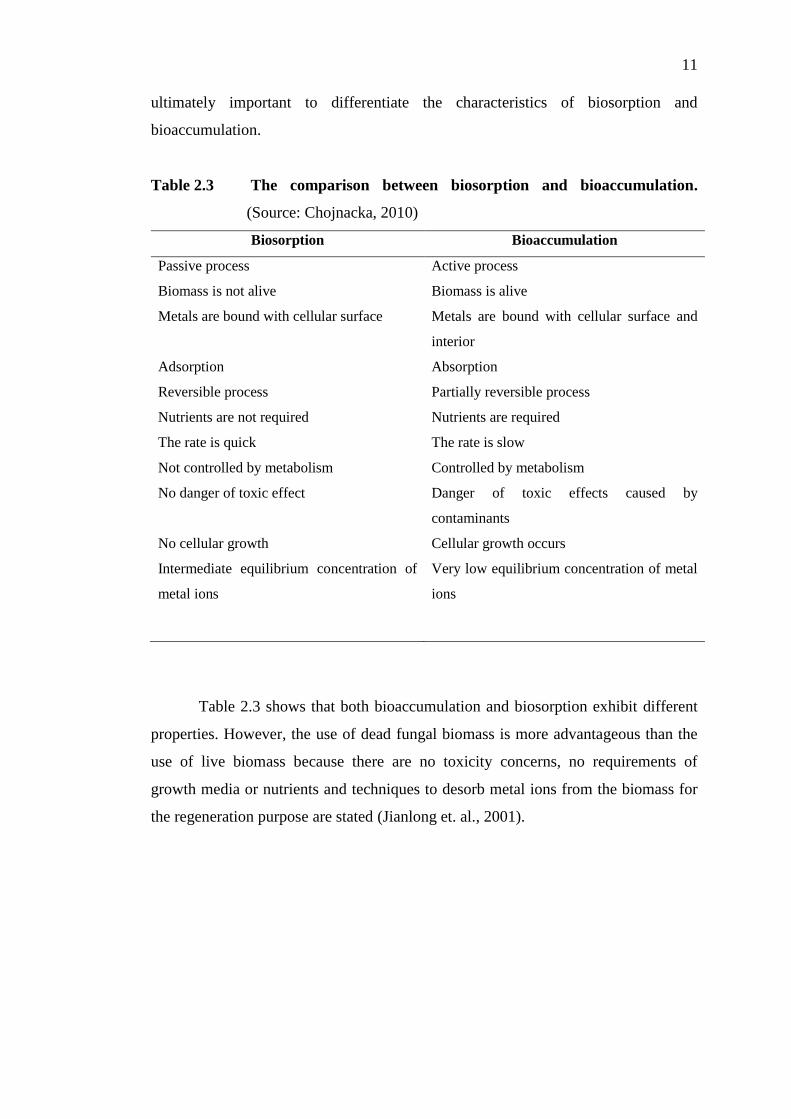
11
ultimately important to differentiate the characteristics of biosorption and
bioaccumulation.
Table 2.3 The comparison between biosorption and bioaccumulation.
(Source: Chojnacka, 2010)
Biosorption Bioaccumulation
Passive process
Biomass is not alive
Metals are bound with cellular surface
Adsorption
Reversible process
Nutrients are not required
The rate is quick
Not controlled by metabolism
No danger of toxic effect
No cellular growth
Intermediate equilibrium concentration of
metal ions
Active process
Biomass is alive
Metals are bound with cellular surface and
interior
Absorption
Partially reversible process
Nutrients are required
The rate is slow
Controlled by metabolism
Danger of toxic effects caused by
contaminants
Cellular growth occurs
Very low equilibrium concentration of metal
ions
Table 2.3 shows that both bioaccumulation and biosorption exhibit different
properties. However, the use of dead fungal biomass is more advantageous than the
use of live biomass because there are no toxicity concerns, no requirements of
growth media or nutrients and techniques to desorb metal ions from the biomass for
the regeneration purpose are stated (Jianlong et. al., 2001).

12
2.5 Mechanism of metal uptake
The understanding of the mechanisms by which microorganisms accumulate metals
is crucial to the development of microbial processes for concentration, removal and
recovery of metals from aqueous solution. According to Kapoor and Viraraghavan
(1995), the uptake of heavy metals by biomass can take place by an active mode
(dependent on the metabolic activity) known as bioaccumulation or by a passive
mode. This passive mode is independent of metabolic activity and thus this type of
mechanism only happens to cell walls and external surfaces in the case of non-living
biomass. Metabolism-independent uptake essentially involves chemisorptions such
as ionic exchange, complexation and/or chelation, physical adsorption, micro-
precipitation and oxidation or reduction (biosorption).
Apart from that, the concentration gradient and diffusion through cell walls
and membranes also causes ion entrapment in inter- and intra-fibrillar capillaries and
spaces of the structural polysaccharide network (Volesky & Holan, 1995). Thus, the
identification of microorganisms’ functional groups involved in the biosorption
process is important upon exploring the mechanism of metal uptake.
The cell wall structure of certain algae, fungi and bacteria was found to be
particularly responsible for biosorption mechanism. The several chemical groups that
could attract and sequester the metals in biomass are: acetamido groups of chitin,
structural polysaccharide of fungi, amino and phosphate groups in nucleic acids,
amino, amido, sulfhydryl, and carboxyl groups in proteins, hydroxyls in
polysaccharides, and mainly carboxyls and sulphates in the polysaccharides of
marine algae (Volesky & Holan, 1995). Volesky (2007) also stated that the amine,
NH2 group that is active and ubiquitous in fungal cell walls are also capable of
sequestering anions.
Apart from the functional groups of fungal and algae mentioned above,
bacteria have quantities of peptidoglycan (Gram “+”) and teichoic acid (Gram “-”) in
their cell walls. All of these components featuring an important ion exchange active
groups in the structure (Treen-Sears, Volesky, & Neufeld, 1984). Thus, these

13
constituents stated have contributed to the discovery of the active chemical groups
are involved in the metal binding mechanism.
Both living and dead fungi are able to remove heavy metal ions from aqueous
solutions. The metal uptake by dead cells occurs as a result of reaction with the
functional groups present in cells specifically in the cell wall. According to (Kapoor
& Viraraghavan, 1995), the metal uptake by the cell wall has been broadly based on
two mechanisms: uptake directed by functional groups like phosphate, carboxyl,
amine and phosphate diesther species of these compounds. The second up
mechanism results from physicochemical inorganic interactions directed by
adsorption phenomena.
The mechanism of uptake depends on the type of removal: radionuclides
results from the combination of the above two processes. However, the removal of
heavy metal ions is mainly affected by one of the processes; where the uptake is
directed by functional groups which agree with literatures stated above. Meanwhile,
the efficiency depends on the capacity and affinity including physico-chemical
nature (Goyal & Ahluwalia, 2007) which is contributed by the environmental factors
such as the solution pH, temperature and initial metal ions concentration
(Vijayaraghavan & Yun, 2008).
These functional groups mainly consist of several types of ionisable sites
including carboxyls, amines, hydroxyls, phosphates and sulfhydryls (Viraraghavan &
John Peter, 2008).The major binding groups for biosorption are shown in Table 2.4.
The table also depicted that the functional groups with different values of pKa that are
indicative of the binding properties of a given group.

14
Table 2.4 Functional groups of biosorbents. (Source: Volesky, 2007)
Binding
group
Structural
formula pKa
Ligand
atom
Occurrence in
selected biomolecules
Hydroxyl
Carbonyl (ketone)
Carbonyl
–OH
–C=O
|
OH
9.5-13
-
1.7-4.7
O
O
O
Polysaccharides, Uronic
acid, Sulfated PS
Peptide bond
Uronic acids,
Amino acid
Sulfhydryl (thiol)
Sulfonate
–SH
O
||
–S=O
||
O
8.3-10.8
1.3
S
O
Amino acid
Sulphated PS
Thioether
Amine
Secondary anime
Amide
>S
–NH2
>NH
–C=O
|
NH2
-
8-11
13
-
S
N
N
N
Amino acid
Chitosan, Amino acid
Cti, PG, Peptide bond
Amino acid
Imine
Imidazole
= NH
–C-N-H
|| >CH
H-C-N
11.6-
12.6
6.0
N
N Amino acid
Phosphonate
OH
|
–P=O
|
OH
0.9-2.1
6.1-6.8
O
Phospholipids
Phosphodiester
>P=O
|
OH
1.5
O Teichoic acid
LipoPS

15
The mechanism of metal binding is not well understood yet due to the
complex nature of microbial biomass, which is not readily amenable to instrumental
analysis. However, there is a breakthrough of using spectroscopic techniques such as
Infrared Spectroscopy (IR), Raman, Electron Dispersive Spectroscopy (EDS), X-ray
Photoelectron Spectroscopy (XPS), Electron Microscopy (Scanning, Transmission),
Nuclear Magnetic Resonance (NMR) and X-ray Diffraction Analysis (XRD) to
enable identification of biosorptive sites which are also known as functional groups.
According to Gupta et. al., (2000), instrumental analysis is a relatively new technique
for determination of binding energy of electrons in atoms/molecules which depends
on the distribution of valence charges and thus gives information about the oxidation
state of an atom/ion.
Fourest et. al., (1996) stated that the properties of a biosorbent can be
obtained by using simpler techniques such as titration which enables to determine
acidic disassociation constants and also metal affinity constants. These properties
play a major role in describing the selectivity of the biomass for different metal ion
(Chojnacka, 2010).
2.6 Fungi
2.6.1 Classification and general characteristics
Microscopic fungi include yeasts with spherical budding cells and molds with
elongated filamentous hyphae in mycelia. The molds are filamentous fungi, such as
Penicillium, Aspergillus and Candida. The body or vegetative structure of a fungus is
called thallus, which varies in complexity and size from single cell microscopic
yeasts to multicellular molds. A single filament is called a hypha. Hyphae usually
grow together, collectively called a mycelium (Prescott, Harley & Klein, 2002). The
classification of fungi is shown in Table 2.5.

16
Table 2.5 The classification of fungi. (Source: Wang & Chen, 2006)
Group Common Name Hyphae Typical Representative
Ascomycetes Sac fungi Septate Neurospora
Saccharomyces
Morchella
Basidiomycetes Club fungi, mushroom Septate Amanita
Agaricus
Zygomycetes Bread molds Coenocytic Mucor
Rhizopus
Oomycetes Water molds Coenocytic Allomyces
Deuteromycetes Fungi imperfecti Septate Penicillium
Aspergillus
Candida
Most fungi are filamentous. The hyphae are typically 5-10 μm wide but may
vary from 0.5 μm to 1.00 mm, depending on the species (Lester & Birkettn, 1999).
The mycelium is composed of a complex mass of filaments or hyphae while the
hyphae have walls which are composed of cellulose and/or chitin as depicted in
Figure 2.1. A common cytoplasm exists throughout the hyphae. Thus fungi cellular
organization has three types. The types of fungi cellular organization is tabulated in
Table 2.6. Generally, the principle of classification for fungi is mainly based on the
type of sexual spores.
Table 2.6 Cellular organization in filamentous fungi.
(Source: Lester & Birkettn, 1999)
Cellular organization Description
Coenocytic Hypha contains a mass of multi-nucleate cytoplasm
which also known as aseptate.
Septate with uni-nucleate
protoplasts
Hypha is divided by crosswalls or septa, each
compartment containing a single nucleus.
Septate with multi-nucleate
protoplasts between septa
Central pore in the septum connecting the cytoplasm of
neighbouring cells and permitting the migration of both
cytoplasm and nuclei.

17
2.6.2 Cell wall and its main composite
Various polysaccharides are the main constituents of the fungal cell wall, typically in
the range of 80-90% constituent. The polysaccharides are often complexes with
proteins, lipids, polyphosphates, and inorganic ions, making up the wall-cementing
matrix (Sag & Kutsal, 2001).
Figure 2.1 The cell wall composition of fungi.
Source: (Kapoor & Viraraghavan, 1995)
Figure 2.1 shows the general structure of cell wall and other features of the
cross section of a fungal cell. It can be seen that the fungal cell wall is rigid and
structurally composed of complex layers of polysaccharides and protein. In general,
the fungal cell wall can be regarded as a two-phase system consisting of the chitin
skeleton framework embedded in an amorphous polysaccharide matrix (Wang &
Chen, 2009).
2.6.3 Fungal biomass as biosorbent
Gadd (1986) mentioned that fungi and yeasts able to accumulate micronutrients such
as Cu, Zn and Mn, and non-nutrient metals like U, Ni, Cd, Sn and Hg in amounts
higher than the nutritional requirement. Besides, the potential of fungal biomass
18
asadsorbents for the removal of heavy metals and radio nuclides from polluted
waters were regonized by Shumate et. al., (1978) and Jilek et. al., (1975). Azab and
Peterson (1989) have shown in their studies that fungi outperformed activated carbon
and ion exchange resins for cadmium removal from aqueous solutions (Viraraghavan
et. al., 2008).
Meanwhile, fungi are also recognized for their ability to produce a large
variety of extra cellular proteins, organic acids, enzymes and other metabolites, and
their used biomass may be used as effective biosorbent material for removal,
reduction and detoxification of industrial effluent composition (Tsekova et. al., 2010).
Various fungal species under the genus Aspergillus, Penicillium and Rhizopus
have been shown to be effective in biosorption of heavy metals from polluted
effluents both as immobilized cells and in the mobilized state (Tsekova et. al., 2010).
There are reports that fungi belonging to the Rhizopus and Penicillium have already
been studied as potential biomass for removal of heavy metals from aqueous solution.
Meanwhile, fungus of Aspergillus species has also been studied with different
influential parameters vastly in the research since decades (Akar & Tunali, 2006;
Dursun, 2006; Tsekova et. al., 2010).
2.6.4 Aspergillus niger
In this study, A. niger is the biosorbent of interest. It is a filamentous ascomycete
fungus and it is one of the most common species in the genus Aspergillus (Lesmana
et al., 2009). It is an important microorganism in biotechnology applications (Bapat,
Kundu & Wangikar, 2003).
A.niger has been extensively used in processes of organic acid production
such as citric acid and oxalic acid due to its ability of organic acids excretion in
abundant amount. Citric acid and several enzymes produced by A.niger are
considered GRAS (generally regarded as safe) by the United States Food and Drug
Administration (FDA).
19
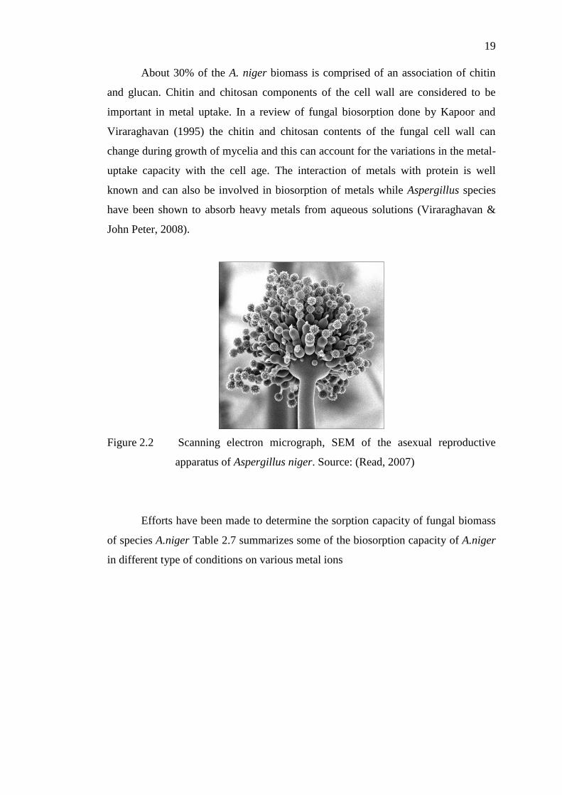
About 30% of the A. niger biomass is comprised of an association of chitin
and glucan. Chitin and chitosan components of the cell wall are considered to be
important in metal uptake. In a review of fungal biosorption done by Kapoor and
Viraraghavan (1995) the chitin and chitosan contents of the fungal cell wall can
change during growth of mycelia and this can account for the variations in the metal-
uptake capacity with the cell age. The interaction of metals with protein is well
known and can also be involved in biosorption of metals while Aspergillus species
have been shown to absorb heavy metals from aqueous solutions (Viraraghavan &
John Peter, 2008).
Figure 2.2 Scanning electron micrograph, SEM of the asexual reproductive
apparatus of Aspergillus niger. Source: (Read, 2007)
Efforts have been made to determine the sorption capacity of fungal biomass
of species A.niger Table 2.7 summarizes some of the biosorption capacity of A.niger
in different type of conditions on various metal ions

20
Table 2.7 Biosorption capacity of A. niger (mg/g) on metal ions.
Condition Metal
ions
Biosorption capacity
(mg/g) Reference(s)
Pretreated with
NaOH
Cu
Pb
Cu
Pb
Pb
Cd
Cu
Ni
28.7
32.6
25.5
28.9
7.24
3.43
2.66
0.96
Dursun (2006)
Dursun (2006)
Dursun (2003)
Dursun (2003)
Kapoor et al. (1999)
Kapoor et al. (1999)
Kapoor et al. (1999)
Kapoor et al. (1999)
Growing Cu
Pb
15.6
34.4
Dursun et al.(2003a)
Dursun et al.(2003a)
Live Pb
Cd
Cu
Ni
2.25
1.31
0.75
1.75
Kapoor et al. (1999)
Kapoor et al. (1999)
Kapoor et al. (1999)
Kapoor et al. (1999)
Attached to
wheat bran
Cu
Zn
-
-
Modak et al. (1996)
Modak et al. (1996)
There is an obvious phenomena observed from the above tabulation of the
biosorption capacity. Alkali treatment (pretreated with NaOH) of fungal have shown
to increase significantly the metal uptake capacity compared to the rest of the A.
niger.

CHAPTER 3
3 METHODOLOGY
The first stage of the project is the cultivation of fungi, A.niger and dried the
obtained biomass of the cultivation. Then the following step is the preparation of
synthetic lead solution with 0.15M of blank solution and 0.1M of stock solution,
adjusted to the initial concentration and pH of interest. Then, the studies began with
batch isotherm and kinetics studies for the investigation of pH and initial metal ion
concentration on biosorption. Lastly, the results obtained from the studies were
evaluated and analyzed by using isotherm and kinetic models.
3.1 Preparation of adsorbent
The fungus used in this project was A. niger. The biomass was cultivated in a liquid
medium using the shake flask method. The culture medium contained the following
composition: 10g/L peptone (R&M Chemicals); 20g/L sucrose (R&M Chemicals)
and 3g/L yeast extract (BactoTM)
. The medium was transferred to several 250ml
Erlenmeyer flasks and autoclaved at 121ºC for 15mins and cooled to room
temperature.
The A.niger spores obtained from the Department of Science in University of
Tunku Abdul Rahman were inoculated into the medium and allowed to grow for a
period of 7 days on an orbital shaker at 225 rpm. After seven days the harvested cells

22
are washed several times with deionised distilled water. The biomass was then dried
in oven at for four days, grounded and sieved to millimetre size of particles.
The prepared dry biomass was used in all adsorption experiment of this study.
3.2 Preparation of metal ion solutions
The stock solution can be prepared using Plumbum Nitrate, (R&M Chemicals with
M= 331.02 g/mol), where adequate amount of plumbum nitrate, Pb(NO3)2 is
dissolved into 500ml of deionized water to obtain 0.1M of stock solution.
3.3 Preparation of blank solution
The blank solution can be prepared using Sodium Nitrate, NaNO3(Merck). Blank
solution is prepared by dissolving adequate amount of sodium nitrate, NaNO3 into
deionized water to obtain 0.15M of blank solution.
3.4 Metal ions solution
The metal ions solution of different working concentrations is prepared by diluting
the 0.1M stock solution in accordance to the investigated range of concentration. The
ranges of concentrations of both metal ions prepared from stock and blank solutions
varied between 5 and 200ppm.

23
3.5 Batch biosorption studies
The effect of initial metal ions and pH on the biosorption of Pb(II) on fungal biomass
was investigated in the pH range of 1 - 6 and metal ion concentration in the range of
5 to 200 mg/L. By using the 0.1M stock solution and 0.15M blank solution, 100 ml
of metal ion solutions is prepared into several Erlenmeyer flask in accordance to the
initial concentration of interest. The initial pH of each metal ion solution was
adjusted to the required pH value by using either 0.1M of HCl or NaOH or 1M of
HCl.
Figure 3.1 Schematic diagram of bath biosorption equilibrium study.
Figure 3.1 depicts the procedure of the batch biosorption study. Firstly,
100mg of A. niger dried biomass was added to each of the metal ion solution and
sealed with aluminium foil. The reaction mixtures were shaken in incubator shaker at
225rpm, 25 and up to 24 hours. The content of the flasks was separated by filtration
using filter paper and collected in centrifugal tubes. The final concentration, Ce of the
metal ion solutions were then measured with ICP (Optical Emission Spectrometer,
Optima 7000DV).

24
3.6 Equipments
There are several equipments used throughout the biosorption of A. niger from the
preparation of fungal biomass, biosorption and kinetics studies and the analysis of
metal ions concentrations. The figures are arranged chronologically based on the
experimental steps.
Figure 3.2 Orbital shaker (SSL1;Stuart®)
Figure 3.3 Oven (Beschickung-Loading Modell 100-800;Memmert)

25
Figure 3.4 ICP (Optima 7000DV;Perkin Elmer, Uberlingen, Germany).
3.7 Metal uptake capacity
Volesky (2004) states that the quality of the biosorbent is judged according to the
amount of metal ions it can attract and retain in an “immobilized” form. Therefore,
the uptake of metal ions must be determined as the amount of metal ions bound by
the unit of biosorbent and calculated from a metal mass balance yielding:
(3.1)
where,
q = mg metal ions per g dry biosorbent
V = reaction volume
Ci = initial concentration (mg/L)
Cf = residual concentration (mg/L)
m = amount of dry biosorbent (g)
The efficiency of heavy metal removal was calculated from the amount of
metal ions adsorbed on the biosorbent and the amount of metal ions available in the

26
synthetic lead solutions. The percentage removal of Pb(II) ions from the synthetic
lead solution can be calculated by using the equation shown below:
(3.2)
However, the percentage removal is an approximation that it could lead to
outright misleading conclusions on the relative sorption performance. The equation
for the “percentage removal” can only serve the purpose of crude orientation,
perhaps adequate only for quick and very approximate screening biosorbent
materials (Kratochvil & Volesky, 1998).
3.8 Biosorption isotherms
The sorption performance of the biomass can be assessed by using several isotherm
models. Table 3.1 exhibits the advantages and disadvantages of the adsorption
isotherms typically used in the biosorption equilibrium studies.
Table 3.1 Adsorption Isotherms. Source: (Kapoor & Viraraghavan, 1995).
Isotherm Equation Advantages Disadvantages
Langmuir
Interpretable
parameters
Not structured;
monolayer
sorption
Freundlich
Simple
expression
Not structured;
multilayered
sorption
Redlich-
Peterson
Approaches
Freundlich at
higher
concentrations
-
BET
Multilayer
sorption -
27
The two widely accepted and easily linearized equilibrium adsorption
isotherm models are Langmuir and Freundlich model which describe monolayer
adsorption and the latter, multilayer adsorption.
3.8.1 Langmuir isotherm model
Researches on biosorption of heavy metals have mainly focus on the adsorption
efficiencies and adsorption equilibrium for different biosorbent materials, where the
analysis of experimental data is important to serve as a basis for the development of
continuous or large scale biosorption process (Sag & Kutsal, 2001). The Langmuir
adsorption isotherm has traditionally been used to quantify and contrast the
performance of different biosorbents (Davis, Volesky & Alfonso, 2003).
Biosorption equilibrium is more frequently done with Langmuir than
Freundlich equation, because a clear plateau can be distinguished on the sorption
isotherms (Chojnacka, 2010). In the searching of new biosorbents, the equilibrium
sorption curve is useful in identifying the better sorption performance of different
biosorbents based on the fitting of data into Langmuir isotherm model.
Figure 3.5 Comparative example of sorption isotherm curve.
(Source: Volesky, 2007)

28
A sorbent is considered as a “good” sorbent if it has a high Qmax and a steep
initial sorption isotherm slope as indicated by low values of Langmuir parameter b
(Volesky, 2004). Figure 2.3 shows that “B” performed better than “A” at lower
equilibrium concentrations. The following assumptions are valid for Langmuir model:
i. Fixed number of adsorption sites; at equilibrium, at any temperature and gas
pressure a fraction of the surface sites, is occupied by adsorbed molecules,
and the fraction is free.
ii. All sorption sites are uniform (i.e. constant heat of adsorption).
iii. Only one sorbate.
iv. One sorbate molecules reacts with one active sites.
v. No interaction between sorbed species.
(Langmuir, 1918)
The equation was developed by Irving Langmuir in 1916. It is expressed in a
hyperbolic form:
(3.3)
The Langmuir equation can be transformed to the linear form:
(3.4)
where,
Qe = metal ion sorbed, mg/g
Ce = equilibrium concentration of metal ion solutions, mg/L
Qmax = maximum amount of metal ion which can be taken up by biosorbent, mg/g
KL = Langmuir constant, L/mg (affinity)
(Langmuir, 1918)

29
3.8.2 Freundlich isotherm model
The Freundlich equation is an empirical relationship describing the adsorption of
solutes from a liquid to a solid surface. In other words, it does not indicate a finite
uptake capacity of the biosorbent and thus can only be applied in the low to
intermediate concentration ranges. Freundlich (1907) stated that Freundlich isotherm
relationship is exponential with the following general form of:
(3.5)
The Freundlich equation can be linearized by taking logarithm of both sides
of the equation to give:
(3.6)
where,
KF and 1/n = Freundlich constant
(Source: Freundlich, 1907)
3.9 Biosorption kinetic studies
Batch kinetic studies were carried out to determine the equilibrium time which is
defined as the time needed to reach equilibrium (point beyond which there is very
limited removal of metal).
The initial lead concentration is adjusted to 50 mg/L. Then 0.5 mg of dry
fungal biomass was added to the plumbum solution in each of the 34 conical flasks
of 250mL volume. The samples were placed on the rotary and orbital shaker at
225rpm. The samples were collected tentatively in a duplicate in duration of 48 hours,

30
in the intervals of 20mins for the first hour, and then on an hourly basis. Upon
collection, samples are immediately filtered through filter papers of 0.45 µm pore
sized into centrifugal tubes. The final pH values of each sample were noted and the
lead concentrations in the filtrates are then determined by using ICP. The kinetics
studies of biosorption were usually described and analyzed with pseudo-first or
pseudo-second order model (Chojnacka, 2010).
3.9.1 Pseudo-first order kinetics model
The reaction order is related with the mechanism of biosorption, which is most
frequently ion-exchange or surface precipitation (metal hydroxide, sulphide or
carbonate). Literatures have reported that the rate limiting step is chemisorption
which involves valent forces by sharing or exchange of electrons between sorbent
and sorbate. In kinetic modelling, the pseudo-first order kinetic equation which also
known as Lagergren (1898) equation has the following equation:
(3.7)
Taking the logarithm of both sides:
(3.8)
where,
Qt = amount of adsorbed ions on the biosorbent at time t,
k = rate constant of Lagergren first-order biosorption
The equation above assumes that metal cation binds only to one sorption site
on the sorbent surface (Lagergren, 1898).

31
3.9.2 Pseudo-second order kinetics model
Ho and McKay (1999) used a pseudo-second order rate equation for a comparison of
chemisorption kinetic models applied to pollutant removal on various sorbents. The
pseudo-second order equation has the following equation:
(3.9)
The equation can be linearized to:
(3.10)
where,
k2 = rate constant of second-order biosorption
The equation assumes that metal cations are bound to two binding sites on the
sorbent surface (Ho & McKay, 1999).

32
CHAPTER 4
4 RESULTS AND DISCUSSIONS
This chapter presents the results of the experiments conducted using different initial
lead concentrations and pH of synthetic solutions. The performance of the biosorbent
is evaluated using two adsorption isotherm models, Langmuir and Freundlich. The
kinetic studies were carried out based on the pseudo first-order and pseudo second-
order reaction models. The effect of the two parameters: the initial lead ions
concentration and pH of the metal solutions is also discussed in this chapter.
4.1 Batch isotherm studies
The equilibrium of biosorption of heavy metal was modelled using adsorption-type
isotherms, Langmuir and Freundlich models, to describe the biosorption equilibrium.
The studies of isotherm of lead adsorption on the dry fungal biomass, A. niger
were carried out by varying the initial lead ions concentration and pH of solutions
with the methods stated in section 3.5 (page 35). Adsorption isotherms were plotted
as a function of the adsorbed quantity per unit weight of adsorbent, Qe. at equilibrium
and the final equilibrium concentration of residual sorbate remaining in the solution,
Ce.
Figure 4.1 shows the adsorption isotherm for A.niger biosorption of lead ions
at pH 1 - 6. The sorption isotherms depict the experimentally determined
performance of biosorbent. The biosorbents’ performance in lead uptake can be

33
assessed using the Langmuir and Freundlich isotherm to determine mechanism of
monolayer or multilayer adsorption, respectively.
Figure 4.1 The adsorption isotherms for lead at different pHs.
There are several research studies on biosorption of metals using A.niger
fungal biomass from year 2006 to 2008. Table 4.1 shows the adsorption equilibrium
models applied for the biosorption processes in these studies. Dursun (2006) had
demonstrated that the adsorption isotherms of A. niger to remove Pb(II) was of a
saturated type kinetic model where the experimental data fitted to three different
adsorption isotherms namely, Langmuir, Freundlich and Redlich-Peterson. The latter,
Redlich-Peterson isotherm model is normally proposed as an isotherm compromising
the features of the Langmuir and Freundlich isotherms (Yu & Ya-Juan, 2008).
Table 4.1 Equilibrium and kinetic studies of heavy metal adsorption onto
Aspergillus niger. (Source: Lesmana et. al., 2009)

34
Biosorbent Heavy
metal Adsorption Equilibria
Adsorption
kinetic Reference
A. niger Cu(II)
Pb (II)
Cr(VI)
Cr(VI)
Langmuir, Freundlich &
Redlich-Peterson
Langmuir & Freundlich
Langmuir & Freundlich
Saturation type
kinetic model
Pseudo-first &
pseudo-second
order
-
Dursun et. al., (2006)
Mungasavalli et. al.,
(2007)
Kumar et. al., (2008)
Isotherm models provide information on the biosorbent uptake capabilities
and also reflect the equilibrium behaviour. Langmuir isotherm includes parameter KL
which relates to the energy of adsorption. KL value is obtained from the reciprocal of
b value, which represents the affinity of the biosorbent towards the heavy metal ions.
Meanwhile, Freundlich isotherm is an indication of a finite uptake capacity of
the sorbent (Volesky & Holan, 1995). The magnitude of KF and n shows the ease of
uptake of heavy metal ions from an aqueous solution and high adsorption capacity
(Akar & Tunali, 2006). In a comparative study by Dursun (2006), KF was used as a
relative measure of adsorption capacity while n was related to intensity of adsorption.
The Langmuir constant, KL and maximum adsorption capacity, Qm were
obtained from the intercept and slope of Figure 4.2 and 4.3. The results showed that
the maximum adsorption happened at pH 6, and it decreased as the pH of lead ion
solutions decreased. The KL values obtained from the Langmuir isotherm models
indicated that the affinity of biosorbent towards lead ions increased when pH
increased from 1 to 6.

35
Figure 4.2 Langmuir isotherms for lead removal at pH 1 – 3.
Figure 4.3 Langmuir isotherms for lead removal at pH 4 – 6.
The Freundlich constants Kf and n were calculated from the intercept and
slope of plot ln Qe versus ln Ce respectively, as shown in Figure 4.4 and 4.5. These
values were listed in Table 4.2 with their correlation coefficients, R2. The constant n
values (pH 3 – 6) obtained for Freundlich isotherm were above 1.0, which indicated

36
that Pb(II) ions are favourably adsorbed by the biomass at these pH values (Dursun,
2006).
Figure 4.4 Freundlich isotherms for lead removal at pH 1 – 3.
Figure 4.5 Freundlich isotherms for lead removal at pH 4 – 6.

37
The experimental data obtained in this study were well fitted into both
Langmuir and Freundlich isotherm models, except at pH 2 and pH 3. This study’s
analysis agreed with the study by Dursun et. al., (2006) describing the biosorption
process involving both monolayer and multilayer mechanisms.
Table 4.2 Langmuir and Freundlich isotherm model constants.
pH Langmuir isotherm Freundlich isotherm
Qmax, (mg/g) KL R2 KF n R
2
1.0 - - 0.97 - 0.757 0.88
2.0 0.10 0.11 0.69 - 0.831 0.83
3.0 18.59 20.41 0.84 - 1.118 0.38
4.0 23.98 26.34 0.99 0.871 1.780 0.93
5.0 31.65 34.75 0.96 0.469 2.410 0.97
6.0 66.23 72.73 0.99 0.450 1.833 0.97
4.2 Batch kinetic studies
The kinetics of heavy metal adsorption was modelled using pseudo first-order and
pseudo second-order equations described in details in section 3.9 (page 41).
According to Yu & Ya-Juan (2008), pseudo first-order and second-order kinetic
equations have been widely used to describe time evolution of biosorption under
non-equilibrium conditions. The criterion in the determination of the adequacy of
kinetic model is based on the value of the correlation coefficient, R2 of the plot
(Ahmed & Mohammed, 2008).
The reaction rate can be obtained by plotting a linear plot of
against t. The slope obtained is the rate constant of Lagergren first-order biosorption,
k from equation 3.8 (page 42).

38
Meanwhile, the rate constant of second-order biosorption, can be obtained
from the slope of curve plotted with t/Qe versus t.
The parameters of the kinetics models (pseudo-first, pseudo-second) with
their correspondent coefficients of determination are calculated from the slopes and
intercepts of the linear plot of these models as shown in Figure 4.6 and 4.7,
respectively.
Figure 4.6 The pseudo first-order kinetics data for lead adsorption on A.niger.
The results obtained for the linearized pseudo second-order showed
correlation coefficient of 0.99. Thus, the adsorption of Pb(II) ions onto the A.niger
biomass is regarded as pseudo-second order rather than pseudo-first order.

39
Figure 4.7 The pseudo second-order kinetics for lead adsorption on A.niger.
The rate constants were 8.52x10⁻⁴
and 9.2x10⁻³
g/mg·min for the pseudo first
order and second-order reaction, respectively. In the kinetic studies of biosorbents,
the biosorption is similar to the conventional sorption processes, involves inherently
very fast sorption reaction mechanisms based predominantly on chemisorptions.
4.3 Effect of pH on the metal uptake
Since earlier studies on biosorption phenomena, it has been known that the uptakes
of heavy metal cations by most biomass types decrease dramatically as pH of the
metal solutions decreases from pH 6 to 2.5 (Sag & Kutsal, 2001). The environmental
factor, which is the pH of metal ion solutions, influenced the surface metal binding
sites of the biosorbents and the chemistry of the cells walls, thus the
physicochemistry and hydrolysis of the metals are influenced as well (Tsekova et. al.,
2010).
In this study, the effect of pH was investigated by varying the pH of metal ion
solutions from 1.0 to 6.0. The results obtained were analysed by Langmuir isotherm

40
equation to compare the maximum adsorption values for each pH value as tabulated
in Table 4.2.
Figure 4.8 depicts the maximum adsorption capacity for lead at pH 1 to pH 6.
It shows the influence of pH in terms of adsorption uptake. It shows that the
adsorption happens in pH range of 4 – 6. The biosorption capacity is reduced as the
pH of lead ion solutions is changed towards acidic range since there was little or no
biosorption of lead determined for pH range less than 2. The heavy metal removal
capacity increased greatly from 31.65 to 66.23 mg/g with an increase in pH from 5.0
to 6.0 at, respectively.
Figure 4.8 The maximum adsorption of Pb on A.niger at different pHs.
According Kapoor et. al., (1999) a sudden increase in sorption with a slight
increase in pH is often referred to as an “adsorption edge”. The results also showed
that the dead fungal biomass, A. niger favoured the pH 6 metal ion solution and
possessed higher affinity towards the lead metal ions in this environment.

41
The results of the experiments indicated that pH is an important parameter
affecting the biosorption of heavy metals. As depicted in Figure 4.8, at low pH
(lower than 3.0) heavy metal removal was inhibited; this phenomenon is possibly a
result of a positive charge density on the surface binding sites due to a high
concentration of protons in solution.
In other words, at highly acidic pH, the overall surface charge on cells
became positive and metal cations and protons compete for binding sites on cell wall,
which resulted in lower metal uptake rates. According to Dursun (2006), it has been
suggested that at low pH values, cell wall ligands would be closely associated with
H3O+ that restricts access to ligands by metal ions as a result of growing repulsive
force.
Besides of the ionic state of the solutions and adsorbents, highly acidic
environment may also serve as desorbing conditions for the purpose of releasing the
metal ions from the binding sites (Chojnacka, 2010). This theory has been proved by
Jianlong et. al., (2001), where 0.1M of nitric acid was able to effectively elute the
biosorbed lead ions from the fungal biomass. The acidic environment was playing a
role as to desorb and regenerate the biomass to be reused (Volesky, 2007).
However, with an increase in pH, the negative charge density on the cell
surface increases due to deprotonation of the metal binding sites and thus biosorption
increased. At pH values above the iso-electric point, there is a net negative charge on
the cell surface and the ionic state of ligands such as carboxyl, phosphate and amino
groups will be such that so as to promote reaction with metal ions, hence the rapid
binding efficiency was obtained.
4.4 Effect of initial concentration on metal uptake
The effect of initial metal ion concentration on the biosorption capacity of A.niger
was studied under the ambient condition at 25 and pH 1 – 6 range. The biosorption
of Pb(II) ions on A.niger increased with increasing initial concentration of metal ions.

42
Figure 4.9 depicts the metal ions removal percentage ranging from pH 1.0 to
6.0. There is a general trend where the initial metal ions increases, the percentage
removal increases as well.
Figure 4.9 Effect of initial lead concentration on biosorption capacity of A.niger.
Figure 4.9 shows the effect of initial metal ion concentration on lead
biosorption by fungal biomass. There is an obvious trend shown on the graph: the
biosorption curves of pH 4 to 6 starts with a slope and reaches equilibrium when the
initial metal ion concentrations increase. Despite of the rule stated, these phenomena
may be explained by an increase in the number of metal ions competing for the
available binding sites on the biomass surface and the lack of binding sites for
complexation of Pb(II) ions at higher concentration levels.

43
Table 4.3 shows data on the increase in the metal ions to biomass ratio which
results in decrease of the biosorption efficiencies. For instance, the percentage
removal of lead metal ions decreases from 71.20 % to 22.10 % when the metal ions
concentration increases from 5 to 200 mg/L. There is a rule stated Dursun (2006): an
increase in the initial metal concentration results in an inrease in the biosorption
capacity because the initial metal concentration provides a driving force to overcome
mass transfer resistance between the biosorbent and biosorption medium.
Table 4.3 Percentage removal of lead by biomass of A. niger.
Lead Percentage Removal, %
Initial Concentration
Ci, mg/L pH 1 pH 2 pH 3 pH 4 pH 5 pH 6
5 37.94 3.70 0.40 59.40 63.20 71.20
10 0.70 2.00 0.40 41.02 56.90 60.05
15 2.87 3.67 4.20 35.53 50.20 53.87
25 4.04 0.04 2.68 35.08 54.96 50.72
35 2.97 1.03 0.20 32.60 29.09 50.91
45 2.73 0.76 2.36 28.40 49.49 45.24
55 1.13 0.09 0.67 23.62 23.20 38.89
70 2.31 3.86 1.26 20.89 20.74 39.06
85 0.84 1.65 0.61 17.62 29.20 34.39
100 1.91 0.60 2.00 16.12 17.36 32.75
150 4.07 8.73 8.27 9.55 13.00 28.40
200 4.55 8.20 1.35 16.80 13.20 22.10
In other words, at high concentration levels, more Pb(II) ions are left in
solution compared to the adsorbed metal ions may have caused the saturation of
binding sites on the biomass surface. When the biomass surface are saturated with
metal ions, the ion-exchange mechanism could not take place, thus the biosorption is
inhibited. On the other hand, at low concentration of metal ions the number of
available binding sites on the biomass surface is high and hence biosorption of
metals was very effective.

44

45
CHAPTER 5
5 CONCLUSION AND RECOMMENDATIONS
5.1 Conclusion
The ability of A. niger biomass to adsorb lead, Pb(II) ions was investigated in a batch
system. The equilibrium of biosorption of lead, Pb(II) ions on the biosorbent was
evaluated using Langmuir and Freundlich models. Both of the adsorption models
applied agreed well with the experimental data. However, Langmuir equation was
the best isotherm model that represented the biosorption data in this study because
the obtained correlation coefficients were higher. The equilibrium data of lead
adsorption fitted well to both Langmuir and Freundlich, thus the adsorption is
characterized as monolayer and multilayer.
The maximum removal efficiency of Pb(II) ions occurs at pH 6 where the
maximum adsorption capacity obtained was 66.23 mg/g using the Langmuir isotherm
model.
This study also showed that the dried fungal biomass of A.niger is very
sensitive to pH changes especially below pH 3. The biosorption favoured the alkaline
environment as the maximum adsorption capacity happened at pH 6.
It was also found that the pseudo-second order model was applicable for the
whole period of contact time. As a conclusion, the results of this study indicated that
A.niger biomass is a suitable biosorbent for the removal of Pb(II) ions from aqueous
solution.

46
5.2 Recommendations
It is obvious that many different endeavours and challenging contributions have to
made on the path of developing biosorption from a scientific curiosity to useful
applications for the sake of protecting the environment with low cost.
The analysis of experimental data in this study shows that the adsorption of
metal ions was best at the pH range of 4 to 6. Thus, it is suggested that the pH range
of the study can be narrowed down to this pH range but adding more parameters of
interest in this study, for example the biosorbent dosage, agitation rate and
temperatures. Meanwhile, the technigue of elution may also be added in the study for
the purpose experimenting on the regeneration and reuse of biosorbents.
Volesky, (2007) also stated the importance of the affinity of adsorbent
towards the metal ions. This “affinity” is largely contributed by swirl of the electrons,
which is thermodynamics. Therefore, more technical and feasibility data are required
for the better understanding and effective use of fungal biosorbent. Therefore, it is
recommended that the thermodynamics studies can be included as well for the
biosorption of Pb(II) ions.

47
5 REFERENCES
Akar, T., & Tunali, S. (2006). Biosorption characteristics of Aspergillus flavus
biomass for removal of Pb(II) and Cu(II) ions from an aqueous solution.
Bioresource Technology (97), 1780-1787.
Bapat, P., Kundu, S., & Wangikar, P. (2003). An optimized method for Aspergillus
niger spore production on natural carrier substrates. Biotechnology Prog. ,
1683-1688.
Biosorption. (n.d.). Retrieved August 14, 2010, from Cheresources:
http://www.cheresources.com/biosorption.shtml
Chojnacka, K. (2010). Biosorption and bioaccumulation - the prospects for practical
application. Environment International (36), 299-307.
Dursun, A. (2006). A comparative study on determination of the equilibrium, kinetic
and thermodynamic parameters of biosorption of copper(II) and lead(II) ions
onto pretreated Aspergillus niger. Biochemical Engineering Journal (28), 187-
195.
EPA. (n.d.). Retrieved February 28, 2011, from Drinking Water Contaminants:
http://water.epa.gov/drink/contaminants/index.cfm
FDA. (n.d.). Retrieved August 2010, from
www.accessdata.fda.gov/scripts/fcn/gras_notices/309902A.PDF
Fourest, E., Serre, A., & Roux, J. (1996). Contribution of carboxyl groups to heavy
metal binding sites in fungal wall. Toxicology Environ. Chem. (54), 1-10.
Freundlich, H. (1907). Z. Physik. Chem. (57), 385-470.

48
Gadd, G. (1986). The uptake of heavy metals by fungi and yeast: the chemistry and
physiology of the process and applications for biotechnology. In H. Eccles, &
S. Hunt, Immobilisation of ions by biosorption. Chichester, UK: Ellis Horwood.
Goyal, D., & Ahluwalia, S. S. (2007). Microbial and plant derived biomass for
removal of heavy metals from wastewater. Bioresource Technology (98), 2243-
2257.
Gupta, R., Ahuja, P., Khan, S., Saxena, R. K., & Mohapatra, H. (2000). Microbial
biosorbents: Meeting challenges of heavy metal pollution in aqueous solutions.
Current Science , 967-973.
Ho, Y., & McKay, G. (1999). Pseudo-second order model for sorption processes.
Process Biochemistry (34), 451-65.
Jianlong, W., Xinmin, Z., Decai, D., & Ding, Z. (2001). Bioadsorption of lead(II)
from aqueous solution by fungal biomass of Aspergillus niger. Journal of
Biotechnology , 273-277.
Kapoor, A., & Viraraghavan, T. (1995). Fungal biosorption - An alternative
treatment option for heavy metal bearing wastewaters: a review. Bioresource
Technology (53), 195-206.
Kapoor, A., Viraraghavan, T., & Roy, C. D. (1999). Removal of heavy metals using
the fungus Aspergillus niger. Bioresource Technology (70), 95-104.
Kratochvil, D., & Volesky, B. (1998). Advances in biosorption of heavy metals.
Trends Biotechnology , 291-300.
Lagergren, S. Z. (1898). theorie der sogenannten adsorption geloester stoffe.
Kungliga Svenska Vetenskapsakademiens. Handlingar (24), 1-39.
Langmuir, I. (1918). Journal of the American Chemical Society (40), 1361.

49
Lester, J., & Birkettn, J. (1999). Microbiology and chemistry for environmental
scienctists and engineers. London. UK: Spon Press.
Naja, G. M., & Volesky, B. (2009, July 2). Publications. Retrieved August 4, 2010,
from Biosorption: http://biosorption.mcgill.ca
Prescott, L., Harley, J., & Klein, D. (2002). Microbiology. McGraw-Hill.
Sag, Y., & Kutsal, T. (2001). Recent trends in the biosorption of heavy metals: a
review. Biotechnology Bioprocess Engineering (6), 376-385.
Treen-Sears, M., Volesky, B., & Neufeld, R. (1984). Ion exchange/complexation of
the uranyl ion by Rhizopus biosorbent. Biotechnol. Bioeng. (26), 1323-1329.
Tsekova, K., Todorova, D., & Ganeva, S. (2010, 05 3). Removal of heavy metals
from industrial wastewater by free and immobilized cells of Aspergillus niger.
International Biodeterioration & Biodegradation , pp. 1-5.
Vijayaraghavan, K., & Yun, Y.-S. (2008). Bacterial biosorbents and biosorption.
Biotechnology Advances (26), 266-291.
Viraraghavan, T., & John Peter, A. (2008). Removal of thallium from aqueous
solution by modified Aspergillus niger biomass. Bioresource Technology (99),
618-625.
Volesky, B. (2007). Biosorption and me. Water Research , 41, 4017-4029.
Volesky, B. (2000). Detoxification of metal-bearing effluents: biosorption for the
next century. Hydrometallurgy , 203-216.
Volesky, B. (2004). Sorption and Biosorption. Montreal, Canada: BV-Sorbex, Inc.
Volesky, B., & Holan. (1995). Biosorption of heavy metals. Biotechnology
Programming , 235-250.

50
Wang, J., & Chen, C. (2009). Biosorbents for heavy metal removal and their future.
Biotechnology Advances , 195-226.
Wang, J., & Chen, C. (2006). Biosorption of heavy metals by Sacchoromyces
cerevisae: a review. Biotechnol Adv (24), 427-451.
Yu, L., & Ya-Juan, L. (2008). Biosorption isotherms, kenetics and thermodynamics.
Separation and Purification Technology (61), 229-242.
Zaid Ahmed, A.-A., & Mohammed, A. S.-A. (2008). Thermodynamics and Kinetic
Studies of Iron(III) Adsorption by Olive Cake in a Batch System. J. Mex. Chem.
Soc. (52), 108-115.