Embed Size (px)
Citation preview

1521-0103/351/2/413–422$25.00 http://dx.doi.org/10.1124/jpet.114.217331THE JOURNAL OF PHARMACOLOGY AND EXPERIMENTAL THERAPEUTICS J Pharmacol Exp Ther 351:413–422, November 2014Copyright ª 2014 by The American Society for Pharmacology and Experimental Therapeutics
Remediation of Hemorrhagic Shock-Induced Intestinal BarrierDysfunction by Treatment with Diphenyldihaloketones EF24and CLEFMA s
Vivek R. Yadav, Alamdar Hussain, Kaustuv Sahoo, and Vibhudutta AwasthiDepartment of Pharmaceutical Sciences, University of Oklahoma Health Science Center, Oklahoma City, Oklahoma
Received June 9, 2014; accepted September 8, 2014
ABSTRACTGut is very sensitive to hypoperfusion and hypoxia, and derangedgastrointestinal barrier is implicated in systemic failure of variousorgans. We recently demonstrated that diphenyldihaloketoneEF24 [3,5-bis(2-fluorobenzylidene)piperidin-4-one] improves sur-vival in a rat model of hemorrhagic shock. In this study, we testedEF24 and its other analogCLEFMA (4-[3,5-bis(2-chlorobenzylidene)-4-oxo-piperidine-1-yl]-4-oxo-2-butenoic acid) for their effect onintestinal barrier dysfunction in hypovolemic shock. Hypo-volemia was induced in rats by withdrawing 50% of blood. EF24or CLEFMA (0.4 mg/kg i.p.) treatment was provided, withoutvolume resuscitation, after 1 hour of hemorrhage. Ileum wascollected 5 hours after the treatment to investigate the expressionof tight junction proteins (zonula occludens, claudin, and occludin)and epithelial injury markers [myeloperoxidase, ileal lipid-bindingprotein (ILBP), CD163, and plasma citrulline]. The ileal perme-ability for dextran-fluoroisothiocyanate and Evan’s blue dye was
determined. EF24 and CLEFMA reduced the hypovolemia-induced plasma citrulline levels and the ileal expression ofmyeloperoxidase, ILBP, and CD163. The drugs also restoredthe basal expression levels of zonula occludens, claudin, andoccludin, which were substantially deranged by hypovolemia.In ischemic ileum, the expression of phospho(tyrosine)-zonulaoccludens-1 was reduced, which was reinstated by EF24 andCLEFMA. In contrast, the drug treatments maintained thehypovolemia-induced expression of phospho(threonine)-occludin,but reduced that of phospho(tyrosine)-occludin. Both EF24 andCLEFMA treatments reduced the intestinal permeability en-hanced by hypovolemia. EF24 and CLEFMA attenuate hypo-volemic gut pathology and protect barrier function by restoringthe status of tight junction proteins. These effects were observedin unresuscitated shock, implying the benefit of EF24 andCLEFMA in prehospital care of shock.
IntroductionModerate to severe blood loss is accompanied by a systemic
compensation to maintain cardiac output. This compensation ischaracterized by increased sympathetic outflow that results inan increase in heart rate and vasoconstriction in nonessentialtissues. The resultant hypoperfusion causes a disproportionatedecrease in portal blood flow that adversely affects the barrierand absorptive functions of the intestine (Rhodes et al., 1973;Fink and Delude, 2005). The physical boundary of the intestinalbarrier ismaintained by a columnar epithelium characterized bythe presence of adherens junctions and tight junctions (TJ)(Groschwitz and Hogan, 2009). Whereas adherens junctions areresponsible for maintaining cell-cell contacts, the TJs control theparacellular movement of ions and solutes. The TJs consist ofthe transmembrane proteins occludin and claudins and thecytoplasmic scaffolding proteins zonula occludens (ZO) (Hartsock
and Nelson, 2008). At steady state, gut barrier is maintained byoptimal turnover of these molecular components of TJs.Therapeutic interventions that assist in maintaining or re-storing gut homeostasis may be of immense benefit as adjuncttreatment in hemorrhagic shock and ischemia/reperfusioninjury. The genesis of barrier dysfunction is not limited tohemorrhagic shock, because the same is also observed in thevictims of burn, trauma, sepsis, and radiation injury.Because gut dysfunction is a trigger in the pathogenesis of
multiple organ failure (Moore, 1999; Rotstein, 2000; Senthilet al., 2006; Hauer-Jensen et al., 2007; Groschwitz and Hogan,2009), there exists a need to address loss of barrier functionearly after traumatic injury. Apart from prompt and adequateresuscitation (Shi et al., 2002; Vega et al., 2008), no one specificmedication alone is known to support circulatory deficit inhemorrhagic shock. Therefore, pharmacologic agents have beeninvestigated as part of a comprehensive resuscitation regimen(Kao andFink, 2010; Cotton, 2011). For instance, ethyl pyruvate,a free radical scavenger, has been shown to improve survivaland/or reduce organ dysfunction in a wide variety of preclinicalmodels of critical illnesses (Fink and Delude, 2005; Fink, 2007).
This work was supported by the National Institutes of Health NationalHeart, Lung, and Blood Institute [Grant R01-HL104286].
dx.doi.org/10.1124/jpet.114.217331.s This article has supplemental material available at jpet.aspetjournals.org.
ABBREVIATIONS: CLEFMA, 4-[3,5-bis(2-chlorobenzylidene)-4-oxo-piperidine-1-yl]-4-oxo-2-butenoic acid; EBD, Evan’s blue dye; EF24, 3,5-bis(2-fluorobenzylidene)piperidin-4-one; FITC, fluoroisothiocyanate; HRP, horseradish peroxidase; IHC, immunohistochemistry; IL, interleukin; ILBP,Ileal lipid-binding protein; LPS, lipopolysaccharide; NF-kB, nuclear factor kB; PCR, polymerase chain reaction; PEG, poly(ethylene glycol); TJ, tightjunction; ZO, zonula occludens.
413
http://jpet.aspetjournals.org/content/suppl/2014/09/09/jpet.114.217331.DC1Supplemental material to this article can be found at:
at ASPE
T Journals on A
ugust 5, 2020jpet.aspetjournals.org
Dow
nloaded from

It has been shown to ameliorate barrier dysfunction in lipopoly-saccharide (LPS)-treated Caco-2 monolayers in vitro (Sappingtonet al., 2003).Recently, we reported that a diphenyldihaloketone, EF24 [3,5-
bis(2-fluorobenzylidene)piperidin-4-one] (Fig. 1A), suppresses in-flammatory phenotype in lung of rats hemorrhaged by 50% ofcirculating blood (Yadav et al., 2013). In the process, EF24 alsosignificantly improved the survival in this preclinical model ofhemorrhagic shock (Yadav et al., 2013). We have also shown thatEF24 inhibits LPS-induced nuclear factor kB (NF-kB) and reducessecretion of proinflammatory cytokines inLPS-stimulateddendriticcells used as an in vitro model of sterile inflammation (Vilekaret al., 2012). Another potent analog of EF24 is CLEFMA (4-[3,5-bis(2-chlorobenzylidene)-4-oxo-piperidine-1-yl]-4-oxo-2-butenoic acid)(Fig. 1A), which has bis-2-chloro in place of bis-2-fluoro functionalgroups and carries an additional maleic acid chain at thepiperidonyl nitrogen (Lagisetty et al., 2010). In this study, wehypothesized that the salutary effects of diphenyldihaloketonesin unresuscitated hemorrhagic shock would involve remedia-tion of intestinal barrier function loss. The putative mechanismof EF24 action is based on its ability to suppress NF-kBactivation by inhibiting the catalytic activity of inhibitory NF-kBkinase (Kasinski et al., 2008). The results unraveled for the firsttime that EF24 and CLEFMA administration dramaticallyimproved histologic, functional, and molecular signatures ofintestinal barrier in a rat model of fixed (50%) volume hem-orrhagic shock. Significantly, these effects were observedwithout any accompanying volume resuscitation.
Materials and MethodsEF24 and CLEFMA were synthesized in-house by the procedures
published elsewhere (Lagisetty et al., 2009, 2010). Sterile solutions of
these drugs were prepared in normal saline using poly(ethyleneglycol) (PEG)-400 as a cosolvent (3 parts saline1 1 part PEG400). Theprimary rabbit antibodies against rat antigens were obtained fromSanta Cruz Biotechnology (Dallas, TX). Horseradish peroxidase (HRP)–conjugated secondary goat anti-rabbit IgG was from Sigma-Aldrich(St. Louis,MO). All other chemicals were obtained from diverse vendorsrepresented by VWR (Radnor, PA).
Rat Model of Hypovolemic Shock. The animal experimentswere performed according to the National Institutes of Health AnimalUse and Care Guidelines and were approved by the Institutional AnimalCare and Use Committee of the University of OklahomaHealth SciencesCenter. Male Sprague Dawley rats (250–300 g) were purchased fromHarlan Laboratories (Indianapolis, IN). The rats were housed in regular12-hour light/dark cycles. Before initiating the experiment, we allowedthe rats to acclimatize for at least 5 days. The method of femoral arterycatheterization in rats has been described elsewhere (Awasthi et al.,2007). Briefly, left femoral artery was cannulated with a Teflon-tippedcatheter and the catheter was subcutaneously tunneled and securedto the nape; the rats were allowed 2 days to recover from surgery.The cannulated ratswere clustered apriori in four groups (n5 4–6/group),as follows: control (CTRL), hemorrhagic shock1 vehicle (HS1 V),hemorrhagic shock 1 EF24 (HS 1 EF), and hemorrhagic shock 1CLEFMA (HS 1 CL). On the day of the experiment, the rats werehandled under isoflurane (2–3%) anesthesia inmedical air stream (2 l/min).The rats were heparinized with 100 U heparin to prevent catheterblockade. Hemorrhagic shock was induced by withdrawing approxi-mately 50% of circulating blood at the rate of 1.0 ml/min. The totalvolume of blood was estimated approximately 6% of the total body weight(Weiss et al., 2000). The hypovolemic rats were allowed to wake up andfreely compensate for 1 hour, before drug solution was administeredintraperitoneally. The drug treatment consisted of approximately 0.4mg/kgbodyweight, whereas HS 1 V and CTRL groups received equivalentamounts of vehicle (25% PEG400 in saline) or 0.9% saline, respectively,in an identical fashion. The treatment volume was approximately 100 mlfor all groups, irrespective of small differences in body weight. Bloodpressure was digitally monitored by instrumenting the rats to an iWorx
Fig. 1. Structures of (A) EF24 and CLEFMA. (B) Experimental design: Hemorrhagic shock was induced by withdrawing 50% of blood through indwellingfemoral artery catheter. The hemorrhaged rats were treated with EF24 or CLEFMA (abbreviated as EF and CL) after 1 hour (0.4 mg/kg b.wt. i.p.). Theuntreated hemorrhaged rats (HS + V group) received equal volume of vehicle (100 ml). The control group consisted of normal rats subjected to catheterimplantation, but no hemorrhage or drug treatment was provided. At 6 hours, the rats were euthanized and ileum was collected for evaluationsdescribed in this article. (C) A representative blood pressure profile at baseline and after hemorrhagic shock. (D) Mean arterial pressure (MAP) invarious groups of rats. Blood pressure was recorded at baseline (BL, n = 12), immediately after hemorrhage (HS, n = 12), and after 5 hours of treatmentwith EF24 (n = 4), CLEFMA (n = 4), or vehicle (V, n = 4). (E) Mean hematocrit values at baseline (BL, n = 12), immediately after shock (HS, n = 12), andafter 5 hours of treatment with EF24, CLEFMA, or vehicle (n = 12). The hematocrit values of rats belonging to the three treatment groups were combinedto obtain a composite value for HS + EF/CL/V group (*P , 0.05 versus BL; #P , 0.05 versus HS).
414 Yadav et al.
at ASPE
T Journals on A
ugust 5, 2020jpet.aspetjournals.org
Dow
nloaded from

data acquisition system (Dover, NH). After 6 hours of hemorrhage, thesurviving rats were euthanized with an overdose of SOMNASOL,Euthanasia-III Solution (Butler Schein Animal Health, Dublin, OH).Small intestine was immediately isolated and cleaned of luminal debriswith ice-cold saline.
Real-Time Polymerase Chain Reaction. The total RNA wasextracted using RNA-STAT60 (TEL-TEST, Friendswood, TX) andquantified by absorbance values at 260 nm. The reverse-transcriptasereaction was performed for 1 hour at 42°C using 2 mg total RNA, 1 mgoligo(dT), 200 U Moloney murine leukemia virus reverse-transcriptaseenzyme, 500 mM dNTP mix, and 25 U RNase inhibitor (Promega,Madison,WI). The resultant cDNAwas used to carry out 40 polymerasechain reaction (PCR) cycles consisting of 15 seconds at 95°C, 30 secondsat 58°C, and 30 seconds at 72°C on an ABIPrism 7000 sequencedetection system (Applied Biosystems, Foster City, CA). The reactionswere performed using SybrGreen II (Qiagen, Valencia, CA) and Go TaqColorless master mix (Promega). Each PCR was set up in triplicatewells in a total volume of 25 ml, containing cDNA equivalent of 20 ngtotal RNA. The quantitative values of the genes of interest werenormalized using b-actin as the endogenous reference, and fold increaseover control values was calculated using the relative quantificationmethod of 2-DD cycle threshold. All the primers were of 20 base pairs(Supplemental Table 1).
Immunohistochemistry. The protein expression levels of ZO-1,ZO-2, claudin, and occludin were assessed in formalin-fixed ileal tissuesby using an immunohistochemistry (IHC) kit from DakoCytomation(Carpinteria, CA). In brief, the tissue samples were fixed with para-formaldehyde and embedded in paraffin. The tissues were sectioned andprocessed in the imaging core facility of the Oklahoma Medical ResearchFoundation (Oklahoma City, OK). Briefly, the slides were blocked witha protein-block solution for 20 minutes and incubated overnight withprimary antibody dilutions (Supplemental Table 2). The slides werewashed and incubated with biotinylated link universal antiserum, followedby HRP-streptavidin conjugate. The slides were rinsed, and the color wasdeveloped using 3,3-diaminobenzidine hydrochloride as a chromogen.Finally, the sections were rinsed in distilled water, counterstained withMayer’s hematoxylin solution, and mounted with DPX-mounting mediumfor evaluation. For histopathological examination, the paraffin-embeddedtissues were sectioned, stained with H&E stain, observed with Olympusmicroscope IX701, and digitally recorded using an Olympus DP70 camera(Olympus, Center Valley, PA).
Immunoblotting. The isolated frozen tissues were minced andincubated on ice for 30 minutes in ice-cold whole-cell lysate bufferconsisting of 10% Nonidet P-40, 5 M NaCl, 1 M HEPES, 0.1 Methyleneglycoltetraacetic acid, 0.5 M EDTA, 0.1 M phenylmethylsulfonylfluoride, 0.2 M NaOV, 1 M NaF, 2 mg/ml aprotinin, and 2 mg/mlleupeptin. The protein was extracted by homogenization using a douncehomogenizer and centrifugation at 14,000 rpm for 10 minutes. The proteinswere fractionated by SDS-PAGE, electrotransferred onto nitrocellulosemembranes, blotted with primary antibodies, followed by HRP-conjugated secondary antibody. The primary antibodies were obtainedfrom Santa Cruz Biotechnology and used at a dilution of 1:1000(Supplemental Table 2). The immunoreactive bands were detected bySuperSignal West Femto detection reagent (Thermo Fischer Scientific,Rockford, IL). The blots were imaged using Ultraquant image acquisitionmachine (Claremont, CA), and the densitometric readings for proteinswere normalized with those of actin.
Immunoprecipitation. Ileal tissues were lysed in ice-cold homog-enization buffer containing 25mMTris-HCl (pH7.4), 2mMEDTA, 1mMphenylmethylsulfonyl fluoride, 0.2 mM NaOV, 50 mM NaF, 150 mMNaCl, and 1mg/ml each of leupeptin and pepstatin. After a 10-minute spinat 20,000g, 4°C, the supernatant was collected andprotein concentrationwas determined (Bio-Rad Bradford protein assay; Bio-Rad, Hercules,CA). Homogenate volumes corresponding to 0.2 mg total protein wereincubated overnight with 2 mg monoclonal anti–phospho-Tyr oranti–phospho-Thr antibodies (Supplemental Table 2) at 4°C andthen with 20 ml bead-immobilized protein A (GBiosciences, St. Louis, MO)for 2 hours to collect the immunocomplex. The beads were washed three
times, and the bound complex was eluted from the agarose beads withSDS-PAGE sample buffer for electrophoresis and immunoblotting withrabbit anti–ZO-1 and anti-occludin antibodies at 1:1000 dilution in TBST.
Intestinal Permeability. We assessed intestinal permeability bytwo methods, as follows: one in situ and the other in vitro. For in situassessment of intestinal permeability in hypovolemic rats, the ratswere injected with 50 ml solution of Evan’s blue dye (EBD) in saline(30 mg/kg via the arterial catheter) 1 hour prior to euthanasia. Theaccumulation of dye in intestinal tissue was quantitated by determiningthe absorbance of tissue lysate at 620 nm. Briefly, approximately 100mgileal tissue was extracted in dimethylformamide (500 ml). The extractswere centrifuged at 12,000g, and the absorbance values of supernatantswere determined. The amount of EBD accumulated in intestinal tissuewas normalized with the concentration of EBD in plasma for eachindividual rat. The EBD concentration in plasma was estimated bycomparing the absorbance (620 nm) of appropriate dilutions of plasmawith a standard curve.
The in situ method was accompanied by a determination ofpermeability in isolated ileal segments following a method describedelsewhere (Cruz et al., 2011). Briefly, the harvested small intestinewas carefully cleaned of luminal debriswith ice-cold saline and invertedover a plastic pipette. Approximately 5-cm segments were cut and tiedon one end to create a pouch. After filling the pouch with 1 ml saline,the other end was also tied and the segments were immersed in asaline bath containing 0.1 mg/ml fluoroisothiocyanate (FITC)-dextran(mol. wt. 4000). The bath was kept at 37°C, with constant bubbling ofoxygen and gentle mixing. After allowing 45 minutes of incubation, theexternal surface of intestinal pouches was thoroughly washed withsaline, and the internal content was collected for fluorescence reading.The fluorescence values were calculated on per unit volume per hourbasis and expressed as percentage of that in the external bath. Atleast three intestinal segments from each rat (n 5 4 per group) wereassayed to determine the mean 6 S.E.M. values.
Myeloperoxidase Assay. The frozen ileal tissue samples werethawed, diced with a razor blade, and homogenized in 50 mM potassiumphosphate buffer (pH6.0). The homogenatewas centrifuged at 10,000 rpmfor 15minutes, and the pellet was resuspended in 0.3ml phosphate buffercontaining 50 mM hexadecyltrimethylammonium bromide. The mixturewas subjected to three cycles of bath sonication (20 seconds), snap-freezingin liquid nitrogen, and thawing to room temperature. The supernatantswere collected by centrifuging the mixture at 10,000 rpm for 10 minutes.The aliquots of supernatants were diluted 1:2 and 1:10 with thehexadecyltrimethylammonium bromide phosphate buffer and addedwith 30 times the volume of phosphate buffer containing 0.167 mg/mlO-dianisidine dihydrochloride and 0.0005% hydrogen peroxide. Theabsorbance was monitored at 460 nm. At least four ileal tissues pergroup were assayed, each in triplicate.
Plasma Citrulline Enzyme-Linked Immunosorbent Assay.Concentration of citrulline in plasma samples was determined by usinga commercial sandwich enzyme-linked immunosorbent assay test kit(MyBioSource, San Diego, CA). The manufacturer-recommendedmethod was followed.
Data Analysis. The results were analyzed by one-way analysis ofvariance applying the Bonferroni post-test using Prism 6 software(GraphPad, San Diego, CA). A value P , 0.05 was consideredstatistically significant. The densitometry of immunoreactive bandswas performed from three replicates using Image J 1.46r freeware(National Institutes of Health, Bethesda, MD).
ResultsThe gut barrier dysfunction stimulates the activation of
gut-liver-lung axis, and it often precedes multiple organ dysfunc-tion in severe hemorrhagic shock. In our previous study, wehad found that administration of a diphenyldihaloketone EF24improved the survival rate in a rat fixed-volume hemorrhagicshock model (Yadav et al., 2013). CLEFMA, an analog of EF24,
Hemorrhagic Shock-Induced Intestinal Barrier Dysfunction 415
at ASPE
T Journals on A
ugust 5, 2020jpet.aspetjournals.org
Dow
nloaded from

was not tested in the same manner. To substantiate the ther-apeutic effect of these diphenyldihaloketones, we now presentthe histologic, molecular, and functional evidences demonstrat-ing the improvement of gut barrier function in fixed-volumehemorrhagic shock. The structure of EF24 andCLEFMAwork isprovided in Fig. 1A, whereas the experimental design is depictedin Fig. 1B. The characteristics of this rat model are summarizedin Fig. 1, C–E. The blood withdrawal caused a drop in meanarterial pressure (mm Hg) from 97.5 6 6.9 to 43 6 4.1. After6 hours of the treatment with EF24, CLEFMA, or vehiclecontrol (saline), mean arterial pressure was significantlyincreased, but remained well below the basal level (Fig. 1D).The corresponding mean hematocrit value at 6 hours afterhemorrhage was 26 6 0.9 as compared with 45 6 3.7 ofcontrol rats (Fig. 1E).EF24 and CLEFMA Treatments Ameliorate Hemorrhage-
Induced Ileal Injury. H&E-stained pictures of ileum at loworiginal magnification (100�) showing global injury inducedby hemorrhagic shock are provided in Supplemental Fig. 1,whereas high original magnification pictures (400�) areshown in Fig. 2A. The control rats showed a normal ilealstructure, characterized by intact villi and a normal epithelialcell lining. Rats sacrificed 6 hours after shock showed sub-stantial extension of the subepithelial space, edema, anddenudation as well as massive lifting and sloughing of thevilli. The villi heights were considerably shorter in the case of
untreated rats as compared with the control rats, and thecolumnar architecture of ileal barrier was completely lost inuntreated rats. There appeared to be increased number andactivity of goblet cells in ileal epithelium of hemorrhaged rats.Treatments with EF24 and CLEFMA considerably reducedthis injury, with ileal epithelium showing close to normalarchitecture.EF24 and CLEFMA Treatments Reduce Myeloperox-
idase Activity and CD163 Expression in Ileal Tissue.The influx of neutrophils in the ileal epitheliumwas determinedby estimating the activity of myeloperoxidase, an enzymereleased during degranulation of neutrophils and monocytes.Treatments with EF24 and CLEFMA markedly amelioratedthe hemorrhage-induced increase in ileal myeloperoxidase activ-ity (Fig. 2B). We also estimated cytokine-induced neutrophilchemoattractant-1, the rat equivalent of interleukin (IL)-8,which is a powerful neutrophil chemoattractant expressed bymacrophages in inflamed intestinal tissue (MacDermott, 1999).There was no change in ileal IL-8 levels after hemorrhage ordrug treatment (Supplemental Fig. 2), suggesting a possibilityof some other chemoattractant for neutrophil accumulation inileal tissue. CD163 is a hemoglobin-heptaglobin scavengerreceptor, which marks the presence of monocytes/macrophagesin the subepithelial lamina propria; it is highly expressed onresident tissue macrophages and to a lesser extent on monocytes(Fabriek et al., 2005). Figure 2C shows that the expression of
Fig. 2. (A) A representative set of H&E-stained ileal tissues of rats subjected to hemorrhagic shock. The arrows point to the disruption of epithelialintegrity. (B) Effect of EF24 and CLEFMA on HS-induced myeloperoxidase (MPO) activity in ileal tissue (*P , 0.05 versus CTRL; #P, 0.05 versus HS,n = 4/group). The groups are defined in Fig. 1. (C) CD163 expression in ileal tissue of rats subjected to hemorrhagic shock (upper panel). CD163 expressionwas quantitated by densitometry (lower panel; P = 0.24, CTRL versus HS + V; P = 0.13, HS + V versus HS + EF; and P = 0.07, HS + V versus HS + CL, n =3/group). CL, CLEFMA; CTRL, control; HS, hemorrhagic shock; V, vehicle.
416 Yadav et al.
at ASPE
T Journals on A
ugust 5, 2020jpet.aspetjournals.org
Dow
nloaded from

CD163 in ileal tissue was increased in rats subjected tohemorrhagic shock. This hemorrhagic shock–induced expressionof CD163 was suppressed by EF24 and CLEFMA treatments.EF24 and CLEFMA Treatments Restore the Expres-
sion of TJ and Antimicrobial Proteins in Ileal Tissue.One of the early events in the failure of gut wall integrity is theTJ loss. We found that the mRNA expression of ZO-1, occludin,and claudin-4 was increased, but that of ZO-2, ZO-3, claudin-1,claudin-2, and claudin-3 was suppressed (Fig. 3A). In general,the mRNAs that were reduced by hemorrhagic shock showedlarge increase by EF24 and CLEFMA treatments, whereas theeffect of these drugs was moderately suppressive on themRNAs that were increased by hemorrhage.Among the major defenses against potential posthemorrhage
infectious insult, the secretions of Paneth cells play animportant role (Gunther et al., 2013). They contribute to theintestinal antibacterial defense by releasing antibacterialfactors stored in their cytoplasmic granules. Lysozyme is anearlymarker of Paneth cells’ health.We estimated the status ofthe rat homologs of defensins lysozyme, a-defensin–relatedsequence-1, angiopoetin-4, and defensin-5 by real-time PCR,and found that, although hemorrhage did not significantlyalter the mRNA levels of antimicrobial proteins, CLEFMAtreatment increased the expression levels of all four antimi-crobial proteins in a significant manner; the effect of EF24treatment on their mRNA levels was not significant (Fig. 3B).The protein expression of ZO-1, ZO-2, occludin, and claudin-1
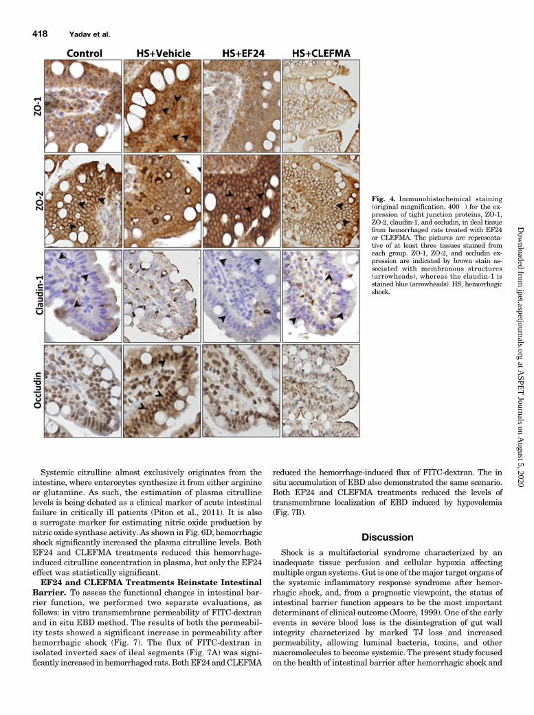
was first assessed by IHC (Fig. 4). At protein levels also, theexpression of ZO-1 and occludin was enhanced, whereas that ofZO-2 and claudin-1 was reduced by hypovolemia. These effectsof blood loss on the expression of various TJ proteins were
reversed when the hypovolemic rats were treated with EF24 orCLEFMA (Fig. 4). The results from IHC analyses of tissueswere confirmed by immunoblotting of ileal lysates (Fig. 5, Aand B). The expression-inducing effect of hypovolemic shock onZO-1 and occludin was contrary to what was expected in aderanged ileal epithelium. Because ZO-1 is post-transcriptionallyregulated by tyrosine (Tyr) phosphorylation, we investigatedwhether hypovolemia and EF24 treatment have any effect onthe expression of phosphorylated form of ZO-1. To this end, weimmunoprecipitated the tissue lysate with anti-phosphotyrosineantibody and probed the blot with anti–ZO-1 antibody. We foundthat hypovolemia significantly reduced the Tyr-phosphorylatedZO-1. EF24 as well as CLEFMA treatment recovered thephospho(Tyr)–ZO-1 to its basal levels (Fig. 5C). In case of occludin,the Tyr phosphorylation was not much affected by hemorrhagicshock, but it was substantially reduced by EF24 and CLEFMAtreatments. At the same time, threonine (Thr) phosphorylation ofoccludin was found to be increased by hemorrhagic shock.Treatments with EF24 and CLEFMA appeared to have main-tained the hemorrhage-induced Thr phosphorylation of occludin(Fig. 5C).EF24 and CLEFMA Treatments Diminish Epithelial
Cell Injury. Injury to intestinal epithelia is marked by anincreased shedding of ILBP, resulting in lowered detectionof cell-associated ILBP. ILBP is a small cytosolic protein(15 kDa) belonging to a family of fatty acid–binding proteins.We examined ILBP levels in ileal tissue by IHC and immuno-blotting. As shown in Fig. 6, A and B, hemorrhagic shocksubstantially decreased the expression of ILBP in ileum, butEF24 and CLEFMA treatments recovered the basal level ofILBP expression.
Fig. 3. Messenger RNA levels of (A) tight junction proteins (ZO-1, ZO-2, ZO-3, occludin, claudin-1, claudin-2, claudin-3, and claudin-4) and (B)antimicrobial proteins Defa-1 (defensin a-1), Defa-5 (defensin a-5), angiopoietin 4 (Angpt-4), and lysozyme-2 (Lyz-2) in ileal tissues. At least three tissuesper group were assayed, each in triplicate (*P , 0.05 versus control [CRTL]; #P , 0.05 versus hemorrhagic shock [HS]).
Hemorrhagic Shock-Induced Intestinal Barrier Dysfunction 417
at ASPE
T Journals on A
ugust 5, 2020jpet.aspetjournals.org
Dow
nloaded from
Systemic citrulline almost exclusively originates from theintestine, where enterocytes synthesize it from either arginineor glutamine. As such, the estimation of plasma citrullinelevels is being debated as a clinical marker of acute intestinalfailure in critically ill patients (Piton et al., 2011). It is alsoa surrogate marker for estimating nitric oxide production bynitric oxide synthase activity. As shown in Fig. 6D, hemorrhagicshock significantly increased the plasma citrulline levels. BothEF24 and CLEFMA treatments reduced this hemorrhage-induced citrulline concentration in plasma, but only the EF24effect was statistically significant.EF24 and CLEFMA Treatments Reinstate Intestinal
Barrier. To assess the functional changes in intestinal bar-rier function, we performed two separate evaluations, asfollows: in vitro transmembrane permeability of FITC-dextranand in situ EBD method. The results of both the permeabil-ity tests showed a significant increase in permeability afterhemorrhagic shock (Fig. 7). The flux of FITC-dextran inisolated inverted sacs of ileal segments (Fig. 7A) was signi-ficantly increased in hemorrhaged rats. BothEF24andCLEFMA
reduced the hemorrhage-induced flux of FITC-dextran. The insitu accumulation of EBD also demonstrated the same scenario.Both EF24 and CLEFMA treatments reduced the levels oftransmembrane localization of EBD induced by hypovolemia(Fig. 7B).
DiscussionShock is a multifactorial syndrome characterized by an
inadequate tissue perfusion and cellular hypoxia affectingmultiple organ systems. Gut is one of themajor target organs ofthe systemic inflammatory response syndrome after hemor-rhagic shock, and, from a prognostic viewpoint, the status ofintestinal barrier function appears to be the most importantdeterminant of clinical outcome (Moore, 1999). One of the earlyevents in severe blood loss is the disintegration of gut wallintegrity characterized by marked TJ loss and increasedpermeability, allowing luminal bacteria, toxins, and othermacromolecules to become systemic. The present study focusedon the health of intestinal barrier after hemorrhagic shock and
Fig. 4. Immunohistochemical staining(original magnification, 400�) for the ex-pression of tight junction proteins, ZO-1,ZO-2, claudin-1, and occludin, in ileal tissuefrom hemorrhaged rats treated with EF24or CLEFMA. The pictures are representa-tive of at least three tissues stained fromeach group. ZO-1, ZO-2, and occludin ex-pression are indicated by brown stain as-sociated with membranous structures(arrowheads), whereas the claudin-1 isstained blue (arrowheads). HS, hemorrhagicshock.
418 Yadav et al.
at ASPE
T Journals on A
ugust 5, 2020jpet.aspetjournals.org
Dow
nloaded from

treatment with two diphenyldihaloketones, EF24 and CLEFMA.Earlier, we have shown that EF24 treatment suppresseshemorrhage-induced pulmonary markers of inflammation,namely NF-kB, Toll-like receptor 4, cyclooxygenase-2, andIL-1 receptor 1, as well as systemic proinflammatory cytokines,such as tumor necrosis factor-a and IL-6 (Yadav et al., 2013,2014). We also reported that EF24 potently suppressed NF-kBactivation, modulated dendritic cell phenotype, and reducedsecretion of proinflammatory cytokines (Vilekar et al., 2012).These initial observations led us to hypothesize that EF24 andCLEFMA will protect ileum from barrier function loss inhemorrhagic shock.
The TJ proteins belonging to the ZO family not only providescaffolding for the assembly of other TJ proteins via their PDZdomains, but also act as membrane-associated guanylate kinase-like signaling proteins in cellular growth pathways (Bauer et al.,2010). We found that hemorrhage affected the expression of ZO-1and ZO-2 differently—whereas ZO-1 was upregulated, ZO-2showed downregulation after hemorrhagic shock; EF24 andCLEFMA both recovered ZO-1 and ZO-2 expressions to theirbasal levels. The reasons for differential response of ZO-1 andZO-2 to hemorrhagic shock are not clear, but literature providessome clues about the behavior of ZO-1. It turns out that appro-priate localization of these PDZ-containing proteins close to the
Fig. 5. (A) Expression of tight junction proteins ZO-1, ZO-2,claudin, and occludin in ileal tissue from various groups(n $ 3 per group). (B) The immunoblots were analyzed bydensitometry. Actin expression was probed to ensure equalloading of total protein in each well and to normalize thedensitometry values. (C) The effect of hemorrhagic shockand drug treatments on the phosphorylation status ofZO-1 and occludin proteins. Tyrosine phosphorylation wasprobed for both ZO-1 and occludin, whereas threonine phos-phorylation was examined only for occludin. The ileal tissuehomogenate was immunoprecipitated by anti-phosphotyrosineor anti-phosphothreonine antibodies, and the blots were probedwith anti–ZO-1 and anti-occludin antibodies. The immuno-precipitation was performed on two separate tissues fromeach group. CL, CLEFMA; CTRL, control; HS, hemorrhagicshock; V, vehicle.
Fig. 6. (A) A representative immunohis-tochemical staining of ILBP expression(blue stain, arrowheads) in rat ileum. (B)The ileal tissue lysates were immunoblot-ted with anti-rat ILBP antibody. (C) Theimmunoblots were analyzed by densitom-etry. At least three randomly selectedileal tissues per group were assayed. Actinexpression was probed to ensure equalloading of total protein in each well and tonormalize the ILBP densitometry values.(D) Plasma levels of citrulline (n = 4 pergroup). CL, CLEFMA; EF, EF24; HS,hemorrhagic shock; V, vehicle (*P , 0.05versus control [CRTL]; #P , 0.05 versusHS).
Hemorrhagic Shock-Induced Intestinal Barrier Dysfunction 419
at ASPE
T Journals on A
ugust 5, 2020jpet.aspetjournals.org
Dow
nloaded from

plasma membrane in the enterocytes is more important thanmere cellular expression levels for effective barrier function.This restricted localization is dependent on post-translationalmodification, such as tyrosine phosphorylation (Rao et al., 2002).Even when the expression levels of ZO-1 remain unaltered, thetyrosine phosphorylation and relocalization from apical mem-brane of intestinal villi could occur in breached intestinal barrier(Hamada et al., 2010). Phosphorylation also affects mutualinteraction among TJ proteins. For instance, the interactionbetween ZO-1 and occludin is phosphorylation-dependent(Tash et al., 2012). Dephosphorylated ZO-1 has been shownto cause absence of membranous localization of occludin inactive celiac disease (Ciccocioppo et al., 2006). Our observationthat hemorrhage induces the expression of ZO-1, but decreasesthe phosphorylated form of ZO-1, also points to the importanceof phospho–ZO-1 in the formation of intestinal barrier.The claudins and occludins are two major transmembrane
proteins that interact with ZO proteins and directly determinethe paracellular permeability to different ions and largemolecules(Hu et al., 2013). The function of claudins in epithelial barrier isalso subject to modulation by Ser/Thr phosphorylation andinteraction with PDZ-binding domains (Groschwitz and Hogan,2009). We found that hemorrhage downregulated the expressionof claudins 1, 2, and 3, but upregulated the expression of claudin 4.Even though such differential expression of various claudinproteins in response to ischemia/reperfusion injury of the intestinehas also been noted by others (Takizawa et al., 2012), the reasonsare not clearly understood. Among 27 known members of claudinfamily, claudins-1 and -3 have sealing functions, claudin-2 formsa channel providing selectivity to the cations, whereas the role ofclaudin 4 in barrier function is not known (Gunzel and Fromm,2012). Recent reports have associated claudin-4 expression withepithelial malignancies and premalignant precursor lesions(Neesse et al., 2012). Interestingly, transforming growthfactor-b has been reported to transcriptionally upregulateclaudin-4 expression via a Smad-4–dependent pathway (Kotleret al., 2013). Because transforming growth factor-b is increasedin hemorrhagic shock (Ayala et al., 1993), its role in hypovolemia-induced claudin-4 expression could not be ruled out.Unlike claudins, occludin protein has the greatest effect on
the flux of large macromolecules (Al-Sadi et al., 2011). Theexpression of occludin is markedly decreased in intestinal
permeability disorders, such as Crohn’s disease and ulcera-tive colitis (Gassler et al., 2001), suggesting that decreasedoccludin expression is associated with an increase in intestinalpermeability. Our finding is contrary to this conjecture, andwe found that hemorrhagic shock increased occludin ex-pression at both mRNA and protein levels. The effect ofphosphorylation status of occludin in maintaining ilealhomeostasis has been elegantly investigated by Rao’sgroup (Seth et al., 2007; Rao, 2009). The phosphorylationof occludin at Ser/Thr helps, whereas phosphorylation at Tyrresidue deters its interaction with ZO-1 (Seth et al., 2007; Rao,2009). Our results from phospho-Tyr coimmunoprecipitationshow that the ileum-preserving EF24 and CLEFMA treat-ments reduce ileal phospho(Tyr)-occludin in hypovolemic ratsand support these in vitro findings in Caco-2 monolayers. Wealso found that hemorrhagic shock induced Thr phosphoryla-tion of occludin, and treatments with EF24 and CLEFMApreserved this modification. The occludin-specific effect ofEF24 and CLEFMA may be explained by their ability toreduce Tyr phosphorylation and maintain Thr phosphoryla-tion of occludin, thereby aiding its interaction with ZO-1.Earlier, Seth et al. (2007) predicted that the net role ofoccludin is determined by the relative levels of Tyr and Thrphosphorylation. It is noteworthy that the Tyr/Thr phosphor-ylation ratio of occludin is regulated by protein phosphatases2A and 1 (Seth et al., 2007; Sheth et al., 2009). Overall, theseresults imply that mere increased expression of occludin maynot be sufficient, but phosphorylation at appropriate aminoacid residue may be important in regulating its interactionwith other TJ proteins.The remarkable restoration of TJ proteins and accompany-
ing histology by treatments with EF24 and CLEFMA wasmanifested in the resurrection of ileal barrier function. Thetreatments significantly reduced hemorrhagic shock-inducedincrease in the intestinal permeability. We also found that onlyCLEFMA treatment upregulatedmRNA levels of antimicrobialproteins; neither hypovolemia nor EF24 significantly alteredthe antimicrobial protein mRNA levels. The hypothesis thatthe variation in chemical structure of EF24 and CLEFMAresults in these differences is a subject of further investigation.Notmuch is known about the changes in antimicrobial proteinssecreted by Paneth cells in hemorrhagic shock, but in one
Fig. 7. Effect of EF24 and CLEFMA on hemorrhagic shock-induced increase in intestinal permeability for dextran-FITC4000 and EBD. (A) Approximately5-cm saline-filled inverted sacs of ileum (n = 4 segments per rat, 4 rats per group) were allowed to equilibrate with dextran-FITC solution at 37°C. After45 minutes, the inner contents of the segments were assayed for accumulation of fluorescent dextran. Permeability index was expressed as fluorescenceunits/cm per hour. (B) EBD was injected 1 hour before euthanasia, and its accumulation in the ileal tissues was estimated by colorimetry (n = 3 pergroup). The EBD accumulation in ileal tissue was quantitated with respect to the plasma EBD concentration. *P, 0.05 versus control (CTRL); #P, 0.05versus HS. CL, CLEFMA; HS, hemorrhagic shock; V, vehicle.
420 Yadav et al.
at ASPE
T Journals on A
ugust 5, 2020jpet.aspetjournals.org
Dow
nloaded from

report rat enteric a-defensin gene was found to be upregulatedimmediately after termination of shock (Condon et al., 1999).Because no long-termassessmentwas performed in the publishedstudy (Condon et al., 1999), our results could be reconciled byspeculating that the defensemechanisms originating fromPanethcells are progressively salvaged over time. Paneth cells developfrom epithelial progenitor cells and are restricted to crypt base inthe gut epithelium (Gunther et al., 2013).The above-described salutary effects of EF24 and their time
course pointed toward a rather rapid localization in theintestinal tissue. We examined the biodisposition of EF24 bylabeling it with imageable radionuclide technetium-99m (140KeV g ray, 6-hour decay half-life) for g camera imaging. For thiswe synthesized a N-HYNIC conjugate of EF24 by employinga procedure described elsewhere (Lagisetty et al., 2012). Asshown in Supplemental Fig. 3, intravenously-injected technetium-99m–EF24 rapidly accumulated in rat liver and intestine. The firstsign of EF24 localization in gut was observed within 5 minutes ofinjection. The accumulation increased over time, and EF24appeared to be retained in intestinal tissue up to 6 hours afterinjection. No other organ except liver and intestines accumu-lated significant radioactivity, suggesting that its clearancedepended on hepatobiliary route. The image-derived knowl-edge of drug accumulation suggests that EF24 is naturallycleared from circulation into the intestine, which might explainthe remarkable effects of EF24 and CLEFMA on intestinalintegrity in hemorrhagic shock. What molecular pathways areinvolved in these actions remains a subject of continued in-vestigation in our laboratory.Conclusions. Besides hemorrhagic shock, dysregulated
barrier function is a hallmark in many other disorders, suchas inflammatory bowel diseases, food allergy, celiac disease,type I diabetes, etc. (Groschwitz and Hogan, 2009; Salim andSoderholm, 2011). The intestinal pathology of hemorrhagicshock is also similar to that observed in victims of burn, sepsis,radiation exposure (MacNaughton, 2000; Hauer-Jensen et al.,2007), or drug toxicities (Meng et al., 2013; Russo et al., 2013).Despite such widespread impact, the current therapies for themanagement of barrier function loss remain inadequate. Theresults of this study show that the administration of EF24 orCLEFMA significantly improved the intestinal integrity of ratssubjected to hemorrhage. These encouraging observations ledus to speculate about the additive benefits of EF24 or CLEFMAwhen administered in combination with conventional resusci-tation fluids used to treat hemorrhagic shock.
Acknowledgments
The authors thank Andria Hedrick, research associate in theDepartment of Pharmaceutical Sciences, University of OklahomaHealth Sciences Center, for technical help.
Authorship Contributions
Participated in research design: Awasthi.Conducted experiments: Yadav, Hussain, Sahoo, Awasthi.Contributed new reagents or analytic tools: Awasthi.Performed data analysis: Yadav, Awasthi.Wrote or contributed to the writing of the manuscript: Yadav,
Hussain, Awasthi.
References
Al-Sadi R, Khatib K, Guo S, Ye D, Youssef M, and Ma T (2011) Occludin regulatesmacromolecule flux across the intestinal epithelial tight junction barrier. Am JPhysiol Gastrointest Liver Physiol 300:G1054–G1064.
Awasthi V, Yee SH, Jerabek P, Goins B, and Phillips WT (2007) Cerebral oxygendelivery by liposome-encapsulated hemoglobin: a positron-emission tomographicevaluation in a rat model of hemorrhagic shock. J Appl Physiol (1985) 103:28–38.
Ayala A, Meldrum DR, Perrin MM, and Chaudry IH (1993) The release of trans-forming growth factor-beta following haemorrhage: its role as a mediator of hostimmunosuppression. Immunology 79:479–484.
Bauer H, Zweimueller-Mayer J, Steinbacher P, Lametschwandtner A, and Bauer HC(2010) The dual role of zonula occludens (ZO) proteins. JBiomedBiotechnol 2010:402593.
Ciccocioppo R, Finamore A, Ara C, Di Sabatino A, Mengheri E, and Corazza GR(2006) Altered expression, localization, and phosphorylation of epithelial junctionalproteins in celiac disease. Am J Clin Pathol 125:502–511.
Condon MR, Viera A, D’Alessio M, and Diamond G (1999) Induction of a rat entericdefensin gene by hemorrhagic shock. Infect Immun 67:4787–4793.
Cotton BA (2011) Alternative fluids for prehospital resuscitation: “pharmacological”resuscitation fluids. J Trauma 70:S30–S31.
Cruz RJ, Jr, Harada T, Sasatomi E, and Fink MP (2011) Effects of ethyl pyruvate andother a-keto carboxylic acid derivatives in a rat model of multivisceral ischemiaand reperfusion. J Surg Res 165:151–157.
Fabriek BO, Dijkstra CD, and van den Berg TK (2005) The macrophage scavengerreceptor CD163. Immunobiology 210:153–160.
Fink MP (2007) Ethyl pyruvate: a novel anti-inflammatory agent. J Intern Med 261:349–362.
Fink MP and Delude RL (2005) Epithelial barrier dysfunction: a unifying theme toexplain the pathogenesis of multiple organ dysfunction at the cellular level. CritCare Clin 21:177–196.
Gassler N, Rohr C, Schneider A, Kartenbeck J, Bach A, Obermüller N, Otto HF,and Autschbach F (2001) Inflammatory bowel disease is associated with changes ofenterocytic junctions. Am J Physiol Gastrointest Liver Physiol 281:G216–G228.
Groschwitz KR and Hogan SP (2009) Intestinal barrier function: molecular regula-tion and disease pathogenesis. J Allergy Clin Immunol 124:3–20, quiz 21–22.
Günther C, Neumann H, Neurath MF, and Becker C (2013) Apoptosis, necrosis andnecroptosis: cell death regulation in the intestinal epithelium. Gut 62:1062–1071.
Günzel D and Fromm M (2012) Claudins and other tight junction proteins. ComprPhysiol 2:1819–1852.
Hamada K, Shitara Y, Sekine S, and Horie T (2010) Zonula Occludens-1 alterationsand enhanced intestinal permeability in methotrexate-treated rats. Cancer Che-mother Pharmacol 66:1031–1038.
Hartsock A and Nelson WJ (2008) Adherens and tight junctions: structure, functionand connections to the actin cytoskeleton. Biochim Biophys Acta 1778:660–669.
Hauer-Jensen M, Kumar KS, Wang J, Berbee M, Fu Q, and Boerma M (2007) In-testinal toxicity in radiation- and combined injury: significance, mechanisms, andcountermeasures, in Global Terrorism Issues and Developments (Larche RA ed) pp1–40, Nova Science Publishers, New York.
Hu YJ, Wang YD, Tan FQ, and Yang WX (2013) Regulation of paracellular perme-ability: factors and mechanisms. Mol Biol Rep 40:6123–6142.
Kao KK and Fink MP (2010) The biochemical basis for the anti-inflammatory andcytoprotective actions of ethyl pyruvate and related compounds. Biochem Phar-macol 80:151–159.
Kasinski AL, Du Y, Thomas SL, Zhao J, Sun SY, Khuri FR, Wang CY, Shoji M, SunA, Snyder JP, et al. (2008) Inhibition of IkappaB kinase-nuclear factor-kappaBsignaling pathway by 3,5-bis(2-flurobenzylidene)piperidin-4-one (EF24), a novelmonoketone analog of curcumin. Mol Pharmacol 74:654–661.
Kotler BM, Kerstetter JE, and Insogna KL (2013) Claudins, dietary milk proteins,and intestinal barrier regulation. Nutr Rev 71:60–65.
Lagisetty P, Powell DR, and Awasthi V (2009) Synthesis and structural determinationof 3 3,5-bis(2-fluorobenzylidene)-4-piperidone analogs of curcumin. J Mol Struct936:23–28.
Lagisetty P, SubramaniamD, Sahoo K, Anant S, and Awasthi V (2012) Anticancer activityof an imageable curcuminoid 1-[2-aminoethyl-(6-hydrazinopyridine-3-carbamidyl)-3,5-bis-(2-fluorobenzylidene)-4-piperidone] (EFAH). Chem Biol Drug Des 79:194–201.
Lagisetty P, Vilekar P, Sahoo K, Anant S, and Awasthi V (2010) CLEFMA - an anti-proliferative curcuminoid from structure-activity relationship studies on 3,5-bis(benzylidene)-4-piperidones. Bioorg Med Chem 18:6109–6120.
MacDermott RP (1999) Chemokines in the inflammatory bowel diseases. J ClinImmunol 19:266–272.
MacNaughton WK (2000) Review article: new insights into the pathogenesis ofradiation-induced intestinal dysfunction. Aliment Pharmacol Ther 14:523–528.
Meng J, Yu H, Ma J, Wang J, Banerjee S, Charboneau R, Barke RA, and Roy S (2013)Morphine induces bacterial translocation in mice by compromising intestinalbarrier function in a TLR-dependent manner. PLoS One 8:e54040.
Moore FA (1999) The role of the gastrointestinal tract in postinjury multiple organfailure. Am J Surg 178:449–453.
Neesse A, Griesmann H, Gress TM, and Michl P (2012) Claudin-4 as therapeutictarget in cancer. Arch Biochem Biophys 524:64–70.
Piton G, Manzon C, Cypriani B, Carbonnel F, and Capellier G (2011) Acute intestinalfailure in critically ill patients: is plasma citrulline the right marker? IntensiveCare Med 37:911–917.
Rao R (2009) Occludin phosphorylation in regulation of epithelial tight junctions.Ann N Y Acad Sci 1165:62–68.
Rao RK, Basuroy S, Rao VU, Karnaky KJ, Jr, and Gupta A (2002) Tyrosine phos-phorylation and dissociation of occludin-ZO-1 and E-cadherin-beta-catenin com-plexes from the cytoskeleton by oxidative stress. Biochem J 368:471–481.
Rhodes RS, Depalma RG, and Robinson AV (1973) Intestinal barrier function inhemorrhagic shock. J Surg Res 14:305–312.
Rotstein OD (2000) Pathogenesis of multiple organ dysfunction syndrome: gut origin,protection, and decontamination. Surg Infect (Larchmt) 1:217–223.
Russo F, Linsalata M, Clemente C, D’Attoma B, Orlando A, Campanella G, Giotta F,and Riezzo G (2013) The effects of fluorouracil, epirubicin, and cyclophosphamide(FEC60) on the intestinal barrier function and gut peptides in breast cancerpatients: an observational study. BMC Cancer 13:56.
Hemorrhagic Shock-Induced Intestinal Barrier Dysfunction 421
at ASPE
T Journals on A
ugust 5, 2020jpet.aspetjournals.org
Dow
nloaded from

Salim SY and Söderholm JD (2011) Importance of disrupted intestinal barrier ininflammatory bowel diseases. Inflamm Bowel Dis 17:362–381.
Sappington PL, Han X, Yang R, Delude RL, and Fink MP (2003) Ethyl pyruvateameliorates intestinal epithelial barrier dysfunction in endotoxemic mice andimmunostimulated caco-2 enterocytic monolayers. J Pharmacol Exp Ther 304:464–476.
Senthil M, Brown M, Xu DZ, Lu Q, Feketeova E, and Deitch EA (2006) Gut-lymphhypothesis of systemic inflammatory response syndrome/multiple-organ dysfunc-tion syndrome: validating studies in a porcine model. J Trauma 60:958–965..
Seth A, Sheth P, Elias BC, and Rao R (2007) Protein phosphatases 2A and 1 interactwith occludin and negatively regulate the assembly of tight junctions in the CACO-2 cell monolayer. J Biol Chem 282:11487–11498.
Sheth P, Samak G, Shull JA, Seth A, and Rao R (2009) Protein phosphatase 2A playsa role in hydrogen peroxide-induced disruption of tight junctions in Caco-2 cellmonolayers. Biochem J 421:59–70.
Shi HP, Deitch EA, Da Xu Z, Lu Q, and Hauser CJ (2002) Hypertonic saline improvesintestinal mucosa barrier function and lung injury after trauma-hemorrhagicshock. Shock 17:496–501.
Takizawa Y, Kishimoto H, Kitazato T, Tomita M, and Hayashi M (2012) Changes inprotein and mRNA expression levels of claudin family after mucosal lesion byintestinal ischemia/reperfusion. Int J Pharm 426:82–89.
Tash BR, Bewley MC, Russo M, Keil JM, Griffin KA, Sundstrom JM, Antonetti DA,Tian F, and Flanagan JM (2012) The occludin and ZO-1 complex, defined by small
angle X-ray scattering and NMR, has implications for modulating tight junctionpermeability. Proc Natl Acad Sci USA 109:10855–10860.
Vega D, Badami CD, Caputo FJ, Watkins AC, Lu Q, Xu da Z, Berezina TL, Zaets SB,Feketeova E, and Deitch EA (2008) The influence of the type of resuscitation fluidon gut injury and distant organ injury in a rat model of trauma/hemorrhagic shock.J Trauma 65:409–414..
Vilekar P, Awasthi S, Natarajan A, Anant S, and Awasthi V (2012) EF24 suppressesmaturation and inflammatory response in dendritic cells. Int Immunol 24:455–464.
Weiss J, Taylor GR, Zimmermann F, and Nebendahl K (2000) Collection of bodyfluids, in The Laboratory Rat (Krinke GJ ed) pp 485–510, Academic Press, London.
Yadav VR, Sahoo K, Roberts PR, and Awasthi V (2013) Pharmacologic suppression ofinflammation by a diphenyldifluoroketone, EF24, in a rat model of fixed-volumehemorrhage improves survival. J Pharmacol Exp Ther 347:346–356.
Yadav VR, Vilekar P, Awasthi S, and Awasthi V (2014) Hemorrhage-inducedinterleukin-1 receptor pathway in lung is suppressed by 3,5-bis(2-fluorobenzylidene)-4-piperidone in a rat model of hypovolemic shock. Artif Organs 38:675–683.
Address correspondence to: Dr. Vibhudutta Awasthi, Department ofPharmaceutical Sciences, 1110 North Stonewall Avenue, Oklahoma City, OK73117. E-mail: [email protected]
422 Yadav et al.
at ASPE
T Journals on A
ugust 5, 2020jpet.aspetjournals.org
Dow
nloaded from