Embed Size (px)
Citation preview
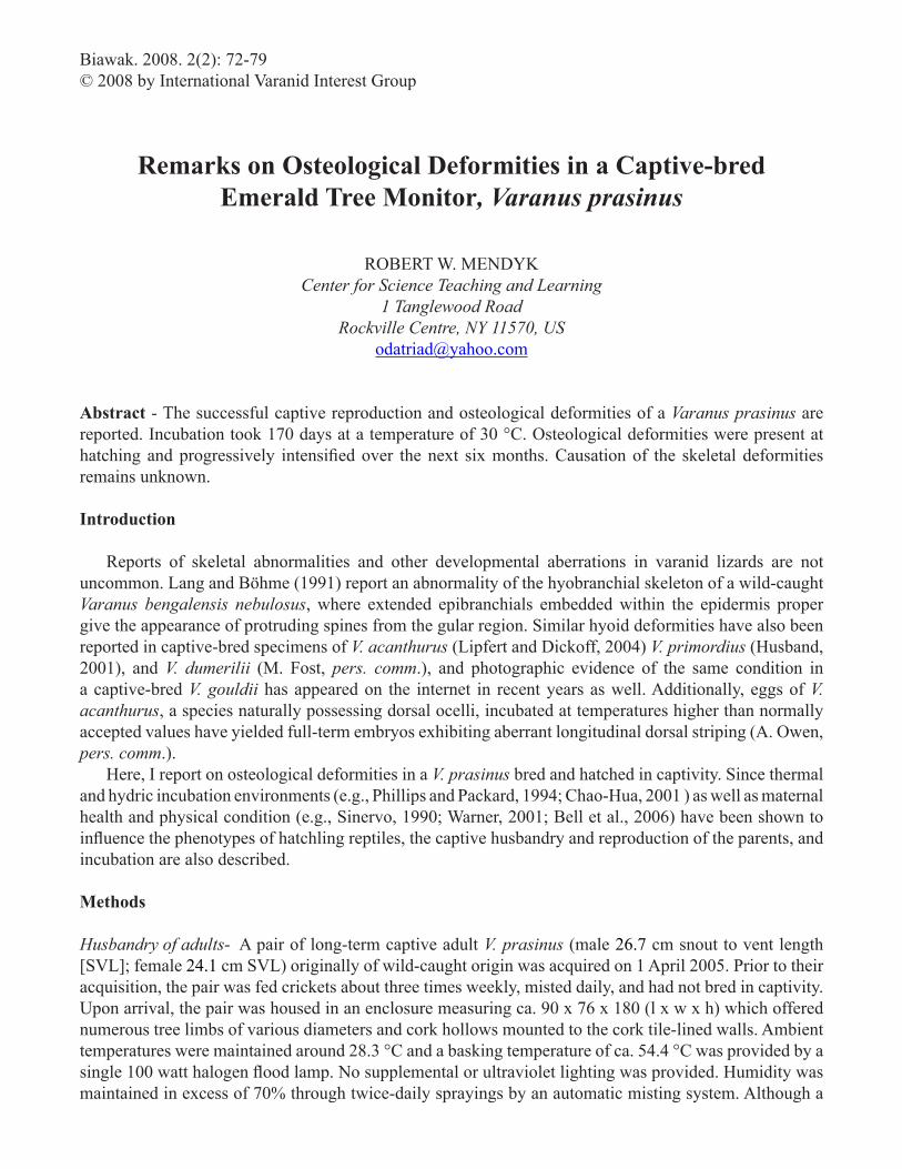
Biawak. 2008. 2(2): 72-79© 2008 by International Varanid Interest Group
Remarks on Osteological Deformities in a Captive-bred Emerald Tree Monitor, Varanus prasinus
ROBERT W. MENDYKCenter for Science Teaching and Learning
1 Tanglewood RoadRockville Centre, NY 11570, US
Abstract - The successful captive reproduction and osteological deformities of a Varanus prasinus are reported. Incubation took 170 days at a temperature of 30 °C. Osteological deformities were present at hatching and progressively intensified over the next six months. Causation of the skeletal deformities remains unknown.
Introduction
Reports of skeletal abnormalities and other developmental aberrations in varanid lizards are not uncommon. Lang and Böhme (1991) report an abnormality of the hyobranchial skeleton of a wild-caught Varanus bengalensis nebulosus, where extended epibranchials embedded within the epidermis proper give the appearance of protruding spines from the gular region. Similar hyoid deformities have also been reported in captive-bred specimens of V. acanthurus (Lipfert and Dickoff, 2004) V. primordius (Husband, 2001), and V. dumerilii (M. Fost, pers. comm.), and photographic evidence of the same condition in a captive-bred V. gouldii has appeared on the internet in recent years as well. Additionally, eggs of V. acanthurus, a species naturally possessing dorsal ocelli, incubated at temperatures higher than normally accepted values have yielded full-term embryos exhibiting aberrant longitudinal dorsal striping (A. Owen, pers. comm.). Here, I report on osteological deformities in a V. prasinus bred and hatched in captivity. Since thermal and hydric incubation environments (e.g., Phillips and Packard, 1994; Chao-Hua, 2001 ) as well as maternal health and physical condition (e.g., Sinervo, 1990; Warner, 2001; Bell et al., 2006) have been shown to influence the phenotypes of hatchling reptiles, the captive husbandry and reproduction of the parents, and incubation are also described.
Methods Husbandry of adults- A pair of long-term captive adult V. prasinus (male 26.7 cm snout to vent length [SVL]; female 24.1 cm SVL) originally of wild-caught origin was acquired on 1 April 2005. Prior to their acquisition, the pair was fed crickets about three times weekly, misted daily, and had not bred in captivity. Upon arrival, the pair was housed in an enclosure measuring ca. 90 x 76 x 180 (l x w x h) which offered numerous tree limbs of various diameters and cork hollows mounted to the cork tile-lined walls. Ambient temperatures were maintained around 28.3 °C and a basking temperature of ca. 54.4 °C was provided by a single 100 watt halogen flood lamp. No supplemental or ultraviolet lighting was provided. Humidity was maintained in excess of 70% through twice-daily sprayings by an automatic misting system. Although a
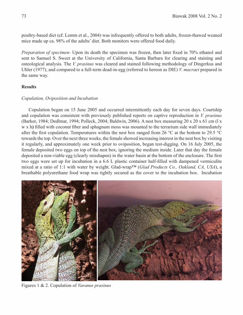
Figures 1 & 2. Copulation of Varanus prasinus
Biawak 2008 Vol. 2 No. 273
poultry-based diet (cf. Lemm et al., 2004) was infrequently offered to both adults, frozen-thawed weaned mice made up ca. 98% of the adults’ diet. Both monitors were offered food daily.
Preparation of specimen- Upon its death the specimen was frozen, then later fixed in 70% ethanol and sent to Samuel S. Sweet at the University of California, Santa Barbara for clearing and staining and osteological analysis. The V. prasinus was cleared and stained following methodology of Dingerkus and Uhler (1977), and compared to a full-term dead-in-egg (referred to hereon as DIE) V. macraei prepared in the same way.
Results Copulation, Oviposition and Incubation
Copulation began on 15 June 2005 and occurred intermittently each day for seven days. Courtship and copulation was consistent with previously published reports on captive reproduction in V. prasinus (Barker, 1984; Dedlmar, 1994; Polleck, 2004; Baldwin, 2006). A nest box measuring 20 x 20 x 61 cm (l x w x h) filled with coconut fiber and sphagnum moss was mounted to the terrarium side wall immediately after the first copulation. Temperatures within the nest box ranged from 26 °C at the bottom to 29.5 °C towards the top. Over the next three weeks, the female showed increasing interest in the nest box by visiting it regularly, and approximately one week prior to oviposition, began test-digging. On 16 July 2005, the female deposited two eggs on top of the nest box, ignoring the medium inside. Later that day the female deposited a non-viable egg (clearly misshapen) in the water basin at the bottom of the enclosure. The first two eggs were set up for incubation in a 6.6 L plastic container half-filled with dampened vermiculite mixed at a ratio of 1:1 with water by weight. Glad-wrap™ (Glad Products Co., Oakland, CA, USA), a breathable polyurethane food wrap was tightly secured as the cover to the incubation box. Incubation
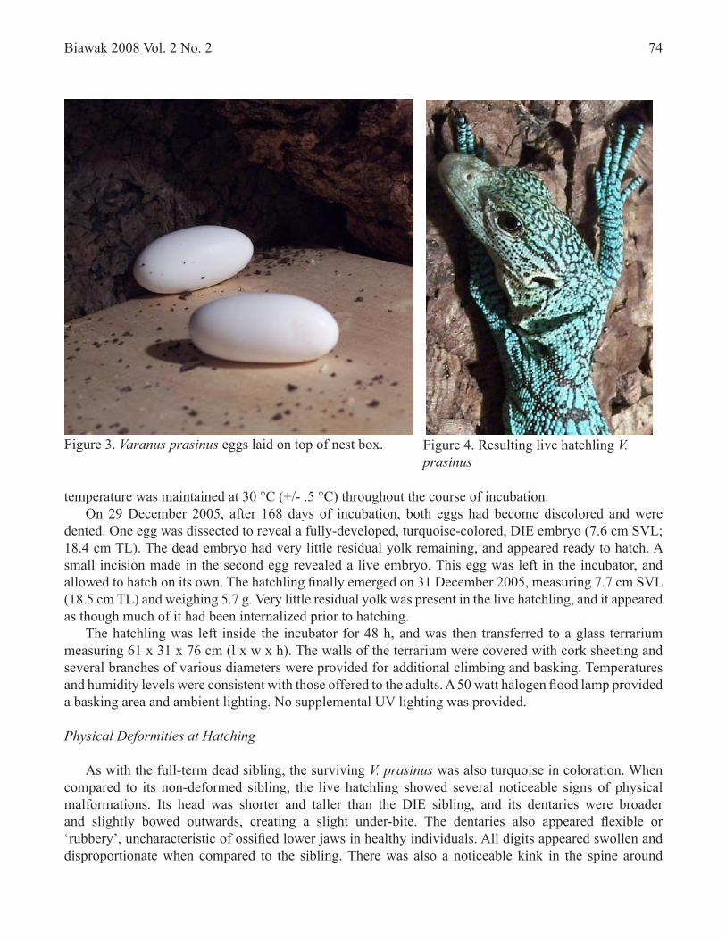
Figure 3. Varanus prasinus eggs laid on top of nest box. Figure 4. Resulting live hatchling V. prasinus
Biawak 2008 Vol. 2 No. 2 74
temperature was maintained at 30 °C (+/- .5 °C) throughout the course of incubation. On 29 December 2005, after 168 days of incubation, both eggs had become discolored and were dented. One egg was dissected to reveal a fully-developed, turquoise-colored, DIE embryo (7.6 cm SVL; 18.4 cm TL). The dead embryo had very little residual yolk remaining, and appeared ready to hatch. A small incision made in the second egg revealed a live embryo. This egg was left in the incubator, and allowed to hatch on its own. The hatchling finally emerged on 31 December 2005, measuring 7.7 cm SVL (18.5 cm TL) and weighing 5.7 g. Very little residual yolk was present in the live hatchling, and it appeared as though much of it had been internalized prior to hatching. The hatchling was left inside the incubator for 48 h, and was then transferred to a glass terrarium measuring 61 x 31 x 76 cm (l x w x h). The walls of the terrarium were covered with cork sheeting and several branches of various diameters were provided for additional climbing and basking. Temperatures and humidity levels were consistent with those offered to the adults. A 50 watt halogen flood lamp provided a basking area and ambient lighting. No supplemental UV lighting was provided. Physical Deformities at Hatching
As with the full-term dead sibling, the surviving V. prasinus was also turquoise in coloration. When compared to its non-deformed sibling, the live hatchling showed several noticeable signs of physical malformations. Its head was shorter and taller than the DIE sibling, and its dentaries were broader and slightly bowed outwards, creating a slight under-bite. The dentaries also appeared flexible or ‘rubbery’, uncharacteristic of ossified lower jaws in healthy individuals. All digits appeared swollen and disproportionate when compared to the sibling. There was also a noticeable kink in the spine around
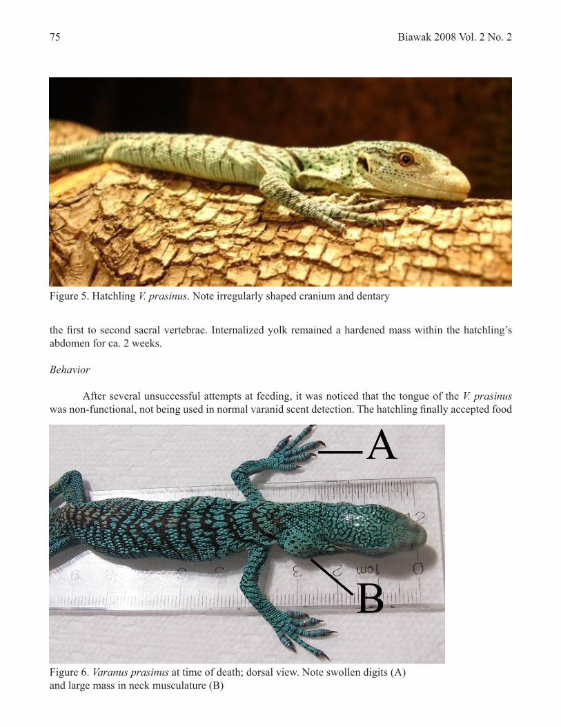
Figure 6. Varanus prasinus at time of death; dorsal view. Note swollen digits (A) and large mass in neck musculature (B)
Biawak 2008 Vol. 2 No. 275
the first to second sacral vertebrae. Internalized yolk remained a hardened mass within the hatchling’s abdomen for ca. 2 weeks. Behavior
After several unsuccessful attempts at feeding, it was noticed that the tongue of the V. prasinus was non-functional, not being used in normal varanid scent detection. The hatchling finally accepted food
Figure 5. Hatchling V. prasinus. Note irregularly shaped cranium and dentary
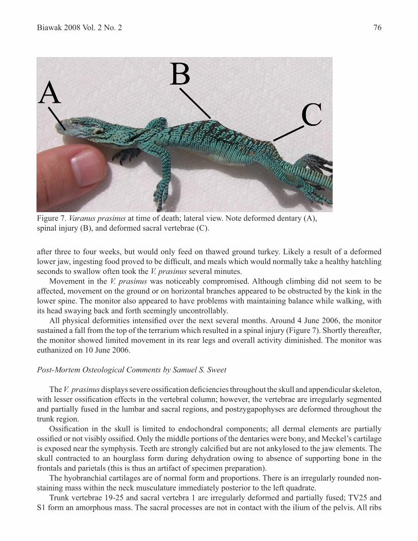
Figure 7. Varanus prasinus at time of death; lateral view. Note deformed dentary (A), spinal injury (B), and deformed sacral vertebrae (C).
Biawak 2008 Vol. 2 No. 2 76
after three to four weeks, but would only feed on thawed ground turkey. Likely a result of a deformed lower jaw, ingesting food proved to be difficult, and meals which would normally take a healthy hatchling seconds to swallow often took the V. prasinus several minutes. Movement in the V. prasinus was noticeably compromised. Although climbing did not seem to be affected, movement on the ground or on horizontal branches appeared to be obstructed by the kink in the lower spine. The monitor also appeared to have problems with maintaining balance while walking, with its head swaying back and forth seemingly uncontrollably. All physical deformities intensified over the next several months. Around 4 June 2006, the monitor sustained a fall from the top of the terrarium which resulted in a spinal injury (Figure 7). Shortly thereafter, the monitor showed limited movement in its rear legs and overall activity diminished. The monitor was euthanized on 10 June 2006.
Post-Mortem Osteological Comments by Samuel S. Sweet
The V. prasinus displays severe ossification deficiencies throughout the skull and appendicular skeleton, with lesser ossification effects in the vertebral column; however, the vertebrae are irregularly segmented and partially fused in the lumbar and sacral regions, and postzygapophyses are deformed throughout the trunk region. Ossification in the skull is limited to endochondral components; all dermal elements are partially ossified or not visibly ossified. Only the middle portions of the dentaries were bony, and Meckel’s cartilage is exposed near the symphysis. Teeth are strongly calcified but are not ankylosed to the jaw elements. The skull contracted to an hourglass form during dehydration owing to absence of supporting bone in the frontals and parietals (this is thus an artifact of specimen preparation). The hyobranchial cartilages are of normal form and proportions. There is an irregularly rounded non-staining mass within the neck musculature immediately posterior to the left quadrate. Trunk vertebrae 19-25 and sacral vertebra 1 are irregularly deformed and partially fused; TV25 and S1 form an amorphous mass. The sacral processes are not in contact with the ilium of the pelvis. All ribs
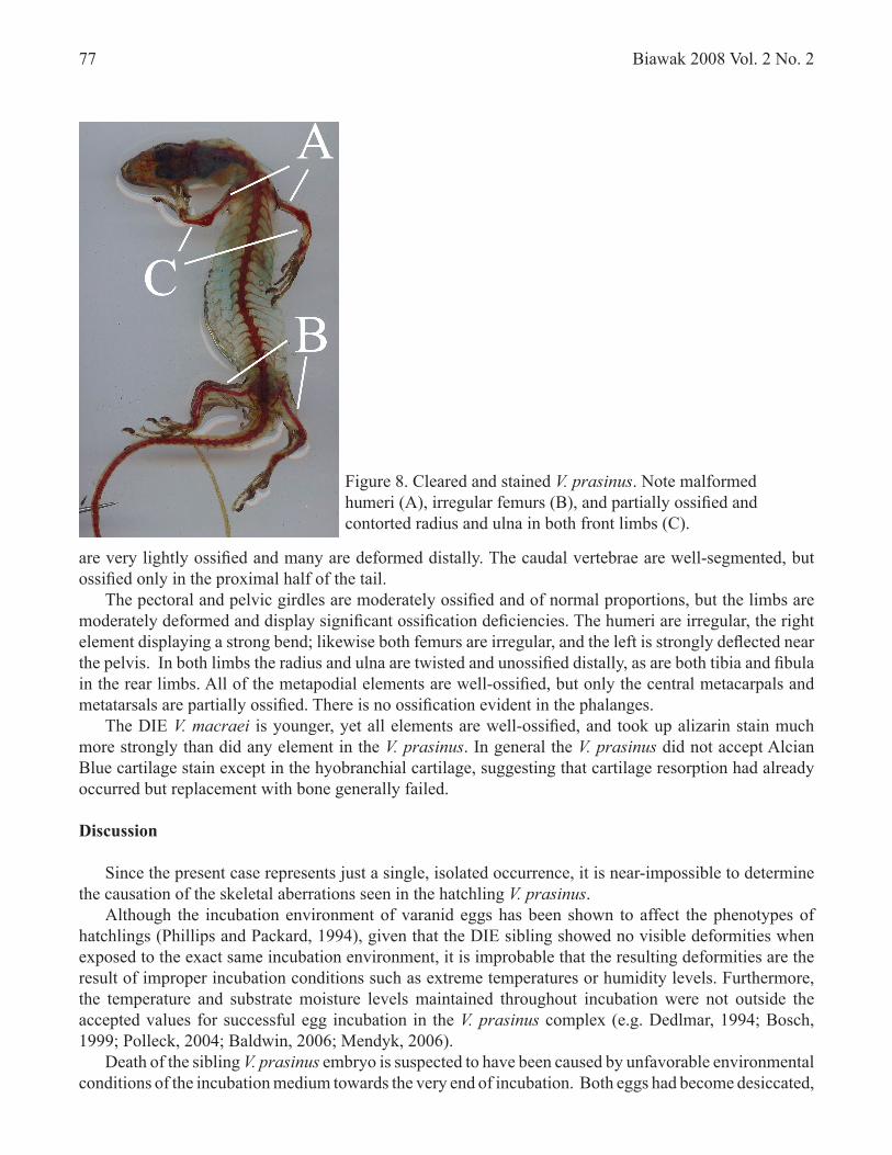
are very lightly ossified and many are deformed distally. The caudal vertebrae are well-segmented, but ossified only in the proximal half of the tail. The pectoral and pelvic girdles are moderately ossified and of normal proportions, but the limbs are moderately deformed and display significant ossification deficiencies. The humeri are irregular, the right element displaying a strong bend; likewise both femurs are irregular, and the left is strongly deflected near the pelvis. In both limbs the radius and ulna are twisted and unossified distally, as are both tibia and fibula in the rear limbs. All of the metapodial elements are well-ossified, but only the central metacarpals and metatarsals are partially ossified. There is no ossification evident in the phalanges. The DIE V. macraei is younger, yet all elements are well-ossified, and took up alizarin stain much more strongly than did any element in the V. prasinus. In general the V. prasinus did not accept Alcian Blue cartilage stain except in the hyobranchial cartilage, suggesting that cartilage resorption had already occurred but replacement with bone generally failed.
Discussion
Since the present case represents just a single, isolated occurrence, it is near-impossible to determine the causation of the skeletal aberrations seen in the hatchling V. prasinus. Although the incubation environment of varanid eggs has been shown to affect the phenotypes of hatchlings (Phillips and Packard, 1994), given that the DIE sibling showed no visible deformities when exposed to the exact same incubation environment, it is improbable that the resulting deformities are the result of improper incubation conditions such as extreme temperatures or humidity levels. Furthermore, the temperature and substrate moisture levels maintained throughout incubation were not outside the accepted values for successful egg incubation in the V. prasinus complex (e.g. Dedlmar, 1994; Bosch, 1999; Polleck, 2004; Baldwin, 2006; Mendyk, 2006). Death of the sibling V. prasinus embryo is suspected to have been caused by unfavorable environmental conditions of the incubation medium towards the very end of incubation. Both eggs had become desiccated,
Biawak 2008 Vol. 2 No. 277
Figure 8. Cleared and stained V. prasinus. Note malformed humeri (A), irregular femurs (B), and partially ossified and contorted radius and ulna in both front limbs (C).
Biawak 2008 Vol. 2 No. 2 78
indicating that the vermiculite had become too dry, with one embryo perishing. Since both embryos incubated for the entire term, using up virtually all of the nutrients and reserves provided by the female within each egg, it is also unlikely that maternal health or investment had anything to do with the cause of the deformities in the live hatchling. Physical deformities in the V. prasinus were evident upon hatching, however they progressively intensified with age. Although there have been few studies which assess the need for UV light by varanids (Gillespie et al., 2001), given the importance of UV exposure in many other diurnal lizard groups, and the prevalence of bone deficiencies in specimens of these taxa maintained in the absence of supplemental UV light (e.g., Mader, 1996), it is possible that a lack of UV exposure by the juvenile V. prasinus in the present case intensified its osteological deformities. Aside from the skeletal aberrations seen in the V. prasinus, of particular interest was the difference in body coloration between both adults and their offspring (Figures 1 & 4, respectively). Both the live hatchling and DIE embryo were turquoise in coloration, whereas both adults were emerald green and classical representatives of V. prasinus. Turquoise offspring resulting from captive breedings of V. prasinus have previously been reported (e.g., see images in Polleck, 2004), and long-term captives originating from the wild have been known to lose yellow pigmentation over time in captivity, resulting in turquoise body coloration (pers. obs.). The true cause of this loss of pigmentation remains to be seen, however a deficient diet and or lack of adequate UV exposure may be suspect. This green to turquoise color transformation appears to be common in several green-colored reptile and amphibian species such as Morelia viridis, Physignathus cocincus, Basiliscus plumifrons, Abronia graminea., and Litoria caerulea (pers. obs.), but has received very little attention in the herpetological or herpetocultural literature (Switak, 2006). Future investigations are needed to determine the cause of this color loss in captive herpetological collections and whether or not the same agent is responsible across all groups in which it occurs, and if such a deficiency may have any lasting physiological effects on captive specimens.
Acknowledgements – I would like to thank Samuel S. Sweet for preparing the V. prasinus and for analyzing its osteology.
Literature Cited
Baldwin, B. 2006. Successful care and reproduction of Green Tree monitors (Varanus prasinus) at the San Diego Zoo. Iguana 13(4): 283-287.
Barker, D.G. 1984. Maintenance and reproduction of Green Tree monitors at the Dallas Zoo. Proceedings of the 8th International Herpetological Symposium on Captive Propagation and Husbandry 8: 91-92.
Bell, B., J.R. Spotila and J. Congdon. 2006. High incidence of deformity in aquatic turtles in the John Heinz National Wildlife Refuge. Environmental Pollution 142(3): 457- 465.
Bosch, H. 1999. Successful breeding of the Emerald monitor (Varanus p. prasinus) in the Löbbecke Museum + Aquazoo, Düsseldorf (Germany). Mertensiella 11: 225-226.
Chao-Hua, J.I.X.Z, 2001. Effects of thermal and hydric environments on incubating eggs, hatching success, and hatchling traits in the Chinese skink (Eumeces chinensis). Acta Zoologica 47(3): 256-265.
Biawak 2008 Vol. 2 No. 279
Dedlmar, A. 1994. Haltung und Nachzucht des Smaragdwarans (Varanus [Odatria] prasinus). Salamandra 30(4): 234-240.Dingerkus, G. and L.D. Uhler. 1977. Enzyme clearing of alcian blue stained whole small vertebrates for demonstration of cartilage. Stain Technology 52: 229-232.
Gillespie, D, F.L. Frye, S.L. Stockham and T. Fredeking. 2001. Blood values in wild and captive Komodod dragons (Varanus komodoensis). Zoo Biology 19(6): 495-509.
Husband, G. 2001. Natural history and captive maintenance of the Northern Bluntspined monitor Varanus primordius. Herpetofauna 31(2): 126-131.
Lang, M. and W. Böhme. 1991. Remarks on a hyoid abnormaility in Varanus bengalensis nebulosus Gray, 1931. Mertensiella 2: 233-239.
Lemm, J.M., M.S. Edwards, T.D. Grant and A.C. Alberts. 2004. Comparison of growth and nutritional status of juvenile Komodo monitors (Varanus komodoensis) maintained on rodent or poultry-based diets. Zoo Biology 23: 239-252.
Lifert, J. and A. Dickhoff. 2004. Der Stachelschwanzwaran Varanus acanthurus. Natur und Tier, Münster.
Mader, D.R. 1996. Reptile Medicine and Surgery. W.B. Saunders Company, Philadelphia.
Mendyk, R.W. 2006. The Green Tree monitor: a herpetological gem. Reptiles 4(8): 44-53.
Phillips, J.A. and G.C. Packard. 1994. Influence of temperature and moisture on eggs and embryos of the White-throated Savannah monitor Varanus albigularis: implications for conservation. Biological Conservation 69: 131-136.
Polleck, R. 2004. Haltung und Nachzucht von Varanus prasinus prasinus (Schlegel, 1839). Sauria 26(2): 43-45.
Sinervo, B. 1990. The evolution of maternal investment in lizards: an experimental and comparative analysis of egg size and its effects on offspring performance. Evolution 44: 279-294.
Switak, K.-H. 2006. Adventures in Green Python Country. Natur und Tier, Münster.Natur und Tier, Münster.ünster.nster.
Warner, D.A. 2001. Phenotypes and survival of hatchling lizards. Unpublished Thesis, VirginiaPhenotypes and survival of hatchling lizards. Unpublished Thesis, Virginia Polytechnic Institute and State University.