Embed Size (px)
Citation preview
Remarkable preservation of brain tissues in an Early
Cretaceous iguanodontian dinosaur
MARTIN D. BRASIER1†, DAVID B. NORMAN2*, ALEXANDER G. LIU2,3*,
LAURA J. COTTON4, JAMIE E. H. HISCOCKS5, RUSSELL J. GARWOOD6,7,
JONATHAN B. ANTCLIFFE8,9,10 & DAVID WACEY3,11
1Department of Earth Sciences, University of Oxford, South Parks Road, Oxford OX1 3AN, UK2Department of Earth Sciences, University of Cambridge, Downing Street,
Cambridge CB2 3EQ, UK3School of Earth Sciences, University of Bristol, Life Sciences Building,
24 Tyndall Avenue, Bristol BS8 1TQ, UK4School of Biological Sciences, The University of Hong Kong, Kadoorie Biological Sciences
Building, Pokfulam Road, Hong Kong SAR, China5Cantelupe Road, Bexhill-on-Sea, East Sussex TN40 1PP, UK
6School of Earth and Environmental Sciences, University of Manchester, Manchester M13 9PL, UK7Department of Earth Sciences, Natural History Museum, Cromwell Road, London SW7 5BD, UK
8Institute of Earth Sciences, University of Lausanne, 1015 Lausanne, Switzerland9Department of Zoology, University of Oxford, The Tinbergen Building,
South Parks Road, Oxford OX1 3PS, UK10Oxford University Museum of Natural History, Parks Road, Oxford OX1 3PW, UK
11Centre for Microscopy Characterisation and Analysis, and Australian Research
Council Centre of Excellence for Core to Crust Fluid Systems, The University of
Western Australia, 35 Stirling Highway, Perth, WA 6009, Australia
*Correspondence: [email protected]; [email protected]
Abstract: It has become accepted in recent years that the fossil record can preserve labile tissues.We report here the highly detailed mineralization of soft tissues associated with a naturally occur-ring brain endocast of an iguanodontian dinosaur found in c. 133 Ma fluvial sediments of the Weal-den at Bexhill, Sussex, UK. Moulding of the braincase wall and the mineral replacement of theadjacent brain tissues by phosphates and carbonates allowed the direct examination of petrifiedbrain tissues. Scanning electron microscopy (SEM) imaging and computed tomography (CT) scan-ning revealed preservation of the tough membranes (meninges) that enveloped and supported thebrain proper. Collagen strands of the meningeal layers were preserved in collophane. The blood ves-sels, also preserved in collophane, were either lined by, or infilled with, microcrystalline siderite.The meninges were preserved in the hindbrain region and exhibit structural similarities withthose of living archosaurs. Greater definition of the forebrain (cerebrum) than the hindbrain (cere-bellar and medullary regions) is consistent with the anatomical and implied behavioural complexitypreviously described in iguanodontian-grade ornithopods. However, we caution that the observedproximity of probable cortical layers to the braincase walls probably resulted from the settling ofbrain tissues against the roof of the braincase after inversion of the skull during decay and burial.
Supplementary material: Information regarding associated fossil material, and additionalimages, can be found at https://doi.org/10.6084/m9.figshare.c.3519984
Gold Open Access: This article is published under the terms of the CC-BY 3.0 license.
†Deceased 16 December 2014.
From: Brasier, A. T., McIlroy, D. & McLoughlin, N. (eds) Earth System Evolution and Early Life:a Celebration of the Work of Martin Brasier. Geological Society, London, Special Publications, 448,http://doi.org/10.1144/SP448.3# 2016 The Author(s). Published by The Geological Society of London.Publishing disclaimer: www.geolsoc.org.uk/pub_ethics
by guest on February 12, 2018http://sp.lyellcollection.org/Downloaded from
The fossil record of animal soft tissues is remark-ably extensive, spanning the entire Phanerozoic(Allison & Briggs 1993) and potentially the latestNeoproterozoic (cf. Budd & Jensen 2015). Discus-sion of whole organism biology, including the con-sideration of soft tissues, is now commonplace,particularly in the study of marine invertebrates.The soft tissues of vertebrates (perhaps with theexception of those from the Mesozoic) and terres-trial organisms in particular are, by comparison,rarely preserved. Brain tissues are among the leastcommonly preserved soft tissues in the fossil recordbecause fossilized brains themselves are extremelyrare and, more importantly, because most braintissues are highly labile. The vast majority of ourknowledge of the brains of ancient organisms comesnot from preserved brain tissue (although see Pra-del et al. 2009), but from indirect sources. Theseinclude comparative anatomical studies of closelyrelated extant taxa, the study of fossilized endocasts(the natural internal casts of braincases; e.g. Edinger1929, 1941; Kurochkin et al. 2007) and three-dimensional digital reconstructions of the spacewithin fossilized braincases. The exceptional pres-ervation of neural tissues is known from a range ofCambrian marine arthropods from the Chengjiangand Burgess Shale Lagerstatte (e.g. Ma et al. 2012,2015; Tanaka et al. 2013; Ortega-Hernandez 2015),providing a critical window into understanding theevolution of the nervous system during the Cam-brian explosion. By contrast, although neural tissuecan (rarely) be preserved in fossil vertebrates (Tri-najstic et al. 2007), the direct replication of thesoft tissue of the vertebrate brain has not previouslybeen reported. Little information has been available(either directly or indirectly) regarding structuressuch as the meningeal and cortical tissues or associ-ated brain vasculature. Fossilized brain soft tissuehas never been reported for any terrestrial organism.
Perhaps unexpectedly, our knowledge of dino-saurian braincases and the structure of their endo-cranial cavities has a surprisingly long history. Awell-preserved braincase (NHMUK R2501) wasfound almost 150 years ago in Wealden exposureson the Isle of Wight and was described as probablybelonging to Iguanodon (Hulke 1871). Almost 30years later, Andrews (1897) longitudinally sec-tioned the same specimen, this time unequivocallyidentifying it as Iguanodon, and prepared it so thatthe endocranial cavity could be cast and its princi-pal features described (Norman & Weishampel1990, fig. 25.8; Norman 2004, fig. 19.7). Work onthis specimen was augmented by the preparation,by DBN in 1976, of a second natural iguanodon-tian (Iguanodon-like) endocast from the Weald(NHMUK R8306). The latter included better pre-served details of the morphology of the intracranialvascular, neural and vestibular systems (Norman
1977, 2004; see also Norman & Weishampel1990). The relatively smooth topography of the cra-nial endocast described by Hulke (1871) andAndrews (1897) suggested that dinosaurian brains,and in particular their lobes and surface convolu-tions, were not closely pressed against the cranialwall so as to leave detailed impressions of theirshape (as is known to be the case in pterosaurs, birdsand mammals; Jerison 1970, 1971, 1973). Work byDendy (1910) on extant reptiles, and others such asOstrom (1961) and Hopson (1979), tended to rein-force the general opinion that reptile-grade verte-brates had brains that were not packed tightlywithin the braincase. Romer (1956) had previouslyobserved that the embryonic expansion of the reptilebrain is rapid and far advanced before skull develop-ment begins, so, at best, the interior walls of reptilebraincases reflect the shape of the brain at an early(and comparatively poorly differentiated) state ofits development, rather than at a later, more differ-entiated state. More recently, imaging techniquessuch as tomography have been used to create accu-rate three-dimensional representations of endo-cranial cavities in a variety of dinosaurs (Rogers1999; Brochu 2000; Witmer et al. 2008; Witmer& Ridgely 2009; Lautenschlager et al. 2012), aswell as other fossil taxa extending back into thePalaeozoic (e.g. Pradel et al. 2009; Giles & Fried-man 2014; Marek et al. 2015).
Here we report for the first time that braintissue preservation at a microscopic scale can takeplace within a braincase. The fossil cranial endocastdescribed here was salvaged by one of us (JEHH)from intertidal exposures of the Tunbridge WellsSandstone, Hastings Group (Upper Valanginian),east of Cooden, near Bexhill in Sussex (Fig. 1).Scanning electron microscopy (SEM) revealeddetailed structures, interpreted as meningeal fabrics,blood vessels (including capillaries) and potentiallysuperficial cortical tissues, which have beenreplaced by calcium phosphate (collophane) ormoulded by microcrystalline iron carbonate (sider-ite). The organism from which this endocast origi-nates has been referred to informally as‘Iguanodon’. The taxon Iguanodon has, for histori-cal reasons, a rather tortuous history that is unfortu-nate given its importance as one of the foundingmembers of Richard Owen’s Dinosauria (Owen1842). As Norman (2010, 2011, 2015) has demon-strated, a number of taxa of iguanodontian ornitho-pod dinosaurs have been collected from the EnglishWealden succession and referred to by the genericname Iguanodon. The type genus was first estab-lished on a range of disassociated material collectednear Cuckfield in West Sussex, primarily by Mantell(1825, 1827). Following revision and re-descriptionof Iguanodon-like material from the Weald of Eastand West Sussex by Norman (2010, 2011, 2015),
M. D. BRASIER ET AL.
by guest on February 12, 2018http://sp.lyellcollection.org/Downloaded from
two Iguanodon-like taxa are now recognized fromthe Lower Wealden outcrops of Valanginian age:Barilium dawsoni (Norman 2011) and Hypselospi-nus fittoni (Norman 2015). The dimensions of thenew specimen indicate that it came from an individ-ual with a body length of 4–5 m and could thereforeoriginate from either Barilium (up to 8 m long) orHypselospinus (up to 6 m long).
Methods
The endocast specimen was analysed using con-ventional photographic methods, SEM and X-raymicrotomography (mCT). The uncoated surface ofthe entire specimen was examined using a Philips
XL30S environmental scanning electron micro-scope in the Centre for Microscopy Characterisa-tion and Analysis at The University of WesternAustralia. The analysis conditions were an acceler-ating voltage of 15–20 kV, a working distance ofc. 20 mm and a chamber pressure of 0.3–0.4 Torr.Qualitative elemental and mineral analyses wereundertaken on small loose fragments of the endo-cast. These were carbon-coated and examined usinga Zeiss Supra 1555 field-emission scanning electronmicroscope equipped with an Oxford InstrumentsX-Maz 80 silicon drift energy-dispersive x-rayspectrometry (EDS) detector and Aztec analysissoftware at the Centre for Microscopy Characterisa-tion and Analysis. The entire specimen was scannedusing mCT at the Natural History Museum, London
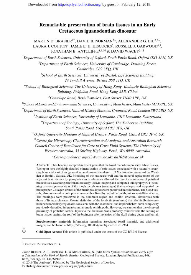
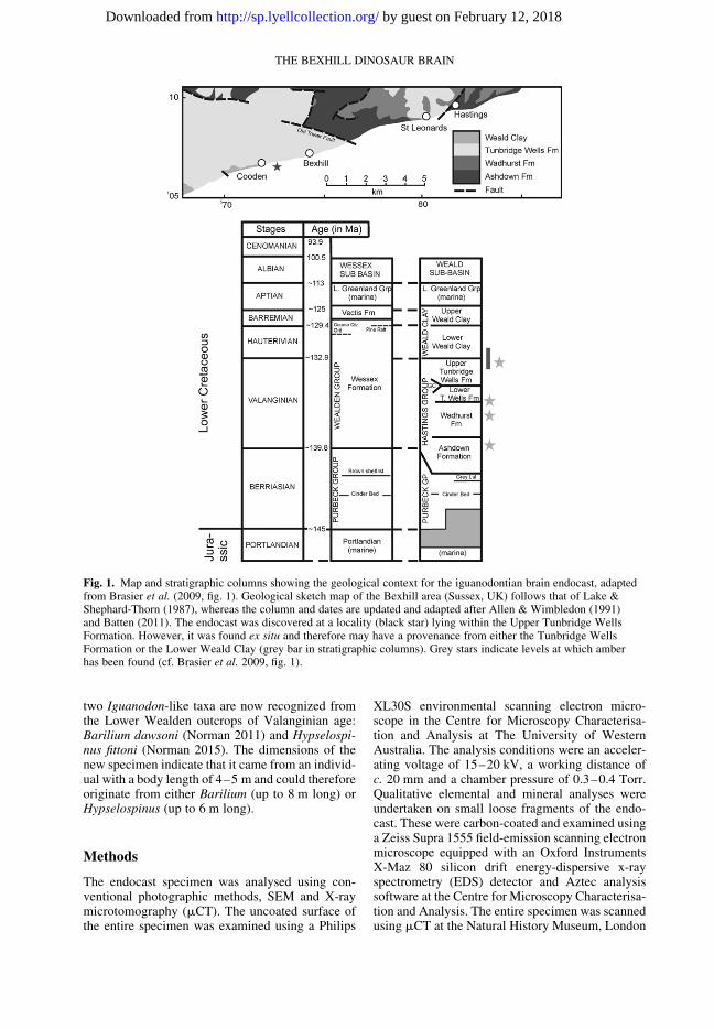
Fig. 1. Map and stratigraphic columns showing the geological context for the iguanodontian brain endocast, adaptedfrom Brasier et al. (2009, fig. 1). Geological sketch map of the Bexhill area (Sussex, UK) follows that of Lake &Shephard-Thorn (1987), whereas the column and dates are updated and adapted after Allen & Wimbledon (1991)and Batten (2011). The endocast was discovered at a locality (black star) lying within the Upper Tunbridge WellsFormation. However, it was found ex situ and therefore may have a provenance from either the Tunbridge WellsFormation or the Lower Weald Clay (grey bar in stratigraphic columns). Grey stars indicate levels at which amberhas been found (cf. Brasier et al. 2009, fig. 1).
THE BEXHILL DINOSAUR BRAIN
by guest on February 12, 2018http://sp.lyellcollection.org/Downloaded from
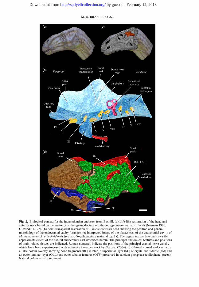
Fig. 2. Biological context for the iguanodontian endocast from Bexhill. (a) Life-like restoration of the head andanterior neck based on the anatomy of the iguanodontian ornithopod Iguanodon bernissartensis (Norman 1980;OUMNH T.127). (b) Semi-transparent restoration of I. bernissartensis head showing the position and generalmorphology of the endocranial cavity (orange). (c) Interpreted image of the plaster cast of the endocranial cavity ofMantellisaurus cf. atherfieldensis (see also Supplementary material fig. 1a). The region in pale blue indicates theapproximate extent of the natural endocranial cast described herein. The principal anatomical features and positionsof brain-related tissues are indicated. Roman numerals indicate the positions of the principal cranial nerve canals,which have been superimposed with reference to earlier work by Norman (2004). (d) Natural cranial endocast witha false-colour overlay showing bone fragments (BF) in blue, a superficial layer (SL) of crystalline siderite (red) andan outer laminar layer (OLL) and outer tubular features (OTF) preserved in calcium phosphate (collophane; green).Natural colour ¼ silty sediment.
M. D. BRASIER ET AL.
by guest on February 12, 2018http://sp.lyellcollection.org/Downloaded from
(NHMUK) with a Nikon XTH-225 instrument.X-rays were generated using a tungsten targetwith an accelerating voltage of 225 kV, a currentof 190 mA and no added filtration. To maximize res-olution, two scans were undertaken to cover thelength of the specimen. For each scan, 3142 projec-tions of 0.5 s exposure were collected and thenreconstructed to create volumes with 38.9 mm vox-els. These were converted to two image stacks thatwere cropped and then aligned in SPIERSalign (Sut-ton et al. 2012), surfaced in SPIERSedit, rendered inBlender (cf. Garwood & Dunlop 2014) and volumerendered in Drishti (Limaye 2012).
The specimen is currently in the private posses-sion of Jamie Hiscocks, but negotiations are under-way to have it housed in a public museum. The loosefragments analysed using SEM-EDS (OUMNHK59010/p1–p2) are housed alongside associatedpost-cranial material from the same site at theOxford University Museum of Natural History(OUMNH K.59010/1–8). Copies of the mCT scandatasets are also available as OUMNH K59010/p3–p4. All SEM and mCT images have additionallybeen archived on the open access server Zenodo(doi: 10.5281/zenodo. 50499).
Geological context
The cranial endocast (Figs 2d & 3) was exposed bytidal erosion and found among fluvial sedimentaryunits of the c. 133 Ma Early Cretaceous Upper Tun-bridge Wells Formation (Fig. 1; see also Lake &Shephard-Thorn 1987; Allen & Wimbledon 1991;Radley 2006a, b; Batten 2011). The petrifiedendocast had been eroded from its matrix duringthe winter of 2004 and was collected, ex situ, froma tidal pool (Ordnance Survey coordinates TQ72498 06692). It is possible that the specimen hadbeen transported by longshore drift and was derivedfrom the nearby Weald Clay, which outcrops c.1 km to the west. The cranial endocast was foundnear other ornithopod remains that included limbfragments, a tarsal bone and broken vertebrae(OUMNH K.59010/4–8). Fossilized footprintsand trackways of Iguanodon-like ornithopods werefound at a similar stratigraphic level (cf. Beckles1854), as well as amber with probable microbialinclusions. This level lies above beds in the Ash-down Formation containing amber with the oldestknown spider silk (Brasier et al. 2009).
All the fossil bones recovered from this loca-tion appear to originate from a fine-grained, cross-bedded siltstone that forms part of the infill of afluvial channel. The surrounding sedimentary unitsconsist of rippled sandstones with charcoalifiedwood, thin tabular ironstones with charcoal, biotur-bated mudstones containing freshwater molluscs,
fossil soil fabrics, lignites and amber nodules withfossil inclusions. These are consistent with a sea-sonal wetland depositional environment subject tooccasional forest fires (cf. Brasier et al. 2009).
Large-scale endocast morphology and
preservation
Superficial morphology
The natural cranial endocast (Figs 2d & 3) is closein both shape and size to the endocranial cavityseen within a specimen of ‘Iguanodon’ on displayin the OUM (OUMNH K.59015a/p-c/p; Fig. 4;see also Norman & Weishampel 1990, fig. 25.8).The latter, to judge from its size, is most likelyfrom a fully grown individual of Mantellisaurusatherfieldensis (Norman 1986) and was collectedfrom the Isle of Wight in the 1860s. As the Wealdeniguanodontian taxa are closely comparable anatom-ically, it is not unreasonable to use one as a tem-plate for the other. Comparisons between the newspecimen and other Wealden natural endocasts
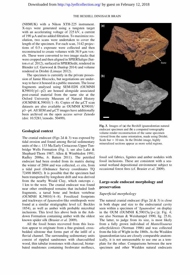
Fig. 3. Images of (a) the Bexhill iguanodontian naturalendocast specimen and (b) a computed tomographyvolume render reconstruction of the same specimenviewed from the same orientation, made using Drishti.Scale bar ¼ 10 mm. In the Drishti image, highlymineralized sections appear as more solid regions.
THE BEXHILL DINOSAUR BRAIN
by guest on February 12, 2018http://sp.lyellcollection.org/Downloaded from
(such as NHMUK R2501 and R8306; Norman &Weishampel 1990, figs 25.11 and 25.12; Norman2004) allow the identification of major regionsoccupied by the brain within the endocranial cavity,along with the cranial nerves and parts of the venoussinus drainage system (Fig. 2c).
The Bexhill endocast lacks the olfactory bulbregion at the extreme anterior end of the cranialcavity, as well as most of the floor and adjacent lat-eral walls and the area that would have been occu-pied by the medulla oblongata posteriorly (Fig. 2c,the pale blue shading indicates what is actually pre-served in this natural cast). The ventral surface ofthe endocast, which would, in life, have been sup-ported by the floor of the braincase, is weatheredand much eroded. The dorsal and lateral portionsof the forebrain and hindbrain (the areas occupiedby the cerebral and cerebellar expansions) exhibitthe best preservation (Figs 2d & 3; Supplementarymaterial figs 3, 4). The mid-brain region, whichwould have been occupied by the optic lobes in life,is completely obscured in endocasts because theselobes were overlain by the cerebral and cerebellarexpansions, as well as large vascular sinuses.
Preservation
Our SEM and CT studies of the entire Bexhillendocast reveal four structurally distinct regions(Fig. 2d):
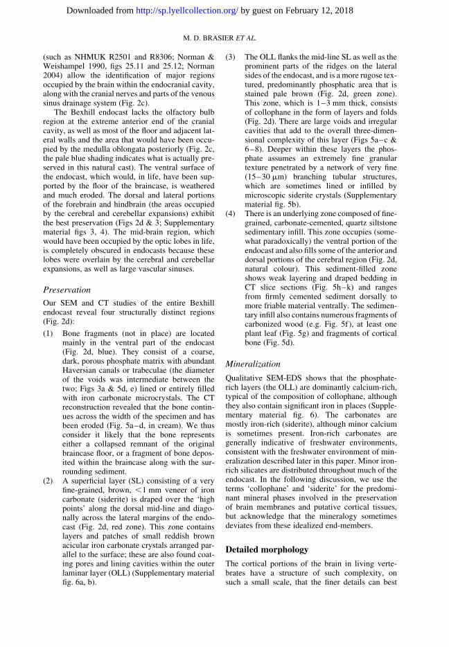
(1) Bone fragments (not in place) are locatedmainly in the ventral part of the endocast(Fig. 2d, blue). They consist of a coarse,dark, porous phosphate matrix with abundantHaversian canals or trabeculae (the diameterof the voids was intermediate between thetwo; Figs 3a & 5d, e) lined or entirely filledwith iron carbonate microcrystals. The CTreconstruction revealed that the bone contin-ues across the width of the specimen and hasbeen eroded (Fig. 5a–d, in cream). We thusconsider it likely that the bone representseither a collapsed remnant of the originalbraincase floor, or a fragment of bone depos-ited within the braincase along with the sur-rounding sediment.
(2) A superficial layer (SL) consisting of a veryfine-grained, brown, ,1 mm veneer of ironcarbonate (siderite) is draped over the ‘highpoints’ along the dorsal mid-line and diago-nally across the lateral margins of the endo-cast (Fig. 2d, red zone). This zone containslayers and patches of small reddish brownacicular iron carbonate crystals arranged par-allel to the surface; these are also found coat-ing pores and lining cavities within the outerlaminar layer (OLL) (Supplementary materialfig. 6a, b).
(3) The OLL flanks the mid-line SL as well as theprominent parts of the ridges on the lateralsides of the endocast, and is a more rugose tex-tured, predominantly phosphatic area that isstained pale brown (Fig. 2d, green zone).This zone, which is 1–3 mm thick, consistsof collophane in the form of layers and folds(Fig. 2d). There are large voids and irregularcavities that add to the overall three-dimen-sional complexity of this layer (Figs 5a–c &6–8). Deeper within these layers the phos-phate assumes an extremely fine granulartexture penetrated by a network of very fine(15–30 mm) branching tubular structures,which are sometimes lined or infilled bymicroscopic siderite crystals (Supplementarymaterial fig. 5b).
(4) There is an underlying zone composed of fine-grained, carbonate-cemented, quartz siltstonesedimentary infill. This zone occupies (some-what paradoxically) the ventral portion of theendocast and also fills some of the anterior anddorsal portions of the cerebral region (Fig. 2d,natural colour). This sediment-filled zoneshows weak layering and draped bedding inCT slice sections (Fig. 5h–k) and rangesfrom firmly cemented sediment dorsally tomore friable material ventrally. The sedimen-tary infill also contains numerous fragments ofcarbonized wood (e.g. Fig. 5f), at least oneplant leaf (Fig. 5g) and fragments of corticalbone (Fig. 5d).
Mineralization
Qualitative SEM-EDS shows that the phosphate-rich layers (the OLL) are dominantly calcium-rich,typical of the composition of collophane, althoughthey also contain significant iron in places (Supple-mentary material fig. 6). The carbonates aremostly iron-rich (siderite), although minor calciumis sometimes present. Iron-rich carbonates aregenerally indicative of freshwater environments,consistent with the freshwater environment of min-eralization described later in this paper. Minor iron-rich silicates are distributed throughout much of theendocast. In the following discussion, we use theterms ‘collophane’ and ‘siderite’ for the predomi-nant mineral phases involved in the preservationof brain membranes and putative cortical tissues,but acknowledge that the mineralogy sometimesdeviates from these idealized end-members.
Detailed morphology
The cortical portions of the brain in living verte-brates have a structure of such complexity, onsuch a small scale, that the finer details can best
M. D. BRASIER ET AL.
by guest on February 12, 2018http://sp.lyellcollection.org/Downloaded from
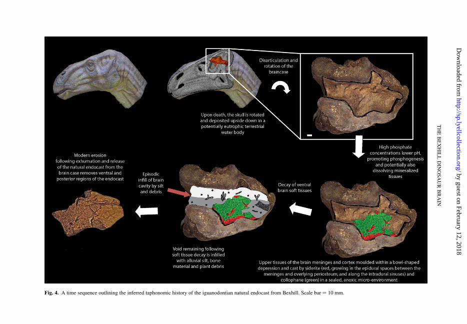
Fig. 4. A time sequence outlining the inferred taphonomic history of the iguanodontian natural endocast from Bexhill. Scale bar ¼ 10 mm.
TH
EB
EX
HIL
LD
INO
SA
UR
BR
AIN
by guest on February 12, 2018http://sp.lyellcollection.org/
Dow
nloaded from
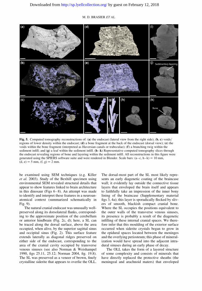
be examined using SEM techniques (e.g. Killeret al. 2003). Study of the Bexhill specimen usingenvironmental SEM revealed structural details thatappear to show features linked to brain architecturein this dinosaur (Figs 6–8). An attempt was madeto identify and interpret these features in a neuroan-atomical context (summarized schematically inFig. 9).
The natural cranial endocast was unusually well-preserved along its dorsolateral flanks, correspond-ing to the approximate position of the cerebellumor anterior hindbrain (Fig. 2c, d); here, a SL canbe traced along the dorsal surface, above the areaoccupied, when alive, by the superior sagittal sinusand occipital sinus (Fig. 2). This surface featureextends laterally as diagonal ridges preserved oneither side of the endocast, corresponding to thearea of the cranial cavity occupied by transversevenous sinuses (see also Norman & Weishampel1990, figs 25.11, 25.12; Norman 2004, fig. 19.8).The SL was preserved as a veneer of brown, finelycrystalline siderite that appears to overlie the OLL.
The dorsal-most part of the SL most likely repre-sents an early diagenetic coating of the braincasewall; it evidently lay outside the connective tissuelayers that enveloped the brain itself and appearsto faithfully take an impression of the inner bonylining of the braincase (Supplementary materialfigs 3, 4a); this layer is sporadically flecked by sliv-ers of smooth, blackish compact cranial bone.Where the SL occupies the positions equivalent tothe outer walls of the transverse venous sinuses,its presence is probably a result of the diageneticinfilling of these internal cranial spaces. We there-fore infer that this moulding of the exterior surfaceoccurred when siderite crystals began to grow inthe epidural spaces located between the meningesand the overlying periosteum; this phase of mineral-ization would have spread into the adjacent intra-dural sinuses during an early phase of decay.
The OLL takes the form of a layered structureof some complexity and consists of minerals thathave directly replaced the protective sheaths (themeningeal and arachnoid maters) that enveloped
Fig. 5. Computed tomography reconstructions of: (a) the endocast (lateral view from the right side); (b, c) voids/regions of lower density within the endocast; (d) a bone fragment at the back of the endocast (dorsal view); (e) thevoids within the bone fragment (interpreted as Haversian canals or trabeculae); (f) a branching twig within thesediment infill; and (g) a leaf within the sediment infill. (h–k) Representative computed tomography slices throughthe endocast revealing regions of bone and layering within the sediment infill. All reconstructions in this figure weregenerated using the SPIERS software suite and were rendered in Blender. Scale bars: (a–c, h–k) ¼ 10 mm,(d, e) ¼ 5 mm, (f, g) ¼ 2 mm.
M. D. BRASIER ET AL.
by guest on February 12, 2018http://sp.lyellcollection.org/Downloaded from

the brain cortex (grey matter). The OLL is thickeston the flanks of the endocast beneath the ‘duralpeak’ (Fig. 2). Both optical microscopy and SEM
revealed this layered region to be constructed ofthin, interwoven laminar sheets of phosphate (Figs6–8) that were either planar or, more commonly,corrugated into ribbon-like folds and troughs.These structures range from microns to millimetresin diameter and are conspicuously aligned acrossthe brain axis. SEM reveals that these ribbonswere themselves composed of aligned, micrometre-sized filaments (Fig. 6, Dm). Some of the junctionsbetween these ribbons contain small intraduralspaces (Fig. 6a, Ids). Small elongate voids, infilledlargely with reddish brown, rod-shaped siderite cry-stals 20–30 mm in length occur within and betweenthese ribbons (Fig. 6b, Si), which also generallylie parallel to the endocast surface. This OLL fabricof fibrous ribbons with occasional voids has all themicroscopic features expected of either the perios-teum or meningeal mater (which together form thedura mater; see Fig. 9). These form the tough protec-tive outer coatings seen in living vertebrate brains(cf. Runza et al. 1999 – as interpreted in Fig. 9).The ribbons and filaments are taken to representthe remains of bundles of fibrous collagen thathave been replaced by phosphate before significantorganic decay took place.
The web-like structure of phosphatized ribbonsin the OLL is punctured locally by well-definedapertures (Fig. 6c, Ls); this web-like fabric closelyresembles that seen in the arachnoid mater of themeninges (cf. Reina et al. 2002). The arachnoidmater in the dorsal hindbrain of living avian archo-saurs is a thin sheet of interwoven collagen rib-bons that comprises the innermost layer of thedura mater (Fig. 6b, c, Am; Fig. 8a, Am). In archo-saurs, this layer directly overlies the cortex (greymatter) of the brain (Fig. 6b, Gm) without any inter-vening subarachnoid space and pia mater as found inthe more complex brains of mammals (Runza et al.1999). In this petrified natural cranial endocast, theinner, arachnoid-like layer seemingly exhibits con-volutions (Fig. 6b, Gy); these structures are inter-preted as ‘gyri’ (see also Wilson 1971), reflectingin part the topographic complexity that existsbetween the arachnoid mater and the underlying cer-ebellar cortex (¼grey matter) (Fig. 6b, c, Gm; seealso summary Fig. 9).
The outer tubular features within the cranialendocast mainly lie within the fabric of the OLLand are predominantly arranged parallel to the endo-cast surface, just above what is suggested here to bethe arachnoid mater (Figs 7 & 8a, Bv). The tubularstructures are either rounded or compressed incross-section and typically range up to 100 mm indiameter (compare Figs 7 & 8). Several examplesshow finely layered walls (Fig. 7) and their inter-nal spaces are either lined or infilled with microcrys-talline siderite formed during early diagenesis.These tubes are very similar in size and shape to
Fig. 6. Environmental scanning electron microscopyimages of the iguanodontian natural cranial endocast.Images of the outer laminar layer on the flanks of thecerebellar area. (a) Well-preserved textures reflectingthe organization of the meningeal layers lyingimmediately above the cortex; the pit-like structuresrepresent intradural spaces lying between the thickerribbons of meningeal tissue. (b) Features interpretedas folds and valleys of the meninges, lying above acomplex fabric with recesses (Gy) possiblyrepresenting some of the superficial features of theunderlying grey matter (Gm) of the cerebellar cortex.(c) Features interpreted as fossilized dura mater (Dm),the outermost membranes that surround the brain.Scale bars ¼ 100 mm. All images in this figure wereobtained from scans of the uncoated complete brainendocast. Am, probable arachnoid mater (traces ofcollagenous sheets and ribbons reinforcing themeningeal layer); Dm, probable dura mater preservedas micron-sized phosphatized filaments; Gy, potentialgyri – these structures are normally associated withdeeper layers in the cortex (i.e. the grey matter) andreflect some of the convolutions near the surface of thecortex; Ids, intradural spaces; Ls, lesions in themeningeal fabric; Si, lozenge-shaped siderite crystals.
THE BEXHILL DINOSAUR BRAIN
by guest on February 12, 2018http://sp.lyellcollection.org/Downloaded from

blood vessels and form a network that extendsacross the surface of the meninges and, in someinstances, penetrates the cortex as part of theblood supply (arterial) or drainage (venous) systems(see Fig. 9).
Immediately beneath the meninges some dee-per areas of the endocast lack these laminar orribbon-like features and exhibit a texture of consid-erable fine-scale complexity (Fig. 6b, c). This areacould be interpreted as the mineralized remnantsof cortical tissue (grey matter), representing animperfect record of the complexity of the cerebellarcortex.
Figure 9 provides an idealized summary andinterpretation of the dorsal cranium and endo-cranial cavity soft tissues that appear to have beenpreserved in the natural cranial endocast fromBexhill. Such soft tissue preservation is compara-tively rare in non-avian dinosaurs and, indeed,in any terrestrial vertebrate, but this specimenclearly demonstrates that even brain-associatedtissues can be preserved under exceptional tapho-nomic conditions.
Taphonomic history
The three zones of preservation that we recognize(carbonate, phosphate–carbonate, and siltstone), aswell as their distribution on the natural endocast, aresuggestive of a specific set of taphonomic con-ditions acting on the specimen during the periodshortly after death, consistent with burial of thedinosaur braincase in an aqueous medium.
Conditions necessary for mineralization
The brain structures described here were preservedin phosphate and carbonate; however, to preservesoft tissue as phosphate, a locally anoxic environ-ment is required to promote bacterially mediatedmineralization (Briggs et al. 1993). In the predo-minantly fluvial system suggested by the sedimentsassociated with this specimen, eutrophication(algal blooms) and/or stratification of the water col-umn is required to result in water column anoxia.Under freshwater conditions, eutrophication addsphosphate to the water column in the form of a
Fig. 7. Environmental scanning electron microscopy images of tubular structures on the exterior of the Bexhilliguanodontian cranial endocast and within the outer laminar layer, interpreted here as meningeal blood vessels.Arrows in (a) point to areas where the hollow nature of the vessels can be seen; arrows in (b) point to branching ofthe vessels; (c–f) additional images of hollow tubular structures within the outer laminar layer. All images in thisfigure were obtained from scans of the uncoated complete brain endocast.
M. D. BRASIER ET AL.
by guest on February 12, 2018http://sp.lyellcollection.org/Downloaded from
phosphoric acid series. Ionized phosphoric acids(hydrogen phosphate22, dihydrogen phosphate12)and orthophosphoric acid (H3PO4) drasticallyreduce the pH of the water, rapidly fixing soft tissues(the equivalent of pickling) and degrading and dis-solving the surrounding mineralized tissues. Conse-quently, the soft tissues associated with the braincould have been preserved and cast prior to com-plete burial by sediment, which would completethe sealing process before the bone of the braincasehad been completely dissolved away. Any phospho-genic layer is likely to have formed at the base of thewater column or within the upper layer of soft sedi-ment beneath the sediment–water interface.
Taphonomic scenario
The death of the dinosaur may have occurredadjacent to or within a temporarily eutrophic waterbody. The body most likely collapsed into the
water column and the head came to lie, invertedand partially buried, in sediment at the bottom ofthe water body. With the head in the proximity ofthe anoxic and phosphogenic layer, the processesof soft tissue preservation (linked to decay), dissolu-tion of the surrounding bone and phosphatizationcould proceed. In an inverted position the upper(dorsal) portion of the brain was, in effect, ponded,because it lay within a bowl-shaped containerformed by the occipital, parietal and lateral brain-case wall bones. This container was lined by theperiosteum and meninges – membranes that formtough sheaths surrounding the cortical portions ofthe brain.
The lower parts of the braincase floor and ventralbrain tissues – overturned (Fig. 4) – decayed toform a stagnant (anoxic) pool of decomposing tissueand fluid enriched in phosphate and iron. The high-fidelity replacement of durable collagen proteinsand blood vessels associated with the meninges on
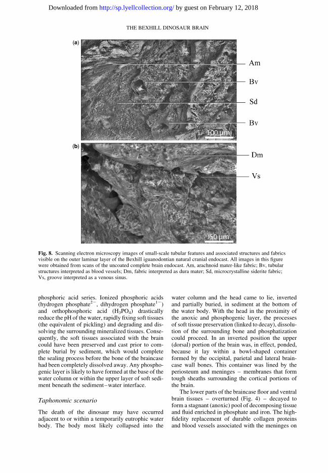
Fig. 8. Scanning electron microscopy images of small-scale tubular features and associated structures and fabricsvisible on the outer laminar layer of the Bexhill iguanodontian natural cranial endocast. All images in this figurewere obtained from scans of the uncoated complete brain endocast. Am, arachnoid mater-like fabric; Bv, tubularstructures interpreted as blood vessels; Dm, fabric interpreted as dura mater; Sd, microcrystalline siderite fabric;Vs, groove interpreted as a venous sinus.
THE BEXHILL DINOSAUR BRAIN
by guest on February 12, 2018http://sp.lyellcollection.org/Downloaded from

the opposite, dorsal surface was facilitated by thevery rapid growth of amorphous microcrystals ofcalcium phosphate (cf. Martill 2001). Autolithifiedbacteria were not observed associated with thesemicrocrystals, suggesting that mineral replacementprobably proceeded quickly. Phosphatization ofthe meningeal layers probably took place under con-ditions of low pH and low oxygen tension in fluidslow in sulphate, but rich in ferrous ions (Allison &Pye 1994). Such fluids are typical of environ-ments influenced by Fe3+-reducing bacteria andwith high concentrations of calcium, phosphateand ferric ions (released from the adjacent biologi-cal tissues, such as bone, brain tissue and bloodcells; cf. Allison 2001). The local removal of fer-rous ions by the formation of siderite may havefurther encouraged the rapid precipitation of cal-cium phosphate. Excess ferrous ions were alsolikely to have been incorporated into the phosphatephase in places, as suggested by our SEM-EDSanalyses.
Deeper within the endocranial cavity, less refrac-tory nervous tissues of the cerebellar cortex arepresumed to have decayed away, or experiencedmuch lower fidelity moulding via the precipitationof amorphous phosphate and carbonate microcrys-tals. The latter would probably have occurredunder conditions that were relatively more alkaline,
with more freely available bicarbonate ions. Thesiderite microcrystals in this region were typicallyrod-shaped and c. 30–50 mm long (Fig. 6b, Si);they were composed of nanocrystals of regularshape and size, perhaps reflecting some degree ofbacterial mediation. Moulds and casts of heterotro-phic bacteria are more usually coccoid (spheroidal)or bacillate (rod-shaped) in form, so the observedstructures are unlikely to be bacterially derivedartefacts (cf. Wilby & Briggs 1997; Liebig 2001;Martill 2001).
The ventral portion of the braincase (upper interms of burial orientation) was evidently filled byepisodic infiltrations of alluvial silt, carrying withit carbonized plant debris and broken bone. Inplaces, phases of internal sediment deposition alter-nated with thin partings coated with siderite. Thereis a persistent, exposed suture line in iguanodontiandinosaurs between the bones that form the lateralwalls and floor of the braincase (Norman 1977);this may have resulted in the floor of the braincasebecoming detached from the remainder of thebraincase as decay and dissolution proceeded,allowing sediment access to the floor of the endocra-nium. As the specimen was discovered ex situ, wecannot comment on the preservational processesthat affected the rest of the body, or on the potentialpreservational fidelity they may or may not exhibit.
Fig. 9. An idealized reconstruction of the head of an iguanodontian dinosaur showing the dorsal braincase andassociated soft tissue features as evidenced by examination of the natural endocast from Bexhill. (a) Reconstructionof the head of an iguanodontian (OUMNH T.127) in the oblique anterior view showing the area of the skull roofthat has been ‘dissected’ to the right. (b) Partly ‘exploded’ restoration of the underlying braincase and brain tissuesidentified following detailed examination of the structures preserved in the natural cranial endocast. Am, arachnoidmater (meninge); Av, arachnoid villus (protruding into a mid-line venous sinus); Bc, braincase bone (parietal); Bv,blood vessels; Co, collagenous sheath enclosing the brain structures and lining the bones of braincase (combinedperiosteal and meningeal sheets); Dm, dura mater (meninge); Es, epidural space; Gm, grey matter (brain cortex);Ids, intradural space (between the meninges); Sd, subdural space; Vs, sagittal venous sinus.
M. D. BRASIER ET AL.
by guest on February 12, 2018http://sp.lyellcollection.org/Downloaded from
How intelligent was the Bexhill
iguanodontian?
Previous studies have suggested that dinosaurbrains may have shared general similarities withthose of modern crocodilians in having a thickouter packaging of dural tissue and extensive lym-phatic and venous sinuses (Dendy 1909; Romer1956; Ostrom 1961; Hopson 1979). It has beenargued that the latter structures are very likely toobscure details of the brain tissue beneath, meaningthat the topography of cranial endocasts of dino-saurs reflects (at best) an early ontogenetic stagein brain development (Romer 1956). As a conse-quence, dinosaur cranial endocasts typically reveala generalized brain morphology, rather than (moredesirable) fine details about the size, structure andtherefore relative biological importance of thebrain lobes and their bearing on probable behaviou-ral repertoires (‘intelligence’) (Dendy 1909; Hopson1979; Rogers 1999; Witmer et al. 2008).
Previous measurements of dinosaur endocastvolumes estimate them to have contained as littleas 50% of actual brain (Hopson 1979; Evans et al.2009). Exceptions to this general assumption havebeen proposed: hadrosaurs (lambeosaurines, deri-ved iguanodontian ornithopods) have been arguedto have had brain tissues that filled rather more ofthe endocranial cavity (Evans 2005, cited in Evanset al. 2009), especially in the anterior and ventralregions of the endocasts where more detailed lobe-like structures (e.g. cerebral hemispheres and thehypothalamus) are discernible to the naked eye.More posterior regions of the endocranium in
dinosaurs, adjacent to the cerebellum and medulla,are, by contrast, relatively poorly defined and mayindeed have been overlain by extensive sinuses, asis the case in extant crocodilians (Ostrom 1961;Hopson 1979; Evans et al. 2009).
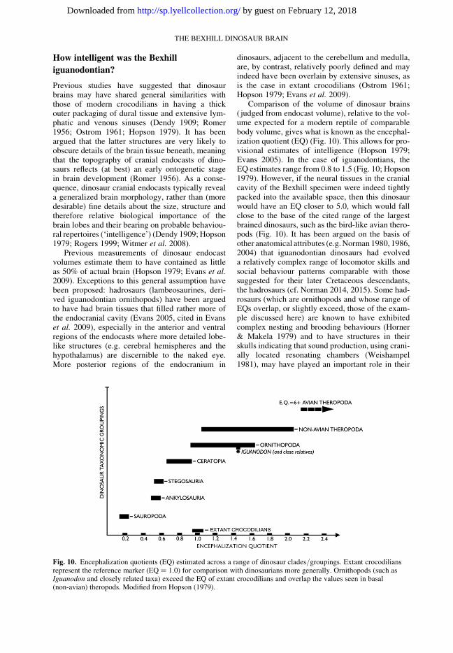
Comparison of the volume of dinosaur brains( judged from endocast volume), relative to the vol-ume expected for a modern reptile of comparablebody volume, gives what is known as the encephal-ization quotient (EQ) (Fig. 10). This allows for pro-visional estimates of intelligence (Hopson 1979;Evans 2005). In the case of iguanodontians, theEQ estimates range from 0.8 to 1.5 (Fig. 10; Hopson1979). However, if the neural tissues in the cranialcavity of the Bexhill specimen were indeed tightlypacked into the available space, then this dinosaurwould have an EQ closer to 5.0, which would fallclose to the base of the cited range of the largestbrained dinosaurs, such as the bird-like avian thero-pods (Fig. 10). It has been argued on the basis ofother anatomical attributes (e.g. Norman 1980, 1986,2004) that iguanodontian dinosaurs had evolveda relatively complex range of locomotor skills andsocial behaviour patterns comparable with thosesuggested for their later Cretaceous descendants,the hadrosaurs (cf. Norman 2014, 2015). Some had-rosaurs (which are ornithopods and whose range ofEQs overlap, or slightly exceed, those of the exam-ple discussed here) are known to have exhibitedcomplex nesting and brooding behaviours (Horner& Makela 1979) and to have structures in theirskulls indicating that sound production, using crani-ally located resonating chambers (Weishampel1981), may have played an important role in their
Fig. 10. Encephalization quotients (EQ) estimated across a range of dinosaur clades/groupings. Extant crocodiliansrepresent the reference marker (EQ ¼ 1.0) for comparison with dinosaurians more generally. Ornithopods (such asIguanodon and closely related taxa) exceed the EQ of extant crocodilians and overlap the values seen in basal(non-avian) theropods. Modified from Hopson (1979).
THE BEXHILL DINOSAUR BRAIN
by guest on February 12, 2018http://sp.lyellcollection.org/Downloaded from
social interactions. However, living crocodilianarchosaurs also exhibit vocalization as part of theirbehavioural repertoire and have complex nesting,nest-tending and brooding behaviours (Guggisberg1972), but exhibit a relatively low EQ.
Literal interpretation of the surface featuresexhibited by the Bexhill fossil suggests that themeninges were not very thick and that the neurallayers of the cerebellar cortex lay immediately(,1 mm) beneath the meningeal tissues. Thiscould easily be taken as evidence that iguanodontianornithopods had a greater volume of neural tissuethan has been argued on the basis of comparisonwith crocodiles (Hopson 1979). In other words,iguanodontians such as Barilium and Hypselospinuswere comparatively ‘intelligent’ dinosaurs, with agreater volume of neural tissue packed into theendocranium than previously predicted. The coun-ter-argument to this interpretation is that the surfacefeatures on the natural endocast indicate that themeningeal tissues and decaying brain had collapsed,under gravity, into the bowl-like braincase. Conse-quently, the proximity of meningeal tissues to thebraincase wall is here interpreted as an artefact ofpreservation, rather than an indication of the densepacking of neural tissue into the cranial cavity.
Conclusions
We have reported a remarkably preserved partialnatural endocast of the cranial cavity of an iguano-dontian ornithopod dinosaur, which, to our knowl-edge, is the first described example of mineralizedbrain soft tissues from a fossilized terrestrial verte-brate. This endocast was collected from an intertidalexposure of Valanginian (Early Cretaceous) age.The endocast topography closely reflects the mor-phology of the anterior and dorsal portions ofknown iguanodontian cranial cavities. The endocastconsists of a complex of siderite, collophane andlayered siltstone, the latter having accumulatedwithin the cranial cavity after the death of the dino-saur and during later phases of organic decay andsub-aerial burial of the braincase. Superficial exam-ination of the specimen allowed comparison withbraincases and endocasts (both natural and artificial)attributable to closely related, but geologicallyyounger (Barremian–Aptian), iguanodontians suchas Iguanodon bernissartensis (Norman 1980) andM. atherfieldensis (Norman 1986). More detailedexamination of the natural endocast using lightmicroscopy, as well as SEM and mCT imaging, hasrevealed hitherto unexpected details about themembranous tissues lining the braincase walls andadjacent tissues that invested the brain itself.Comparisons drawn with brain-associated softtissues in living species suggest that portions of
the tissue space between the braincase wall andtough tissues (meninges) surrounding the brainitself were mineralized by siderite early in thedecay process. The actual meninges (meningeal andarachnoid maters) appear to have been preservedby phosphate replacement of the original tissues(including some of the larger blood vessels thatmay be lined or infilled by microcrystalline sider-ite). The meningeal structures that could be dis-cerned appear to show similarities with those seenin living archosaurs (crocodiles and birds). A fine-textured collophane matrix lying seemingly beneaththe meningeal structures suggests the partial preser-vation of deeper cortical tissues of the brain itself.
As is common with many dinosaur endocasts, itappears that the anterior portion of the endocast(equivalent to the areas occupied by the forebrainlobes and hypothalamus) was well developed andmoulded the developing braincase walls. More pos-teriorly, the mid- and hindbrain were enveloped byextensive blood and lymphatic sinuses that maskedthe details of brain morphology to a far greaterextent. Behavioural complexity is strongly associ-ated with forebrain development and it is reasonableto suppose that iguanodontian dinosaurs of this typewere moderately complex behaviourally (no lessso than modern crocodilians, for example). Thissuggestion is reinforced by our knowledge of igua-nodontian anatomy and the complexity of theirknown or implied range of locomotor, social andreproductive repertoires.
JEHH found the specimen. MDB and DBN collaborated todevelop this research project and co-ordinated the investi-gation. DW, AGL and RG conducted the analyses. DBN,MDB, JBA and AGL developed the manuscript and, fol-lowing the untimely death of MDB, all the authors wereinvolved in data interpretation and the final redrafting ofthe manuscript. We acknowledge the facilities, scientificand technical assistance of the Australian Microscopy &Microanalysis Research Facility at the Centre for Micros-copy Characterisation and Analysis, The University ofWestern Australia, a facility funded by the university,state and Commonwealth governments. R. Callow under-took early petrological and SEM studies in Oxford.D. Siveter and E. Howlett kindly facilitated imaging andstudy of ‘Iguanodon’ casts in the OUMNH collections.MDB acknowledges funds provided by V. and T. Brasier.DBN was supported by the Odell Trust Fund from Christ’sCollege Cambridge. AGL is supported by the NaturalEnvironment Research Council (grant number NE/L011409/1). JBA acknowledges the ongoing support ofthe Department of Zoology and OUMNH, University ofOxford. RG is a Scientific Associate at the Natural HistoryMuseum, London, and a member of the InterdisciplinaryCentre for Ancient Life. DW acknowledges funding fromthe Australian Research Council and the EuropeanCommission. This is CCFS paper 861. The authorswould like to thank D. Martill and A. Iwaniuk for helpfulsuggestions during the review process.
M. D. BRASIER ET AL.
by guest on February 12, 2018http://sp.lyellcollection.org/Downloaded from

References
Allen, P. & Wimbledon, W.A. 1991. Correlation ofNW European Purbeck–Wealden (nonmarine LowerCretaceous) as seen from the English type-areas.Cretaceous Research, 12, 511–526.
Allison, P.A. 2001. Decay. In: Briggs, D.E.G. &Crowther, P. (eds) Paleobiology II. Blackwell Sci-ence, Oxford, 237–255.
Allison, P.A. & Briggs, D.E. 1993. Exceptional fossilrecord: distribution of soft-tissue preservation throughthe Phanerozoic. Geology, 21, 527–530.
Allison, P.A. & Pye, K. 1994. Early diagenetic mineral-ization and fossil preservation in modern carbonateconcretions. Palaios, 9, 561–575.
Andrews, C.W. 1897. Note on the cast of the brain ofIguanodon. Annals of the Magazine of Natural History,XIX, 585–591.
Batten, D.J. 2011. Wealden geology. In: Batten, D.J.(ed.) English Wealden Fossils. The PalaeontologicalAssociation, London, 7–14.
Beckles, S.H. 1854. On the Ornithoidichnites of the Weal-den. Quarterly Journal of the Geological Society ofLondon, 10, 456–464.
Brasier, M.D., Cotton, L. & Yenney, I. 2009. Firstreport of amber with spider webs and microbialinclusions from the earliest Cretaceous (c. 140 Ma)of Hastings, Sussex. Journal of the Geological Society,London, 166, 989–997, http://doi.org/10.1144/0016-76492008-158
Briggs, D.E.G., Kear, A.J., Martill, D.M. & Wilby,P.R. 1993. Phosphatisation of soft-tissue in experi-ments and fossils. Journal of the Geological Society,London, 150, 1035–1038, http://doi.org/10.1144/gsjgs.150.6.1035
Brochu, C.A. 2000. A digitally rendered endocast forTyrannosaurus rex. Journal of Vertebrate Paleontol-ogy, 20, 1–6.
Budd, G.E. & Jensen, S. 2015. The origin of animals anda ‘Savannah’ hypothesis for early bilaterian evolution.Biological Reviews, first published online November20, 2015, http://doi.org/10.1111/brv.12239
Dendy, A. 1909. The intracranial vascular system ofSphenodon. Philosophical Transactions of the RoyalSociety of London B, 200, 403–426.
Dendy, A. 1910. On the structure, development andmorphological interpretation of the pineal organs andadjacent parts of the brain of the tuatara (Sphenodon).Philosophical Transactions of the Royal Society ofLondon B, 201, 227–331.
Edinger, T. 1929. Die fossilen Gehirne. Ergebnisse derAnatomische Entwicklungsgeschichte, 28, 1–249.
Edinger, T. 1941. The brain of Pterodactylus. AmericanJournal of Science, 239, 665–682.
Evans, D.C. 2005. New evidence on brain-endocranialcavity relationships in ornithischian dinosaurs. ActaPalaeontologica Polonica, 50, 617–622.
Evans, D.C., Ridgely, R. & Witmer, L.M. 2009. Endo-cranial anatomy of lambeosaurine hadrosaurids (Dino-sauria; Ornithischia): a sensorineural perspective oncranial crest function. The Anatomical Record, 292,1315–1337.
Garwood, R. & Dunlop, J. 2014. The walking dead:Blender as a tool for paleontologists with a case
study on extinct arachnids. Journal of Paleontology,88, 735–746.
Giles, S. & Friedman, M. 2014. Virtual reconstruction ofendocast anatomy in early ray-finned fishes (Osteich-thyes, Actinopterygii). Journal of Paleontology, 88,636–651.
Guggisberg, C.A.W. 1972. Crocodiles: Their NaturalHistory, Folklore and Conservation. David & Charles,Newton Abbott.
Hopson, J.A. 1979. Paleoneurology. In: Cans, C., North-
cutt, R.G. & Ulinksi, P. (eds) Biology of the Reptilia(Neurology A). Academic Press, New York, 9,39–146.
Horner, J.R. & Makela, R. 1979. Nest of juveniles pro-vides evidence of family structure among dinosaurs.Nature, 282, 256–257.
Hulke, J.W. 1871. Note on a large reptilian skull fromBrook, Isle of Wight, probably dinosaurian and refer-able to the genus Iguanodon. Quarterly Journal ofthe Geological Society of London, XXVII, 199– 206,http://doi.org/10.1144/GSL.JGS.1871.027.01-02.27
Jerison, H.J. 1970. Brain evolution: new light on old prin-ciples. Science, 170, 1224–1225.
Jerison, H.J. 1971. More on why birds and mammals havebig brains. The American Naturalist, 105, 185–189.
Jerison, H.J. 1973. Evolution of the Brain and Intelli-gence. Academic Press, New York.
Killer, H.E., Laeng, H.R., Flammer, J. & Groscurth,P. 2003. Architecture of arachnoid trabeculae, pillars,and septa in the subarachnoid space of the humanoptic nerve: anatomy and clinical considerations. Brit-ish Journal of Ophthalmology, 87, 777–791.
Kurochkin, E.N., Dyke, G.J., Saveliev, S.V., Per-
vushov, E.M. & Popov, E.V. 2007. A fossil brainfrom the Cretaceous of European Russia and avian sen-sory evolution. Biology Letters, 3, 309–313.
Lake, R.D. & Shephard-Thorn, E.R. 1987. Geologyof the Country Around Hastings and Dungeness.British Geological Survey, Sheet Memoirs, 320/321.HMSO, London.
Lautenschlager, S., Rayfield, E.J., Altangerel, P.,Zanno, L.E. & Witmer, L.M. 2012. The endocranialanatomy of Therizinosauria and its implications forsensory and cognitive function. Plos One, e52289.
Liebig, K. 2001. Bacteria. In: Briggs, D.E.G. &Crowther, P. (eds) Paleobiology II. Blackwell Sci-ence, Oxford, 253–255.
Limaye, A. 2012. Drishti: a volume exploration and pre-sentation tool. In: Stock, S.R. (ed.) Developments inX-Ray Tomography VIII. Proceedings, SPIE Confer-ence, 8506. International Society for Optics and Pho-tonics, San Diego, CA, 85060X– 85060X.
Ma, X., Hou, X., Edgecombe, G.D. & Strausfeld, N.J.2012. Complex brain and optic lobes in an early Cam-brian arthropod. Nature, 490, 258–261.
Ma, X., Edgecombe, G.D., Hou, X., Goral, T. &Strausfeld, N.J. 2015. Preservational pathways ofcorresponding brains of a Cambrian Euarthropod. Cur-rent Biology, 25, 2969–2975, http://doi.org/10.1016/j.cub.2015.09.063
Mantell, G.A. 1825. Notice on the Iguanodon, a newlydiscovered fossil reptile from the sandstone of TilgateForest, in Sussex. Philosophical Transactions of theRoyal Society of London, CXV, 179–186.
THE BEXHILL DINOSAUR BRAIN
by guest on February 12, 2018http://sp.lyellcollection.org/Downloaded from
Mantell, G.A. 1827. Illustrations to the Geology of Sus-sex: With Figures and Descriptions of the Fossil of Til-gate Forest. Lupton Relfe, London.
Marek, R.D., Moon, B.C., Williams, M. & Benton,M.J. 2015. The skull and endocranium of a LowerJurassic ichthyosaur based on digital reconstructions.Palaeontology, 58, 723–742.
Martill, D.M. 2001. The Santana Formation. In: Briggs,D.E.G. & Crowther, P. (eds) Paleobiology II. Black-well Science, Oxford, 351–355.
Norman, D.B. 1977. On the anatomy of the ornithischiandinosaur Iguanodon. PhD thesis, King’s CollegeLondon.
Norman, D.B. 1980. On the ornithischian dinosaur Iguan-odon bernissartensis from Belgium. Memoires del’Institut Royal des Sciences Naturelles de Belgique,178, 1–105.
Norman, D.B. 1986. On the anatomy of Iguanodon athe-rfieldensis (Ornithischia: Ornithopoda). Bulletin del’Institut Royal des Sciences Naturelles de Belgique,56, 281–372.
Norman, D.B. 2004. Basal Iguanodontia. In: Weisham-
pel, D.B., Dodson, P. & Osmolska, H. (eds) TheDinosauria. University of California Press, Berkeley,CA, 413–437.
Norman, D.B. 2010. A taxonomy of iguanodontians(Dinosauria: Ornithopoda) from the lower WealdenGroup (Valanginian) of southern England. Zootaxa,2489, 47–66.
Norman, D.B. 2011. On the osteology of the lower Weal-den Group (Valanginian) ornithopod Barilium dawsoni(Iguanodontia: Styracosterna). Special Papers inPalaeontology, 86, 165–194.
Norman, D.B. 2014. Iguanodonts from the Wealden ofEngland: do they contribute to the discussion concern-ing hadrosaurs origins? In: Evans, D. & Eberth, D.(eds) Hadrosaurs. Indiana University Press, Bloom-ington, IN, 10–43.
Norman, D.B. 2015. On the history, osteology and sys-tematic position of the Wealden (Hastings Group)dinosaur Hypselospinus fittoni (Iguanodontia: Styra-costerna). Zoological Journal of the Linnean Societyof London, 173, 92–189.
Norman, D.B. & Weishampel, D.B. 1990. Iguanodontiaand related ornithopods. In: Weishampel, D.B., Dod-
son, P. & Osmolska, H. (eds) The Dinosauria. Univer-sity of California Press, Berkeley, CA, 510–533.
Ortega-Hernandez, J. 2015. Homology of head scleritesin Burgess Shale Euarthropods. Current Biology, 25,1625–1631.
Ostrom, J.H. 1961. Cranial anatomy of the hadrosauriandinosaurs of North America. Bulletin of the AmericanMuseum of Natural History, 122, 35–196.
Owen, R. 1842. Report on British Fossil Reptiles. Part ii.Report of the British Association for the Advancementof Science, 1841.
Pradel, A., Langer, M., Maisey, J.G., Gefard-Kuiyama,D., Cloetens, P., Janvier, P. & Tafforeau, P.
2009. Skull and brain of a 300-million-year-old chimae-roid fish revealed by synchrotron holotomography.Proceedings of the National Academy of Sciences ofthe United States of America, 106, 5224–2228.
Radley, J.D. 2006a. A Wealden guide I: the Weald Sub-basin. Geology Today, 22, 109–118.
Radley, J.D. 2006b. A Wealden guide II: the Wessex Sub-basin. Geology Today, 22, 187–193.
Reina, M.A., Casola, O.D.L., Lopez, A., Andres, J.A.,Mora, M. & Fernandez, A. 2002. The origins ofthe spinal subdural space: ultrastructural findings.Anesthesia and Analgesia, 94, 991–995.
Rogers, S.W. 1999. Allosaurus, crocodiles and birds:evolutionary clues from spiral computed tomo-graphy of an endocast. Anatomical Record, 257,162–173.
Romer, A.S. 1956. Osteology of the Reptiles. University ofChicago Press, Chicago.
Runza, M., Pietrabissa, R., Mantero, S., Albani, A.,Quaglini, V. & Contro, R. 1999. Lumbar duramater biomechanics; experimental characterizationand scanning electron microscopy observations. Anes-thesia and Analgesia, 88, 1317–1321.
Sutton, M.D., Garwood, R.J., Siveter, D.J. & Siveter,D.J. 2012. SPIERS and VAXML; a software toolkit fortomographic visualisation and a format for virtualspecimen interchange. Palaeontologia Electronica,15, 1–14.
Tanaka, G., Hou, X., Ma, X., Edgecombe, G.D. &Strausfeld, N.J. 2013. Chelicerate neural ground pat-tern in a Cambrian great appendage arthropod. Nature,502, 364–367.
Trinajstic, K., Marshall, C., Long, J. & Bifield, K.2007. Exceptional preservation of nerve and muscletissues in Late Devonian placoderm fish and theirevolutionary implications. Biology Letters, 3,197–200.
Weishampel, D.B. 1981. Acoustic analyses of potentialvocalization in lambeosaurine dinosaurs (Reptilia:Ornithischia). Paleobiology, 7, 252–261.
Wilby, P.R. & Briggs, D.E.G. 1997. Taxonomic trendsin the resolution of detail preserved in fossil phospha-tized soft tissues. Geobios, 30 (Suppl. 1), 493–502.
Wilson, J.A. 1971. Early Tertiary vertebrate faunas, ViejaGroup. Trans-Pecos Texas: Agriochoeridae and Mery-coidodontidae. Texas Memorial Museum Bulletin, 18,1–83.
Witmer, L.M. & Ridgely, R.C. 2009. New insights intothe brain, braincase, and ear region of tyrannosaurs(Dinosauria, Theropoda), with implications for sensoryorganization and behavior. The Anatomical Record,292, 1266–1296.
Witmer, L.M., Ridgely, R.C., Dufeau, D.L. & Simones,M.C. 2008. Using CT to peer into the past: 3D visual-ization of the brain and ear regions of birds, crocodilesand nonavian dinosaurs. In: Endo, H. & Frey, R. (eds)Anatomical Imaging: Towards a New Morphology.Springer, Berlin, 67–97.
M. D. BRASIER ET AL.
by guest on February 12, 2018http://sp.lyellcollection.org/Downloaded from