Embed Size (px)
Citation preview
Paraatoiogy Today, Austraban Supplement, july I986
Remarkable Parasites
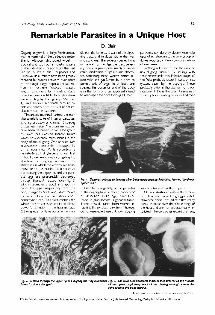
Dugong dugon IS a large herbivorous marine mammal of the distinctrve order Sirenia. Although distributed wrdely in tropical and subtropical coastal waters of the Indo-Pacific regron from the Red Sea to Australia, the Philrppines and Okinawa, rts numbers have been greatly reduced by human actrvitres over most of this range. Large populatlons stlll re- main in northern Australian waters where specimens for sclentific study have become avarlable through subsrs- tence huntlng by Aboriglnal people (Fig. I), and through accldental Capture by nets and trawls or as a result of natura1 drsasters such as cyclones.
Thls unrque mammal harbours 1t.s own characterrstlc suite of internal parasrtes. Ignoring probably synonyms, 15 species of dlgenean flukes l4 and one nematode5 have been descrlbed so far. One group of flukes has evolved bizarre forms whlch now occupy many nlches in the body of the dugong. One species lives In abscesses deep wtthin the upper lip of rts host (Fig. 2). It resembles a nematode at frrst glance, and was first notlced by an anatomist investlgating the structure of dugong vlbrissae. The abscesses In whlch the worms live com- municate to the outsrde by a series of pores along the upper lip, and the para- site ergs are presumably dlscharged through these. A related fluke (Fig. 3) whlch resembles a bowl In shape, In- habits the upper respiratory tract. The body margln bears a skirt whrch makes the worm look Ikke an old-fashloned housemard’s cap. This skirt enables the whole body to act as a sucker and allows powetful adheslon to the host mucosa. Other species of fluke occur In the mld-
D. Blair
521
in a Unique Host
dle ear, the lumen and walls of the diges- tive tract, and In ducts within the liver and pancreas. The several species living in the walls of the digestive tract gener- ally occur in pairs, presumably to allow cross-fet-trlization. Capsules and absces- ses contalning these worms communi- cate with the gut lumen by a pore to permit exit of eggs. In at least one specres, the postenor end of the body is in the form of a tail apparently used to keep open the pore to the gut lumen.
parasites, nor do they closely resemble eggs of schistosomes, the only group of flukes reported in the clrculatory system of mammals.
Nothrng is known of the Irfe cycle of any dugong paraslte. By analogy with thelr nearest relatives, infective stages of the fluke probably occur in cysts on sea grasses eaten by the dugongs. These probably exlst in the stomach or smal1 intestlne. If thrs is the case, It remains a mystery how rnvadrng parasrtes find thelr
Fig. 1. Dugong swtäcing to breathe afrer being harpooned by Aboriginal hunter, Northern Queensland.
Desplte its large tally, not all parasrtes way to srtes such as the upper lip. of the dugong have yet been discovered Outside Australran waters there have or described. Fluke ergs have been been few collections of dugong parasrtes. found In granulomata In gonadal tissue. However, these few indicate that many These possibly came from worms in- parasltes occur over the whole range of habitrng the circulatory system. The ergs the host and are not geographically re- do not resemble those of known dugong strlcted. The only other extant sirenians,
Fig. 2. Section through the upper lip of a dugong showing numerous flukes (Labicola elongata).
Fig. 3. The fluke Cochleotrema indicum that adheres to the mucosa of rhe upper respimtory tract of the dugong through a muscular skirt around the body margin.
For technia1 reasons we are unable to reproduce this figure in colour. See the July issue of Parasitology Today for full colour illustration.
s22 Parasitology Today, Australian Supplement, ju/y / 986
the manatees of the tropical Americas and West Africa, are known to host a relatively limited number of parasites. Only one genus is common to dugongs and manatees, but the parasite fauna of manatees has not yet been extensively explored except at the extreme north- ern edge of their range. Referen«rr l Blalr. D. (1979) Ann/. Parasit Hum. Comp. 54.
5 19-526
2 Blair. D. (1980) Ann/. Parasit Hum. Comp. 55, 51 1-525
3 Blar. D. (1981) Aust, 1. Zool Suppl. Scr. No. 81. 1-54
4 Blw, D. (1984) I” The DU&TXI~ (Marsh, H. ed.) 2nd edn.. pp. 157-163, James Cook Unwersity of North Queensland. Australia
5 Sprent. J.F.A. ( 1980) /. Helminth. 54. 309-327
David Blair IS at the Department of Zoology, Un/vers/ty of Canterbury, Chrktchurch, New Zealand
Parasitology in CSIRO The Science and Industry Act of 1926 created the Council for Scientific and Industrial Research (CSIR) and, with a substantial donation from F.D. McMaster, the McMaster Laboratory opened its doors in 193 1. In the pre-war years ( 1926~ 1939) CSIR research was centred on the problems of primary production with notable parasitological problems such as liver fluke. gastro-intestinal worm parasites and blow-fly strike in sheep. It may be a tribute to the para- sitological way of life that all three pests continue to figure as important com- ponents of current research.
In 1949 CSIR was reconstituted as the Commonwealth Scientific and Indus- trial Research Organization (CSIRO) under the dlrection of Sir lan Clunies Ross. Clunies Ross had been the fitst Officer in Charge of the McMaster Laboratory before his knighthood and before his appearance on the $50 note.
Clunies Ross investigated hydatids, trk paralysis in dogs and the gastro- intetiinal parasltes of ruminants. He was soon joined at the McMaster by Hugh McLeod Gordon and together they published in 1936 the classic book The Internal farasites and Parasitic Diseases of Sheep; their Cause and Control. This became ‘bible’ for veterinary students and others for many years, and although several generations of new anthelmintics have made the treatment recommen- dations quaint and outmoded, a great deal of the basic control philosophy expressed by Ross and Gordon con- tinues to make good sense. Gordon also taught the parasitology course in the burgeoning Veterinary School and through this alone has had a profound influence on veterinary parsitology in Australia. He pioneered Australian work on the new anthelmintics as they came to hand, the advent of phenothiazine and thiabendazole being particularly important landmarks. Even more important were his epidemiological studies that underpinned the develop- ment of the concept of strategie and tactical anthelmintic programs for worm control.
In trials in the early thirties, Graham and Clunies-Ross found that sheep on improved fertilised pastures not only grew better and produced more wool than those on natura1 pastures, but also appeared less likely to suffer from parasitism. Further experiments in NSW (CSIR Pamphlet No. 71, 1937) con- firmed these trends and showed that the better nutrition afforded by the im- proved pastures did not cause a thicken- ing of the wool fïbre or a deterioration in the important characteristics of colour, handie and value. These experiments were reassuring to graziers uncertain about the use of improved pastures and no doubt influenced the greater use of fertilizers during the next 30 years. Graham was a shrewd scientist whose other interests included the control of blow-fly strike and external parasites of sheep.
The McMaster nematode egg count- ing slide is known world wide. Its development owed much to the technological expertise of Harold Whltlock, who also developed many more techniques based on his wide experience as a demonstrator to veter- inary students.
A marked expansion in agricultural research took place after the conclusion of the wat-. At this time DDT and BHC were being investigated for the control of lice and keds of sheep and for cattle tick and buffalo fly. The apparent early magie of such preparations against the pests promised, but did not finally de- liver, a new era in control and the leve1 of infestations of sheep lice and buffalo fly is now no better than 50 years ago. An earlier over-reliance on acaricides for cattle tick control bas changed to an acceptance of the value of selective breeding for less susceptible cattle breeds.
Douglas Stewar-t had become a member of the CSIRO team by 1946 with the task of unravelling the mysteries of helminth resistance and immunity With equipment and techniques much less sophisticated than those currently in vogue, Stewart made important con-
tributions to the understanding of hel- minth resistance, including elucidation of the self-cure phenomenon. The poor correspondence between levels of serum antibody and leve1 of resistance to Haemonchus contortus was con- firmed and the dramatic response mat could be evoked in skln sensitivity tests was recorded. Some sheep appeared completely refractory to massive chal- lenge infections. An irradiated H. contor- tus larve1 vaccine was shown to induce a measurable leve1 of resistance but ap- peared not to offer a practica1 avenue for exploitation. Stewart became Officer in Charge of the McMaster Laboratory in 1954; John Dineen then led the immunology group.
Sydney University had established a College at Armidale in 1938 and Hugh Gordon soon had a smal1 parasitology group working from the College cam- pus. The early parasitological work at Armidale showed the need for a fully controlled area for further studies, and led to the development of the 1280 ha. Chiswick research station. The initial stocking rate and rotational grazing experiments in collaboration with the Division of Plant Industry were models for field research at that time. A special interest was the interaction with parasitism of management factors such as nutrition, stocking rates, rotational grazing, and alternate grazing.
Parasitology in CSIRO bas been a con- tinuous activity for over fifty years during which time the importante of agriculture to the Australian economy bas steadily declined. In turn this bas seen an erosion of funds and staff available for parasitological and other primary pro- duction research. But in 1986, parasites continue as an important factor in losses of productivity in domestic animals and it is hard to envisage a CSIRO research portfolio that failed to continue to address these problems.
W.H. Southcott Honorary Research fellow CSIRO Pastoral Research Laboratory, Armidale, NSW
Wel1 I hope they take all theirfikhy bugs awa .wth them when 1% all over!