Embed Size (px)
Citation preview

Review
Remaining Hurdles for Tissue-Engineeringthe Temporomandibular Joint Disc
Ryan P. Donahue,1 Jerry C. Hu,1 and Kyriacos A. Athanasiou1,*
HighlightsCurrent treatments for TMDs lacklong-term efficacy and are palliative,motivating tissue-engineering forrepair or replacement of the injuredor ailing tissues in the TMJ, such asthe disc.
Scaffold-based or scaffold-freeapproaches, cell sources, biochemicalstimuli, and mechanical stimuli are allelements of the tissue-engineeringprocess that need to be consideredto tailor TMJ disc construct properties.
Large animals can serve as models of
Thetemporomandibular joint (TMJ)disc, afibrocartilaginousstructurebetweenthemandible and temporal bone, is implicated in temporomandibular disorders(TMDs). TMDs symptomatically affect approximately 25% of the population, ofwhich 70% have internal derangement of the disc. Treatments lack efficiency,motivating novel therapies, including tissue-engineering toward TMJ disc regen-eration. Recent developments in scaffold-based or scaffold-free approaches, cellsources, and biochemical and mechanical stimulation have resulted in constructsexhibiting native tissue mechanics. Safety and efficacy of tissue-engineeredimplants have shown promising results in orthotopic animal studies. However,many hurdles need to be overcome in tissue-engineering approaches, and clinicaland regulatory pathways. Future studies present an opportunity for clinicians andresearchers to work together toward safe and effective clinical trials.
human TMDs; orthotopic implantationin large animal models is a necessarytranslational step.
The first successful orthotopic study ofthe TMJ disc in a large animal modelhas primed the field for translation oftissue-engineered constructs; how-ever, there are still numerous hurdlesprior to human clinical trials.
1Department of BiomedicalEngineering, University of California,Irvine, Irvine, CA, USA
*Correspondence:[email protected] (K.A. Athanasiou).
Motivation for Tissue-Engineering of the TMJ DiscThe temporomandibular joint (TMJ) is a ginglymoarthrodial joint (see Glossary), central tospeaking and chewing functions [1]. The TMJ contains a disc between a condyle and the glenoidfossa-articular eminence region [2] (Figure 1). The TMJ disc is biconcave and fibrocartilaginous innature [2]. As the TMJ articulates, the TMJ disc may distribute the stresses that develop within thejoint [3] (Figure 1). Trauma [4] and age-related degeneration [5] can cause abnormal loading in theTMJ, leading to temporomandibular disorders (TMDs). TMDs are characterized by orofacial painand/or limitation in jaw movement [6–8], and symptoms are present in approximately 25% of thepopulation [9]. Perplexingly, TMDs affect womenup to eightfold more than men[9–12]. In addition,TMDs affectmostly younger patientsbetween 20and 50yearsofage [12–14]. Asthesecondmostcommon musculoskeletal condition resulting in pain and disability, TMDs cost an estimated $4billion per annum in the US (https://www.nidcr.nih.gov/research/data-statistics/facial-pain).
A specific subset of TMDs involves discal pathologies such as internal derangement (ID), discthinning, and disc perforation. ID affects about 70% of TMD patients [15]. Severe cases of IDpresentwithdisc thinningand eventualdiscperforation (Figure2) in5–15%of IDpatients [5,16,17].However, ID and disc perforation can occur independently; the independent cases of discperforation can be due to age-related wear [5]. These discal pathologies are the most prevalentmanifestation of TMDs [15]. Osteoarthritis (OA) is also commonly seen in conjunction with ID[16,18], but the relationship between ID and OA is not understood; it is not known whether oneprecedes the other or if both share common causative events [18]. However, it is thought that TMJdisc pathologies such as ID or disc perforation are the first steps in a series of degenerativechanges (i.e., OA) seen throughout the adjacent articulating, soft tissue surfaces [19].
Management of disc-related TMDs varies with disease severity [20]. Non- and minimallyinvasive strategies include physical therapy [21], occlusal splints or adjustments [22],
Trends in Molecular Medicine, March 2019, Vol. 25, No. 3 https://doi.org/10.1016/j.molmed.2018.12.007 241© 2018 Elsevier Ltd. All rights reserved.
GlossaryGinglymoarthrodial joint: jointfunctioning in both rotation andtranslation.Internal derangement (ID):misalignment or displacement of theTMJ disc from a normal anatomicalposition.Mastication: mechanical grinding offood into smaller pieces by teeth.Osteoarthritis (OA): slowlyprogressing joint diseasecharacterized by degenerativechanges in the cartilage andsubchondral bone; presents throughwear of the cartilage or underlyingbone and presence of osteophytes;commonly affects large diarthrodialjoints such as the knee, but alsojoints such as the TMJ.OA score: semiquantitative measureof severity of OA based onhistomorphological analysis ofcartilage, underlying bone, anddegenerative marks such asosteophytes; a higher numberindicates increased degeneration;standardized by various groupsincluding the Osteoarthritis ResearchSociety International (OARSI) and theInternational Cartilage Regenerationand Joint Preservation Society
Openposi on
Closedposi on
Ar cular eminence
Lateral pterygoid
Mandibular condyle
Mandibularfossa
Posterior capsulewall
TMJ disc
TMJ disca achments
TMJ disc
Posterior band Anterior band
Intermediate zone
AP
I
S
AP
L
M
(A)
(B)
Figure 1. TMJ Disc Anatomy. (A)Depending on the open or closed positionof the joint, the TMJ disc is situatedbetween the mandibular condyle andthe articular eminence-mandibular fossaregion. In this sagittal view, the disc is heldin place by disc attachments, present atall angles (e.g., lateral, medial, posterior,anterior), surrounding the disc. The joint isseparated into two joint capsules deli-neated by the TMJ disc. (B) The disc isregionally composed of two bands in theanterior and posterior portions of the disc.The middle portion of the disc is referredto as the intermediate zone. Abbrevia-tions: A, anterior; I, inferior; L, lateral; M,medial; P, posterior; S, superior; TMJ,temporomandibular joint.
pharmacologic agents [23], sodium hyaluronate and corticosteroid injections [24], arthrocent-esis [25], and arthroscopy [16]. However, these treatments are only palliative. Only 5% of TMDsare candidates for surgical intervention [26]; surgeries for TMDs include discectomy with orwithout disc replacement [27] and partial or full joint reconstruction with autologous [28] or
(ICRS).Young’s modulus: material propertydefining the stiffness of a materialwhen deformed by uniaxial tension orcompression; measured as the ratioof stress (force per unit area) tostrain (change in length divided byoriginal length).
Healthy closed posi�on Internal derangement(A) (B)
Disc thinning(C)Disc perfora�on(D)
Figure 2. Internal Derangement of the TMJ Disc. (A) A healthy closed jaw position is shown. (B) The most commontype of internal derangement is shown, where the disc is displaced anteriorly. Progression of the joint in this configurationoften causes (C) disc thinning and (D) eventual disc perforation.
242 Trends in Molecular Medicine, March 2019, Vol. 25, No. 3

alloplastic materials [29]. Discectomy has shown promise for symptom reduction but hasshown degenerative remodeling of the joint as a result [30,31]. Costochondral rib grafts areused to reconstruct the mandibular condyle [28], but no autologous grafts exist for thecomplete joint [14]. Alloplastic total joint prostheses have been indicated for severe ankylosis,failure of autologous grafts, failure of Proplast–Teflon implants, or severe OA [32]. Most TMDpatients range between 20 and 50 years of age [12–14], but the typical lifetime of alloplastictotal joint prostheses is 10–15 years [33], making revisions likely within a patient’s lifetime [14].The use of alloplastic total joint prostheses is reserved as an option of last resort for a smallsubset of patients, creating a gap in terms of treatment options between non- or minimallyinvasive strategies and end-stage surgical techniques.
The treatments described above do not provide mid-stage intervention for patients. To fill thisgap, novel treatment strategies to improve patient outcomes must be developed. Tissue-engineering aims to regenerate the pathological tissues in TMDs with biological neotissues torestore long-term function. Here, we focus on TMJ disc pathologies due to their overarchingprevalence in TMDs [15]. In particular, we discuss recent tissue-engineering efforts (Table 1)and remaining hurdles for TMJ disc tissue-engineering.
Recent Tissue-Engineering EffortsTissue-engineering uses scaffolds, cells, and various signals such as biochemical and mechan-ical stimuli (Figure 3). As discussed in this section, advances in materials engineering haveresulted in a variety of scaffolds [34–36], while scaffold-free approaches, such as the self-assembling process [37–39], have also emerged in TMJ disc tissue-engineering. In terms of cellsources, primary chondrocytes, mesenchymal stem cells (MSCs), and cell expansion technol-ogies are also reviewed below (Table 1). Signals such as biochemical and mechanical stimuli formechanical improvement of the TMJ disc (Table 1) are also discussed. This section alsoexamines small animal models that have been used for examining the performance of theseimplants [36,39–43].
Novel Scaffold-Based and Scaffold-Free ApproachesThe primary purpose of scaffolds is to provide a template for cells to form tissues. Scaffolds canbe functionalized with biomolecules to direct cell behavior and manufactured with mechanicalproperties similar to the tissues they are intended to replace. Ideally, scaffold degradation ratesmatch the rate of tissue formation. Scaffolds recently used in tissue-engineering the TMJ discinclude natural materials and synthetic materials (Table 1). Two particularly interesting develop-ments include novel scaffold fabrication methods and the emergence of scaffold-freeapproaches.
New fabrication methods allow for surface modifications of scaffolding materials. Layer-by-layer nanoassembly is one such fabrication method [34,44]. Titanium dioxide nanofilms areused to modify surfaces of scaffolds for tissue-engineering of bone [44] as well as cartilage [34].These nanofilms are created by layer-by-layer nanoassembly, based on the principle ofelectrostatic charge, to coat various surfaces allowing for increased cell attachment, controlof cell phenotype, and control of differentiation. In a study using titanium dioxide surfacemodification with seeded TMJ disc cells, cell proliferation and extracellular matrix (ECM)deposition increased with increasing thickness of nanofilms [34]. The matrix was reminiscentof a fibrous ECM, in contrast to a cartilaginous ECM. Type I collagen and decorin, approxi-mately 0.34 and 0.31 mg/mL, were present in higher amounts than type II collagen andaggrecan, approximately 0.14 and 0.28 mg/mL, after 14 days of culture on 20 layers of titaniumdioxide nanofilms [34]. Additional work needs to be performed to couple layer-by-layer
Trends in Molecular Medicine, March 2019, Vol. 25, No. 3 243
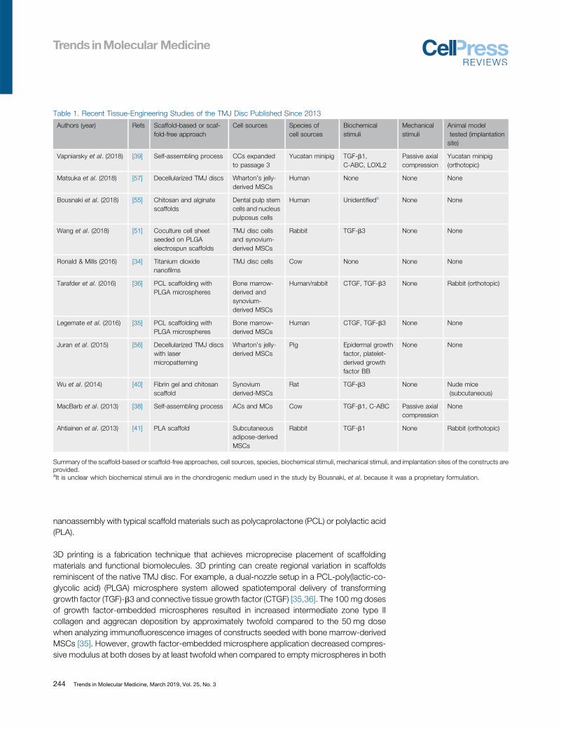
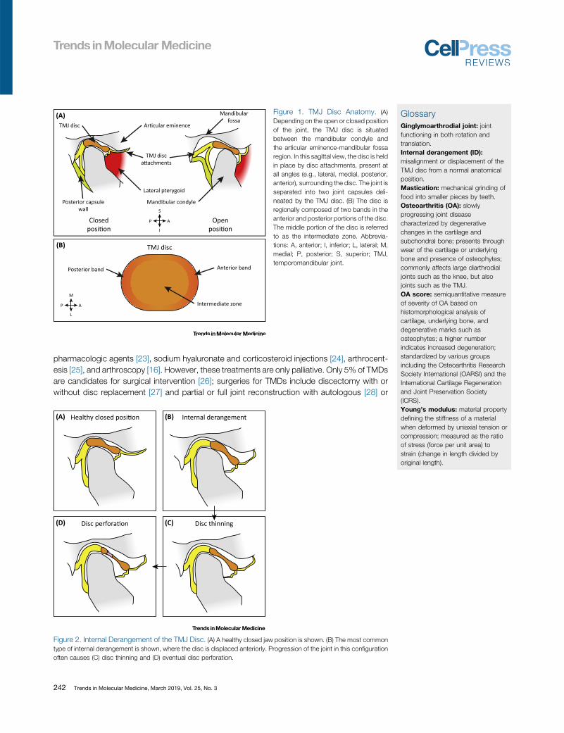
Table 1. Recent Tissue-Engineering Studies of the TMJ Disc Published Since 2013
Authors (year) Refs Scaffold-based or scaf-fold-free approach
Cell sources Species ofcell sources
Biochemicalstimuli
Mechanicalstimuli
Animal modeltested (implantationsite)
Vapniarsky et al. (2018) [39] Self-assembling process CCs expandedto passage 3
Yucatan minipig TGF-b1,C-ABC, LOXL2
Passive axialcompression
Yucatan minipig(orthotopic)
Matsuka et al. (2018) [57] Decellularized TMJ discs Wharton’s jelly-derived MSCs
Human None None None
Bousnaki et al. (2018) [55] Chitosan and alginatescaffolds
Dental pulp stemcells and nucleuspulposus cells
Human Unidentifieda None None
Wang et al. (2018) [51] Coculture cell sheetseeded on PLGAelectrospun scaffolds
TMJ disc cellsand synovium-derived MSCs
Rabbit TGF-b3 None None
Ronald & Mills (2016) [34] Titanium dioxidenanofilms
TMJ disc cells Cow None None None
Tarafder et al. (2016) [36] PCL scaffolding withPLGA microspheres
Bone marrow-derived andsynovium-derived MSCs
Human/rabbit CTGF, TGF-b3 None Rabbit (orthotopic)
Legemate et al. (2016) [35] PCL scaffolding withPLGA microspheres
Bone marrow-derived MSCs
Human CTGF, TGF-b3 None None
Juran et al. (2015) [56] Decellularized TMJ discswith lasermicropatterning
Wharton’s jelly-derived MSCs
Pig Epidermal growthfactor, platelet-derived growthfactor BB
None None
Wu et al. (2014) [40] Fibrin gel and chitosanscaffold
Synoviumderived-MSCs
Rat TGF-b3 None Nude mice(subcutaneous)
MacBarb et al. (2013) [38] Self-assembling process ACs and MCs Cow TGF-b1, C-ABC Passive axialcompression
None
Ahtiainen et al. (2013) [41] PLA scaffold Subcutaneousadipose-derivedMSCs
Rabbit TGF-b1 None Rabbit (orthotopic)
Summary of the scaffold-based or scaffold-free approaches, cell sources, species, biochemical stimuli, mechanical stimuli, and implantation sites of the constructs areprovided.aIt is unclear which biochemical stimuli are in the chondrogenic medium used in the study by Bousnaki, et al. because it was a proprietary formulation.
nanoassembly with typical scaffold materials such as polycaprolactone (PCL) or polylactic acid(PLA).
3D printing is a fabrication technique that achieves microprecise placement of scaffoldingmaterials and functional biomolecules. 3D printing can create regional variation in scaffoldsreminiscent of the native TMJ disc. For example, a dual-nozzle setup in a PCL-poly(lactic-co-glycolic acid) (PLGA) microsphere system allowed spatiotemporal delivery of transforminggrowth factor (TGF)-b3 and connective tissue growth factor (CTGF) [35,36]. The 100 mg dosesof growth factor-embedded microspheres resulted in increased intermediate zone type IIcollagen and aggrecan deposition by approximately twofold compared to the 50 mg dosewhen analyzing immunofluorescence images of constructs seeded with bone marrow-derivedMSCs [35]. However, growth factor-embedded microsphere application decreased compres-sive modulus at both doses by at least twofold when compared to empty microspheres in both
244 Trends in Molecular Medicine, March 2019, Vol. 25, No. 3

Cell sources
ACs
Scaffold-based
LBL nanoassembly & 3D prin ng
TMJ disc construct
Mechanicals muli
Biochemicals muli
Shear
Growth factors
TGF-β1
TGF-β3
Enzymes
C-ABC
LOXL2
Enhanced TMJ disc constructImproved mechanical proper es
Increased collagen contentDirec onal anisotropy
MSCs
TMJ disc cells & MCs
Scaffold-free
Self-assembly
Tension
Hydrosta cpressure
Compression
Enzymes
C-ABC TGF-β1
LOXL2
β
Figure 3. Tissue-Engineering Paradigm of TMJ Disc Constructs. Combination of an appropriate cell source andscaffold-based or scaffold-free approaches can be used for fabrication of a TMJ disc construct (upper panels). Via theapplication of various biochemical and mechanical stimuli, an enhanced, biomimetic construct can be tissue-engineered(lower panels). Abbreviations: ACs, hyaline articular chondrocytes; C-ABC, chondroitinase ABC; LBL, layer-by-layer;LOXL2, lysyl oxidase-like 2; MCs, knee meniscus cells; MSCs, mesenchymal stem cells; TGF-b, transforming growthfactor b; TMJ, temporomandibular joint.
areas analyzed [35]. Similar trends were apparent in instantaneous and relaxation moduliindicating that mechanical properties did not necessarily trend with growth factor applicationand ECM content [35]. Compared to traditional scaffold-based approaches, 3D printing offersthe ability to create regional variation which can resemble native ECM content.
Scaffold-free approaches, such as the self-assembling process [37–39], have been developedto bypass issues [45] related to scaffold degradation products [46] (e.g., acidity due to PLAdegradation), fabrication byproducts [46] (e.g., crosslinkers and plasticizers), and stressshielding of cells [47]. The self-assembling process recapitulates developmental aspects ofcartilage formation to generate functional neotissues with characteristics resembling those of
Trends in Molecular Medicine, March 2019, Vol. 25, No. 3 245

native tissues [45,48]. Specifically, it is the most prominent of these techniques for TMJ disctissue-engineering because it has generated mechanically robust tissue [37]. Stimulation ofself-assembled TMJ disc constructs by bioactive agents and mechanical compression resultedin values of approximately 3.5%, 2.75 MPa, and 2.25 MPa for collagen per wet weight, tensileYoung’s modulus, and ultimate tensile strength (UTS), respectively. Additional analysis ofconstructs created from cocultures of hyaline articular chondrocytes (ACs) and knee meniscuscells (MCs) found collagen fibril alignment reminiscent of native TMJ discs, exhibiting direction-dependent strains in finite element analysis. This was promising because it showed anisotropictissue on par with the alignment of native tissue [38], which further substantiates scaffold-freetissue-engineering as an alternative to scaffold-based approaches.
While scaffold-free approaches do not necessarily have the flexibility of scaffold-basedapproaches, for example, scaffold functionalization with biomolecules, these limitations canbe overcome with exogenous stimulation, which can have various effects on scaffold-freeconstructs such as increased mechanical properties [49,50]. In addition, variation of the cellsource can also have a large influence on the eventual properties of the resulting constructs.
Cell SourcesSelection of a cell source is one of the most important considerations for TMJ disc tissue-engineering (Table 1). Options for primary cells range from native TMJ disc cells [34,51] to othercells from hyaline articular cartilage and the knee meniscus [38]. In addition, recent advances incell expansion technologies [52–54] have allowed exploration of costal cartilage-derived cells[39]. MSCs are also heavily used [35,36,40,41,51,55–57].
Potential primary cell sources for TMJ disc tissue-engineering include TMJ disc cells, ACs,MCs, and costal chondrocytes (CCs). TMJ disc cells have been used in multiple studies [34,51],but the dearth of available, healthy tissue raises concerns for this source [58]. Thus, ACs andMCs have been considered [38]. Using AC–MC coculture with the self-assembling processresulted in a functional, anisotropic TMJ disc as discussed above [38]. With recent advances incell expansion technologies that preserve chondrogenic phenotype [52–54], CCs might allowfor either an autologous or allogeneic approach to replacing cartilages, as demonstratedpreviously in articular cartilage [59,60] and the TMJ disc [39]. Allogeneic CCs can be harvestedfrom cadaveric tissue, while autologous tissue harvest procedures are conducted routinely forrhinoplasty and autologous TMJ reconstruction. Thus, existing surgical procedures may besufficient for tissue regeneration purposes. The use of CCs can also remove or reduce donorsite morbidity and virtually eliminate the potential of harvesting cells from OA tissue. When usedin a hyaline articular cartilage model, CC constructs have attained a functionality index (FI; Box1) of 55% compared to the medial condyle cartilage properties [60]. These techniques and
Box 1. FI Compares Construct Properties to Native Tissue Values
Values for biochemical content, such as overall collagen (Col) and GAG content, accompany values for variousmechanical properties such as UTS, tensile Young’s modulus (ET), compressive relaxation modulus (Er), and com-pressive instantaneous modulus (Ei). Ranging from 0 to 100%, a value of 100% represents perfect recapitulation ofnative values. Subscripts serve to designate native (N) or tissue-engineered (TE) values.
FI TEjNð Þ ¼ 16
1 � jGAGN � GAGTE
GAGNj
� �þ 1 � jColN � ColTE
ColNj
� �þ 1 � jE
iN � Ei
TE
EiN
j !
þ 1 � jErN � Er
TE
ErN
j� �
þ 1 � jETN � ET
TE
ETN
j !
þ 1 � jUTSN � UTSTE
UTSNj
� �266664
377775 � 100%
246 Trends in Molecular Medicine, March 2019, Vol. 25, No. 3

results offer promise of an alternative source of chondrocytes that can create mechanicallystable constructs for other parts of the body such as the TMJ disc.
An array of MSCs from both adult and fetal tissues have been used, as previously reviewed [61].MSCs from various tissues (Table 1) offer an autologous or allogeneic approach and can beisolated in large quantities, making these sources clinically relevant for construct formation.Perhaps the most interesting MSCs are those derived from the synovium because they wereshown to synthesize cartilage oligomeric matrix protein, link protein, and glycosaminoglycans(GAGs), similar to ACs [62]. For example, synovium-derived MSCs on fibrin–chitosan scaffoldsincreased type I collagen expression approximately twofold in vitro and ECM deposition in vivoas demonstrated by histological analysis when compared to pure chitosan scaffolds [40].Progress using MSCs has resulted in morphological and biochemical biomimicry evaluated viahistology, gene expression, and other biochemical assays [36,40,41,51], but future researchshould focus on assaying functional properties of MSC-derived constructs via mechanicaltesting.
The choice of cell source remains a challenge within the field of TMJ disc tissue-engineering.Lack of standardization of mechanical testing modalities makes it difficult to compare sourceshead-to-head and to determine if one cell source is more suitable than another. Perhaps themost important characteristic to consider when choosing a cell source is mechanical stability ofthe resulting tissue-engineered construct due to the dynamic joint environment.
Improvement of Mechanical Properties of TMJ Disc FibrocartilageThe TMJ disc functions in a dynamic environment of compression, tension, and shear [63,64].Finite element analysis shows stresses in the TMJ disc during mouth opening to be greater than7 MPa in compression, 4 MPa in tension, and 1 MPa in shear [65]. For comparison, the hipexperiences 7–10 MPa in compression and up to 18 MPa during stressful activities such asstanding up [66,67]. Characterization of the native tissue should aim to define the goldstandard, design criteria for tissue-engineered TMJ disc constructs; the expectation is thatreplicating the mechanical properties of native tissue would allow for restoration of mechanicalfunction. Thus, to engineer constructs with physiological levels of mechanical stresses in mind,various biochemical and mechanical stimuli, and also changes in scaffold processing (Figure 3)have been developed. For scaffold-free approaches, self-assembled constructs haveapproached native values in mechanical properties due to synergistic effects of biochemicaland mechanical stimulation [38,39].
A majority of recent scaffold-based studies use only biochemical stimuli to improve constructmechanical properties (Table 1). Constructs stimulated with biochemical stimuli have beenpreviously found to exhibit native tissue structure–function relationships. For example, insulin-like growth factor I and TGF-b applied to constructs created from TMJ disc cells increasedcollagen synthesis by greater than 400% at 3 weeks of culture, leading to higher aggregatemoduli of 5 kPa [68]. However, constructs sometimes do not follow native tissue structure–function relationships [35] (e.g., increased matrix deposition leading to increased mechanicalproperties). To overcome such deficiencies, mechanical stimulation may be considered.However, mechanical stimulation has not been used in scaffold-based TMJ disc approaches,although it has been used in other fibrocartilages such as the knee meniscus. For example,hydrostatic pressure combined with TGF-b1 led to fourfold higher collagen deposition andthreefold higher GAG deposition, as compared to the unpressurized growth factor controls inMC-seeded PLA scaffolds [69]. Studies showing recapitulation of native tissue structure–function relationships should serve as models for future studies toward identifying additional
Trends in Molecular Medicine, March 2019, Vol. 25, No. 3 247

stimuli. Biochemical stimuli must continue to be investigated, but, additionally, mechanicalstimuli can be used to increase mechanical properties of engineered discs to withstand thedynamic in vivo environment.
Scaffold-free approaches have combined biochemical and mechanical stimuli to generatestiffer, stronger, anisotropic constructs, followed by examination of the resulting constructs inlarge animal models. Using a scaffold-free approach with AC–MC coculture, TGF-b1, chon-droitinase ABC (C-ABC), and lysyl oxidase-like 2 (LOXL2) have been identified in the past asefficacious for fibrocartilage tissue-engineering, enhancing tensile Young’s modulus and UTSby 245% and 186%, respectively [70]. In a self-assembled TMJ disc model using AC–MCcoculture stimulated with only TGF-b1 and C-ABC, tensile Young’s modulus, UTS, andcollagen per wet weight increased by twofold or greater in the intermediate zone of the disc,as compared to controls [38]. Passive axial compression and these biochemical stimuli werecombined and noted to exhibit synergism, showing 5.8-, 14.7-, and 13.8-fold increases incollagen per wet weight, tensile Young’s modulus, and UTS, respectively, compared tounstimulated controls [38]. Moving to in vivo studies, TMJ discs engineered using all threestimuli (TGF-b1, C-ABC, and LOXL2) coupled with passive axial compression, yielded an FI(Box 1) of 42% of native properties with a passaged, allogeneic CC source [39]. By combiningthese three biochemical stimuli with mechanical stimulation, increased functional propertieswere achieved as compared to either alone. Thus, further synergistic effects of other biochemi-cal and mechanical stimuli should be explored.
As reviewed elsewhere [49], strategies for other tissues, such as hyaline articular cartilage, canhelp inform further mechanical improvement of constructs. Similar designs and models can beused to engineer the fibrocartilage of the TMJ disc. For example, in a recent study on tensionand its effects for articular cartilage engineering, continuous stimulation combined with abioactive regimen increased the tensile properties by 5.8-fold over unstimulated controls inAC-derived, self-assembled constructs [71]. By improving mechanical stability using biochemi-cal and mechanical stimuli, constructs continue to approach native tissue values. Attainingmechanical biomimicry is a crucial characteristic for constructs to perform satisfactorily whenimplanted into the orthotopic environment.
Current Animal ModelsPrior to human clinical trials, tissue-engineered implants should be examined in relevant animalmodels to demonstrate initial safety and efficacy. Similar to TMJ disc tissue-engineering, devel-opment of animal models is based on design criteria. For the TMJ, similar anatomy, chewingpatterns, and diets compared to humans, and ease of surgical access are included in the designcriteria. In addition, relative size of TMJ structures and animal cost may also determine whichmodel touse. Animalmodels exist forvarious purposes such asobserving theadverse reactions toan implant subcutaneously to examining surgically induced pathologies in orthotopic studies.Small animals such as mice and rats are economical, serve as pain models [72,73], and simulateOA and associated degenerative changes in the joints [74,75]. However, their small TMJ disc sizelimits studies to simple subcutaneous implantation as opposed to orthotopic studies in largeranimals such as rabbits [43]. Moving toward orthotopic studies, rabbits allow for additionalbiochemical and histological analysis, and reliable mechanical testing [42], but present substantialdifferences from human size and loading conditions [43]. This motivates the use of large animalmodels that more closely resemble human anatomies and conditions [42].
Many preliminary studies involve subcutaneous implantation to examine possible adversereactions and establish proof-of-concept. These studies, as reviewed [43], are commonly
248 Trends in Molecular Medicine, March 2019, Vol. 25, No. 3
performed in mice or rats due to their low cost, without much consideration of anatomical ordietary similarities. For example, a fibrin–chitosan scaffold with synovium-derived rat MSCs wasembedded into explanted TMJ discs with perforation defects and implanted into nude micesubcutaneously in a xenogeneic approach [40]. Histological analysis showed increased type Iand II collagen deposition in the fibrin–chitosan scaffold, compared to the pure chitosanscaffold [40]. Although this study represents a disc perforation model, additional biochemicaland mechanical analyses must be performed in larger animals to show reparative ability in thefully loaded orthotopic environment.
Recent studies used rabbits for orthotopic evaluation of tissue-engineered TMJ discs [36,41].For example, 3D printed PCL–PLGA microsphere scaffolds seeded with allogeneic, synovium-derived MSCs were implanted into the disc and noted histologically to degrade by 6 weeks [36].Cells retained their chondrocyte-like phenotype in vivo [36]. Scoring of the condylar surfaceswith an OA score resulted in values of approximately 3.9 and 2.4 for the scaffolds without andwith growth factors, respectively, where a lower score represents a better outcome [36]. Whilethese studies [36,41] demonstrate feasibility for implantation of tissue-engineered TMJ discs viahistological analysis, mechanical testing is of paramount importance to show the integrity oftissue-engineered constructs.
Strides in animal studies are promising to the research community as they point to a feasibletranslation pathway for tissue-engineered constructs. The use of ectopic small animal andlarger orthotopic models (e.g., mouse and rabbit models) is a crucial first step in proof-of-concept work for the field. However, it will ultimately be regenerative studies in orthotopicanimal models in species such as the minipig that will be most impactful for translation of tissue-engineered TMJ discs toward human clinical studies.
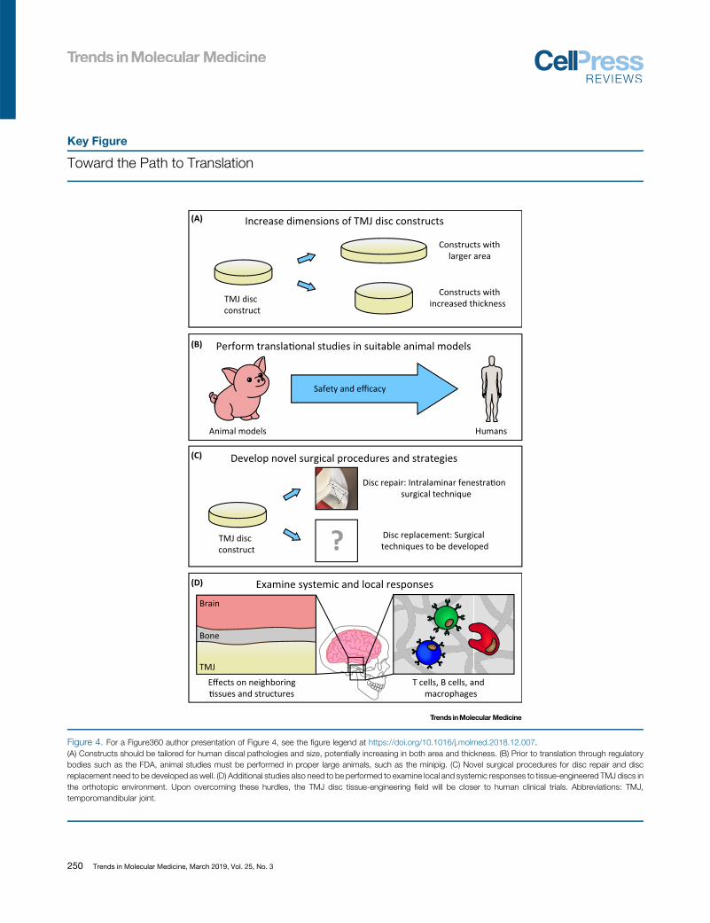
Path to TranslationTranslational hurdles that remain include tuning of construct mechanical properties toward bio-mimicry (Figure3)aswellasscale-upofareaandthicknessof implants (Figure4,KeyFigure).Arecentminipig study, showing safe and efficacious implantation of TMJ constructs [39], establishes thisorthotopic largeanimalmodelasacogentelement in the translationalpathway (Figure4).Clinicalandregulatory hurdles are also significant for translation of TMJ disc constructs (Figure 4).
Application of Proper Tissue-Engineering Parameters for Tuning of TMJ Disc Constructs tothe TMJ Mechanical EnvironmentConstructs must be tuned to the mechanical environment of the TMJ disc because they will besubject to compressive, tensile, and shear forces [63,64]. Theoretically, the required mechanicalproperties will depend on surgical technique, model, and animal. For example, it was shown thatan FI (Box 1) of 42% was sufficient when implanted via the intralaminar fenestration surgicaltechnique (Figure 5) in a focal thinning model in the Yucatan minipig [39]. When moving towardperforation or larger defects, this implant might be insufficient. In contrast, some constructs mightbe too stiff or strong compared to native values, as observed in some scaffold-based approaches[35], causing stress concentrations and possible degeneration on the articulating surfaces. Also, amismatch in the rates ofscaffold degradation versus tissue formation can lead to failure. Therefore,it is important to consider tuning mechanical properties by application of proper stimulationregimens, whether using a scaffold-based or scaffold-free tissue-engineering approach (Figure 3).
Tailoring of Tissue-Engineered TMJ Discs to Human Discal Pathologies and SizeAs the translational direction points to additional large animal orthotopic studies before humanclinical trials commence, defect models must increase in size. As such, constructs must also
Trends in Molecular Medicine, March 2019, Vol. 25, No. 3 249
Key Figure
Toward the Path to Translation
Increase dimensions of TMJ disc constructs
Perform transla�onal studies in suitable animal models
Constructs withlarger area
Constructs withincreased thicknessTMJ disc
construct
Animal models Humans
Safety and efficacy
(A)
(B)
T cells, B cells, andmacrophages
Examine systemic and local responses
Effects on neighboring�ssues and structures
Brain
TMJ
Bone
(D)
TMJ discconstruct
Develop novel surgical procedures and strategies
Disc repair: Intralaminar fenestra�onsurgical technique
Disc replacement: Surgicaltechniques to be developed?
(C)
Figure 4. For a Figure360 author presentation of Figure 4, see the figure legend at https://doi.org/10.1016/j.molmed.2018.12.007.(A) Constructs should be tailored for human discal pathologies and size, potentially increasing in both area and thickness. (B) Prior to translation through regulatorybodies such as the FDA, animal studies must be performed in proper large animals, such as the minipig. (C) Novel surgical procedures for disc repair and discreplacement need to be developed as well. (D) Additional studies also need to be performed to examine local and systemic responses to tissue-engineered TMJ discs inthe orthotopic environment. Upon overcoming these hurdles, the TMJ disc tissue-engineering field will be closer to human clinical trials. Abbreviations: TMJ,temporomandibular joint.
250 Trends in Molecular Medicine, March 2019, Vol. 25, No. 3

(A) (B)
(C) (D)
(E) (F)
(G) (H)
(I) (J)
Figure 5.
(Figure legend continued on the bottom of the next page.)
The Intralaminar Fenestration Surgical Technique. (A, B) Through a preauricular incision, the tempor-omandibular joint (TMJ) was exposed. (C–E) Surgeons filleted the disc open with a scalpel, and (F, G) created a one-sided
Trends in Molecular Medicine, March 2019, Vol. 25, No. 3 251

scale-up (Figure 4). In the recent minipig study [39], a one-sided, 3 mm defect, mimicking discthinning, was used. Future studies need to scale-up to a larger defect area to mimic increaseddisc thinning, in addition to two-sided defects to mimic disc perforation. To scale-up constructsto larger thicknesses, one might consider using larger scaffolds. However, as scaffolds andconstructs trend upward in thickness, it should be kept in mind that diffusion limitationsincrease. Decreased diffusion can result in shell-like neotissues with necrotic centers, thatdisplay inadequate mechanics. However, scaffold-free approaches might prove advantageousfor creation of larger constructs to mimic disc thinning. Self-assembled articular cartilageconstructs made of passaged ACs up to 25 mm in diameter have been made by combiningcytochalasin D, TGF-b1, C-ABC, and LOXL2, under a compressive load and in mechanicalconfinement [76]. This approach may allow for examining TMJ disc healing in larger defects thatmimic clinically observed disc thinning. As such, a significant portion of future TMJ disc studiesshould investigate the scale-up of defects and constructs for relevance to human TMJanatomy.
Novel and Cogent Translational StudiesOrthotopic large animal models need to be performed to examine the safety and efficacy oftissue-engineered constructs prior to translation. Possible species for performing regenerativestudies include sheep [77], goats [78], dogs [79], farm pigs [80], and minipigs [81]. The farm pigand minipig are two suitable models that have been recently used for regenerative studies dueto their similarities to humans in chewing patterns, diet, and anatomy [3,81–85].
In a recent study demonstrating safety and efficacy of a self-assembled, allogeneic, tissue-engineered implant for disc repair, a novel TMJ disc thinning model was created in the Yucatanminipig [39]. Because the implants were created from a CC source, implantation into the TMJdisc represented nonhomologous use. Implants approaching native tissue values were formedby a regimen of biochemical and mechanical stimulation. To affix implants securely, theintralaminar fenestration surgical technique was developed [39] (Figure 5). Although thiswas an allogeneic, nonhomologous use which has potential to elicit an immune response,implant safety was shown by minimal to no immune response to the constructs, as assayed byhistological staining for CD3, CD20, and CD68 for T cells, B cells, and macrophages. However,it was specified that additional work needs to further elucidate the immunological response [39],such as macrophage activation due to tissue-engineered implants [86–88] (Figure 4). In termsof efficacy, results showed that the tensile Young’s modulus, integration at the repair-to-nativetissue interface, and percent of defect closure were 3.4-, 3.2-, and 4.4-fold higher, respectively,compared to empty defect controls [39]. OA scores of the condylar surface treated withimplants were threefold less than the empty defect controls [39], yielding a better clinicaloutcome overall. Together, these results demonstrate the feasibility of allogeneic TMJ disctissue-engineered constructs in the orthotopic environment and pave the way for additionalorthotopic large animal studies and future human clinical trials (Figure 4).
Overcoming Additional Clinical and Regulatory HurdlesIn stark contrast to diarthrodial joints such as the knee, there is limited knowledge surroundingthe TMJ, especially when it comes to developing new processes and products for repair orreplacement of the TMJ disc. Compared to the TMJ, a greater variety of products, treatments,
thinning defect via a biopsy punch. (H) A tissue-engineered disc was placed between the two laminae and (I) sutured backtogether. Sutures attached to the side of the disc instead of on the articulating surfaces allowed for continued loading of theTMJ disc while healing; this placement avoided possible stress concentrations and resulting degeneration. (J) The lateralattachment was recreated by use of an anchoring system. Reprinted, with permission, from [39].
252 Trends in Molecular Medicine, March 2019, Vol. 25, No. 3
Outstanding QuestionsHow do researchers achieve tuning oftissue-engineered constructs to themechanical environment of the TMJdisc?
Can researchers scale-up constructs,in area and thickness, to be relevant tohuman discal pathologies and size?
For what cases will tissue-engineeredproducts be indicated (orcontraindicated)?
Can novel surgical procedures bedeveloped for accessing the TMJ,and fixing and implanting tissue-engi-neered TMJ disc constructsorthotopically?
What are the local and systemicresponses to tissue-engineered TMJdiscs in vivo?
How would tissue-engineered con-structs for the TMJ disc be regulatedby the FDA?
and studies exist for the knee. To illustrate these differences, one can consider indications andcontraindications in the TMJ versus the knee. For example, in the knee, there are clearguidelines as to what constitutes small, large, partial-thickness, and full-thickness defectswith concomitant treatment algorithms [89]. In contrast, it is not clear when a tissue-engineeredtreatment would be indicated in the TMJ. Currently, in the knee, tissue-engineered products arecontraindicated for the OA milieu [90]. This has not been confirmed for the TMJ, although theexpectation is that the constructs under OA conditions might succumb to the same fate as thenative tissue [91]. Development of treatment guidelines and additional studies specific to theTMJ should continue, toward bringing TMJ-related knowledge to levels of other diarthrodialjoints.
One must also consider fixation and associated surgical approaches. The intralaminar fenes-tration surgical technique (Figure 5) was successful in treating early-stage disc thinning, but inthe minipig [39]. However, in 5% of TMD cases requiring surgery [26], it is not yet obvious howone may be able to attach a partial or whole, tissue-engineered disc (Figure 4). Surgeons andresearchers must continue to collaborate to develop surgical approaches for implantation oftissue-engineered implants, as they are of utmost importance to the success of the tissue-engineered treatment.
With regard to clearing the regulatory hurdle, proximity of the TMJ to the brain (Figure 4) maynecessitate more stringent safety requirements than products for other joints such as the knee.These requirements may include analysis of the synovial fluid in the TMJ, but also theneighboring cerebrospinal fluid. Notoriously, mechanical failure and resulting degradation ofthe Proplast–Teflon disc implants resulted in exposure of the brain cavity [92–94]. Additionally,current large animal work has yet to investigate fully the immunological implications related toTMJ disc implants (Figure 4) or how immunomodulation may be used in a proinflammatoryenvironment [95]. In terms of regulation, the US Food and Drug Administration (FDA) has notpreviously guided a tissue-engineered TMJ disc product [96], thus raising the question ofestablishing TMJ-specific safety and efficacy guidance documents. Future research in the fieldneeds to establish the safety of tissue-engineered TMJ discs by elucidating the immuneresponse. Additionally, researchers need to communicate with regulatory bodies, such asthe FDA, to obtain guidance on how tissue-engineered TMJ disc products need to bedemonstrated as safe and efficacious.
Concluding RemarksWhile recent advances propel TMJ disc tissue-engineering forward, many hurdles still exist. Tosummarize, the pressing challenges include improvement of mechanical properties of con-structs, scale-up of implant dimensions, determination of indications for tissue-engineereddiscs, development of surgical techniques, analysis of the immunological response, andregulation by the FDA (see Outstanding Questions). Tissue-engineering and basic scienceinvestigations for TMDs will continue to drive the field. The field should focus toward addressingquestions in the clinical and regulatory spaces. Specifically, studies should pay attention todeveloping novel surgical techniques and associated fixation methods toward human clinicaltrials. For each new tissue-engineering approach, regulatory requirements need to be satisfiedby demonstration of TMJ-specific safety and efficacy in large animal models. As regulatorybodies turn their attention toward clinical trials, these data will be the primary preclinicalassessment of implants. Considering the momentum toward significant preclinical studies,it is an exciting time to be in the field of TMJ disc tissue-engineering. After the early successshown in the orthotopic study performed in the Yucatan minipig [39] and the identification ofclinical and regulatory hurdles discussed here, there is new impetus to develop tissue-
Trends in Molecular Medicine, March 2019, Vol. 25, No. 3 253

engineering solutions to begin addressing the various intractable TMJ trauma and degenerativeailments. The possibility of translating tissue-engineered TMJ discs is increasingly beingrealized.
AcknowledgmentsThe authors would like to acknowledge support from the following funding sources: National Institutes of HealthR01
DE015038.
References
1. Aryaei, A. et al. (2016) Recent tissue-engineering advances for thetreatment of temporomandibular joint disorders. Curr. Osteo-poros. Rep. 14, 269–279
2. Alomar, X. et al. (2007) Anatomy of the temporomandibular joint.Semin. Ultrasound CT MRI 28, 170–183
3. Sindelar, B.J. and Herring, S.W. (2005) Soft tissue mechanics ofthe temporomandibular joint. Cells Tissues Organs 180, 36–43
4. Arnett, G.W. et al. (1996) Progressive mandibular retrusion-idio-pathic condylar resorption. Part II. Am J. Orthod. Dentofac.Orthopeics 110, 117–127
5. Katzberg, R.W. and Westesson, P.-L. (1993) Diagnosis of theTemporomandibular Joint, W.B. Saunders
6. Dubner, R. et al. (2016) The evolution of TMD diagnosis: past,present, future. J. Dent. Res. 95, 1093–1101
7. Slade, G.D. et al. (2016) Painful temporomandibular disorder: decade ofdiscovery from OPPERA studies. J. Dent. Res. 95, 1084–1092
8. Scrivani, S.J. et al. (2008) Temporomandibular disorders. N. Engl.J. Med. 359, 2693–2705
9. Solberg, W.K. et al. (1979) Prevalence of mandibular dysfunctionin young adults. J. Am. Dent. Assoc. 98, 25–34
10. Gonçalves, D.A. de G. et al. (2010) Symptoms of temporoman-dibular disorders in the population: an epidemiological study. J.Orofac. Pain 24, 270–278
11. Martins-Júnior, R.L. et al. (2010) Temporomandibular disorders: areport of 124 patients. J. Contemp. Dent. Pract. 11, 71–78
12. Wilkes, C.H. (1989) Internal derangements of the temporoman-dibular joint: pathological variations. Arch. Otolaryngol. HeadNeck Surg. 115, 469–477
13. Warren, M.P. and Fried, J.L. (2001) Temporomandibular disordersand hormones in women. Cells Tissues Organs 169, 187–192
14. Murphy, M.K. et al. (2013) Temporomandibular joint disorders: areview of etiology, clinical management, and tissue-engineeringstrategies. Int. J. Oral Maxillofac. Implants 28, 393–414
15. Farrar, W.B. and McCarty, W.L.J. (1979) The TMJ dilemma. J.Alabama Dent. Assoc. 63, 19–26
16. Muñoz-Guerra, M.F. et al. (2013) Temporomandibular joint discperforation: long-term results after operative arthroscopy. J. OralMaxillofac. Surg. 71, 667–676
17. Kuribayashi, A. et al. (2008) MRI findings of temporomandibularjoints with disk perforation. Oral Surg. Oral Med. Oral Pathol. OralRadiol. Endod. 106, 419–425
18. American Society of Temporomandibular Joint Surgeons (2003)Guidelines for diagnosis and management of disorders involvingthe temporomandibular joint and related musculoskeletal struc-tures. Cranio 21, 68–76
19. Ballesteros, L.E. and León-S, F.E. (1999) Anatomical and patho-logical study of the temporomandibular joint disk in Colombianindividuals. Rev. Med. Chil. 127, 1469–1474 (in Spanish)
20. Romero-Reyes, M. and Uyanik, J.M. (2014) Orofacial pain man-agement: current perspectives. J. Pain Res. 7, 99–115
21. Armijo-Olivo, S. et al. (2016) Effectiveness of manual therapy andtherapeutic exercise for temporomandibular disorders: system-atic review and meta-analysis. Phys. Ther. 96, 9–25
22. Pficer, J.K. et al. (2017) Occlusal stabilization splint for patientswith temporomandibular disorders: meta-analysis of short andlong term effects. PLoS One 12, e0171296
254 Trends in Molecular Medicine, March 2019, Vol. 25, No. 3
23. Kurita Varoli, F. et al. (2015) Analgesia evaluation of 2 NSAIDdrugs as adjuvant in management of chronic temporomandibulardisorders. Sci. World J. 2015, 359152
24. Mountziaris, P.M. et al. (2009) Emerging intra-articular drug deliv-ery systems for the temporomandibular joint. Methods 47, 134–140
25. De Riu, G. et al. (2013) Arthrocentesis and temporomandibularjoint disorders: clinical and radiological results of a prospectivestudy. Int. J. Dent. 2013, 790648
26. Dolwick, M.F.F. and Dimitroulis, G. (1994) Is there a role fortemporomandibular joint surgery? Br. J. Oral Maxillofac. Surg.32, 307–313
27. Eriksson, L. and Westesson, P.L. (2001) Discectomy as an effec-tive treatment for painful temporomandibular joint internalderangement: a 5-year clinical and radiographic follow-up. J.Oral Maxillofac. Surg. 59, 750–758
28. Sharma, H. et al. (2015) Costochondral graft as interpositionalmaterial for TMJ ankylosis in children: a clinical study. J. Max-illofac. Oral Surg. 14, 565–572
29. Wolford, L.M. et al. (2003) Comparison of 2 temporomandibularjoint total joint prosthesis systems. J. Oral Maxillofac. Surg. 61,685–690
30. Hinton, R.J. (1992) Alterations in rat condylar cartilage followingdiscectomy. J. Dent. Res. 71, 1292–1297
31. Agerberg, G. and Lundberg, M. (1971) Changes in the temporo-mandibular joint after surgical treatment. A radiologic follow-upstudy. Oral Surg. Oral Med. Oral Pathol. 32, 865–875
32. Mercuri, L.G. (2000) The use of alloplastic prostheses for temporo-mandibular joint reconstruction. J. Oral Maxillofac. Surg. 58, 70–75
33. Ingawalé, S. and Goswami, T. (2009) Temporomandibular joint:disorders, treatments, and biomechanics. Ann. Biomed. Eng. 37,976–996
34. Ronald, S. and Mills, D. (2016) Fibrochondrocyte growth andfunctionality on TiO2 nanothin films. J. Funct. Biomater. 7, 15
35. Legemate, K. et al. (2016) Engineering human TMJ discs withprotein-releasing 3D-printed scaffolds. J. Dent. Res. 95, 800–807
36. Tarafder, S. et al. (2016) Micro-precise spatiotemporal deliverysystem embedded in 3D printing for complex tissue regeneration.Biofabrication 8, 025003
37. Hu, J.C. and Athanasiou, K.A. (2006) A self-assembling processin articular cartilage tissue-engineering. Tissue Eng. 12, 969–979
38. MacBarb, R.F. et al. (2013) Engineering functional anisotropy infibrocartilage neotissues. Biomaterials 34, 9980–9989
39. Vapniarsky, N. et al. (2018) Tissue-engineering toward temporo-mandibular joint disc regeneration. Sci. Transl. Med. 10,eaaq1802
40. Wu, Y. et al. (2014) The pilot study of fibrin with temporomandib-ular joint derived synovial stem cells in repairing TMJ disc perfo-ration. BioMed Res. Int. 2014, 454021
41. Ahtiainen, K. et al. (2013) Autologous adipose stem cells andpolylactide discs in the replacement of the rabbit temporoman-dibular joint disc. J. R. Soc. Interface 10, http://dx.doi.org/10.1098/rsif.2013.0287
42. Almarza, A.J. et al. (2018) Preclinical animal models for tempo-romandibular joint tissue-engineering. Tissue Eng. B Rev. 24,171–178

43. Helgeland, E. et al. (2018) Scaffold-based TMJ tissue regenera-tion in experimental animal models: a systematic review. TissueEng. B Rev. 24, 300–316
44. Kommireddy, D.S. et al. (2006) Stem cell attachment to layer-by-layer assembled TiO2 nanoparticle thin films. Biomaterials 27,4296–4303
45. Athanasiou, K.A. et al. (2013) Self-organization and the self-assembling process in tissue-engineering. Annu. Rev. Biomed.Eng. 15, 115–136
46. Liu, X. and Ma, P.X. (2004) Polymeric scaffolds for bone tissue-engineering. Ann. Biomed. Eng. 32, 477–486
47. Bryant, S.J. et al. (2004) Crosslinking density influences themorphology of chondrocytes photoencapsulated in PEG hydro-gels during the application of compressive strain. J. Orthop. Res.22, 1143–1149
48. Lee, J.K. et al. (2017) The self-assembling process and applica-tions in tissue-engineering. Cold Spring Harb. Perspect. Med. 7,a025668
49. Salinas, E.Y. et al. (2018) A guide for using mechanical stimulationto enhance tissue-engineered articular cartilage properties. Tis-sue Eng. B Rev. 24, 345–358
50. Kwon, H. et al. (2016) Articular cartilage tissue-engineering: therole of signaling molecules. Cell. Mol. Life Sci. 73, 1173–1194
51. Wang, C.H. et al. (2018) Layering poly (lactic-co-glycolic acid)-based electrospun membranes and co-culture cell sheets forengineering temporomandibular joint disc. J. Biol. Regul. Home-ost. Agents 32, 55–61
52. Murphy, M.K. et al. (2013) Enhancing post-expansion chondro-genic potential of costochondral cells in self-assembled neocar-tilage. PLoS One 8, e56983
53. Murphy, M.K. et al. (2015) Engineering a fibrocartilage spectrumthrough modulation of aggregate redifferentiation. Cell Trans-plant. 24, 235–245
54. Murphy, M.K. et al. (2015) TGF-b1, GDF-5, and BMP-2 stimulationinduces chondrogenesis in expanded human articular chondro-cytes and marrow-derived stromal cells. Stem Cells 33, 762–773
55. Bousnaki, M. et al. (2018) Fibro/chondrogenic differentiation ofdental stem cells into chitosan/alginate scaffolds towards tempo-romandibular joint disc regeneration. J. Mater. Sci. Mater. Med.29, 97
56. Juran, C.M. et al. (2015) Engineered microporosity: enhancingthe early regenerative potential of decellularized temporomandib-ular joint discs. Tissue Eng. A 21, 829–839
57. Matuska, A.M. et al. (2018) Approaches to improve integrationand regeneration of an ex vivo derived temporomandibular jointdisc scaffold with variable matrix composition. J. Mater. Sci.Mater. Med. 29, 152
58. Johns, D.E. et al. (2008) Clinically relevant cell sources for TMJdisc engineering. J. Dent. Res. 87, 548–552
59. Huwe, L.W. et al. (2017) Using costal chondrocytes to engineerarticular cartilage with applications of passive axial compressionand bioactive stimuli. Tissue Eng. A 24, 516–526
60. Huwe, L.W. et al. (2018) Characterization of costal cartilage andits suitability as a cell source for articular cartilage tissue-engi-neering. J. Tissue Eng. Regen. Med. 12, 1163–1176
61. Zhang, S. et al. (2015) Stem cells for temporomandibular jointrepair and regeneration. Stem Cell Rev. Rep. 11, 728–742
62. Recklies, A.D. et al. (1998) Regulation of cartilage oligomericmatrix protein synthesis in human synovial cells and articularchondrocytes. Arthritis Rheum. 41, 997–1006
63. Tanaka, E. and Van Eijden, T. (2003) Biomechanical behavior ofthe temporomandibular joint disc. Crit. Rev. Oral Biol. Med. 14,138–150
64. Wu, Y. et al. (2015) Viscoelastic shear properties of porcinetemporomandibular joint disc. Orthod. Craniofac. Res. 18,156–163
65. Li, Q. et al. (2014) Effect of jaw opening on the stress pattern in anormal human articular disc: finite element analysis based on MRIimages. Head Face Med. 10, 24
66. Athanasiou, K.A. et al. (2017) Articular Cartilage. (2nd edn), CRCPress
67. Hodge, W.A. et al. (1989) Contact pressures from an instru-mented hip endoprosthesis. J. Bone Joint Surg. Am. 71,1378–1386
68. Detamore, M.S. and Athanasiou, K.A. (2005) Evaluation of threegrowth factors for TMJ disc tissue-engineering. Ann. Biomed.Eng. 33, 383–390
69. Gunja, N.J. et al. (2009) Effects of TGF-b1 and hydrostatic pres-sure on meniscus cell-seeded scaffolds. Biomaterials 30, 565–573
70. Makris, E.A. et al. (2014) Combined use of chondroitinase-ABC,TGF-b1, and collagen crosslinking agent lysyl oxidase to engineerfunctional neotissues for fibrocartilage repair. Biomaterials 35,6787–6796
71. Lee, J.K. et al. (2017) Tension stimulation drives tissue formationin scaffold-free systems. Nat. Mater. 16, 864–873
72. Roveroni, R.C. et al. (2001) Development of a behavioral model ofTMJ pain in rats: the TMJ formalin test. Pain 94, 185–191
73. Nicoll, S.B. et al. (2010) A rat model of temporomandibular jointpain with histopathologic modifications. J. Orofac. Pain 24, 298–304
74. Cledes, G. et al. (2006) Validation of a chemical osteoarthritismodel in rabbit temporomandibular joint: a compliment to bio-mechanical models. Int. J. Oral Maxillofac. Surg. 35, 1026–1033
75. Güler, N. et al. (2011) Sodium iodoacetate induced osteoarthrosismodel in rabbit temporomandibular joint: CT and histologicalstudy (part I). Int. J. Oral Maxillofac. Surg. 40, 1289–1295
76. Huang, B.J. et al. (2018) Overcoming challenges in engineeringlarge, scaffold-free neocartilage with functional properties. TissueEng. A 24, 1652–1662
77. Miyamoto, H. et al. (1999) A sheep model for temporomandibularjoint ankylosis. J. Oral Maxillofac. Surg. 57, 812–817
78. Li, L. et al. (2015) Establishment and histological evaluation of agoat traumatic temporomandibular joint model. J. Oral Maxillofac.Surg. 73, 943–950
79. Miyamoto, H. et al. (2007) The effect of etodolac on experimentaltemporomandibular joint osteoarthritis in dogs. J. Craniomaxillo-fac. Surg. 35, 358–363
80. Lowe, J. et al. (2018) Properties of the temporomandibular joint ingrowing pigs. J. Biomech. Eng. 140, 071002
81. Vapniarsky, N. et al. (2017) The Yucatan minipig temporoman-dibular joint disc structure–function relationships support its suit-ability for human comparative studies. Tissue Eng. C Methods 23,700–709
82. Herring, S.W. and Liu, Z.J. (2001) Loading of the temporoman-dibular joint: anatomic and in vivo evidence from the bones. CellsTissues Organs 169, 193–200
83. Herring, S.W. et al. (2002) Temporomandibular joint in miniaturepigs: anatomy, cell replication, and relation to loading. Anat. Rec.266, 152–166
84. Kalpakci, K.N. et al. (2011) An interspecies comparison of thetemporomandibular joint disc. J. Dent. Res. 90, 193–198
85. Herring, S.W. (1976) The dynamics of mastication in pigs. Arch.Oral Biol. 21, 473–480
86. Mosser, D.M. and Edwards, J.P. (2008) Exploring the full spec-trum of macrophage activation. Nat. Rev. Immunol. 8, 958–969
87. Gaffney, L. et al. (2017) Macrophages’ role in tissue disease andregeneration. In Macrophages: Origin, Functions and Biointer-vention (Kubiak, J.Z. and Kloc, M., eds), pp. 245–271, Springer
88. Ogle, M.E. et al. (2016) Monocytes and macrophages in tissuerepair: implications for immunoregenerative biomaterial design.Exp. Biol. Med. 241, 1084–1097
89. Falah, M. et al. (2010) Treatment of articular cartilage lesions ofthe knee. Int. Orthop. 34, 621–630
90. Basad, E. et al. (2015) Matrix-induced autologous chondrocyteimplantation (MACI) in the knee: clinical outcomes and chal-lenges. Knee Surg. Sports Traumatol. Arthrosc. 23, 3729–3735
Trends in Molecular Medicine, March 2019, Vol. 25, No. 3 255

91. Salash, J.R. et al. (2016) Potential indications for tissue-engineer-ing in temporomandibular joint surgery. J. Oral Maxillofac. Surg.74, 705–711
92. Smith, R.M. et al. (1993) Erosion of a Teflon–Proplast implantinto the middle cranial fossa. J. Oral Maxillofac. Surg. 51, 1268–1271
93. Berarducci, J.P. et al. (1990) Perforation into middle cranial fossaas a sequel to use of a proplast–teflon implant for temporoman-dibular joint reconstruction. J. Oral Maxillofac. Surg. 48, 496–498
256 Trends in Molecular Medicine, March 2019, Vol. 25, No. 3
94. Chuong, R. and Piper, M.A. (1992) Cerebrospinal fluid leak asso-ciated with proplast implant removal from the temporomandibularjoint. Oral Surg. Oral Med. Oral Pathol. 74, 422–425
95. Diehl, R. et al. (2017) Immunosuppression for in vivo research:state-of-the-art protocols and experimental approaches. Cell.Mol. Immunol. 14, 146–179
96. Donahue, R.P. et al. Considerations for translation of tissueengineered fibrocartilage from bench to bedside. J. Biomech.Eng. https://doi.org/10.1115/1.4042201. In press