Embed Size (px)
Citation preview
Reliability and Diagnostic Performance of CT ImagingCriteria in the Diagnosis of Tuberculous MeningitisHugo Botha1, Christelle Ackerman2, Sally Candy3, Jonathan A. Carr1, Stephanie Griffith-Richards2,
Kathleen J. Bateman1*
1Division of Neurology, Tygerberg Hospital, University of Stellenbosch, Cape Town, Western Cape, South Africa, 2Department of Radiodiagnostics, Tygerberg Hospital,
University of Stellenbosch, Cape Town, Western Cape, South Africa, 3Department of Radiology, Groote Schuur Hospital, University of Cape Town, Cape Town, Western
Cape, South Africa
Abstract
Introduction: Abnormalities on CT imaging may contribute to the diagnosis of tuberculous meningitis (TBM). Recently, anexpert consensus case definition (CCD) and set of imaging criteria for diagnosing basal meningeal enhancement (BME) havebeen proposed. This study aimed to evaluate the sensitivity, specificity and reliability of these in a prospective cohort ofadult meningitis patients.
Methods: Initial diagnoses were based on the CCD, classifying patients into: ‘Definite TBM’ (microbiological confirmation),‘Probable TBM’ (diagnostic score $10), ‘Possible TBM’ (diagnostic score 6–9), ‘Not TBM’ (confirmation of an alternativediagnosis) or ‘Uncertain’ (diagnostic score of ,6). CT images were evaluated independently on two occasions by fourexperienced reviewers. Intra-rater and inter-rater agreement were calculated using the kappa statistic. Sensitivities andspecificities were calculated using both ‘Definite TBM’ and either ‘Definite TBM’ or ‘Probable TBM’ as gold standards.
Results: CT scan criteria for BME had good intra-rater agreement (k range 0.35–0.78) and fair to moderate inter-rateragreement (k range 0.20–0.52). Intra- and inter-rater agreement on the CCD components were good to fair (k = ranges0.47–0.81 and 0.21–0.63). Using ‘Definite TBM’ as a gold standard, the criteria for BME were very specific (61.5%–100%), butinsensitive (5.9%–29.4%). Similarly, the imaging components of the CCD were highly specific (69.2–100%) but lackedsensitivity (0–56.7%). Similar values were found when using ‘Definite TBM’ or ‘Probable TBM’ as a gold standard.
Discussion: The fair to moderate inter-rater agreement and poor sensitivities of the criteria for BME suggest that littlereliance should be placed in these features in isolation. While the presence of the CCD criteria of acute infarction ortuberculoma(s) appears useful as rule-in criteria, their absence is of little help in excluding TBM. The CCD and criteria forBME, as well as any new criteria, need to be standardized and validated in prospective cohort studies.
Citation: Botha H, Ackerman C, Candy S, Carr JA, Griffith-Richards S, et al. (2012) Reliability and Diagnostic Performance of CT Imaging Criteria in the Diagnosis ofTuberculous Meningitis. PLoS ONE 7(6): e38982. doi:10.1371/journal.pone.0038982
Editor: Qamaruddin Nizami, Aga Khan University, Pakistan
Received January 25, 2012; Accepted May 15, 2012; Published June 29, 2012
Copyright: � 2012 Botha et al. This is an open-access article distributed under the terms of the Creative Commons Attribution License, which permitsunrestricted use, distribution, and reproduction in any medium, provided the original author and source are credited.
Funding: KB received funding from the Discovery Foundation (Academic Fellowship Award; http://www.discovery.co.za/portal/loggedout-individual/discovery-community-about), College of Neurology of South Africa (K.M. Browse Award; http://www.collegemedsa.ac.za/Default.aspx ) and the University of Stellenbosch.The funders had no role in study design, data collection and analysis, decision to publish, or preparation of the manuscript.
Competing Interests: The authors have declared that no competing interests exist.
* E-mail: [email protected]
Introduction
Tuberculous meningitis (TBM) has a case fatality rate of 15–
68% [1–3], and more than half of the survivors are left with
neurological sequelae [4]. Central nervous system involvement is
estimated to occur in around 1% of patients with active
tuberculosis [5]. In the Western Cape, an area with a high
incidence of tuberculosis, TB is one of the most common causes of
meningitis [6].
Early diagnosis and commencement of treatment confers
a significantly better prognosis [2,4]. However, the diagnosis of
TBM is complicated by its variable clinical presentation and the
lack of specificity of its radiological and laboratory features [7]. For
this reason, in most cases, treatment is initiated before a microbi-
ological diagnosis is confirmed. Physicians have to rely on the
clinical presentation, and the results of investigations that are
available within hours, most notably cerebrospinal fluid (CSF)
biochemistry and cell counts and chest radiography [8]. Although
brain imaging is not mandatory for a diagnosis of probable or
definite TBM, characteristic abnormalities on imaging have been
reported to contribute to the diagnostic certainty. A recent expert
consensus case definition (CCD) for use in research incorporates
the following radiological signs in the diagnosis of TBM:
hydrocephalus, infarcts, tuberculoma(s), basal meningeal enhance-
ment, and the presence of pre-contrast basal hyperdensities [9].
On computed tomography (CT) of the brain, hydrocephalus and
meningeal enhancement are reported to be the most sensitive signs
of TBM, being present in 80% and 75% of paediatric cases [9–
11], and in 45% and up to 34% of adult cases, respectively
[4,10,11]. Although magnetic resonance imaging is superior to CT
imaging in the diagnosis of TBM [1], this modality may not be
readily accessible in resource limited settings with high TB burden.
PLoS ONE | www.plosone.org 1 June 2012 | Volume 7 | Issue 6 | e38982
Furthermore, objective criteria for diagnosing basal meningeal
enhancement on CT have recently been proposed based on case-
control studies of childhood TBM in the Western Cape [12–14].
The authors reported a high sensitivity and specificity for basal
enhancement (89% and 94%, respectively) as well as reasonable
sensitivities/specificities for hydrocephalus and acute infarction
(68%/72% and 62%/78%, respectively). They proposed nine
criteria that suggested basal meningeal enhancement. In their
paediatric case-control study, the presence of one or more of these
criteria provided a sensitivity of 91% and a specificity of 97% in
microbiologically confirmed cases of TBM.
The development of clearly defined criteria for the diagnosis of
TBM is crucial, and both the CCD and abovementioned criteria
for basal meningeal enhancement could greatly aid research into
TBM. However, they have not been assessed prospectively. The
aim of the present study was to evaluate the sensitivity, specificity
inter- and intra-rater reliability of the abovementioned diagnostic
CT criteria for basal enhancement, and the imaging components
proposed in the CCD for the diagnosis of definite or probable
TBM in a well characterised, prospective cohort of adult patients
presenting with clinical meningitis.
Methods
Ethical approval was obtained from the Health Research Ethics
Committee at Stellenbosch University (N10/05/174), and the
project was conducted in accordance with the Declaration of
Helsinki. Written informed consent was obtained from patients
who were fully conscious. In patients who were unable to provide
consent, written, informed consent was obtained from a relative,
or, if no relative was available, two independent physicians. All
participants, or, if relevant, their consenting relative, were given an
original copy of the signed and dated patient information leaflet
and consent form. Written consent was obtained from the next of
kin, carers or guardians on behalf of minors involved in the study.
All of the above forms of consent were approved by the Health
Research Ethics Committee at Stellenbosch University (N10/05/
174).
ParticipantsParticipants in this study are a subgroup of patients enrolled in
the Adult Meningitis Study (AMS) currently underway at the
Tygerberg Hospital, Cape Town, South Africa between June 2010
and May 2011 in whom a CT brain scan with contrast was
performed as part of the diagnostic workup. The AMS aims to
enrol all patients presenting to the hospital with clinical features of
meningitis, with the goal of improving the early diagnosis of TBM
by evaluating current methods of diagnosis and developing
a diagnostic approach appropriate to the setting of high TB and
HIV prevalence. The inclusion criteria for this cohort are: a clinical
suspicion of meningitis, age equal or greater than 15 years and an
abnormal CSF with a cell count (.5 white cells per high power
field) and/or total protein ($0.6 g/ml). Exclusion criteria include
contraindication(s) to lumbar puncture, failure to obtain an
adequate volume of CSF (.5 ml), the diagnosis of subarachnoid
haemorrhage (SAH), more than 4 doses of anti-tuberculous (anti-
TB) therapy in the last 14 days or more than two doses of a third
generation cephalosporin in the past 48 hours. These criteria were
derived from recent similar cohorts [15–18]. Large volumes of
CSF and appropriate ancillary investigations are obtained to
ensure that a definitive diagnosis is obtained in as many patients as
possible [19]. Imaging studies of the brain are performed
according to clinical indication by the attending physician. In
each case, based on presenting features, an initial admission
diagnosis is made. Patients are managed in the hospital according
to routine practice by hospital physicians but followed up at the
Neuroinfectious Disease clinic (or telephonically) after 3 months
when a final diagnosis is made.
Diagnostic ClassificationIn each patient the initial diagnosis was based on the CCD,
which categorises patients as: ‘Definite TBM’ – positive microbi-
ology (smear, culture or PCR) confirming TBM; ‘Not TBM’ – the
confirmed presence of an alternative infectious diagnosis and no
suggestion of dual infection; ‘Probable TBM’ and ‘Possible TBM’
– based on the total number of points obtained based on the
clinical findings, CSF parameters, the presence of tuberculosis
elsewhere and cerebral imaging criteria (see supporting informa-
tion Table S1 for more details regarding the diagnostic classifi-
cation). An adjusted scoring system applies in patients in whom
cerebral imaging is not available, and this was used to classify
patients in order to avoid bias. Patients who were not treated for
tuberculosis, and made a full recovery at three months follow up
were classified as ‘Not TBM’. Participants who had insufficient
criteria for a diagnosis of ‘Possible TBM’ were recorded as
‘Uncertain’.
ImagingAll but 3 CT scans were performed on a Siemens SOMATOM
Emotion 6 scanner. Two of the former were performed on
a Toshiba Aquilion, and one on a Siemens SOMATOM
Sensation 40 apparatus. The scanning technique and dose of
intravenous contrast medium (1 ml/kg body weight) were
standardised. Images were reviewed for purposes of classification
on diagnostic monitors using Philips Brilliance Workspace Portal v
2.6.0.18.
Each set of CT images were reviewed independently on two
separate occasions, two to three weeks apart, by two consultant
radiologists, a neuroradiologist and a neurologist - a total of 8
assessments. The reviewers were blinded to the diagnostic category
of the subject, as well as to the other reviewers’ reports. The scans
were assigned random numbers, and all patient information
removed apart from date of birth. During the interval between
viewings, new random numbers were assigned to each set of
images to remove a recall bias. Reviewers were provided with
a standard reporting form with the CCD and criteria for the
presence of basal enhancement, and their briefing included the
viewing of examples of each of the criteria, and standard
instructions on how the reporting form should be completed.
They reported the presence or absence of each CCD criterion
and, for the category ‘hydrocephalus’ and ‘infarction’, an
additional category of ‘questionable’ was provided. The nine
criteria for basal enhancement (BE) are [12,14]:
1. Contrast filling the cisterns, with obliteration of CSF that surrounds
normal vascular enhancement
2. ‘Double and triple line signs’–identification of two or three lines of
enhancement in the middle cerebral artery cisterns represents
enhancement of the meninges lining the lobes that lie against
each other (frontal and temporal lobes) with and without visible
enhancement of the middle cerebral artery itself, respectively.
This sign should not be assessed at the distal middle cerebral
artery where it divides into its sylvian branches
3. ‘Linear enhancement’ in the middle cerebral artery cistern seen
over two or more contiguous slices. (The middle cerebral artery
itself is too small to be seen in its full horizontal length over
more than one slice and is usually tortuous and therefore is seen
in an interrupted fashion on one slice and not as linearity.)
CT Imaging in Tuberculous Meningitis
PLoS ONE | www.plosone.org 2 June 2012 | Volume 7 | Issue 6 | e38982

4. The ‘Y’ sign at the junction of the suprasellar cistern and middle
cerebral artery cistern. Pure vessel enhancement at this region
lacks an arm of the ‘Y’ because the posterior cerebral artery is
not often seen on CT, as it is small
5. Enhancement of the posterior aspect of the infundibular recess of
the third ventricle in the suprasellar cistern. There is no known
vessel that lies here that can be confused with meningeal
enhancement
6. Ill-defined edge to the enhancement as opposed to sharply
marginated enhancement of normal vessels
7. ‘Join the dots’–normal enhancement of the Sylvian vessels is seen
as separate dots, as the branches are seen in cross section.
Abnormal enhancement is present when the dots are joined by
linear enhancement
8. Nodular enhancement–is always pathological because normal
meninges are smooth
9. Asymmetry of any of the above
Intra-rater reliabilityIntra-rater reliability was calculated using the kappa statistic.
For the intra-rater reliability of the variables ‘acute infarction’ and
‘hydrocephalus’, weighted kappa statistics were calculated, with
the disagreements between a normal result (‘no’) and either of the
abnormal results (‘questionable’ and ‘definitely’) more heavily
penalised than disagreement between the abnormal results.
Furthermore, a variable representing the presence of at least two
of the criteria for basal meningeal enhancement was derived (‘All
BE’, see below). Confidence intervals (95%) were calculated from
the standard error. Given the paradoxical kappa values that can
occur as a result of underlying observer bias or asymmetry of the
marginal totals, the maximal obtainable value of kappa for the
given marginal totals (Kmax) was calculated [20], along with the
bias index (BI), prevalence asymmetry index (PAI) and prevalence
and bias adjusted kappa (PABAK) [21].
Inter-rater reliabilityInter-rater reliability was calculated with Fleiss’ kappa statistic.
The two ordinal variables were collapsed into binary variables
(‘no’ and ‘questionable/definitely’), given that questionable find-
ings would be more likely, in this clinical context, to influence
clinical management in a similar way to definite findings. Again,
the variable representing the presence of at least two of the criteria
for basal meningeal enhancement was included. Bias corrected
confidence intervals were derived through bootstrapping, with 10
000 repetitions, as described by Reichenheim [22].
Kappa was interpreted in the following manner: Poor agree-
ment = Less than 0.20, fair agreement = 0.20 to 0.40, moderate
agreement = 0.40 to 0.60, good agreement = 0.60 to 0.80 and very
good agreement = 0.80 to 1.00 [23].
Diagnostic PerformanceA BE feature was taken as present if it was recorded as present
in more than 4 of the 8 reviewer’s ratings. The derived variable,
‘All BE’, was considered present if at least 2 of the 9 signs of basal
meningeal enhancement were reported in more than 4 of 8
ratings. Sensitivity and specificity for each of the variables were
calculated in cases with ‘Definite TBM’ compared to cases
diagnosed as ‘Not TBM’. In a separate analysis, sensitivity and
specificity were calculated, for cases of either ‘Definite TBM’ or
‘Probable TBM’ compared to cases diagnosed as ‘Not TBM’ or
‘Uncertain’. The proportion of positive findings that were reported
in ‘possible TBM’ cases was also calculated.
All analyses were performed in STATA (version 11.2), except
for BI, PAI, PABAK and Kmax, which were calculated using
WinPepi (Version 11.15) [24].
Results
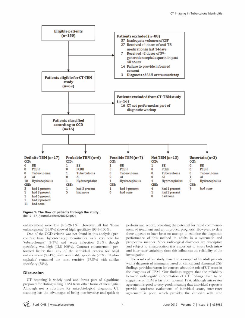
Of the 62 patients assessed over the period of this study, 46 met
inclusion criteria (See Figure 1). Patient demographics were as
follows: 22 male and 24 female; mean age of 33 years (median 31,
range 15–57). Human Immunodeficiency Virus (HIV) status was
known in 35 (out of 46) patients, and 20 were HIV positive and 15,
HIV negative. The status of patients at three months was as
follows: alive (30), dead (12) and unknown (4). Of the unknown
cases, 2 had been classified as ‘Definite TBM’, 1 as ‘Probable
TBM’ and 1 as ‘Unknown’. For the sixteen patients without brain
imaging studies excluded from this analysis, the final diagnoses
were: Definite TBM (3), Probable TBM (1), Possible TBM (2),
Uncertain (2) and Not TBM (6).
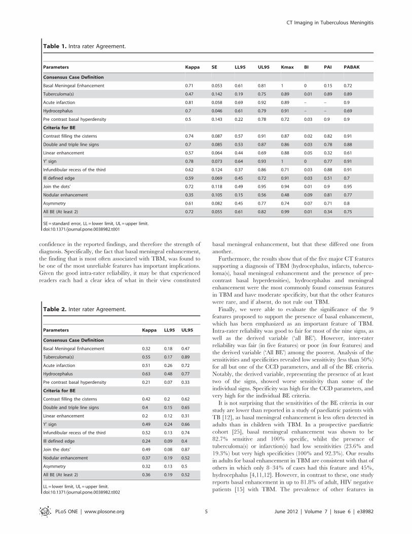
Intra-rater reliabilityComplete results for intra-rater reliability are presented in
Table 1. The reliability of the criteria for basal meningeal
enhancement varied widely, and prevalence of asymmetry was
high for all variables. Good agreement was found for ‘contrast
filling the cisterns’ ‘double and triple line signs’, ‘‘Y’ sign’, ‘join the
dots’, ‘infundibular recess of the third’ and ‘asymmetry’, with all of
the adjusted values suggesting very good agreement. Most of the
criteria for basal meningeal enhancement were significantly
affected by prevalence asymmetry, with adjusted kappas (PABAK)
showing very good agreement for all but three of the parameters.
As for the CCD criteria, ‘acute infarction’ showed very good
agreement, whilst ‘basal meningeal enhancement’ and ‘hydro-
cephalus’ had good agreement that did not change significantly
with adjustment.
Inter-rater reliabilityIn the assessment of inter-rater reliability, the criteria for basal
enhancement (see Table 2), ‘contrast filling the cisterns’ ‘double
and triple line signs’ ‘Y’ sign’, ‘infundibular recess of the third’ and
‘join the dots’ performed the best, showing moderate agreement.
For ‘All BE’ agreement was only fair (0.36, 95%CI 0.19–0.52).
The CCD parameters performed only slightly better, with
‘hydrocephalus’ showing good agreement and ‘tuberculoma(s) and
‘acute infarction’ showing moderate agreement. Pair-wise analysis
failed to identify a particular rater as the cause of the poor
agreement observed.
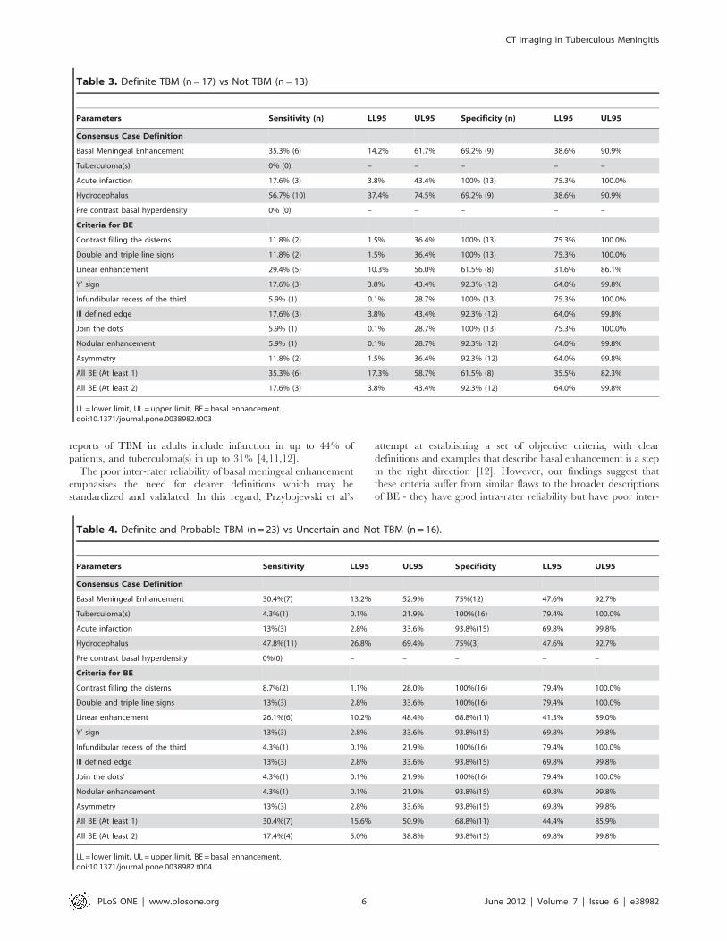
Sensitivity and Specificity.In the analysis of ‘definite TBM’ cases compared with cases of
‘not TBM’ (see Table 3), individual criteria for basal enhancement
were insensitive (0–29.4%). However, when taken individually, all
but ‘linear enhancement’ (61.5%) showed high specificity (92.3–
100%).
Two of the consensus criteria were not present at all in this
cohort (‘tuberculoma(s)’ and ‘pre-contrast basal hyperdensity’).
‘Contrast enhancement’ as defined in the CCD was more sensitive
(35.3%) than any of the individual criteria of basal enhancement
or ‘all BE’, but less specific (69.2%). ‘Acute infarction’ was
insensitive (17.6%) but very specific (100%). ‘Hydrocephalus’ was
the most sensitive (56.7%) with only moderate specificity (69.2%).
Analysis of ‘definite TBM’ and ‘probable TBM’ cases compared
with those classified as ‘uncertain’ and ‘not TBM’ yielded similar
results (see Table 4). Sensitivities of the individual criteria for basal
CT Imaging in Tuberculous Meningitis
PLoS ONE | www.plosone.org 3 June 2012 | Volume 7 | Issue 6 | e38982
enhancement were low (4.3–26.1%). However, all but ‘linear
enhancement’ (68.8%) showed high specificity (93.8–100%).
One of the CCD criteria was not found in this analysis (‘pre-
contrast basal hyperdensity’). Sensitivities were very low for
‘tuberculoma(s)’ (4.3%) and ‘acute infarction’ (13%), though
specificity was high (93.8–100%). ‘Contrast enhancement’ per-
formed better than any of the individual criteria for basal
enhancement (30.4%), with reasonable specificity (75%). ‘Hydro-
cephalus’ remained the most sensitive (47.8%) with similar
specificity (75%).
Discussion
CT scanning is widely used and forms part of algorithms
proposed for distinguishing TBM from other forms of meningitis.
Although not a substitute for microbiological diagnosis, CT
scanning has the advantages of being non-invasive and quick to
perform and report, providing the potential for rapid commence-
ment of treatment and an improved prognosis. However, to date
there appears to have been no attempt to examine the diagnostic
performance of this method in adults in a systematic and
prospective manner. Since radiological diagnoses are descriptive
and subject to interpretation it is important to assess both intra-
and inter-rater variability since this influences the reliability of the
investigation.
The results of our study, based on a sample of 46 adult patients
with a diagnosis of meningitis based on clinical and abnormal CSF
findings, provides reason for concern about the role of CT scans in
the diagnosis of TBM. Our findings suggest that the reliability
between radiologists’ interpretation of CT findings taken to be
suggestive of TBM is far from optimal. First, although intra-rater
agreement is good to very good, meaning that individual reporters
provide consistent evaluations of individual scans, inter-rater
agreement is poor, which provides the clinician with little
Figure 1. The flow of patients through the study.doi:10.1371/journal.pone.0038982.g001
CT Imaging in Tuberculous Meningitis
PLoS ONE | www.plosone.org 4 June 2012 | Volume 7 | Issue 6 | e38982
confidence in the reported findings, and therefore the strength of
diagnosis. Specifically, the fact that basal meningeal enhancement,
the finding that is most often associated with TBM, was found to
be one of the most unreliable features has important implications.
Given the good intra-rater reliability, it may be that experienced
readers each had a clear idea of what in their view constituted
basal meningeal enhancement, but that these differed one from
another.
Furthermore, the results show that of the five major CT features
supporting a diagnosis of TBM (hydrocephalus, infarcts, tubercu-
loma(s), basal meningeal enhancement and the presence of pre-
contrast basal hyperdensities), hydrocephalus and meningeal
enhancement were the most commonly found consensus features
in TBM and have moderate specificity, but that the other features
were rare, and if absent, do not rule out TBM.
Finally, we were able to evaluate the significance of the 9
features proposed to support the presence of basal enhancement,
which has been emphasized as an important feature of TBM.
Intra-rater reliability was good to fair for most of the nine signs, as
well as the derived variable (‘all BE’). However, inter-rater
reliability was fair (in five features) or poor (in four features) and
the derived variable (‘All BE’) among the poorest. Analysis of the
sensitivities and specificities revealed low sensitivity (less than 50%)
for all but one of the CCD parameters, and all of the BE criteria.
Notably, the derived variable, representing the presence of at least
two of the signs, showed worse sensitivity than some of the
individual signs. Specificity was high for the CCD parameters, and
very high for the individual BE criteria.
It is not surprising that the sensitivities of the BE criteria in our
study are lower than reported in a study of paediatric patients with
TB [12], as basal meningeal enhancement is less often detected in
adults than in children with TBM. In a prospective paediatric
cohort [25], basal meningeal enhancement was shown to be
82.7% sensitive and 100% specific, whilst the presence of
tuberculoma(s) or infarction(s) had low sensitivities (23.6% and
19.3%) but very high specificities (100% and 92.3%). Our results
in adults for basal enhancement in TBM are consistent with that of
others in which only 8–34% of cases had this feature and 45%,
hydrocephalus [4,11,12]. However, in contrast to these, one study
reports basal enhancement in up to 81.8% of adult, HIV negative
patients [15] with TBM. The prevalence of other features in
Table 1. Intra rater Agreement.
Parameters Kappa SE LL95 UL95 Kmax BI PAI PABAK
Consensus Case Definition
Basal Meningeal Enhancement 0.71 0.053 0.61 0.81 1 0 0.15 0.72
Tuberculoma(s) 0.47 0.142 0.19 0.75 0.89 0.01 0.89 0.89
Acute infarction 0.81 0.058 0.69 0.92 0.89 – – 0.9
Hydrocephalus 0.7 0.046 0.61 0.79 0.91 – – 0.69
Pre contrast basal hyperdensity 0.5 0.143 0.22 0.78 0.72 0.03 0.9 0.9
Criteria for BE
Contrast filling the cisterns 0.74 0.087 0.57 0.91 0.87 0.02 0.82 0.91
Double and triple line signs 0.7 0.085 0.53 0.87 0.86 0.03 0.78 0.88
Linear enhancement 0.57 0.064 0.44 0.69 0.88 0.05 0.32 0.61
Y’ sign 0.78 0.073 0.64 0.93 1 0 0.77 0.91
Infundibular recess of the third 0.62 0.124 0.37 0.86 0.71 0.03 0.88 0.91
Ill defined edge 0.59 0.069 0.45 0.72 0.91 0.03 0.51 0.7
Join the dots’ 0.72 0.118 0.49 0.95 0.94 0.01 0.9 0.95
Nodular enhancement 0.35 0.105 0.15 0.56 0.48 0.09 0.81 0.77
Asymmetry 0.61 0.082 0.45 0.77 0.74 0.07 0.71 0.8
All BE (At least 2) 0.72 0.055 0.61 0.82 0.99 0.01 0.34 0.75
SE = standard error, LL = lower limit, UL = upper limit.doi:10.1371/journal.pone.0038982.t001
Table 2. Inter rater Agreement.
Parameters Kappa LL95 UL95
Consensus Case Definition
Basal Meningeal Enhancement 0.32 0.18 0.47
Tuberculoma(s) 0.55 0.17 0.89
Acute infarction 0.51 0.26 0.72
Hydrocephalus 0.63 0.48 0.77
Pre contrast basal hyperdensity 0.21 0.07 0.33
Criteria for BE
Contrast filling the cisterns 0.42 0.2 0.62
Double and triple line signs 0.4 0.15 0.65
Linear enhancement 0.2 0.12 0.31
Y’ sign 0.49 0.24 0.66
Infundibular recess of the third 0.52 0.13 0.74
Ill defined edge 0.24 0.09 0.4
Join the dots’ 0.49 0.08 0.87
Nodular enhancement 0.37 0.19 0.52
Asymmetry 0.32 0.13 0.5
All BE (At least 2) 0.36 0.19 0.52
LL = lower limit, UL = upper limit.doi:10.1371/journal.pone.0038982.t002
CT Imaging in Tuberculous Meningitis
PLoS ONE | www.plosone.org 5 June 2012 | Volume 7 | Issue 6 | e38982
reports of TBM in adults include infarction in up to 44% of
patients, and tuberculoma(s) in up to 31% [4,11,12].
The poor inter-rater reliability of basal meningeal enhancement
emphasises the need for clearer definitions which may be
standardized and validated. In this regard, Przybojewski et al’s
attempt at establishing a set of objective criteria, with clear
definitions and examples that describe basal enhancement is a step
in the right direction [12]. However, our findings suggest that
these criteria suffer from similar flaws to the broader descriptions
of BE - they have good intra-rater reliability but have poor inter-
Table 3. Definite TBM (n = 17) vs Not TBM (n = 13).
Parameters Sensitivity (n) LL95 UL95 Specificity (n) LL95 UL95
Consensus Case Definition
Basal Meningeal Enhancement 35.3% (6) 14.2% 61.7% 69.2% (9) 38.6% 90.9%
Tuberculoma(s) 0% (0) – – – – –
Acute infarction 17.6% (3) 3.8% 43.4% 100% (13) 75.3% 100.0%
Hydrocephalus 56.7% (10) 37.4% 74.5% 69.2% (9) 38.6% 90.9%
Pre contrast basal hyperdensity 0% (0) – – – – –
Criteria for BE
Contrast filling the cisterns 11.8% (2) 1.5% 36.4% 100% (13) 75.3% 100.0%
Double and triple line signs 11.8% (2) 1.5% 36.4% 100% (13) 75.3% 100.0%
Linear enhancement 29.4% (5) 10.3% 56.0% 61.5% (8) 31.6% 86.1%
Y’ sign 17.6% (3) 3.8% 43.4% 92.3% (12) 64.0% 99.8%
Infundibular recess of the third 5.9% (1) 0.1% 28.7% 100% (13) 75.3% 100.0%
Ill defined edge 17.6% (3) 3.8% 43.4% 92.3% (12) 64.0% 99.8%
Join the dots’ 5.9% (1) 0.1% 28.7% 100% (13) 75.3% 100.0%
Nodular enhancement 5.9% (1) 0.1% 28.7% 92.3% (12) 64.0% 99.8%
Asymmetry 11.8% (2) 1.5% 36.4% 92.3% (12) 64.0% 99.8%
All BE (At least 1) 35.3% (6) 17.3% 58.7% 61.5% (8) 35.5% 82.3%
All BE (At least 2) 17.6% (3) 3.8% 43.4% 92.3% (12) 64.0% 99.8%
LL = lower limit, UL = upper limit, BE = basal enhancement.doi:10.1371/journal.pone.0038982.t003
Table 4. Definite and Probable TBM (n = 23) vs Uncertain and Not TBM (n = 16).
Parameters Sensitivity LL95 UL95 Specificity LL95 UL95
Consensus Case Definition
Basal Meningeal Enhancement 30.4%(7) 13.2% 52.9% 75%(12) 47.6% 92.7%
Tuberculoma(s) 4.3%(1) 0.1% 21.9% 100%(16) 79.4% 100.0%
Acute infarction 13%(3) 2.8% 33.6% 93.8%(15) 69.8% 99.8%
Hydrocephalus 47.8%(11) 26.8% 69.4% 75%(3) 47.6% 92.7%
Pre contrast basal hyperdensity 0%(0) – – – – –
Criteria for BE
Contrast filling the cisterns 8.7%(2) 1.1% 28.0% 100%(16) 79.4% 100.0%
Double and triple line signs 13%(3) 2.8% 33.6% 100%(16) 79.4% 100.0%
Linear enhancement 26.1%(6) 10.2% 48.4% 68.8%(11) 41.3% 89.0%
Y’ sign 13%(3) 2.8% 33.6% 93.8%(15) 69.8% 99.8%
Infundibular recess of the third 4.3%(1) 0.1% 21.9% 100%(16) 79.4% 100.0%
Ill defined edge 13%(3) 2.8% 33.6% 93.8%(15) 69.8% 99.8%
Join the dots’ 4.3%(1) 0.1% 21.9% 100%(16) 79.4% 100.0%
Nodular enhancement 4.3%(1) 0.1% 21.9% 93.8%(15) 69.8% 99.8%
Asymmetry 13%(3) 2.8% 33.6% 93.8%(15) 69.8% 99.8%
All BE (At least 1) 30.4%(7) 15.6% 50.9% 68.8%(11) 44.4% 85.9%
All BE (At least 2) 17.4%(4) 5.0% 38.8% 93.8%(15) 69.8% 99.8%
LL = lower limit, UL = upper limit, BE = basal enhancement.doi:10.1371/journal.pone.0038982.t004
CT Imaging in Tuberculous Meningitis
PLoS ONE | www.plosone.org 6 June 2012 | Volume 7 | Issue 6 | e38982
rater reliability, which reduces confidence in the contribution of
these radiological signs to the diagnosis of TBM.
Somewhat concerning is the lack of reliability for both acute
infarction - often taken to be of prognostic value - and
hydrocephalus, which might signal the need for surgical in-
tervention. Although the numbers are small, all three patients with
acute infarction had definite TBM when compared with those who
did not have TBM, and this sign may be useful to rule in TBM in
the right context.
The strength of the current study lies in its prospective nature.
Most studies assessing the value of CT in the diagnosis of TBM
have been retrospective. Consequently they have not been able to
standardize imaging techniques, the level of training of the
imaging reviewers, and the timing of the imaging study in relation
to the clinical presentation. Furthermore, the methods for seeking
microbiological confirmation of TBM, the gold standard, have
varied, and if suboptimal, might have created a bias favouring
selection of more severe cases. Similarly, controls were not
necessarily selected for the similarity of their clinical presentations
– the true clinical dilemma. Instead, in some studies, patients with
an established known alternative diagnosis that presented during
the same period and had both imaging and CSF analysis, have
been used as controls [12–14].
There are several limitations to this study: the small sample size
does not allow for a comparison of HIV infected versus uninfected
patients, and it is possible that the imaging criteria may be
different in immunocompromised HIV positive patients. Secondly,
patients with TBM and a normal CSF were excluded (though this
was probably a small number of cases). Finally, the decision to
perform a CT scan in each patient was not according to standard
criteria, but at the judgment of the attending physician. Although
perhaps not ideal for a clinical study, this represented standard
conditions in the study context (a large public referral hospital
serving a community with high risk for tuberculosis).
Given that the sensitivity of CT imaging findings in TBM in our
series of adult patients was low, and given the lack of reliability of
reporting of the features of basal enhancement, the use of separate
scoring systems for patients who have had imaging and those that
have not must be questioned. Those with imaging are likely to
score lower and might be falsely assigned a lower probability of
having TBM. On the basis of this study, we recommend that the
same scoring system should be used for both categories, that is,
regardless of the presence of the CT findings, until the reliability
and test performance of CT findings are more widely assessed in
a number of relevant clinical settings. In addition, clinicians should
be made aware of the need to interpret CT findings in suspected
TBM with caution and to rely on the other diagnostic criteria for
the presence of TBM. Specifically, it must be emphasized that
a normal CT brain scan is not uncommon in early TBM in adult
patients, and clinicians should not be falsely reassured by this
finding.
Despite the widespread use of CT imaging findings in the
diagnosis of tuberculous meningitis, few studies have examined the
reliability and diagnostic performance of the most common
findings among adults. Based on a prospective cohort of patients
with a clinical diagnosis of meningitis, we have shown that the
imaging parameters included in a recent consensus case definition,
including basal meningeal enhancement, suffer from poor inter-
rater reliability and poor to moderate sensitivity and specificity
among adult patients. Furthermore, a recent set of criteria which
aimed to allow for more objective diagnosis of basal meningeal
enhancement was found to be very insensitive in an adult
population, and suffered from poor inter-rater reliability. Whilst
our numbers are relatively small, these findings suggest that
imaging findings should be interpreted with caution, and that
further work is needed to derive imaging criteria which are more
reliable.
Supporting Information
Table S1 Consensus tuberculous meningitis diagnosis.This table from Marais et al. [9] lists the scoring system for the
classification of suspected tuberculous meningitis patients into
definite, probable, possible or not TBM.
(DOCX)
Acknowledgments
The authors would like to thank the patients who participated and the staff
of Tygerberg Hospital who cared for the patients. In addition, the authors
thank Deborah Purdy and Wandile Guma for their help with the
anonymisation and blinding of the radiology.
Author Contributions
Conceived and designed the experiments: KJB HB. Performed the
experiments: CA SC SGR. Analyzed the data: KJB HB JAC. Contributed
reagents/materials/analysis tools: KJB HB JAC. Wrote the paper: CA KJB
HB SC JAC SGR.
References
1. Christensen A-SH, Andersen AB, Thomsen VO, Andersen PH, Johansen IS
(2011) Tuberculous meningitis in Denmark: a review of 50 cases. BMC InfectDis 11: 47.
2. Sheu J-J, Yuan R-Y, Yang C-C (2009) Predictors for outcome and treatment
delay in patients with tuberculous meningitis. Am J Med Sci 338: 134–139.
3. Chapp-Jumbo EN (2006) Neurologic infections in a Nigerian university teachinghospital. African health sciences 6: 55–58.
4. Hosoglu S, Geyik MF, Balik I, Aygen B, Erol S, et al. (2002) Predictors of
outcome in patients with tuberculous meningitis. Int J Tuberc Lung Dis 6: 64–70.
5. Thwaites G, Fisher M, Hemingway C, Scott G, Solomon T, et al. (2009) BritishInfection Society guidelines for the diagnosis and treatment of tuberculosis of the
central nervous system in adults and children. J Infect 59: 167–187.
6. Jarvis JN, Meintjes G, Williams A, Brown Y, Crede T, et al. (2010) Adultmeningitis in a setting of high HIV and TB prevalence: findings from 4961
suspected cases. BMC Infect Dis 10: 67. doi:10.1186/1471–2334–10–67.
7. Thwaites GE, Tran TH (2005) Tuberculous meningitis: many questions, too fewanswers. Lancet neurology 4: 160–170.
8. Thwaites GE, Schoeman JF (2009) Update on tuberculosis of the central nervous
system: pathogenesis, diagnosis, and treatment. Clin Chest Med 30: 745–754, ix.
9. Marais S, Thwaites G, Schoeman JF, Torok ME, Misra UK, et al. (2010)
Tuberculous meningitis: a uniform case definition for use in clinical research.
The Lancet Infectious Diseases 3099: 1–10.
10. Ozates M, Kemaloglu S, Gurkan F, Ozkan U, Hosoglu S, et al. (2000) CT of the
brain in tuberculous meningitis. A review of 289 patients. Acta Radiol 41: 13–
17.
11. Kalita J, Misra UK, Ranjan P (2007) Predictors of long-term neurological
sequelae of tuberculous meningitis: a multivariate analysis. Eur J Neurol 14: 33–
37.
12. Przybojewski S, Andronikou S, Wilmshurst J (2006) Objective CT criteria to
determine the presence of abnormal basal enhancement in children with
suspected tuberculous meningitis. Pediatr Radiol 36: 687–696.
13. Theron S, Andronikou S, Grobbelaar M, Steyn F, Mapukata A, et al. (2006)
Localized basal meningeal enhancement in tuberculous meningitis. Pediatr
Radiol 36: 1182–1185.
14. Andronikou S, Smith B, Hatherhill M, Douis H, Wilmshurst J (2004) Definitive
neuroradiological diagnostic features of tuberculous meningitis in children.
Pediatr Radiol 34: 876–885.
15. Katrak SM, Shembalkar PK, Bijwe SR, Bhandarkar LD (2000) The clinical,
radiological and pathological profile of tuberculous meningitis in patients with
and without human immunodeficiency virus infection. J Neurol Sci 181: 118–
126.
16. Torok ME, Nghia HDT, Chau TTH, Mai NTH, Thwaites GE, et al. (2007)
Validation of a diagnostic algorithm for adult tuberculous meningitis. Am J Trop
Med Hyg 77: 555–559.
CT Imaging in Tuberculous Meningitis
PLoS ONE | www.plosone.org 7 June 2012 | Volume 7 | Issue 6 | e38982
17. Thwaites GE, Chau TTH, Stepniewska K, Phu NH, Chuong LV, et al. (2002)
Diagnosis of adult tuberculous meningitis by use of clinical and laboratoryfeatures. Lancet 360: 1287–1292.
18. Marais S, Pepper DJ, Schutz C, Wilkinson RJ, Meintjes G (2011) Presentation
and outcome of tuberculous meningitis in a high HIV prevalence setting. PloSone 6: e20077.
19. Thwaites GE, Chau TTH, Farrar JJ (2004) Improving the BacteriologicalDiagnosis of Tuberculous Meningitis. Journal of Clinical Microbiology 42: 378–
379.
20. Dunn G (1992) Design and Analysis of Reliability Studies: The StatisticalEvaluation of Measurement Errors. John Wiley & Sons, Incorporated.
21. Byrt T, Bishop J, Carlin JB (1993) Bias, prevalence and kappa. J Clin Epidemiol
46: 423–429.
22. Reichenheim ME (2004) Confidence intervals for the kappa statistic. Stata
Journal: 2004–2004.
23. Altman DG (1991) Practical statistics for medical research. Chapman and Hall.
24. Abramson JH (2011) WINPEPI updated: computer programs for epidemiolo-
gists, and their teaching potential. Epidemiologic perspectives & innovations:
EP+I 8: 1.25. Kumar R, Kohli N, Thavnani H, Kumar A, Sharma B (1996) Value of CT scan
in the diagnosis of meningitis. Indian Pediatr 33: 465–468.
CT Imaging in Tuberculous Meningitis
PLoS ONE | www.plosone.org 8 June 2012 | Volume 7 | Issue 6 | e38982