Embed Size (px)
Citation preview
Perunika 8(2), 281 - 287 (1985)
Relationship Between Watermelon Mosaic Virus (WMV-1) andWatermelon Mosaic Virus 2 (WMV-2) on the basis of
Nucleotide Sequence Homology
NORANI ABU-SAMAHDepartment of Biochemistry and Microbiology,Faculty of Science and Environmental Studies,
Universiti Pertanian Malaysia,Serdang, Selangor, Malaysia.
Key words: Potyvirus; immune electron microscopy; molecular hybridization analysis.
ABSTRAK
Kedua ahli subkumpulan 'watermelon mosaic vines', WMV-1 dan WMV-2 dianggap sebagaivirus-virus yang berlainan berdasarkan kepada bidang hos dan serologi. Immun mikroskop elektrondengan penunjuk pembezaan indeks 2 he 3 mengsahkan perbezaan serologi ini. Analisis hibridisasimolikul dengan menggunakan DNA berkomplimen yang disediakan oleh transkripsi berbalik RNAWMV-1 berprima rawak, menunjukkan tiada homologi di antara WMV-1 dan WMV-2 dan DNAberkomplimen juga menunjukkan tiada homologi dengan RNA dari beberapa 'potyvirus' yangmenjangkiti kekacang dan 'potato virus Y' (PVY). Analisa hibridisasi molikul selanjutnya meng-sahkan yang WMV-1 dan WMV-2 adalah virus-virus yang nyata berbeza.
ABSTRACT
The two members of the watermelon mosaic virus subgroup, WMV-1 and WMV-2, wereconsidered to be different viruses on the basis of host range and serology. Immune electron microscopywith serological differentiation indices of 2 to 3 confirmed this serological difference. Molecularhybridization analysis using complementary DNA prepared by reverse transcription of randomlyprimed WMV-1 RNA showed no homology between WMV-1 and WMV-2 and the complementaryDNA from a range of legume infecting potyvir uses and potato virus Y (PVY). Molecular hybridizationanalysis further confirmed that WMV1 and WMV-2 are distinct viruses.
INTRODUCTION
Watermelon mosaic virus — 1 (WMV-1)and watermelon mosaic virus — 2 (WMV-2)have been differentiated on their biological andserological properties. Using agar double-diffu-sion tests, Milne and Grogan (1969) found aclose serological relationship between variousisolates of WMV-1 and WMV-2 and concludedthat they were strains of WMV even though theirhost ranges differed.
byPurcifull and Hiebert (1979) indicated thatSDS-immunodiffusion tests the Florida
isolates of WMV-1 and WMV-2 were serolo-gically distinct as no cross-reactions were detect-ed.
The Queensland isolates of WMV-1 andWMV-2 were found to have some distinct hostrange differences. WMV-2 Q has a much widerhost range as compared to WMV-1 Q (Greber,1978). They were serologically distinct from oneanother but closely related to Florida isolates ofthe respective types using the SDS-immunodiffu-sion tests with crude sap (Greber, 1978). Physicalproperties of these WMV isolates and electronmicroscopic examination of the virus particles
NORANI ABU-SAMAH
and inclusions were found to be similar to theother isolates reported elsewhere.
Makkouk and Lesemann (1980) found thattheir WMV-1 reacted with the Florida isolate ofWMV-1 but not with WMV-2 Florida isolateantiserum when tested in SDS-immunodiffusiontests and with the decoration technique ofimmune electron microscopy.
Inconsistencies in the reported relationshipsbetween WMV-1 and WMV-2 probably resultedfrom the use of different strains and techniques.
In this paper, the relationship betweenWMV-1 and WMV-2 was reexamined bymolecular hybridization analysis (MHA) whichdiscriminates isolates and strains of the potyvirus,bean yellow mosaic virus (BYMV) (Abu-Samah,1982; Abu-Samah & Randies, 1981; Abu-Samah& Randies, 1983). Molecular hybridizationanalysis (MHA) allows more sensitive discrimina-tion and provides a semi-quantitative estimatebetween closely related isolates. But MHA is toospecific to show relationships between distantlyrelated strains or different viruses. Therefore inthis study serological relationships were alsoexamined by immune electron microscopy(IEM).
MATERIALS AND METHODS
Sources of Virus
Isolates of WMV-1 and WMV-2 weresupplied by a government department* in Indo-oroopilly, Queensland and maintained in theglasshouse on Cucumis satixrus cv. Polaris. In thispaper, these isolates are referred to as WMV-1Qand WMV-2 Q,
Purification of WMV-1 Qa,nd WMV-2 andTheir RNAs
Purification of WMV-1 Q and WMV-2 Qwas from systemically infected leaves of C.sativus cv. Polaris harvested 2 — 3 weeks afterinoculation. Both viruses were purified by
methods based on those of Purcifull and Hiebert(1979) with modifications of the steps after thepolyethylene glycol (PEG 6000) concentrationstep.
For WMV-1 Q, the PEG pellets were resus-pended in 0.05 M potassium phosphate buffer,pH 7.5 The virus was centrifuged through 1 mlof a 30% buffered sucrose cushion in a Spinco 65rotor at 78,000 g for 105 min. The pellet wasresuspended in water.
For WMV-2 Q, after the precipitation stepwith PEG and sedimentation by high speedcentrifugation as above, the virus pellet wasresuspended in 0.05 M potassium phosphatebuffer, pH 7.5. The resuspended material wassubjected to centrifugation in a CsCl gradient( p = 1.28 g/ml) using the SW 50 rotor at40,000 rpm for 18 hrs. The virus containing zonewas removed, diluted with buffer and wasfurther subjected to high speed centrifugation at78,000 g in the Spinco 65 rotor for 120 min. Thepellet was resuspended in water.
The RNAs were extracted from the virussuspension by the pronase procedure as describ-ed for BYMV-RNA (Abu-Samah & Randies,1981). Only WMV-1 was purified furtherthrough two cycles of sucrose density gradients(Abu-Samah & Randies, 1981).
Comparison by Immune Electron Microscopy(IEM)
The antisera of the Florida isolate o]WMV-1 (WMV-1 Fl) and WMV-2 (WMV-2 FTused in this study were kindly provided by DrD.E. Purcifull, University of Florida, Gainesville. The Florida antisera were used in antibodydecoration and dilution end-point clumpinjIEM tests to compare WMV-1 Qand WMV-2 ((Milne and Luisoni, 1977).
Molecular Hybridization Analysis (MHA)
The random primer method of Taylor tal.} (1976) as used for BYMV-RNA (Abu-Sama
•Supplied by Mr. R.S. Greber of the Department of Primary Industries.
282 PERTANIKA VOL. 8 NO. 2, 1985
COMPARISON OF WMVRNA
& Randies, 1981) was used to synthesize comple-mentary DNA (cDNA) to WMV-1. Thehybridization solution contained 0.01 M Tris-HC1, pH 7.0, 0.18 M NaCl, 1 mM EDTA and0.05% SDS (Gould & Symons, 1977; Abu-Samah& Randies, 1981).
Hybridization was carried out in siliconizedglass test tubes, approx. 7 X 50 mm with reactionmixtures overlaid with paraffin oil. To 40 jul ofthe appropriately diluted RNA solution wasadded 2 )Ltl of [ 3H] cDNA (c. 2000 cpm). Thereaction mixtures were immersed in boilingwater for 2 — 3 mins and then incubated at 65 -66°C. Hybridizations were terminated by chill-ing the tubes, removing 30 fil aliquots, andadding them to 300 /jt 1 of a low salt S (assay buffer(0.03 M sodium acetate, 0.05 M NaCl, 1 mMZnSO4, 5% glycerol, pH 4.6) containing 40jug/ml of denaturated calf thymus DNA. Twosamples, each 150 jul, were taken and to one wasadded 2 units of S x nuclease; the other was left asa control. After incubation of both samples at45°C for 30 mins, nuclease resistence was deter-mined by comparing the duplicates incubatedeither with or without enzymes, as described byGould & Symons (1977).
The T mof DNA-RNA hybrid was set up in atotal vol. of 100 jLtl and incubated at 65° to a Ro tvalue exceeding 1.0 mol sec liter '. The mixturewas chilled and placed in a waterbath in whichthe temperature was raised by approx. 1°C permin. At the appropriate temperatures, 5 JULIportions were transferred to 150 fx\ of cold S ]
buffer and the percentage hybrids remaining ateach temperature was determined by S (nucleaseresistence.
RESULTS
Comparison by IEM
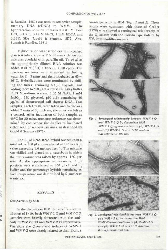
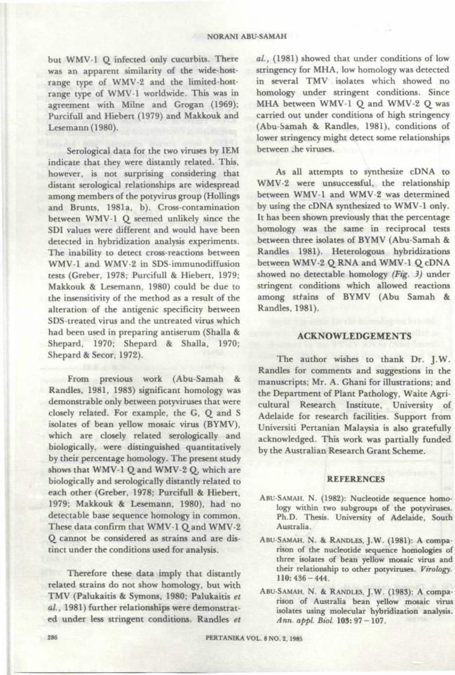
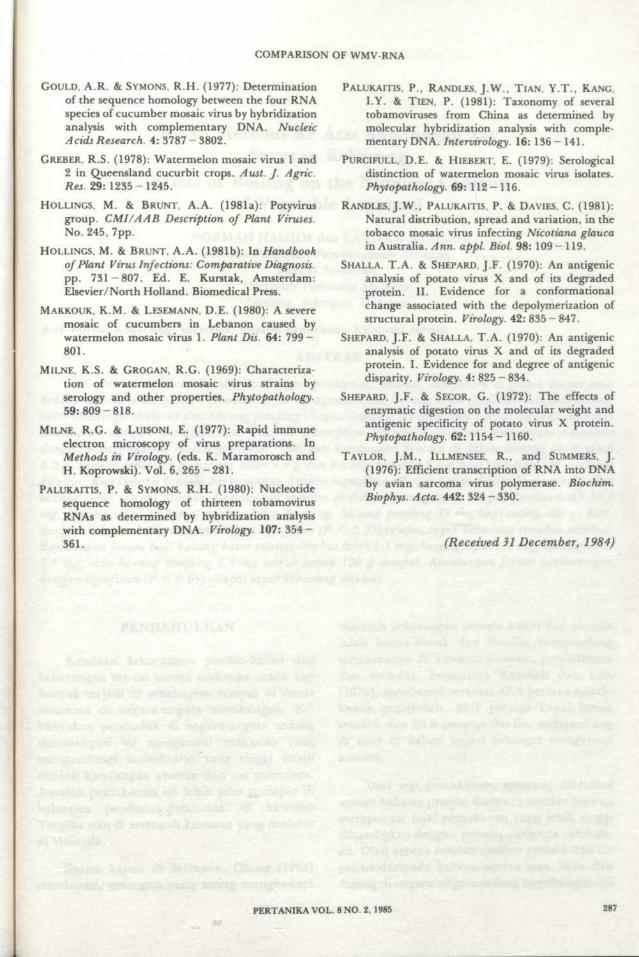
In the decoration IEM test at an antiserumdilution of 1/10, both WMV-1 Qand WMV-2 Qparticles were heavily decorated with the anti-body of WMV-1 Fl and WMV-2 Fl respectively.Therefore the Queensland isolates of WMV-1and WMV-2 were closely related to their Florida
counterparts using IEM (Figs. 1 and 2). Theseresults were consistent with those of Greber(1978) who showed a serological relationship ofthe Q isolates with the Florida type isolates bySDS-immunodiffusion tests.
Fig. 1. Serological relationship between WMV-1 Qand WMV-2 Q. by decoration IEM.WMV-1 Q against antisera to (A) WMV-1 Fland (B) WMV-2 Flat a 1/10 dilution.Bar represents 500 nm.
Fig. 2. Serological relationship between WMV-2and WMV-1 Q by decoration IEM.WMV-2 against antisera to (A) WMV-2 Fland (B) WMV-1 Flat a 1/10 dilution.Bar represents 500 nm.
PERTANIKA VOL. 8 NO. 2. 1985 283
NORANI ABU SAMAH
At the antiserum dilution used, cross-re-actions were detected in reciprocal tests,although with much less decoration for theheterologous tests. The dilution end-points ofboth antisera with both WMV-1 Qand WMV-2 Qby the clumping IEM test are presented in Table1. The serological differentiation index betweenWMV-1 Fl antiserum and WMV-2 Qwas 2 andbetween WMV-2 Fl antiserum and WMV-1 Qwas 3. These antisera failed to decorate the beanisolate of tobacco mosaic virus (B-TMV) andantiserum to Ul isolate of tobacco mosaic virus(Ul-TMV) failed to decorate WMV-1 Q orWMV-2 Q.
TABLE 1Titres of WMV-1 Fl and WMV-2 Fl antisera to
WMV-1 Q and WMV-2 Q, as determined byclumping IEM
Antiserum
WMV-1 Fla
WMV-2 FT
U , - T M V b
WMV-1 Q
2048
512
0
Antigens
I WMV-2 Q
512
4096
0
B-TMV
0
0
0
Antisera kindly supplied by Dr. D.E. Purcifull(a), and Dr.R.I.B. Francki(b).
Figures in the Table are reciprocal titres of antisera.
Comparison by Molecular HybridizationAnalysis (MHA)
Reverse transcription of WMV-1 RNA gavecDNA with 2 X 10⁶count/min (13.4 ng) for aninput of 2 jug of RNA. The specificity of thecDNA is illustraded in Table 2; hybridization ofthe cDNA with other viral RNAs or with nucleicacid extracted from healthy cucumber was notobserved.
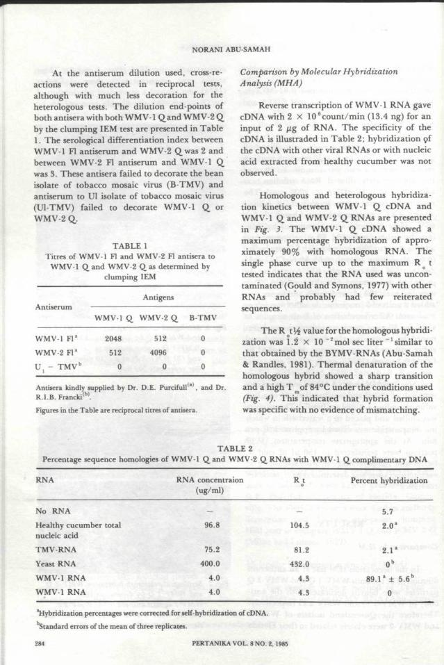
Homologous and heterologous hybridiza-tion kinetics between WMV-1 Q cDNA andWMV-1 Q and WMV-2 Q RNAs are presentedin Fig. 3. The WMV-1 Q cDNA showed amaximum percentage hybridization of appro-ximately 90 % with homologous RNA. Thesingle phase curve up to the maximum R Q ttested indicates that the RNA used was uncon-taminated (Gould and Symons, 1977) with otherRNAs and probably had few reiteratedsequences.
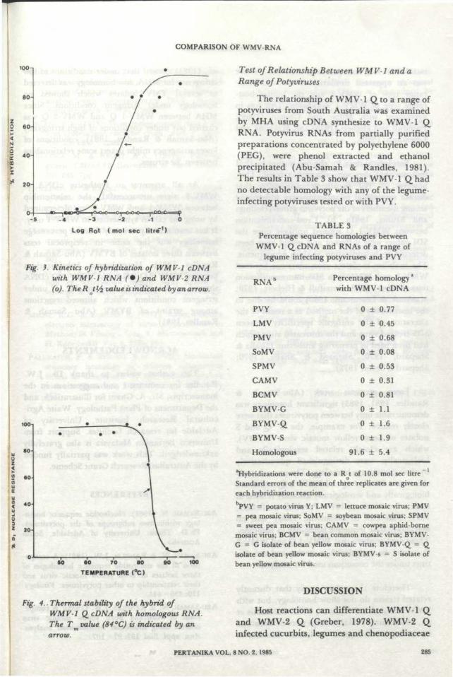
The R t\£ value for the homologous hybridi-zation was 1.2 X 10 ~2mol sec liter ^similar tothat obtained by the BYMV-RNAs (Abu-Samah& Randies, 1981). Thermal denaturation of thehomologous hybrid showed a sharp transitionand a high T of 84°C under the conditions used(Fig. 4). This indicated that hybrid formationwas specific with no evidence of mismatching.
TABLE 2Percentage sequence homologies of WMV-1 Q and WMV-2 Q RNAs with WMV-1 Q complimentary DNA
RNA RNA concentraion(ug/ml)
Percent hybridization
No RNAHealthy cucumber totalnucleic acid
TMV-RNA
Yeast RNA
WMV-1 RNA
WMV-1 RNA
96.8
75.2
400.0
4.0
4.0
104.5
81.2
432.0
4.3
4.3
5.7
2.0a
2.1 a
89.l a ± 5.6b
0
hybridization percentages were corrected for self-hybridization of cDNA.bStandard errors of the mean of three replicates.
284 PERTANIKA VOL. 8 NO. 2, 1985
COMPARISON OF WMVRNA
100n
8 0
CO
6 0
- 5 -4 -3 -2 -1 0
Log Rot ( mol sec litre"1)
Fig. 3. Kinetics of hybridization of WMV-1 cDNAwith WMV 1 RNA (•) and WMV-2 RNA(o). TheR tVz value is indicated by an arrow.
8 0
60-
4 0
50 60 70 80
TEMPERATURE (°C)
9 0 100
Fig. 4.. Thermal stability of the hybrid ofWMV-1 QcDNA with homologous RNA.The T value (84 °C) is indicated by anarrow.
Test of Relationship Between WMV-1 and aRange of Potyviruses
The relationship of WMV-1 Qto a range ofpotyviruses from South Australia was examinedby MHA using cDNA synthesize to WMV-1 QRNA. Potyvirus RNAs from partially purifiedpreparations concentrated by polyethylene 6000(PEG), were phenol extracted and ethanolprecipitated (Abu-Samah & Randies, 1981).The results in Table 3 show that WMV-1 Qhadno detectable homology with any of the legume-infecting potyviruses tested or with PVY.
TABLE 3Percentage sequence homologies between
WMV-1 QcDNA and RNAs of a range oflegume infecting potyviruses and PVY
RNAb
PVY
LMV
PMV
SoMV
SPMV
CAMV
BCMV
BYMV-G
BYMV-Q
BYMV-S
Homologous
Percentage homology3
with WMV-1 cDNA
0 ± 0.77
0 ± 0.45
0 ± 0.68
0 ± 0.08
0 ± 0.53
0 ± 0.31
0 ± 0.81
0 ± 1.1
0 ± 1.6
0 ± 1.9
91.6 ± 5.4
hybridizations were done to a R t of 10.8 mol sec litreStandard errors of the mean of three replicates are given foreach hybridization reaction.bPVY = potato virus Y; LMV = lettuce mosaic virus; PMV= pea mosaic virus; SoMV = soybean mosaic virus; SPMV= sweet pea mosaic virus; CAMV = cowpea aphid-bornemosaic virus; BCMV = bean common mosaic virus; BYMV-G = G isolate of bean yellow mosaic virus; BYMV-Q = Qisolate of bean yellow mosaic virus; BYMV-s = S isolate ofbean yellow mosaic virus.
DISCUSSION
Host reactions can differentiate WMV-1 Qand WMV-2 Q (Greber, 1978). WMV-2 Qinfected cucurbits, legumes and chenopodiaceae
PERTANIKA VOL. 8 NO. 2, 1985 285
NORANI ABU-SAMAH
but WMV-1 Q infected only cucurbits. Therewas an apparent similarity of the wide-host-range type of WMV-2 and the limited-host-range type of WMV-1 worldwide. This was inagreement with Milne and Grogan (1969);Purcifull and Hiebert (1979) and Makkouk andLesemann(1980).
Serological data for the two viruses by IEMindicate that they were distantly related. This,however, is not surprising considering thatdistant serological relationships are widespreadamong members of the potyvirus group (Hollingsand Brunts, 1981a, b). Cross-contaminationbetween WMV-1 Q seemed unlikely since theSDI values were different and would have beendetected in hybridization analysis experiments.The inability to detect cross-reactions betweenWMV-1 and WMV-2 in SDS-immunodiffusiontests (Greber, 1978; Purcifull & Hiebert, 1979;Makkouk & Lesemann, 1980) could be due tothe insensitivity of the method as a result of thealteration of the antigenic specificity betweenSDS-treated virus and the untreated virus whichhad been used in preparing antiserum (Shalla &Shepard, 1970; Shepard & Shalla, 1970;Shepard & Secor, 1972).
From previous work (Abu-Samah &Randies, 1981, 1983) significant homology wasdemonstrable only between potyviruses that wereclosely related. For example, the G, Q and Sisolates of bean yellow mosaic virus (BYMV),which are closely related serologically andbiologically, were distinguished quantitativelyby their percentage homology. The present studyshows that WMV-1 Qand WMV-2 Q, which arebiologically and serologically distantly related toeach other (Greber, 1978; Purcifull & Hiebert,1979; Makkouk & Lesemann, 1980), had nodetectable base sequence homology in common.These data confirm that WMV-1 Q,and WMV-2Q cannot be considered as strains and are dis-tinct under the conditions used for analysis.
Therefore these data imply that distantlyrelated strains do not show homology, but withTMV (Palukaitis & Symons, 1980; Palukaitis etaL, 1981) further relationships were demonstrat-ed under less stringent conditions. Randies et
al, (1981) showed that under conditions of lowstringency for MHA, low homology was detectedin several TMV isolates which showed nohomology under stringent conditions. SinceMHA between WMV-1 Q and WMV-2 Q, wascarried out under conditions of high stringency(Abu-Samah & Randies, 1981), conditions oflower stringency might detect some relationshipsbetween .he viruses.
As all attempts to synthesize cDNA toWMV-2 were unsuccessful, the relationshipbetween WMV-1 and WMV-2 was determinedby using the cDNA synthesized to WMV-1 only.It has been shown previously that the percentagehomology was the same in reciprocal testsbetween three isolates of BYMV (Abu-Samah &Randies 1981). Heterologous hybridizationsbetween WMV-2 QRNA and WMV-1 QcDNAshowed no detectable homology (Fig. 3) understringent conditions which allowed reactionsamong strains of BYMV (Abu Samah &Randies, 1981).
ACKNOWLEDGEMENTS
The author wishes to thank Dr. J. W.Randies for comments and suggestions in themanuscripts; Mr. A. Ghani for illustrations; andthe Department of Plant Pathology, Waite Agri-cultural Research Institute, University ofAdelaide for research facilities. Support fromUniversiti Pertanian Malaysia is also gratefullyacknowledged. This work was partially fundedby the Australian Research Grant Scheme.
REFERENCES
ABU-SAMAH, N. (1982): Nucleotide sequence homo-logy within two subgroups of the potyviruses.Ph.D. Thesis. University of Adelaide, SouthAustralia.
ABU-SAMAH, N. & RANDLES, J.W. (1981): A compa-rison of the nucleotide sequence homologies ofthree isolates of bean yellow mosaic virus andtheir relationship to other potyviruses. Virology.110:436-444.
ABU-SAMAH, N. & RANDLES, J.W. (1983): A compa-rison of Australia bean yellow mosaic virusisolates using molecular hybridization analysis.Ann. appl. Biol 103: 97 - 107.
PERTANIKA VOL. 8 NO. 2, 1985
COMPARISON OF WMV-RNA
GOULD, A.R. & SYMONS, R.H. (1977): Determinationof the sequence homoiogy between the four RNAspecies of cucumber mosaic virus by hybridizationanalysis with complementary DNA. NucleicAcids Research. 4: 3787 - 3802.
GREBER, R.S. (1978): Watermelon mosaic virus 1 and2 in Queensland cucurbit crops. Aust. f. Agric.Res. 29:1235-1245.
HOLLINGS, M. & BRUNT, A.A. (1981a): Potyvirusgroup. CMI/AAB Description of Plant Viruses.No. 245, 7pp.
HOLLINGS, M. & BRUNT, A. A. (1981b): In Handbookof Plant Virus Infections: Comparative Diagnosis.pp. 731-807. Ed. E. Kurstak, Amsterdam:Elsevier/North Holland. Biomedical Press.
MAKKOUK, K.M. & LESEMANN, D.E. (1980): A severemosaic of cucumbers in Lebanon caused bywatermelon mosaic virus 1. Plant Dis. 64: 799 -801.
MILNE, K.S. & GROGAN, R.G. (1969): Characteriza-tion of watermelon mosaic virus strains byserology and other properties. Phytopathology.59:809-818.
MILNE, R.G. & LUISONI, E. (1977): Rapid immuneelectron microscopy of virus preparations. InMethods in Virology, (eds. K. Maramorosch andH. Koprowski). Vol. 6, 265 - 281.
PALUKAITIS, P. & SYMONS, R.H. (1980): Nucleotidesequence homoiogy of thirteen tobamovirusRNAs as determined by hybridization analysiswith complementary DNA. Virology. 107: 3 5 4 -361.
PALUKAITIS, P., RANDLES, J.W., TIAN, Y.T., KANG.
LY. & TIEN, P. (1981): Taxonomy of severaltobamoviruses from China as determined bymolecular hybridization analysis with comple-mentary DNA. Intervirology. 16: 136- 141.
PURCIFULL, D.E. & HIEBERT, E. (1979): Serologicaldistinction of watermelon mosaic virus isolates.Phytopathology. 69: 112 - 116.
RANDLES, J.W., PALUKAITIS, P. & DAVIES, C. (1981):
Natural distribution, spread and variation, in thetobacco mosaic virus infecting Nicotiana glaucain Australia. Ann. appl. Biol. 98: 109- 119.
SHALLA, T.A. & SHEPARD, J.F. (1970): An antigenicanalysis of potato virus X and of its degradedprotein. II. Evidence for a conformationalchange associated with the depolymerization ofstructural protein. Virology. 42: 835 - 847.
SHEPARD, J.F. & SHALLA. T.A. (1970): An antigenicanalysis of potato virus X and of its degradedprotein. I. Evidence for and degree of antigenicdisparity. Virology. 4: 825 - 834.
SHEPARD, J.F. & SECOR, G. (1972): The effects ofenzymatic digestion on the molecular weight andantigenic specificity of potato virus X protein.Phytopathology. 62: 1154- 1160.
TAYLOR, J.M., ILLMENSEE, R., and SUMMERS, J.
(1976): Efficient transcription of RNA into DNAby avian sarcoma virus polymerase. Biochim.Biophys. Acta. 442: 324-330.
(Received 31 December, 1984)
PERTANIKA VOL. 8 NO. 2, 1985 287


![RESEARCHARTICLE ResistancetoSriLankanCassavaMosaic … · 2017. 7. 4. · virus [15],Cucumbermosaic virus,Zucchiniyellow mosaic virusand Watermelon mosaic virus [16–19],Beangolden](https://img.pdfslide.us/doc/110x75/6127a5c32d450a74e22164b0/researcharticle-resistancetosrilankancassavamosaic-2017-7-4-virus-15cucumbermosaic.jpg)