Embed Size (px)
DESCRIPTION
Article focus: • Flexible intra medullary nailing for diaphysial fractures of the femur that are not mid shaft. • The importance of curving the apex of the nails at the fracture site. • How the stiffness of the nail- bone construct changes as the fracture level is moved from distal to proximal when the apex of a nail is constant in the mid- shaft. Key messages: • This study demonstrates that in a synthetic femoral model stabilised with retrograde inserted elastic stable nails curved in a C shaped fashion providing three point contact, the highest stiffness characteristics of the nail-bone construct in antero posterior and medio lateral planes is on having the cortical entry point is on one side and apex & tip of the nail are on other side of the fracture. • The stiffness of the nail-bone construct is higher as the fracture is moved more distally. • More proximal fractures need secondary stabilisation to prevent displacement. Strengths and limitations of this study: • The first study
Citation preview
International Journal of Engineering Research & Science (IJOER) ISSN: [2395-6992] [Vol-2, Issue-2, February- 2016]
Page | 160
Relationship between the Apex of Flexible Nail and the Level of
Fracture:
A Biomechanical Study Ahmed N*
1, Gakhar H
2, Cheung G
3, Sharma A
4
Centre of Orthopaedic biomechanics, Bath University, Bath, UK
Abstract—
Background
The use of flexible nails in paediatric femoral fractures is widely accepted. Flexible nails are uniformly bent in a C-shaped
curve and apex (maximum curve of the nail) is matched to the fracture site. This biomechanical study analyzes how the
relationship of the apex of the elastic nails to the level of the fracture influences the stiffness in a synthetic bone model.
Materials Methods
Twelve synthetic femurs were divided into three groups (n=4/group) depending on the level of fracture. Retrograde elastic
nailing was performed with a pair of pre-bent C-shaped nails and inserted with the apex of the nail in middle of the shaft. We
compared Group A (fracture 2.5cms distal to the middle of the shaft) group B (fracture in the middle of the shaft) and group
C (fracture 2.5cms proximal to the middle of the shaft) using 4-point testing 90º (antero-posterior) and 0º (medio-lateral) to
the plane of the nails.
Results
Group A showed significantly higher stiffness in antero-posterior and medio-lateral planes.
Conclusion
This study demonstrates that when the cortical entry point and the apex of the nails are on either side of fracture has
highest stiffness characteristics.
ARTICLE SUMMARY
Article focus:
Flexible intra medullary nailing for diaphysial fractures of the femur that are not mid shaft.
The importance of curving the apex of the nails at the fracture site.
How the stiffness of the nail- bone construct changes as the fracture level is moved from distal to proximal when the
apex of a nail is constant in the mid- shaft.
Key messages:
This study demonstrates that in a synthetic femoral model stabilised with retrograde inserted elastic stable nails
curved in a C shaped fashion providing three point contact, the highest stiffness characteristics of the nail-bone
construct in antero posterior and medio lateral planes is on having the cortical entry point is on one side and apex &
tip of the nail are on other side of the fracture.
The stiffness of the nail-bone construct is higher as the fracture is moved more distally.
More proximal fractures need secondary stabilisation to prevent displacement.
Strengths and limitations of this study:
The first study comparing apex of the curve to the level of fracture in retrograde inserted C – shaped flexible nails.
Standardised bone- nail construct models tested using American Society for Testing and Materials (ASTM)
standards.
International Journal of Engineering Research & Science (IJOER) ISSN: [2395-6992] [Vol-2, Issue-2, February- 2016]
Page | 161
Synthetic bones used and the effect of cyclical loading, torsion and elastic recoil of the bone nail construct was not
examined in this study.
I. INTRODUCTION
Fixation using elastic stable intra-medullary nails (ESIN) has established itself to be a reliable mode of stabilising long bone
fractures in children [1-3]
. Mechanical stability of a nail-bone construct is essential for satisfactory clinical outcome.
Stability of retrograde inserted elastic stable nails of femoral fractures is created by pre-tensioning of two elastic nails by
curving them in a C- shaped fashion. To get maximum stability at the fracture site it is recommended to curve the nails at the
level of the fracture [4-5]
. To get the maximum curve (apex) of the nails at the level of fracture is technically difficult,
especially if the fracture is proximal or distal to the mid-shaft. Little has been published in comparing positions of the apex of
the nails to the level of fracture in terms of bending stiffness of the construct when the fracture is not in the mid-shaft.
This study aims to understand the biomechanical relationship of the apex of the nail to the level of fracture by studying two C
shaped nails inserted in a synthetic femur when the level of fracture is changed from proximal, midshaft and distal to the
apex of the nails. The apex of the nails is kept constant in the middle of the bone. The change in stiffness characteristics of a
flexible nail-bone construct is studied using four point bending test.
II. MATERIALS AND METHODS
This study used twelve composite anatomical synthetic femoral bones (model no. 2162, Synbone, Switzerland) that had a
length of 45cm and canal diameter of 9.5mm. The flexible nails used in this study were titanium elastic nails (TEN, Synthes).
The nails were circular in cross section and 4mm in diameter and the tip of the nail is flattened and curved. The nails were
contoured using a free hand technique into a uniform 30 degree bow with the nail tip pointing to the concave side of the
bowed nail. This was consistent in all the samples. The apex of the curve corresponded to the middle of the bone in all the
samples. Entry points were made 2cm proximal to the distal metaphyseal flare on either side of the femur. The two pre-bent
nails were inserted in retrograde fashion into the medullary canal of the synthetic femur using standard elastic stable
intramedullary nailing (ESIN) instrumentation and technique. This formed the nail-bone construct for this study.
Simulated transverse fractures were created using handheld saw. The 12 nail-bone constructs analysed were divided into
three groups (n=4 per group) based upon the level of the fracture in relation to the apex of the curve of the nails.
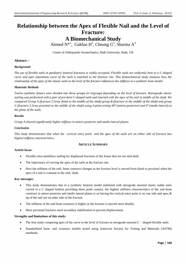
Group A included constructs in which the fracture was 2.5cm distal to the apex of the curve of the nail. Group B included
constructs in which the fracture was at the level of the apex of the curve of the nail. Group C included constructs in which the
fracture was 2.5cm proximal to the apex of the curve of the nail as shown in figure. 1.
FIGURE 1: FEMUR WITH TWO C-SHAPED FLEXIBLE NAILS WITH APEX OF THE CURVE IN THE MIDDLE OF THE
BONE. THE THREE LINES REPRESENT THE THREE LEVELS OF FRACTURES IN THE STUDY.
International Journal of Engineering Research & Science (IJOER) ISSN: [2395-6992] [Vol-2, Issue-2, February- 2016]
Page | 162
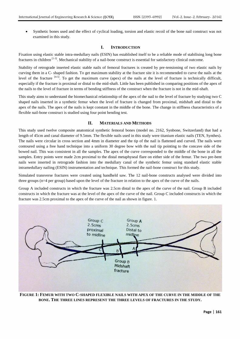
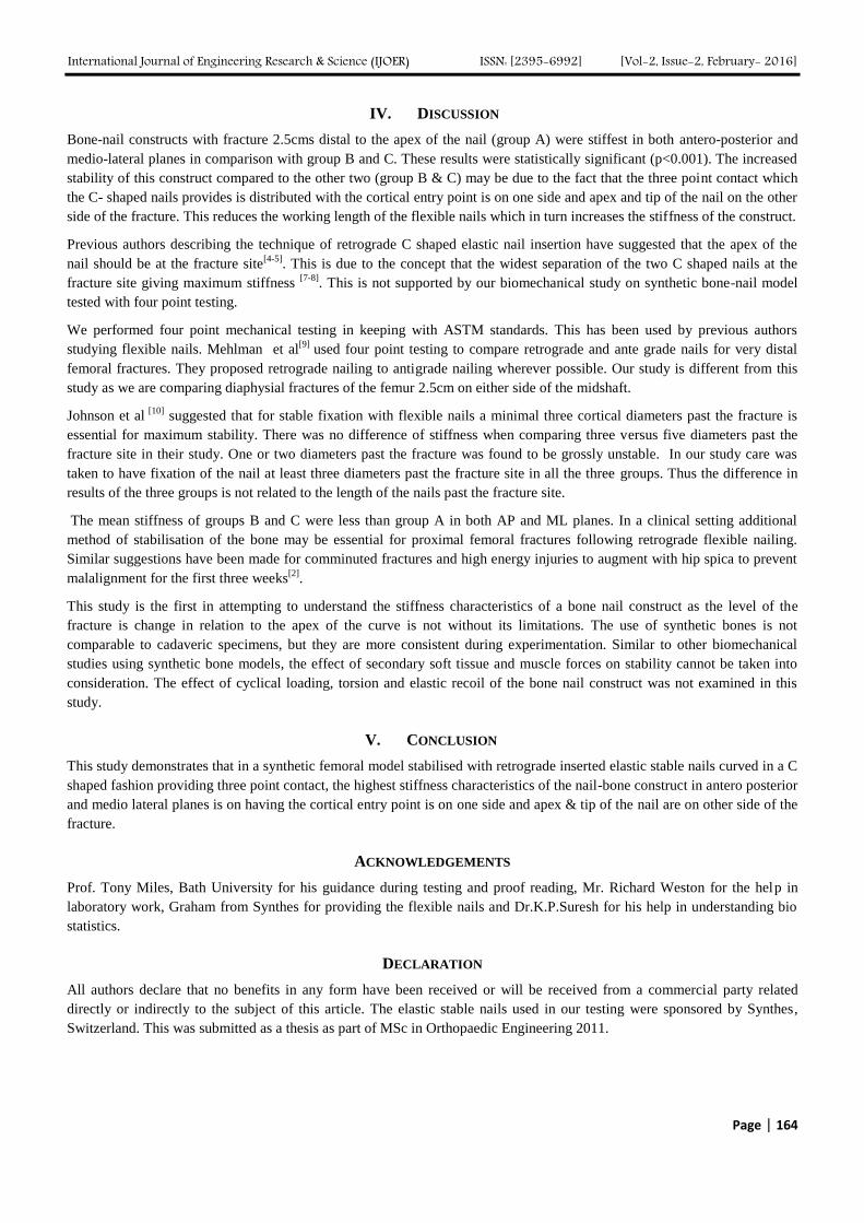
The method of mechanical testing was adapted from the American Society for Testing and Materials (ASTM)[6]
. Four-point
bending was done using a tension compression machine (Instron 3367, dual column tabletop universal testing system) with a
30KN load cell as shown in figure. 2.
Each construct was tested three times in the antero-posterior (AP) plane that is a plane perpendicular to the plane of the nails
as shown in figure 2. Testing was repeated in the medio-lateral (ML) plane that is parallel to the plain of the nails to a
maximum load of 200N. The rate of loading was set at 0.2mm per second with no upper limit of maximum displacement.
Force displacement curves were plotted for testing in both planes.
FIGURE 2: SHOWING FOUR POINT TESTING IN AP PLANE
Analysis of variance (ANOVA) was used to find the significance of study parameters between three groups of constructs.
III. RESULTS
The twelve nail-bone constructs were analysed in antero- posterior and medio-lateral planes. The stiffness of the constructs
was better in the AP plane compared to ML plane in all the three groups.
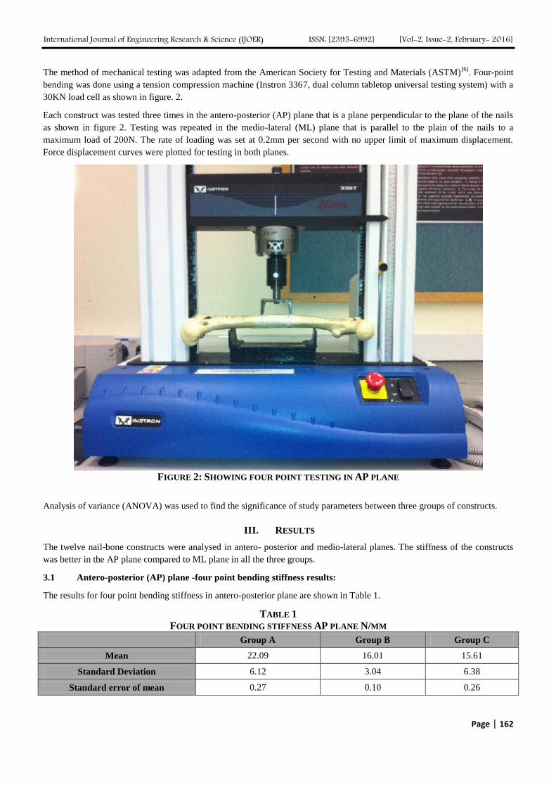
3.1 Antero-posterior (AP) plane -four point bending stiffness results:
The results for four point bending stiffness in antero-posterior plane are shown in Table 1.
TABLE 1
FOUR POINT BENDING STIFFNESS AP PLANE N/MM
Group A Group B Group C
Mean 22.09 16.01 15.61
Standard Deviation 6.12 3.04 6.38
Standard error of mean 0.27 0.10 0.26
International Journal of Engineering Research & Science (IJOER) ISSN: [2395-6992] [Vol-2, Issue-2, February- 2016]
Page | 163
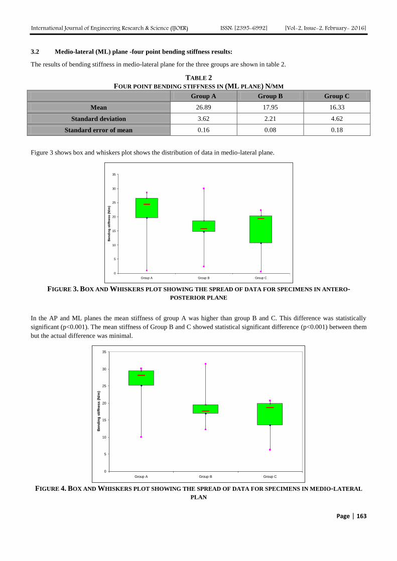
3.2 Medio-lateral (ML) plane -four point bending stiffness results:
The results of bending stiffness in medio-lateral plane for the three groups are shown in table 2.
TABLE 2
FOUR POINT BENDING STIFFNESS IN (ML PLANE) N/MM
Group A Group B Group C
Mean 26.89 17.95 16.33
Standard deviation 3.62 2.21 4.62
Standard error of mean 0.16 0.08 0.18
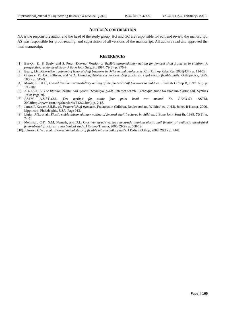
Figure 3 shows box and whiskers plot shows the distribution of data in medio-lateral plane.
0
5
10
15
20
25
30
35
Group A Group B Group C
Ben
din
g s
tiff
ness (
N/m
)
FIGURE 3. BOX AND WHISKERS PLOT SHOWING THE SPREAD OF DATA FOR SPECIMENS IN ANTERO-
POSTERIOR PLANE
In the AP and ML planes the mean stiffness of group A was higher than group B and C. This difference was statistically
significant (p<0.001). The mean stiffness of Group B and C showed statistical significant difference (p<0.001) between them
but the actual difference was minimal.
0
5
10
15
20
25
30
35
Group A Group B Group C
Ben
din
g s
tiff
ness (
N/m
)
FIGURE 4. BOX AND WHISKERS PLOT SHOWING THE SPREAD OF DATA FOR SPECIMENS IN MEDIO-LATERAL
PLAN
International Journal of Engineering Research & Science (IJOER) ISSN: [2395-6992] [Vol-2, Issue-2, February- 2016]
Page | 164
IV. DISCUSSION
Bone-nail constructs with fracture 2.5cms distal to the apex of the nail (group A) were stiffest in both antero-posterior and
medio-lateral planes in comparison with group B and C. These results were statistically significant (p<0.001). The increased
stability of this construct compared to the other two (group B & C) may be due to the fact that the three point contact which
the C- shaped nails provides is distributed with the cortical entry point is on one side and apex and tip of the nail on the other
side of the fracture. This reduces the working length of the flexible nails which in turn increases the stiffness of the construct.
Previous authors describing the technique of retrograde C shaped elastic nail insertion have suggested that the apex of the
nail should be at the fracture site[4-5]
. This is due to the concept that the widest separation of the two C shaped nails at the
fracture site giving maximum stiffness [7-8]
. This is not supported by our biomechanical study on synthetic bone-nail model
tested with four point testing.
We performed four point mechanical testing in keeping with ASTM standards. This has been used by previous authors
studying flexible nails. Mehlman et al[9]
used four point testing to compare retrograde and ante grade nails for very distal
femoral fractures. They proposed retrograde nailing to antigrade nailing wherever possible. Our study is different from this
study as we are comparing diaphysial fractures of the femur 2.5cm on either side of the midshaft.
Johnson et al [10]
suggested that for stable fixation with flexible nails a minimal three cortical diameters past the fracture is
essential for maximum stability. There was no difference of stiffness when comparing three versus five diameters past the
fracture site in their study. One or two diameters past the fracture was found to be grossly unstable. In our study care was
taken to have fixation of the nail at least three diameters past the fracture site in all the three groups. Thus the difference in
results of the three groups is not related to the length of the nails past the fracture site.
The mean stiffness of groups B and C were less than group A in both AP and ML planes. In a clinical setting additional
method of stabilisation of the bone may be essential for proximal femoral fractures following retrograde flexible nailing.
Similar suggestions have been made for comminuted fractures and high energy injuries to augment with hip spica to prevent
malalignment for the first three weeks[2]
.
This study is the first in attempting to understand the stiffness characteristics of a bone nail construct as the level of the
fracture is change in relation to the apex of the curve is not without its limitations. The use of synthetic bones is not
comparable to cadaveric specimens, but they are more consistent during experimentation. Similar to other biomechanical
studies using synthetic bone models, the effect of secondary soft tissue and muscle forces on stability cannot be taken into
consideration. The effect of cyclical loading, torsion and elastic recoil of the bone nail construct was not examined in this
study.
V. CONCLUSION
This study demonstrates that in a synthetic femoral model stabilised with retrograde inserted elastic stable nails curved in a C
shaped fashion providing three point contact, the highest stiffness characteristics of the nail-bone construct in antero posterior
and medio lateral planes is on having the cortical entry point is on one side and apex & tip of the nail are on other side of the
fracture.
ACKNOWLEDGEMENTS
Prof. Tony Miles, Bath University for his guidance during testing and proof reading, Mr. Richard Weston for the help in
laboratory work, Graham from Synthes for providing the flexible nails and Dr.K.P.Suresh for his help in understanding bio
statistics.
DECLARATION
All authors declare that no benefits in any form have been received or will be received from a commercial party related
directly or indirectly to the subject of this article. The elastic stable nails used in our testing were sponsored by Synthes,
Switzerland. This was submitted as a thesis as part of MSc in Orthopaedic Engineering 2011.
International Journal of Engineering Research & Science (IJOER) ISSN: [2395-6992] [Vol-2, Issue-2, February- 2016]
Page | 165
AUTHOR’S CONTRIBUTION
NA is the responsible author and the head of the study group. HG and GC are responsible for edit and review the manuscript.
AS was responsible for proof-reading, and supervision of all versions of the manuscript. All authors read and approved the
final manuscript.
REFERENCES
[1] Bar-On, E., S. Sagiv, and S. Porat, External fixation or flexible intramedullary nailing for femoral shaft fractures in children. A
prospective, randomised study. J Bone Joint Surg Br, 1997. 79(6): p. 975-8.
[2] Beaty, J.H., Operative treatment of femoral shaft fractures in children and adolescents. Clin Orthop Relat Res, 2005(434): p. 114-22.
[3] Gregory, P., J.A. Sullivan, and W.A. Herndon, Adolescent femoral shaft fractures: rigid versus flexible nails. Orthopedics, 1995.
18(7): p. 645-9.
[4] Mazda, K., et al., Closed flexible intramedullary nailing of the femoral shaft fractures in children. J Pediatr Orthop B, 1997. 6(3): p.
198-202.
[5] AO-ASIF, S. The titanium elastic nail system. Technique guide. Internet search, Technique guide for titanium elastic nail, Synthes
1998; Page. 9].
[6] ASTM, A.S.f.T.a.M., Test method for static four point bend test method No. F1264-03. ASTM,
2003(http://www.astm.org/Standards/F1264.htm): p. 2-18.
[7] James R Kasser, J.H.B., ed. Femoral shaft fractures. Fractures in Children, Rookwood and Wilkins', ed. J.H.B. James R Kasser. 2006,
Lippincott: Philadelphia, USA. Page 913.
[8] Ligier, J.N., et al., Elastic stable intramedullary nailing of femoral shaft fractures in children. J Bone Joint Surg Br, 1988. 70(1): p.
74-7.
[9] Mehlman, C.T., N.M. Nemeth, and D.L. Glos, Antegrade versus retrograde titanium elastic nail fixation of pediatric distal-third
femoral-shaft fractures: a mechanical study. J Orthop Trauma, 2006. 20(9): p. 608-12.
[10] Johnson, C.W., et al., Biomechanical study of flexible intramedullary nails. J Pediatr Orthop, 2009. 29(1): p. 44-8.