Embed Size (px)
Citation preview
RELATIONSHIP BETWEEN POSTURE AND PEAK SHOULDER MUSCLE
ACTIVATION IN COLLEGIATE SWIMMERS
A THESIS
Submitted to the Faculty of the School of Graduate Studies
and Research
of
California University of Pennsylvania in partial
fulfillment of the requirements for the degree of
Master of Science
by
Rachel Elizabeth Loan
Research Advisor, Dr. Robert H. Kane
California, Pennsylvania
2010
ii
iii
ACKNOWLEDGEMENTS
Firstly, I would like to thank my family. Mom and dad
thanks for all the support throughout my life, allowing and
helping me to become the person I am today. I would not
have made it this far without your love. Chris, by far the
best brother I could ever ask for, thanks for everything
you do. To my aunts, uncles, cousins, and grandparents,
thank you. Especially my Nannie, who helped me through this
year with cookies, brownies, support and love, and my
Grampie, who I still, and always will, miss and think about
every day.
To my thesis chair, Bob Kane, and committee members
Dr. Hess and Dr. Barnhart, thank you for all your help this
past year. You all pushed me to become a better researcher
and helped me find my strength and drive to finish this
thesis the best that I can. You all helped me look at
things that I wasn‟t finding to help me appreciate the
process more. Tom, I considered you my unofficial committee
member, thank you for all your help this past year.
Mike and Mark, if I get half as much respect, trust
and support from my next employers I will still be happy.
You made this past year such a great experience for me,
allowing myself to grow personally and professionally.
Thank you for believing in anything and everything I do.
To my classmates, I never thought I could come so
close to so many people as quickly as we all did. I
couldn‟t have asked for better friends to make throughout
my time here. It has been quite the “Party in Cal PA” and
always remember to dance with your hands in the air.
To my friends at home, Ariana, Ashley, Jeannie,
Melissa, and Alicia, thank you for your support from 700
miles away and thank you for listening to me complain and
for the faith you gave in me that I could get this done.
Good thing we all had unlimited text messages or I would
never have made it through this year.
And of course, I would like to thank my subjects, the
swimmers of the Washington and Jefferson Swim Team. Thank
you for your interest in my study and your time you
volunteered to come in for my subject testing.

iv
TABLE OF CONTENTS
Page
SIGNATURE PAGE . . . . . . . . . . . . . . . . ii
AKNOWLEDGEMENTS . . . . . . . . . . . . . . . . iii
TABLE OF CONTENTS . . . . . . . . . . . . . . . iv
LIST OF TABLES . . . . . . . . . . . . . . . . vi
INTRODUCTION . . . . . . . . . . . . . . . . . 1
METHODS . . . . . . . . . . . . . . . . . . . 5
Research Design . . . . . . . . . . . . . . . 5
Subjects . . . . . . . . . . . . . . . . . . 5
Preliminary Research . . . . . . . . . . . . . 6
Instruments . . . . . . . . . . . . . . . . 7
Procedures . . . . . . . . . . . . . . . . . 10
Hypotheses . . . . . . . . . . . . . . . . . 14
Data Analysis . . . . . . . . . . . . . . . . 14
RESULTS . . . . . . . . . . . . . . . . . . . 16
Demographic Information . . . . . . . . . . . . 16
Hypotheses Testing . . . . . . . . . . . . . . 17
Additional Findings . . . . . . . . . . . . . . 19
DISCUSSION . . . . . . . . . . . . . . . . . . 21
Discussion of Results . . . . . . . . . . . . . 21
Conclusions . . . . . . . . . . . . . . . . . 25
Recommendations . . . . . . . . . . . . . . . 26
REFERENCES . . . . . . . . . . . . . . . . . . 27
v
APPENDICES . . . . . . . . . . . . . . . . . . 29
APPENDIX A: Review of Literature . . . . . . . . . 30
Shoulder Anatomy . . . . . . . . . . . . . . . 31
Shoulder Musculature . . . . . . . . . . . . 33
Shoulder Activity in Swimming . . . . . . . . . . 35
Freestyle Swimming Techniques . . . . . . . . . 35
Shoulder Issues in Swimmers . . . . . . . . . . 37
Posture Assessment . . . . . . . . . . . . . . 40
EMG Technique . . . . . . . . . . . . . . . . 43
Summary . . . . . . . . . . . . . . . . . . . 44
APPENDIX B: The Problem . . . . . . . . . . . . . 46
Definition of Terms . . . . . . . . . . . . . . 47
Basic Assumptions . . . . . . . . . . . . . . 49
Delimitations of the Study . . . . . . . . . . . 50
Significance of the Study . . . . . . . . . . . 50
APPENDIX C: Additional Methods . . . . . . . . . . 52
Informed Consent (C1) . . . . . . . . . . . . . 53
Photographic Release Form (C2) . . . . . . . . . 57
Demographic Information (C3) . . . . . . . . . . 59
WMPA Scoring Guide (C4) . . . . . . . . . . . . 61
IRB: California University of Pennsylvania (C5) . . . 66
Electrode Placement and MVC (C6) . . . . . . . . . 82
REFERENCES . . . . . . . . . . . . . . . . . . 84
ABSTRACT . . . . . . . . . . . . . . . . . . . 88
vi
LIST OF TABLES
Table Title Page
1. Demographic Information . . . . . . . . 17
2. Subjects Major Stroke . . . . . . . . . 17
3. Descriptive Statistics . . . . . . . . 18
1
INTRODUCTION
It has been estimated that the average college swimmer
completes one million strokes, with each arm, every year.1
In competitive swimming, it is said that 90% or more of the
propulsive force during the freestyle stroke comes from the
upper extremity.2 Swimmers shoulders need to have very
mobile shoulders because mobility has been directly
correlated with greater stroke length and speed.3
The shoulder is a very complex area that is commonly
injured. The fundamental skeletal segments of the shoulder
include the humerus, scapula, clavicle, sternum, and the
first eight thoracic ribs.4 Three synovial joints and one
functional joint makes up the shoulder complex. The only
bony attachment between the axial skeleton and the shoulder
complex is through the sternoclavicular (SC) joint.
Many muscles are involved in moving and stabilizing
the shoulder complex. The majority of the muscles are
involved in moving the various joints of this region. The
serratus anterior is a major muscle of the scapulothoracic
stabilizers and contributes to scapular control and
stability.5,6,8
Control of the scapula is essential for
coordination of the joint and instability can occur.7
The
2
serratus anterior also provides an upward rotation force
during the early phases of elevation.7
The latissimus dorsi is a large muscle whose origin
spans from the lower thoracic vertebrae, all five lumbar
vertebrae, posterior iliac crest, sacrum, and lower three
ribs.8 The latissimus dorsi functions to decrease the
compression in the AC joint.7 The two origins of the
pectoralis major are the clavicular and sternal heads and
the muscle then inserts onto the humerus.4
The sternal
fibers and the clavicular fibers twist so that the sternal
fibers lie underneath the clavicular fibers.4 The short, or
clavicular, fibers are placed under more tension and
stressed than the sternal fibers.4
Pink et al2 described the muscle activity patterns of
12 muscles in the normal shoulder during freestyle
swimming. The muscles examined were the deltoids, rotator
cuff, rhomboids, trapezius, serratus anterior, pectoralis
major, and latissimus dorsi. They found that the serratus
anterior was active during the entire freestyle stroke and
the latissimus dorsi and pectoralis major muscles were
important for the pull through phase of the stroke.
Scovazzo et al9 then did an EMG analysis of the muscle
activity in painful swimmer‟s shoulders and compared the
results to the results from Pink et al.2 They found that
3
the serratus anterior was significantly less active in
painful shoulders when compared to the activity of the
muscle in non painful shoulders, especially after hand
entry into the water. The serratus anterior activity was
most significantly different and was stated as
“dysfunctional and exhibited low levels of activity.” 9
Because of the recent popularity in swimming in the
Olympics, technical advances, improvements in conditioning,
and training equipment have also become more popular.5
Swimmers have been working with team coaches and strength
and conditioning coaches to improve their strength, which
will in turn improve their swimming velocity, stroke
length, and stroke rates. Most traditional weight training
programs for swimming fail to concentrate on the dynamic
strength and the endurance provisions of swimming. This
has been related to painful shoulder incidences because it
causes muscle imbalances, fatigue, improper technique, and
acute and chronic muscle and tendon injuries.
Imbalances in muscles are the major cause of improper
alignment. Shortened muscles pull the origin and
insertions of the muscles closer together, while the weaker
antagonist muscles‟ origins and insertions have a greater
separation than normal.10
Perry et al11 described a variety
of ways to measure posture including photographic analysis
4
and manual measurements. Manual measurements can be done by
using a plumb line.10-12
The plumb line is a line that is
hung from a overhead bar and the “plumb bob” hangs to the
board where the feet are at the standard base point.10
Another tool used to assess posture is the Watson-
MacDonncha Posture Analysis (WMPA). The WMPA is a valid
and reliable way to test posture based on a scoring system
for either 1, 3, or 5 points for 10 different anatomical
places for postural defects.13 The WMPA measures ankle varus
and valgus, knee interspace, knee hyperflexion and
hyperextension, lordosis, kyphosis, scoliosis S and C
curves, rounded shoulders, scapular winging/abduction,
shoulder symmetry, and forward head.12,13
EMG shows the
activation of muscles. Surface EMG measures the muscle
activity if an electrode is placed on the skin directly
above the belly of the muscle. The question that has not
been researched as much as painful versus non painful
shoulders is that of different postures which may cause
muscle imbalances, also cause muscle activity to vary.
5
METHODS
Research Design
A descriptive correlational design was used to measure
shoulder posture and muscle activation levels of collegiate
swimmers. Each athlete was measured for peak muscle
activation during a functional shoulder extension exercise
and a Watson-MacDonncha Posture Analysis (WMPA). The
athletes were measured after the end of their season. The
variables for this study are posture and peak muscle
activation of the latissimus dorsi, pectoralis major, and
serratus anterior during a functional extension exercise
which mimics the pull through phase of the freestyle
stroke.
Subjects
Subjects were volunteers (n=16) from the NCAA
Washington and Jefferson College‟s Division III swim team,
who were recruited through a mass email sent by the
researcher. Before the study was performed, informed

6
consent was administered to the athletes to educate them
about the risks and procedures of the study. By signing
the consent form, each subject indicated that their
participation was completely voluntary and that all results
would remain confidential (Appendix C1). A photographic
release form was also signed since pictures of the subjects
were taken. After consent and photographic release forms
were signed, demographic information was collected
(Appendix C2). Demographic information such as: age, year
in school, number of consecutive years of competitive
swimming, major swimming stroke and distance, and any prior
injuries to the shoulder was also collected (Appendix C3).
All of the subjects‟ information was collected through
self-report.
Preliminary Research
Preliminary research was done for the researcher to
become familiar with the equipment that would be used in
this study, including the Watson-MacDonncha Posture
Analysis (WMPA), which was used to measure total body
posture, and surface electromyography (EMG) which was used
to measure activity in the selected muscles. Also, the
appropriate time frame, of half an hour, for each
7
participant was determined by doing the preliminary
research. Accurate measurements can be ensured with the
researcher becoming proficient with the equipment after
trial runs of the methods.
Instruments
The following measures were taken for the study: the
WMPA and the EMG measured muscle activity.
Posture Analysis
The Watson-MacDonncha Posture Analysis (WMPA) is an
instrument used to measure posture. The WMPA measures
ankle varus and valgus, knee interspace, knee hyperflexion
and hyperextension, lordosis, kyphosis, scoliosis S and C,
rounded shoulders, scapular winging/abduction, shoulder
symmetry, and forward head. Watson et al12 has reported the
WMPA as being valid and the reliability of the posture
scores is 0.85.
During the WMPA, each subject would stand on a
platform that is 20cm high, 60cm long, and 40cm wide.
Adhesive reflective dots were placed on landmarks on the
subject. These markers are placed on the left auricle
pinna, left axis of the glenohumeral joint, left patellar
8
notch, left greater trochanter, both clavicular heads, both
anterior superior iliac spines (ASIS), both tibial
tubercles, the center of both patellae, C7, T3, T6, T9,
T12, L3, L5, the most prominent spot of the sacrum, and the
center of both calcaneus.
Five photographs of each subject were taken. There
was one anterior view photograph, one posterior view
photograph, one lateral view, one at a 45o angle, and one
front view photograph with the subject holding their
subject number. There were three colored lines on the
platform, a red one, blue one, and yellow one. The red one
was placed vertically and is used for placement for the
anterior and posterior views, the blue one was placed
horizontally for the left lateral photograph, and the
yellow one was set at a 45o angle from the left back corner
to the front right corner of the platform for a posterior
left lateral oblique photograph. 13
The suggestion for the camera placement is that it is
placed 10 feet from the platform and the camera lens be 120
cm from the floor.13 For the four photographs the subjects
were asked to stand upright with their chins parallel to
the ground. It was also asked that they fully extend their
elbows and knees for the photographs. 13
9
The criteria for scoring the WMPA are located in
Appendix C2. A score of 5 was given if there were no
postural distortions. A score of 3 was given if there were
moderate postural distortions. A score of 1 was given if
there were moderate to severe postural distortions. Higher
total scores indicate better posture.
Electromyography
In collecting the EMG data, the researcher used three
channels from a Biopac MP150® electromyography machine.
Three channels were designated for the muscles tested
(pectoralis major, latissimus dorsi, serratus anterior).
The Biopac MP150 was connected to a Microsoft Windows based
personal computer with the Biopac‟s AcqKnowledge® program
[Goleta, CA] to collect analyze the data. The study
utilized pre-gelled disposable Ag-AgCl surface electrodes
with a diameter of one centimeter. The electrodes were
placed on the subject‟s dominate arm over the motor points
of each muscle belly with a center-to-center spacing of 2.5
centimeters.14 The raw EMG signal was band pass filtered at
10 and 1000 Hertz (Hz). The researcher utilized a sampling
rate of 2000Hz using the AcqKnowledge software. The
signals were rectified and normalized before the data
analysis was completed.
10
The AcqKnowledge® program computed the muscle activity
in Hertz (Hz). The researcher computed the percentage of
peak muscle activation by first doing a maximum voluntary
contraction (MVC) of each muscle, using a functional
shoulder extension exercise that mimics the freestyle swim
stroke‟s pull through phase. After the testing the maximum
muscle activity during the MVC for functional shoulder
extension exercise, the researcher computed a standard
score for each subject by using the equation:
Max Muscle Activity During Pull Through
Max Muscle Activity During MVC
This will give us a percent of peak muscle activity.
Procedures
Before any descriptive statistics were measured, the
researcher applied for and obtained approval from
California University of Pennsylvania‟s Institutional
Review Board (Appendix C5). Preliminary research was
conducted first so the researcher became proficient in the
procedures of the WMPA and the EMG. All subjects signed an
informed consent Appendix(C1)stating the reasoning for the
study, how it would be conducted, and that they could
withdraw from the study at any time. A photographic
11
release form was also signed Appendix (C2). The subjects
also filled out a demographic form asking questions such as
their years in competitive swimming, prior injury, and
major swimming stroke. The subjects were asked to also
state their dominant
When the subjects arrived for the assessment, they
were first set up for the posture analysis. The subjects
were asked to wear non-baggy shorts and tank tops if
necessary so that the landmarks, curvatures, and any valgus
or varus angles could be photographed.
Five photographs are taken of each subject as per the
recommended procedures.13 The first one was just the subject
number photograph for future referencing. For the second
photograph, the subjects stood facing the camera with their
heels on a red line. Each subject was asked that they keep
their chin parallel to the ground, elbows and knees fully
extended, and heels in contact with each other. The third
photograph was from the posterior view; subjects faced
away from the camera with the heels together on the red
line, chin parallel to the ground and the knees and elbows
fully extended. The fourth photograph was a left lateral
view. Subjects were instructed to stand with their heels
touching on the blue line, chin parallel to the ground and
knees and elbows extended. The fifth photograph was taken
12
at an oblique angle. The subjects stood on a yellow line
which was at a 45o angle from the left back corner to the
front right corner.
The procedure for measuring the WMPA scores is as
follows. First, a grid is placed over the top of each
photograph of each subject with transparency paper. The
grid is lined up with the plumb lines used in the
photographs. A second transparency paper is placed on top
with circle diameters which is used to determine the
degrees of lordosis and kyphosis. A protractor and ruler
were used to measure angles through the adhesive dots. A
score of 5 is given if there is no marked deviation, a 3 is
given if there were moderate deviations, and a 1 is given
for extreme deviations for all body areas. (Appendix C4)
The next part of the process of the study was using
the surface EMG to measure the muscle activation.
Electrode pads were placed on the latissimus dorsi,
pectoralis major, and serratus anterior muscle bellies
(Appendix C4). The subjects then performed an upper body
warm-up, consisting of internal and external rotations and
practice of the functional shoulder extension exercise,
using a Theraband® which helped determine which band would
be used for the subject during testing.
13
After the warm-up was completed, the subject‟s
maximum voluntary contraction was then tested. For the
pectoralis major, the subject was placed in humeral
adduction with the shoulder in 900 of flexion and the palms
together. The subject then applied the resistance by
pressing the palms together. The latissimus dorsi movement
to reach a maximum voluntary contraction was internal
rotation and extension of the humerus with the shoulder in
300 of abduction with the elbow extended. Resistance was
then applied at the distal forearm by the researcher. For
the serratus anterior, the subject‟s dominant arm was
flexed forward to 130o. The researcher then placed one hand
over the dorsal arm and one hand on the lateral scapula for
stability. The subject then isometrically flexed while the
researcher added resistance.13
After the maximal voluntary contraction testing was
complete for the three muscles, subjects performed a
functional shoulder extension exercise against resistance
with a Theraband® which would mimic the pull-through phase
of the freestyle stroke.
The subject would determine which of the three choices
of Theraband® would have the best resistance for them by
going through the motion with each of the three choices.
Once established, the subject stood with enough room from
14
the stationary object holding the bands and bend at the
waist at about 750 to imitate being in the water with their
knees slightly flexed. Shoulder flexion at about 900 of
flexion followed by their pull-through phase with the
resistance of the Theraband®. This motion was repeated ten
times to ensure valid measurements.
Hypothesis
The following hypothesis was tested in this study:
There will be a relationship among shoulder posture
score and peak muscle activity in each of the following
muscles: the pectoralis major, serratus anterior, and
latissimus dorsi, during a functional shoulder extension
exercise.
Data Analysis
A Pearson Product Moment Correlation was used to
analyze the correlation among WMPA score and the peak
muscle activity in each of these muscles. SPSS version
17.0 for Windows analyzed all data at P <.05. All EMG
scores were reported as a percentage of maximal voluntary

15
contraction.
16
RESULTS
The purpose of this study was to examine the
relationship between complete body posture and peak muscle
activation in the shoulders of swimmers. Each subject came
in one time for 30 minutes, and was tested using the WMPA
and Biopac MP150 surface EMG. The WMPA was used to assign
a posture score to each subject and the Biopac MP150 was
used to analyze peak muscle activation in the latissimus
dorsi, serratus anterior, and pectoralis major during a
resisted functional shoulder extension exercise that
mimicked the pull through phase of the freestyle swim
stroke. All data was collected on each subjects Individual
Data Collection Sheet (Appendix C7)
Demographic Information
There were 16 volunteers from Washington and Jefferson
College‟s Men‟s and Women‟s Division III swim teams. The
WMPA and EMG testing were conducted in the same way and by
the same researcher in order to increase internal validity.
17
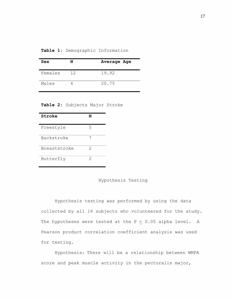
Table 1: Demographic Information
Sex N Average Age
Females 12 19.92
Males 4 20.75
Table 2: Subjects Major Stroke
Stroke N
Freestyle 5
Backstroke 7
Breaststroke 2
Butterfly 2
Hypothesis Testing
Hypothesis testing was performed by using the data
collected by all 16 subjects who volunteered for the study.
The hypotheses were tested at the P < 0.05 alpha level. A
Pearson product correlation coefficient analysis was used
for testing.
Hypothesis: There will be a relationship between WMPA
score and peak muscle activity in the pectoralis major,
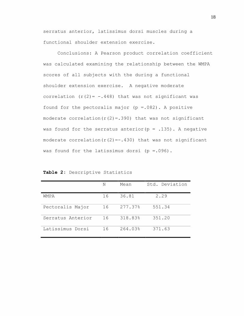
18
serratus anterior, latissimus dorsi muscles during a
functional shoulder extension exercise.
Conclusions: A Pearson product correlation coefficient
was calculated examining the relationship between the WMPA
scores of all subjects with the during a functional
shoulder extension exercise. A negative moderate
correlation (r(2)= -.448) that was not significant was
found for the pectoralis major (p =.082). A positive
moderate correlation(r(2)=.390) that was not significant
was found for the serratus anterior(p = .135). A negative
moderate correlation(r(2)=-.430) that was not significant
was found for the latissimus dorsi (p =.096).
Table 2: Descriptive Statistics
N Mean Std. Deviation
WMPA 16 36.81 2.29
Pectoralis Major 16 277.37% 551.34
Serratus Anterior 16 318.83% 351.20
Latissimus Dorsi 16 264.03% 371.63
19
Additional Findings
An outlier was removed for further testing to see if
it changed any levels of significance. An outlier is
defined as being double the standard deviation away from
the mean. With the outlier removed, there was still no
significant relationship between WMPA scores and percentage
of peak muscle activity.
A one way MANOVA was calculated to examine the
relationship between percentages of peak muscle activation
and if the subject has a history of shoulder injury. No
significant effect was found (Lambda(3,12)= .419, P >.05.)
None of the percentages of peak muscle activation for the
three muscles were significantly influenced by a history of
injury.
The WMPA scores of subjects who were divided into
groups depending on their major stroke were compared using
a one-way ANOVA. No significant difference was found (F
3,12)= .478, P > .05). The subjects WMPA score did not
differ significantly depending on their major stroke.
A one-way MANOVA was calculated examining the effect
of major swim stroke (freestyle, breaststroke, butterfly,
or backstroke) on percentage of peak muscle activation of
each muscle (pectoralis major, serratus anterior,
20
latissimus dorsi). No significant effect was found (Lambda
(9,24.5) = .784, P > .05). None of the percentages of peak
muscle activation of the muscles were significantly
influenced by major swim stroke.
21
DISCUSSION
The following discussion is divided into three
sections: 1) Discussion of Results, 2) Conclusions and 3)
Recommendations.
Discussion of Results
Posture is an important component in making sure the
kinetic chain is operating at its best. It has been stated
that postural distortions can cause discomfort, pain and
disability. Postural problems can lead to extra stress on
joints and muscles which can cause muscle imbalances, such
as shortening or lengthening the distance between the
attachments of the muscles, and can cause athletes to
voluntarily and involuntarily change their body mechanics.
This investigation was performed to determine if there
was any relationship in WMPA score and percentage of peak
shoulder muscle activation in Division III collegiate
swimmers. The primary findings of this study indicated no
significant relationship between posture scores (WMPA) and
percentage of peak muscle activation in swimmers.
22
The results of this study showed that no matter what
the WMPA score is, it should not affect muscle activity at
all. However, if there was a greater array of WMPA scores
our outcomes may have been different.
Overall, based on this study, no matter what the
posture of a swimmer is the muscle activity in the three
main muscles used during the pull through phase of the
freestyle swim stroke, which include: latissimus dorsi,
serratus anterior, and pectoralis major, is not affected.
A particular finding in this study was the difference
in the muscle activity in the serratus anterior when
compared with the latissimus dorsi and pectoralis major.
The most commonly known function of the serratus anterior
is to prevent scapular winging.15
The serratus anterior
works with the trapezius to contribute extensively to
posterior tilt of the scapula as well as scapular upward
rotation.16 The serratus anterior works with the trapezius,
levator scapulae, rhomboids, and pectoralis minor to
control scapular movements.15 With the pectoralis minor,
the serratus anterior abducts the scapula. With the upper
and lower trapezius muscles, the serratus anterior upwardly
rotates the scapula. By itself, the serratus anterior
contributes to all concepts of the scapular movements in

23
all planes of motion during upward rotation, posterior
tilt, and external rotation.15
In this study, the descriptive statistics showed us a
difference in numbers in the serratus anterior compared to
the other two muscles tested. During the primary
hypothesis testing to find a correlation between WMPA
scores and peak percentages of muscle activity in the
muscles there were no significant findings. However, the
serratus anterior had the highest mean peak percent when
compared to the latissimus dorsi and the pectoralis major
mean peak percentages. The serratus anterior also had the
lowest standard deviation of these scores.
It would be assumed that during the freestyle stroke,
the serratus anterior is constantly active to keep the
scapula stabilized throughout the entire stroke which
entails a great deal of shoulder complex and scapular
movement. This descriptive statistic tells us that in the
Division III male and female swimmers, their serratus
anterior activity is working constantly and effectively
during this part of the stroke, this is following the
findings of Pink et al2 who examined the serratus anterior
as being a very active muscle throughout the entire
freestyle stroke.
24
When looking at the descriptive statistics of the one-
way ANOVA comparing peak percentages of muscle activity
with the four strokes the subjects could choose from, the
serratus anterior once again stands out among the three
muscles. For the subjects that choose freestyle, the mean
peak percentage of muscle activity in the serratus anterior
was higher than the mean peak percentages of the latissimus
dorsi and pectoralis major. This demonstrates a difference
in findings then the previous statement and the serratus
anterior is the least active muscle in the pull-through
phase in freestyle swimmers when compared with subjects who
listed their major stroke as butterfly, breaststroke, and
backstroke. The swimming stroke that showed the most
serratus anterior activity was the butterfly swimmers.
If these results are consistent in future research
this will greatly help enhance the knowledge of training
techniques for coaches, athletic trainers, and strength and
conditioning coaches. Posture should be one of the first
components of a program in the first place when in a
corrective exercise phase of training, to prevent any
injuries and biomechanical efficiencies. In swimmer‟s who
have swam all throughout their childhood until college,
their posture may have developed to have the classic
rounded shoulders and forward head that are common in
25
swimmers. These may be harder to correct because the
swimmer is accustomed to doing their activities of daily
living and their swim stroke to be comfortable enough for
them, yet still perform each stroke at maximal
effectiveness.
Conclusions
After completing this study, there was no relationship
found between posture and percentage of peak muscle
activation in swimmers or not. In this current study, all
the subjects had similar posture scores that were slightly
higher than the middle range of scores. Total posture
scores could range from 10-50, which has a median number of
30, and the subjects mean WMPA score was 36.81 with a
standard deviation of + 2.29. This fact made it difficult
to examine any correlation between the posture scores and
percentage of peak shoulder muscle activation because the
subjects WMPA scores were within 6 points of each other.
However, although there were no significant findings
in the hypothesis testing and additional findings, the
descriptive statistics showed a lot of information about
the serratus anterior. Serratus anterior is more active
from the other two muscles when the peak percentages of
26
muscle activity when correlated with the WMPA scores and
when the peak percentages of muscle activity is compared
between the four different swimming strokes.
Recommendations
It is still unknown whether or not posture can effect
muscle activation in swimmers. Further research is
recommended with doing testing closer to the end of the
season. At the time of this testing, subjects had had no
practice in almost two months, unless they swam on their
own. Another recommendation for future research would be to
have more subjects, with more variety of major swim
strokes. Of the subjects in this study, only two listed
their major stroke as breaststroke, and another two listed
their major stroke as butterfly.
If more specific procedures were implemented during the
EMG testing, comparable scores may have been obtained.
Each of the 16 subjects all had different variations of the
freestyle pull through phase.
Also, if there was EMG used to look at muscle firing
patterns rather than peak percentages, we may be able to
find out more information about the serratus anterior
throughout the entire freestyle stroke.
27
REFERENCES
1. Weldon EJ, Richardson AB. Upper extremity overuse
injuries in swimming: A discussion of swimmer‟s
shoulder. Clinics in Sports Med. 2001; 20(3): 423-438.
2. Pink M, Perry J, Browne A, Scovazzo ML, Kerrigan J.
The normal shoulder during freestyle swimming: An
electromyographic and cinematographic analysis of
twelve muscles. Am J Sports Med. 1991; 19(6): 569-576.
3. Borsa PA, Laudner KG, Sauers EL. Mobility and
stability adaptations in the shoulder of the overhead
athlete: A theoretical and evidence-based perspective.
Sports Med. 2008; 38(1): 17-36.
4. Levy O, Rath E. Traumatic soft tissue injuries of the
shoulder girdle. Trauma. 2002; 4: 223-235.
5. Swanik KA, Swanik CB. The effect of functional
training on the incidence of shoulder pain and
strength in intercollegiate swimmers. Sport Rehab.
2002; 11:140-154.
6. Starkey C, Ryan J. Evaluation of orthopedic and
athletic injuries. F.A. Davis Company.2nd ed. 2002
7. Wodsworth D, Bullock-Saxton J. Recruitment patterns of
the scapular rotator muscles in freestyle swimmers
with subacromial impingement. Inter J of Sports Med.
2007; 18(8):618-624.
8. Wickham JB, Brown JM, McAndrew DJ. Muscles within
muscles: Anatomical and functional segmentation of
selected shoulder joint musculature. J of
Musculoskeletal Res.2004;8:57-73.
28
9. Scovazzo M, Brown, A, Pink M, Jobe F, Kerrigan J. An
electromyographic cinematographic analysis of twelve
muscles. Am J Sports Med. 1991; 19(6): 577-582.
10. Kendall FP, McCreary EK, Provance PG, Rodgers MM,
Romani WA. Muscles testing and function with posture
pain. Lippincott Williams & Williams. 2005; 5.
11. Perry M, Smith A, Straker L, Coleman J, O‟Sullivan P.
Reliability of sagital photographic spinal posture
assessment in adolescents. Advances in Physiotherapy.
2008; 10: 66-75.
12. Findlay S. Assessing posture: An essential tool. Sport
Ex. 2005; 6: 18-19.
13. Watson AWS, MacDonncha C. A reliable technique for the
assessment of posture: Assessment criteria for aspects
of posture. J of Sports Med and Phys Fitness. 2000; 9:
260-270.
14. Kibler WB, Sciascia AD, Uhl TL, Tambay N, Cunningham
T. Electromyographic analysis of specific exercises
for scapular control in early phases of shoulder
rehabilitation. Am J Sports Med. 2008; 36(9): 1789-
1797.
15. Scamilla RF, Yamishiro K, Paulos L, Andrews JR.
Shoulder muscle activity and functions in Common
Shoulder Rehabilitation Exercises. Sports Med. 2009;
39(8): 663-685.
16. Ronai P. Exercise modifications and strategies to
enhance shoulder function. Strength Condition J. 2005;
27(4): 36-45.

29
APPENDICES

30
APPENDIX A
Review of Literature
31
Swimmers, throwers, tennis, volleyball, and water polo
players are all included in the classification of overhead
athletes.1 Each of these sports has unique and individual
functional requirements. Swimmers shoulders need to be
very mobile because mobility has been directly correlated
with greater stroke length and speed.1 In competitive
swimming, it is said that 90% or more of the propulsive
force during the freestyle stroke comes from the upper
extremity.2 Also, it has also been estimated that the
average college swimmer completes one million, with each
arm, every year.3
Shoulder Anatomy
The fundamental skeletal segments of the shoulder
include the humerus, scapula, clavicle, sternum, and the
first eight thoracic ribs.4 Three synovial joints and one
functional joint makes up the shoulder complex. The only
bony attachment between the axial skeleton and the shoulder
complex is through the sternoclavicular (SC) joint. The
clavicular notch of the manubrium is the site where the
clavicle articulates with the sternum.5 The other shoulder
32
joints are the acromioclavicular (AC) joint,
scapulothoracic articulation, and the glenohumeral (GH)
joint.
The AC joint is the articulation between the distal
end of the clavicle and the acromion, which is a bony
process of the scapula. This joint is similar to the
sternoclavicular joint because they both are covered with
fibrous cartilage and the AC joint is separated with an
intraarticular disc.4, 6
The scapula is a flat shaped bone
that creates a mobile connection with the thorax.6 This is
not considered a true joint because the synovial
characteristics of a joint are not present.5 The scapular
creates a scapular rhythm that is seen in all movements of
the shoulder. The GH joint is the articulation between the
head of the humerus and the shallow, concave glenoid fossa
of the scapula.5,6 Only 30% of the humeral head fits in
contact with the relatively small glenoid fossa.5,6
The GH
joint is very mobile for unrestricted motion and is
reinforced by the rotator cuff muscles as the dynamic
stabilizers.6
As mobility in the shoulder is increased,
stability is decreased.
33
Shoulder Musculature
Many muscles are involved in moving and stabilizing
the shoulder complex. The majority of the muscles are
involved in moving the various joints of this region. One
group of muscles that receives attention in this region is
the rotator cuff muscle group.
The rotator cuff includes the supraspinatus,
infraspinatus, subscapularis, and teres minor. The
supraspinatus is responsible for the first stage of
abduction and it is said that increased activation of the
supraspinatus can cause upward rotation of the humeral head
in the glenoid fossa.6 The infraspinatus and teres minor
for lateral rotation, and the subscapularis is responsible
for medial rotation.7 The subscapularis may be the most
powerful muscle of the rotator cuff6, and is critical for
normal performance of shoulder motion. EMG shows that the
supraspinatus is a major muscle that is triggered for arm
elevation, overhead throwing, swimming, and golf.8 The four
muscles of the rotator cuff, together, constrains the
humeral head during elevation and work as dynamic
stabilizers of the GH joint by compressing the head of the
humerus into the glenoid fossa, particularly the
supraspinatus6
34
The biceps muscle is also another muscle that acts on
the shoulder and can potentially be a site of injuries.
Collectively, at the shoulder, the biceps motions are
shoulder flexion, abduction and adduction. The long head
is primarily responsible for abduction, while the short
head is primarily responsible for adduction.7 The deltoids
are considered “the most important mover and dynamic
inferior stabilizer of the GH joint”.6 It is composed of
upper, middle, and lower portions. Its actions include
shoulder elevation, adduction, upper rotation, and
depression.7
The primary scapulothoracic muscles include the
rhomboid, serratus anterior and the trapezius.6,9
These
muscles stabilize and control the scapula during GH
movement. These scapulothoracic muscles have the function
of upward rotation of the scapula when the arm is being
elevated, and also are responsible for resisting downward
rotation, which may be produced by the muscles of the GH
joint working on the scapula.10
The serratus anterior and
these scapulothoracic stabilizers contribute to scapular
control and stability.9 Control of the scapula is essential
for coordination of the joint and instability can occur.6
The serratus anterior also provides an upward rotation
force during the early phases of elevation.11
35
The latissimus dorsi is a large muscle whose origin
spans from the lower thoracic vertebrae, all 5 lumbar
vertebrae, posterior iliac crest, sacrum, and lower 3
ribs.12 The latissimus dorsi functions to decrease the
compression in the AC joint.6
The two origins of the pectoralis major are the
clavicular and sternal heads and the muscle then inserts
onto the humerus.4
The sternal fibers and the clavicular
fibers twist so that the sternal fibers lie underneath the
clavicular fibers.4 The short, or clavicular, fibers are
placed under more tension and stressed than the sternal
fibers.4
Shoulder Activity in Swimming
Freestyle Swimming Technique
Competitive freestyle swimming is the act of cycling
the arms as fast as possible, to move through the water, at
a given distance, with a varying amount of kicks per arm
cycle.9,13,14
Freestyle swimming performance depends
exclusively on the propulsive and resistive forces.15,16
The
phases of freestyle swimming consist of early and late
pull-through and early and late recovery. The pull-through
phases are compared to the acceleration phases in throwing
36
sports. Early pull-through begins when the hand enters the
water and ends when the humerus is perpendicular to the
body.2 Late pull through is the completion of the pull
through till the hand exits the water. The recovery
phase‟s first indication is the elbow lift. Once the hand
exits the water till when the humerus is perpendicular to
the body above the water, this is early recovery. From
here until the hand once again enters the water is the late
recovery phase.2
Pink et al described the muscle activity patterns of
12 muscles in the normal shoulder during freestyle
swimming.2 The muscles examined were the deltoids, rotator
cuff, rhomboids, trapezius, serratus anterior, pectoralis
major, and latissimus dorsi. The serratus anterior had a
constant level of activity in non-painful shoulders, except
when activity increased in the pull through phases and when
the hand exited the water.2 The pectoralis major and
latissimus dorsi had sharp peaks of activity during the
pull through, then immediately dropped back to resting
levels.2 These two muscles, the pectoralis major and
latissimus dorsi, are responsible for the increased
strength in swimmers compared to non-swimmers.3
It has been stated that a performance by swimmers can
only be enhanced by reducing the drag that acts on the body
37
or by increasing the propulsive forces.15
The propulsive
forces and resistive forces, or drag, are generated by the
arm movements, which generates a swimming velocity.13,17,18
Along with swimming velocity, studies have been done to
analyze the relationships between stroke length and stroke
rate.13,19,20
The shoulders of swimmers have unique and altered
mobility, stability, and functional requirements to meet
the demands of swimming.1 This shoulder hypermobility is
advantageous to the swimmers because it has been directly
correlated with stroke length, speed, and swimming
performance.1,3,25
Shoulder Issues in Swimmers
Because of the recent popularity in swimming in the
Olympics technical advances, improvements in conditioning,
and training equipment have also become more popular.9
Swimmers have been working with team coaches and strength
and conditioning coaches to improve their strength, which
will in turn improve their swimming velocity, stroke
length, and stroke rates. Most traditional weight training
programs for swimming fail to concentrate on the dynamic
strength and the endurance provisions of swimming.
38
It has been estimated that between 40 to 80 percent of
competitive swimmers have complained about pain in their
shoulders which may influence with training and
competition.9,21-23
This shoulder pain that swimmer‟s
experience is considered „swimmers shoulder‟. This „injury‟
includes symptoms resembling a combination of rotator cuff
tendinitis, subacromial bursitis, impingement syndrome, and
instability.9,21-23
Variables that are probable factors that
lead to swimmers shoulder include shoulder fatigue,
flexibility, improper technique, and strength
imbalances.9,21-23
Swanik et al state that there has been a
limited amount of research done of preventative measures of
this pathology.9
Kennedy stated that overhead athletes develop their
own normal range of motions that are related to their
sport.24,25
This is true in swimmers, who are likely to have
stronger internal rotators and adductors that are
hypertrophied compared to their antagonist muscles.26 These
and other biomechanical changes in swimmers, and all
overhead athletes, are reasons why there are frequent
shoulder injuries.24,25
Muscle fatigue is a contributor to
impingement symptoms because it causes improper techniques,
particularly when the hand enters the water.9
39
Shoulder pain in swimmers is commonly caused by
impingement injuries. McClure et al27 found that their
subjects with the signs and symptoms of impingement had
clear deficits to shoulder range of motion and also the
force production of the shoulder in a variety of
directions. Shoulder pain has been concluded to occur in
swimmer‟s of both genders, in both arms, for all strokes,
at all distances, and at all levels of competition.3 Weldon
and Richardson state that “shoulder pain can be prevented
by spending an equal amount of time stretching the
posterior and anterior capsules and performing scapular
positioning muscle exercises.”3
Scovazzo et al did an electromyographical analysis of
the muscle activity in painful swimmer‟s shoulders.28
They
found that the serratus anterior was significantly less
active in painful shoulders when compared to the activity
of the muscle in non painful shoulders, especially after
hand entry into the water.28
The serratus anterior activity
was most significantly different and was stated as
“dysfunctional and exhibited low levels of activity.”28
40
Posture Assessment
The Posture Committee of the American Academy of
Orthopedic Surgeons defines posture as:
“Posture is usually defined as the relative
arrangement of the parts of the body. Good
posture is that state of muscular and skeletal
balance which protects the supporting structures
of the body against injury or progressive
deformity, irrespective of the attitude (erect,
lying, squatting, or stooping) in which these
structures are working or resting. Under such
conditions the muscles will function most
efficiently and the optimum positions are
afforded for the thoracic and abdominal organs.
Poor posture is a faulty relationship of the
various parts of the body which produces
increased strain on the supporting structures and
in which there is less efficient balance of the
body over its base of support”7
Posture is a main component in assessing and
evaluating acute and chronic injuries, since it can lead to
pain and discomfort and improper mechanics.7 Imbalances in
muscles are the major cause of improper alignment.
Shortened muscles pull the origin and insertions of the
muscles closer together, while the weaker antagonist
muscles‟ origins and insertions have a greater separation
than normal.7 Therefore, therapeutic exercises to restore
proper posture include stretching of the shortened muscles
and strengthening of the weaker muscles.
Perry et al28 described a variety of ways to measure
posture including photographic analysis and manual
41
measurements. Manual measurements can be done by using a
plumb line.7,29,30
The plumb line is a line that is hung from
an overhead bar and the “plumb bob” hangs to the board
where the feet are at the standard base point.7 There are
four types of body postures that are assessed using a plumb
line. They are ideal posture, kyphotic-lordotic posture,
flat back posture, and sway back posture. Swimmers can
present forward head/rounded shoulder postures because of
their tight shoulder internal rotators and adductors.
The plumb line will hypothetically divide the body
into front and back sections with equal weight, and these
sections are not symmetrical. The line of reference for
the plumb line is the ear lobe.7,30
From the side view,
ideally, the plumb line should pass through the shoulder
joint, which the point of reference would be the acromion
process. In the ideal posture, the head is neither tilted
forward or back.7,30
The cervical spine has a slightly
convex curve anteriorly, the thoracic spine has a slight
posterior convex curve, and the lumbar spine is also has a
slightly curve anteriorly like the cervical spine. The
pelvis‟s anterior superior iliac spines (ASIS) are in the
same vertical plane as the pubic symphysis. The hips aren‟t
either flexed or extended, and the knee joints are neither
42
flexed or hyperextended. The ankle joints show a right
angle with the vertical leg and the sole of the foot.7
Another tool used to assess posture is the Watson-
MacDonncha Posture Analysis (WMPA). The WMPA is a valid and
reliable way to test posture based on a scoring system for
either 1, 3, or 5 points for 10 different anatomical places
for postural defects.30 The WMPA measures ankle varus and
valgus, knee interspace, knee hyperflexion and
hyperextension, lordosis, kyphosis, scoliosis S and C
curves, rounded shoulders, scapular winging/abduction,
shoulder symmetry, and forward head.32,33
During the WMPA, the subject stands on a platform that
is 20 cm high, 60 cm long, and 40 cm wide. Adhesive
reflective dots are placed on landmarks on the subject.
These markers are placed on the left auricle pinna, left
axis of the glenohumeral joint, left patellar notch, left
greater trochanter, both clavicular heads, both anterior
superior iliac spines (ASIS), both tibial tubercles, the
center of both patellae, C7, T3, T6, T9, T12, L3, L5, the
most prominent spot of the sacrum, and the center of both
calcaneus.30
Five photographs of each subject are taken for the
WMPA. There is one anterior view photograph, one posterior
view photograph, one left lateral view, one at a 45 degree
43
angle, and one front view photograph with the subject
holding their subject number. There are three colored
lines on the platform, a red one, blue one, and yellow one.
The red one is placed vertically and is used for placement
for the anterior and posterior views, the blue one was
placed horizontally for the left lateral photograph, and
the yellow one was set at a 45o angle from the left back
corner to the front right corner of the platform for a
posterior left lateral oblique photograph.30
The suggestion for the camera placement for the WMPA
is that it is placed 10 ft from the platform and the camera
lens be 120 cm from the floor.29
For each of the four photographs the subjects are
asked to stand upright with their chins parallel to the
ground. It is also asked that they fully extend their
elbows and knees for the photographs.29
EMG Technique
Electromyography (EMG) shows the activation of
muscles. There are two types of EMG for clinical use.
Intramuscular wire electrode EMG is a type where one or two
needles are placed into the belly of the muscle. Surface
EMG measures the muscle activity if an electrode is placed
44
on the skin directly above the belly of the muscle. During
testing using surface EMG, the surface electrodes are
placed directly over the force producing muscle or muscle
group. It then measures the amount of muscle action
potential as it passes under the surface electrodes.31
To generate the maximum voluntary contractions (MVC)
of the pectoralis major, the subject is placed in humeral
adduction with the shoulder in 900 of flexion and the palms
together. The subject then applies the resistance by
pressing the palms together. The latissimus dorsi movement
to reach a MVC is internal rotation and extension of the
humerus with the shoulder in 300 of abduction and the elbow
is extended. Resistance is then applied at the distal
forearm by the clinician.32 For the MVC of the serratus
anterior the subject‟s arm is forward flexed to 130o. The
researcher‟s hands are then placed one over the dorsal arm
and one on the lateral scapula for stability. The subject
then isometrically flexes while the researcher adds
resistance.33
Summary
The shoulder complex plays the most crucial role in
the freestyle swim stroke, creating 90% or more of the
45
propulsive force to carry the swimmer through the water.3
Pink et al2 described the latissimus dorsi and pectoralis
major muscles as being extremely important for the pull-
through phase and the serratus anterior as a very active
muscle throughout the entire stroke.
With recent popularity in swimming, athletes are
working hard to improve their strength which will improve
their swimming velocity, stroke length, and stroke rate.
The most traditional strength training programs do not
concentrate on the dynamic and endurance concepts of
swimming and put the athletes at risk for postural
distortions, especially in the upper body.9
46
APPENDIX B
The Problem
47
THE PROBLEM
Scovazzo, et al27 stated that swimmers with painful
shoulders had shown a difference in the shoulder muscle
activity compared to „normal shoulders‟. A painful
shoulder in this study was one that the subject said he or
she was experiencing pain and then evaluated for
instability, apprehension, and impingement during an
evaluation.28 The purpose of this study was to see if
different types of postures showed a difference in the
shoulder muscle peak activity when compared to „normal
postures‟. EMG testing was done to note any differences in
the production of muscle firing patterns and if the timing
of peak contractions in the latissimus dorsi, pectoralis
major, and serratus anterior were different as well.
Definition of Terms
The following definitions of terms were defined for
this study:
1. Maximum Voluntary Contraction: typically
portrayed in units of kilograms of Newtons,
interval data that shows the greatest muscular
contraction that a person can reach.32
48
2. Muscle Activation: electrical activity of a
muscle during an isometric, eccentric, and/or
concentric contraction.
3. Posture: the relative arrangement of the parts of
the body. Good posture is that state of muscular
and skeletal balance which protects the
supporting structures of the body against injury
or progressive deformity, irrespective of the
attitude (erect, lying, squatting, or stooping)
in which these structures are working or resting.
4. Forward head: the anterior displacement of the
head relative to the thorax.30
5. Rounded Shoulders: shoulders are in front of the
upper chest. 30
6. Shoulder Symmetry: shoulder heights are not
equal. 30
7. Winging Scapula: the medial border of the
shoulder is risen abnormally.30
8. Ankle Valgus/Varus: increased degree between the
Achilles and midline of the heel.30
9. Knee Hyperextension: The greater trochanter is
forward in relationship to the patellar notch and
lateral malleolus.30
49
10. Knee Interspace: testing for genu varum and genu
valgum in the knees.30
11. Lordosis: excessive extension in the lumbar spine
causing excessive lordotic curvature.30
12. Kyphosis: the upper thoracic region has a great
degree of forward flexion.30
13. Scoliosis: The spinous processes do not align,
have lateral shifts.30
14. Plumb Line: used to provide an absolute vertical
line for measuring deviations.30
Basic Assumptions
The following were basic assumptions of this study:
1. All participants are NCAA athletes so they have
received a physical within the last year, are
healthy and fully capable of performing the
required exercises.
2. There was no evidence that the volunteers would
respond differently than random subjects.
3. The subjects answered truthfully on the
demographic sheet.
4. The equipment was working correctly and properly
calibrated.
5. The subjects gave maximum effort during testing.
50
6. The Watson-MacDonncha Posture Analysis (WMPA)is a
valid and reliable way of measuring posture.
Delimitations of the Study
The following were possible delimitations of the
study:
1. The subjects were entirely from Washington and
Jefferson College, ages 18-24.
2. The subjects were active on a NCAA division III
sports team.
Significance of the Study
A variety of postures can be noted when examining the
members of a collegiate swim team. These include, but are
not limited to: normal postures, rounded shoulders, forward
heads, and sway back postures. There have been studies2,28
completed comparing muscle activities in painful shoulders
and non painful (or normal) shoulders. This study
investigated if different types of postures can cause the
muscle firing patterns to change. If there is a
significant difference between the peak muscle activation
patterns depending on WMPA score that shows that posture
can affect the activation of the three muscles tested,
certified athletic trainers, coaches, certified strength
51
and conditioning coaches, etc. may be able to find a way to
train the muscles differently and correct postures. Also,
if a positive relationship is found, strengthening or
stretching certain muscles that can cause the timing of
peak muscle activation occurs at the same time for the
variety of postures.

52
APPENDIX C
Additional Methods

53
Appendix C1
Informed Consent Form
54
Informed Consent Form
1. RACHEL LOAN, who is a Graduate Athletic Training Student
at California University of Pennsylvania, has requested my
participation in a research study at California University
of Pennsylvania. The title of the research is RELATIONSHIP
BETWEEN POSTURE AND PEAK SHOULDER MUSCLE ACTIVATION IN
COLLEGIATE SWIMMERS
2. I have been informed that the purpose of this study is
to see if there are any differences in muscle activation
and timing of peak muscle activation in swimmers with
normal postures and swimmers with abnormal postures, such
as rounded shoulders, forward head, sway back, etc. I
understand that I must be 18 years of age or older to
participate. I understand that I have been asked to
participate along with other Division II and Division III
swimmers who are physically active on their swim team and
have no injuries or neurovascular disorders which could
interfere with the study.
3. I have been invited to participate in this research
project. My participation is voluntary and I can choose to
discontinue my participation at any time without penalty or
loss of benefits. My participation will involve having my
posture assessed using the Watson-MacDonncha Posture
Analysis (WMPA) which is a series of four photographs and
electromyography (EMG) of three of my shoulder muscles
during a repeated set of shoulder exercise against
resistance. I will only have to go in once for about half
an hour.
4. I understand there are foreseeable risks or discomforts
to me if I agree to participate in the study. With
participation in a research program such as this there is
always the potential for unforeseeable risks as well. The
possible risks and/or discomforts include muscle soreness
from doing a shoulder exercise against resistance.
5. I understand that, in case of injury, I can expect to
receive treatment or care in Henry Memorial‟s Athletic
55
Training Facility or Hamer Hall‟s Athletic Training
Facility. This treatment will be provided by the
researcher, RACHEL LOAN, under the supervision of the
Washington and Jefferson or CalU athletic training faculty,
all of which can administer emergency care. Additional
services needed for prolonged care will be referred to the
attending staff at the health services located on campus.
6. There are no feasible alternative procedures available
for this study.
7. I understand that the possible benefits of my
participation in the research is see if a difference in
peak muscle activation in a functional extension exercise
that resembles the pull phase of swimming compared among a
variety of Shoulder Posture Scores. If there is a
significant difference, athletic trainers, coaches,
strength and conditioning coaches, etc. can help train the
shoulder muscles so they reach peak at the same time as
normal shoulders. This may also help decrease injury rates.
8. I understand that the results of the research study may
be published but my name or identity will not be revealed.
Only aggregate data will be reported. In order to maintain
confidentially of my records, RACHEL LOAN, will maintain
all documents in a secure location on campus and password
protect all electronic files so that only the student
researcher and research advisor can access the data. Each
subject will be given a specific subject number to
represent his or her name so as to protect the anonymity of
each subject.
9. I have been informed that I will not be compensated for
my participation.
10. I have been informed that any questions I have
concerning the research study or my participation in it,
before or after my consent, will be answered by:
RACHEL LOAN, ATC
STUDENT/PRIMARY RESEARCHER
978-609-0322
Robert H. Kane, Jr., EdD, ATC, PT
RESEARCH ADVISOR
56
724-938-4562
11. I understand that written responses may be used in
quotations for publication but my identity will remain
anonymous.
12. I have read the above information and am electing to
participate in this study. The nature, demands, risks, and
benefits of the project have been explained to me. I
knowingly assume the risks involved, and understand that I
may withdraw my consent and discontinue participation at
any time without penalty or loss of benefit to myself. In
signing this consent form, I am not waiving any legal
claims, rights, or remedies. A copy of this consent form
will be given to me upon request.
13. This study has been approved by the California
University of Pennsylvania Institutional Review Board.
14. The IRB approval dates for this project are from:
03/04/10 to 03/04/11.
Subject's signature:___________________________________
Date:____________________
Witness signature:___________________________________
Date:____________________

57
Appendix C2
Photographic Release Form

58
Photographic Release Form
Watson-MacDonncha Posture Analysis
Subject #_________
The researcher, Rachel Loan, requests the use of
photographic resources for parts of her current study.
Photography materials will be used for this research study
as the researcher has described in the informed consent
form you signed. These materials will be used for this
study as well as professional publication, professional
conferences, websites and other exhibits related to this
study.
The appearance of these materials on certain media may
require the transfer of copyright of the images. This means
that other individuals may use your image. Regarding the
use of your likeness in photographs please check one of the
following:
I agree _______
I do not agree________
To give the researcher permission to use photographs of me.
_________________________________ ____________

59
Appendix C3
Demographic Information
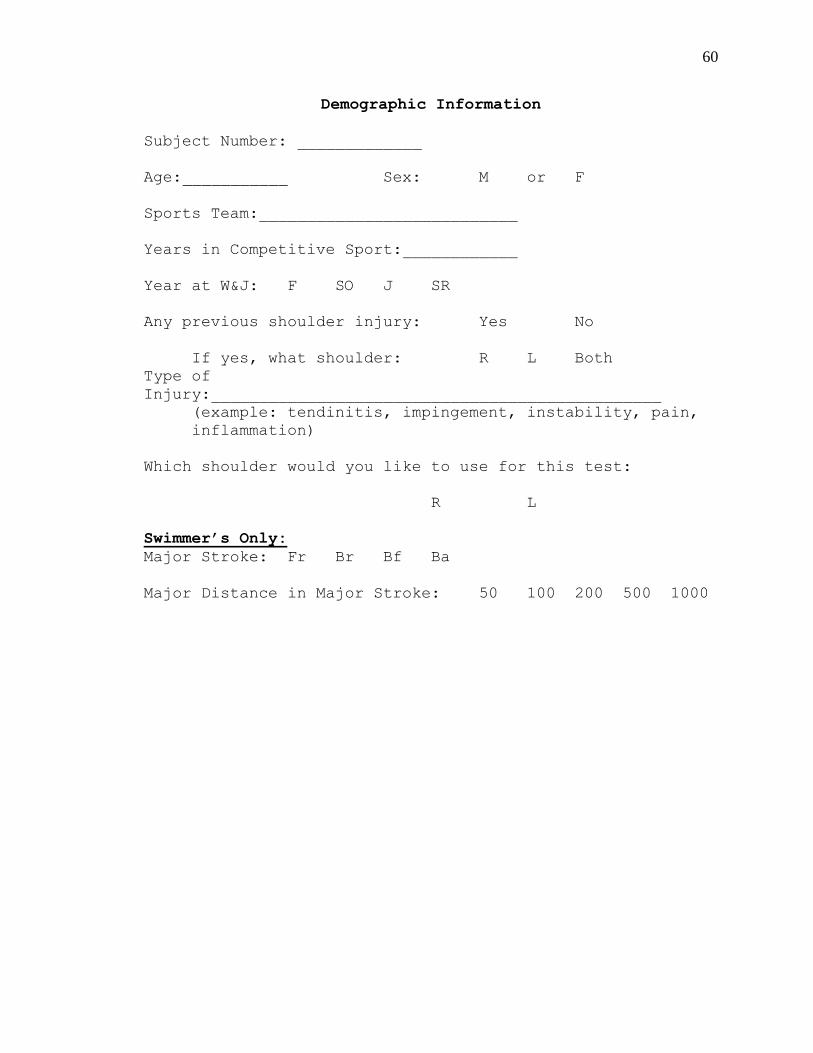
60
Demographic Information
Subject Number: _____________
Age:___________ Sex: M or F
Sports Team:___________________________
Years in Competitive Sport:____________
Year at W&J: F SO J SR
Any previous shoulder injury: Yes No
If yes, what shoulder: R L Both
Type of
Injury:_______________________________________________
(example: tendinitis, impingement, instability, pain,
inflammation)
Which shoulder would you like to use for this test:
R L
Swimmer’s Only:
Major Stroke: Fr Br Bf Ba
Major Distance in Major Stroke: 50 100 200 500 1000

61
Appendix C4
WMPA Scoring Guide
62
1. Ankle Posture (Ankle valgus or varus)
Photograph(s) used: anterior view, posterior view
Scores:
5: the degree of the Achilles tendon is less than
7
3: the degree of the Achilles tendon is between
7-10
1: the degree of the Achilles tendon is greater
than 10
2. Knee Interspace
Photograph(s) used: anterior view, posterior view
Scores:
5: the ankles are together and the medial
epicondyles are touching
3: 1. the medial epicondyles are touching and
the medial malleoli are not touching
2. the medial malleoli are touching but
there is 1-3 mm between the medial
epicondyles
1: 1. the medial epicondyles are more than 4 mm
apart and the medial malleoli are touching.
2. the Q angle is measured more than 15 for
women and more than 12 for men
3. Knee Hyperflexion or Hyperextension
Photograph(s) used: lateral view
Scores:
5: a line can be drawn straight from the greater
trochanter to the lower leg with no marked
deviations
3: there is a moderate deviation from the midline
either with flexion or extension of the knee
63
1: there is extreme deviation from the midline
from the greater trochanter through the thigh and
lower leg with flexion or extension of the knee.
4. Lordosis
Photograph(s) used: lateral view, left oblique view
Scores:
5: a circle with a diameter of 7 cm*
3: a circle with a diameter of 4.5 cm*
1: a circle with a diameter of 3 cm*
*circles of various diameters are used for this
assessment on top of the photographs. They are placed
against the lordosis curvature.
5. Kyphosis
Photograph(s) used: lateral view, left oblique view
Scores:
5: circle with a diameter of 9 cm*
3: circle with a diameter of 7 cm*
1: circle with a diameter of 6 cm*
*circles of various diameters are used for this
assessment on top of the photographs. They are placed
against the lordosis curvature.
6. Scoliosis
Photograph(s) used: posterior view
Scores:
5: a vertical line is made down the vertebrae
with the reflective dot stickers with no marked
deviations
3: a vertical line is made down the vertebrae
with the reflective dot stickers and there is
64
moderate deviation of 1.5-3 degrees from the
vertical line
1: a vertical line is made down the vertebrae
with the reflective dot stickers and there is an
extreme deviation that is greater than 3 degrees
from the vertical line
7. Shoulder; Rounded or Forward
Photograph(s) used: lateral view
Scores:
5: the shoulders are behind the upper chest
3: the front of the shoulders is slightly forward
of the upper chest
1: The fronts of the shoulders are clearly in
front of the upper chest
8. Shoulder abducted/winged scapulae
Photograph(s) used: lateral view, left oblique view
Scores:
5: No deviations
3: the inferior angles and portions of the medial
borders of the scapulae are moderately visible
1: the inferior angles and portions of the medial
borders of the scapulae are excessively Shoulder
Symmetry
9. Shoulder symmetry
Photograph(s) used: anterior view, posterior view
Scores:
5: no deviation when comparing the two shoulders
3: a deviation between 1- 2.5 mm when comparing
the two shoulders
65
1: a deviation greater than 2.5 mm when comparing
the two shoulders
10. Forward Head
Photograph(s) used: lateral view
Scores:
5: the head protraction angle is less than 5
degrees
3: the head protraction angle is between 5-10
degrees
1: the head protraction angle is greater than 10
degrees

66
APPENDIX C5
Institutional Review Board –
California University of Pennsylvania
67
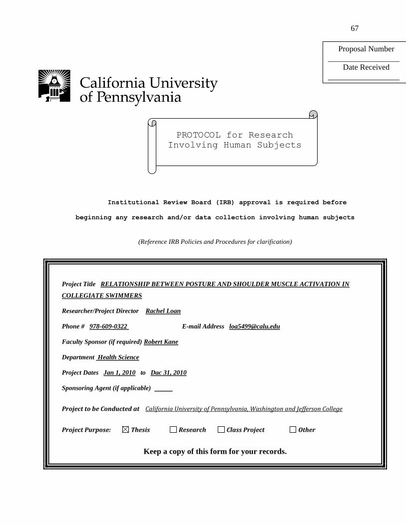
Institutional Review Board (IRB) approval is required before
beginning any research and/or data collection involving human subjects
(Reference IRB Policies and Procedures for clarification)
Project Title RELATIONSHIP BETWEEN POSTURE AND SHOULDER MUSCLE ACTIVATION IN
COLLEGIATE SWIMMERS
Researcher/Project Director Rachel Loan
Phone # 978-609-0322 E-mail Address [email protected]
Faculty Sponsor (if required) Robert Kane
Department Health Science
Project Dates Jan 1, 2010 to Dac 31, 2010
Sponsoring Agent (if applicable)
Project to be Conducted at California University of Pennsylvania, Washington and Jefferson College
Project Purpose: Thesis Research Class Project Other
Keep a copy of this form for your records.
PROTOCOL for Research
Involving Human Subjects
Proposal Number
Date Received
68
Please attach a typed, detailed summary of your project AND complete items 2
through 6.
1. Provide an overview of your project-proposal describing what you plan to do and how you
will go about doing it. Include any hypothesis(ses)or research questions that might be
involved and explain how the information you gather will be analyzed. For a complete list of
what should be included in your summary, please refer to Appendix B of the IRB Policies and
Procedures Manual.
The purpose of this study is to determine if there is a relationship in posture and muscle
activation of certain shoulder muscles that are important during the freestyle's swimming
stroke. The electromyographical (EMG) activity will be measured to evaluate activation of
three shoulder muscles during a functional exercise.
Hypothesis:
There will be a correlation between shoulder posture score and peak muscle activity in the
pectoralis major, serratus anterior, and latissimus dorsi during a functional shoulder extension
exercise.
Procedure:
Before any descriptive statistics were measured, the researcher applied for and obtained
approval from California University of Pennsylvania’s and Washington and Jefferson’s
Institutional Review Board. Preliminary research was conducted first so the researcher
became proficient in the procedures of the WMPA and the EMG. All subjects signed an
informed consent stating the reasoning for the study, how it would be conducted, and that
they could withdraw from the study at any time. A photographic release form was also
signed. The subjects also filled out a demographic form asking questions such as their years
in competitive swimming, hours per week of swimming practices, months per year of the
competitive season, and major swimming stroke.
When the subjects arrived for the assessment, they were first set up for the posture analysis.
The WMPA is a reliable and valid measure of posture. The subjects were asked to wear non-
baggy shorts and tank tops if necessary so that the landmarks, curvatures, and any valgus or
varus angles could be photographed.
Five photographs are taken of each subject. The first one is just the subject number
photograph for future referencing. For the second photograph, the subjects stood facing the
camera and the heels on the red line. Each subject was asked that they keep their chin
parallel to the ground, elbows and knees fully extended, and heels in contact with each other.
The third photograph was for the posterior view. The subject faced away from the camera
with the heels together on the red line; chin parallel to the ground and the knees and elbows
fully extended. The fourth photograph is a left lateral view. Subjects were instructed to stand
with their heels touching on the blue line, chin parallel to the ground and knees and elbows
extended. The fifth photograph was taken at an oblique angle. The subjects stood on the
yellow line which is at a 45 degree angle from the left back corner to the front right corner of
the platform.
The procedure of the measuring the WMPA scores is as follows. First, a grid is placed over
the top of each photograph of each subject with transparency paper. The grid is lined up with
the plumb lines used in the photographs. A second transparency paper is placed on top with
circle diameters which is used to determine the degrees of lordosis and kyphosis. A
protractor and ruler will is used to measure angles through the adhesive dots. A score of 5 is
69
given if there is no marked deviation, a 3 is given if there is moderate deviations, and a 1 is
given for extreme deviations
The next part of the process of the study was using the surface EMG to measure the muscle
activation. Electrode pads were placed on the latissimus dorsi, pectoralis major, and serratus
anterior muscle bellies . The subjects then did an upper body warm up using a Theraband®
which helped determine which band would be used for the subject during testing.
After the warm up was completed, the subject’s maximum voluntary contraction was then
tested. For the pectoralis major, the subject was placed in humeral adduction with the
shoulder in 900 of flexion and the palms together. The subject then applied the resistance by
pressing the palms together. The latissimus dorsi movement to reach a maximum voluntary
contraction is internal rotation and extension of the humerus with the shoulder in 30 degrees
of abduction and the elbow is extended. Resistance is then applied at the distal forearm by
the researcher. For the serratus anterior, the subject’s arm is forward flexed to 130 degrees.
The researcher’s hands are then placed one over the dorsal arm and one on the lateral scapula
for stability. The subject then isometrically flexes while the researcher adds resistance.
After the maximal voluntary contraction testing is complete for the three muscles, the subject
will then perform a functional shoulder extension exercise against resistance with a
Theraband® which would mimic the pull through phase of the freestyle stroke.
The subject would determine which of the three choices of Theraband® would have the best
resistance for them. Once established, the subject would then stand with enough room from
the stationary object holding the bands and bend at the waist at about 75 degrees to imitate
being in the water. They would then start in shoulder flexion at about 90 degrees and
impersonate their pull through phase with the resistance of the Theraband® and would do this
10 times. The procedure of the measuring the WMPA scores is as follows. First, a grid is
placed over the top of each photograph of each subject with transparency paper. The grid is
lined up with the plumb lines used in the photographs. A second transparency paper is placed
on top with circle diameters which is used to determine the degrees of lordosis and kyphosis.
A protractor and ruler will is used to measure angles through the adhesive dots.
The procedure of the measuring the WMPA scores is as follows. First, a grid is placed over
the top of each photograph of each subject with transparency paper. The grid is lined up with
the plumb lines used in the photographs. A second transparency paper is placed on top with
circle diameters which is used to determine the degrees of lordosis and kyphosis. Pictures will
be compared to the set of WMPA guidelines to determine if the postures being looked at will
be given a 1,3, or 5. A score of 1 is given if there is a marked deviation. A score of 3 is given
if there is a moderate deviation measured. A score of 5 is given if there are good body
mechanics that ranges from no deviations to a level that is above moderate deviations.
Data Analysis:
Pearson Product Moment Correlation was used to analyze correlation between shoulder
posture score with the peak muscle activity in swimmers, r values will be computed for each
muscle; pectoralis major, serratus anterior, and latissimus dorsi. SPSS version 17.0 for
Windows analyzed all data at p <.05. All EMG scores were reported as a percentage of
maximal voluntary contraction
2. Section 46.11 of the Federal Regulations state that research proposals involving human
subjects must satisfy certain requirements before the IRB can grant approval. You should
describe in detail how the following requirements will be satisfied. Be sure to address each
area separately.
70
a. How will you insure that any risks to subjects are minimized? If there are potential
risks, describe what will be done to minimize these risks. If there are risks, describe
why the risks to participants are reasonable in relation to the anticipated benefits.
Every precaution possible will be taken to make sure there are minimal risks during
this descriptive study. Subjects include only non-injured and physically active
participants which will decrease the likelihood of injury. If at any time the subjects
experience any pain or discomfort they will be allowed to discontinue the study. If
there is an injury, the research, who is a Certified Athletic Trainer assigned for the
year at Washington and Jefferson College, will be present to evaluate and provide
treatment for the subject. An Athletic Trainer is certified nationally through the
Board of Certification and the responsibilities of an Athletic Trainer encompass the
prevention, diagnosis, and intervention of emergency, acute, and chronic medical
conditions involving impairment, functional limitations, and disabilities. The risks to
the participants are reasonable with respect to the benefits because the risk is very
low. This descriptive study can change the way that athletes with a variety of
postures train for maximal gains of their sport.
b. How will you insure that the selection of subjects is equitable? Take into account
your purpose(s). Be sure you address research problems involving vulnerable
populations such as children, prisoners, pregnant women, mentally disabled persons,
and economically or educationally disadvantaged persons. If this is an in-class
project describe how you will minimize the possibility that students will feel coerced.
The subjects will be volunteer athletes from Washington and Jefferson University.
The study will be emailed to the coaches and announced at practices. The potential
subject will in no way be forced to participate in this study. There will be no research
problems involving vulnerable populations because the subjects will be college aged
students that do not include prisoners, pregnant, mentally disabled, or economically
or educationally disadvantaged persons.
c. How will you obtain informed consent from each participant or the subject’s legally
authorized representative and ensure that all consent forms are appropriately
documented? Be sure to attach a copy of your consent form to the project summary.
The copies of informed consent will be distributed to the coaches for the student
athletes and copies of the informed consent will be available on days when the study
is being done. The paper will inform the participant about their role in the descriptive
study and the procedure of the study.
d. Show that the research plan makes provisions to monitor the data collected to insure
the safety of all subjects. This includes the privacy of subjects’ responses and
provisions for maintaining the security and confidentiality of the data.
The data collection will be in a securely in password protected files on the
researcher's laptop used for collecting data, and the photographs will be kept in the
research advisor’s office, both of which only the researcher and research advisor will
have access to. The subjects' names will never appear on the data and they will be
assigned a number to keep the results anonymous.
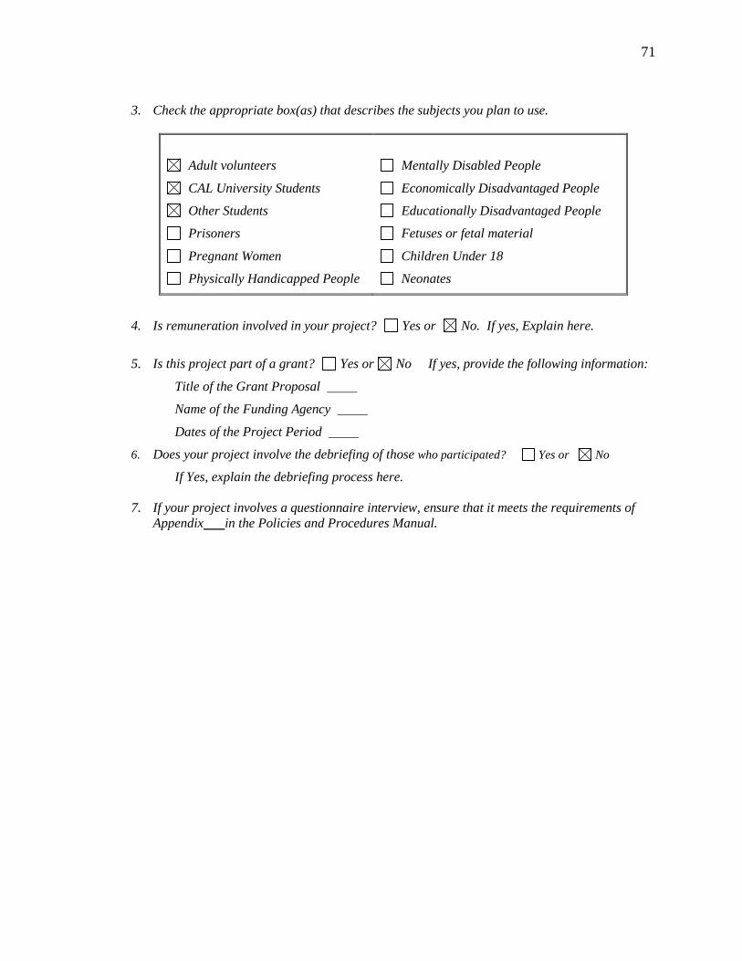
71
3. Check the appropriate box(as) that describes the subjects you plan to use.
Adult volunteers
CAL University Students
Other Students
Prisoners
Pregnant Women
Physically Handicapped People
Mentally Disabled People
Economically Disadvantaged People
Educationally Disadvantaged People
Fetuses or fetal material
Children Under 18
Neonates
4. Is remuneration involved in your project? Yes or No. If yes, Explain here.
5. Is this project part of a grant? Yes or No If yes, provide the following information:
Title of the Grant Proposal
Name of the Funding Agency
Dates of the Project Period
6. Does your project involve the debriefing of those who participated? Yes or No
If Yes, explain the debriefing process here.
7. If your project involves a questionnaire interview, ensure that it meets the requirements of
Appendix in the Policies and Procedures Manual.
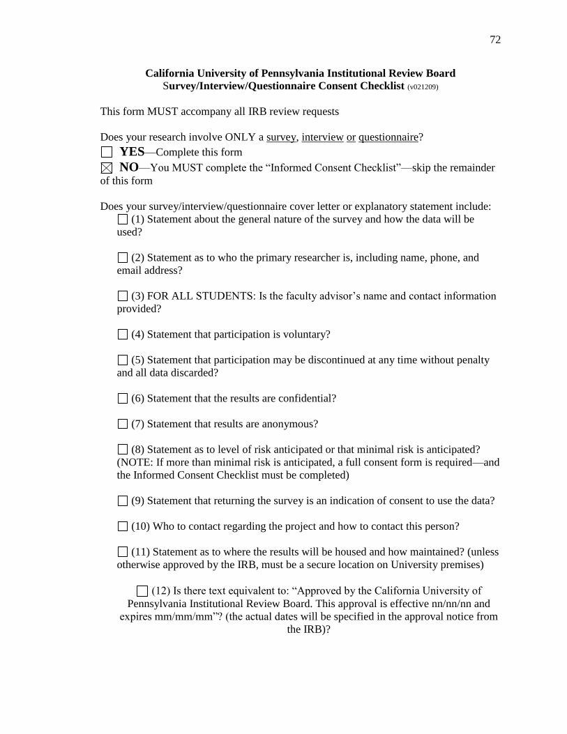
72
California University of Pennsylvania Institutional Review Board
Survey/Interview/Questionnaire Consent Checklist (v021209)
This form MUST accompany all IRB review requests
Does your research involve ONLY a survey, interview or questionnaire?
YES—Complete this form
NO—You MUST complete the “Informed Consent Checklist”—skip the remainder
of this form
Does your survey/interview/questionnaire cover letter or explanatory statement include:
(1) Statement about the general nature of the survey and how the data will be
used?
(2) Statement as to who the primary researcher is, including name, phone, and
email address?
(3) FOR ALL STUDENTS: Is the faculty advisor’s name and contact information
provided?
(4) Statement that participation is voluntary?
(5) Statement that participation may be discontinued at any time without penalty
and all data discarded?
(6) Statement that the results are confidential?
(7) Statement that results are anonymous?
(8) Statement as to level of risk anticipated or that minimal risk is anticipated?
(NOTE: If more than minimal risk is anticipated, a full consent form is required—and
the Informed Consent Checklist must be completed)
(9) Statement that returning the survey is an indication of consent to use the data?
(10) Who to contact regarding the project and how to contact this person?
(11) Statement as to where the results will be housed and how maintained? (unless
otherwise approved by the IRB, must be a secure location on University premises)
(12) Is there text equivalent to: “Approved by the California University of
Pennsylvania Institutional Review Board. This approval is effective nn/nn/nn and
expires mm/mm/mm”? (the actual dates will be specified in the approval notice from
the IRB)?
73
(13) FOR ELECTRONIC/WEBSITE SURVEYS: Does the text of the cover letter
or
explanatory statement appear before any data is requested from the participant?
(14) FOR ELECTONIC/WEBSITE SURVEYS: Can the participant discontinue
participation at any point in the process and all data is immediately discarded?
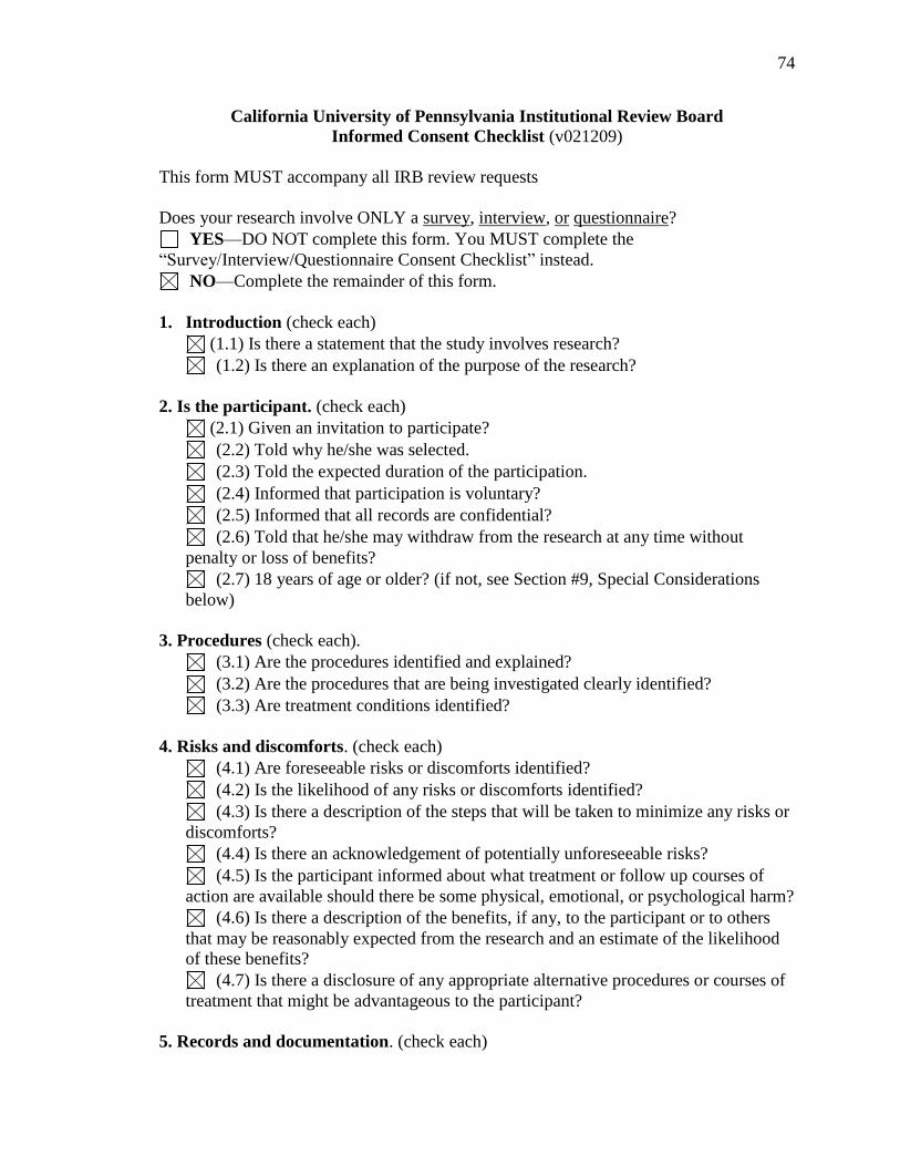
74
California University of Pennsylvania Institutional Review Board
Informed Consent Checklist (v021209)
This form MUST accompany all IRB review requests
Does your research involve ONLY a survey, interview, or questionnaire?
YES—DO NOT complete this form. You MUST complete the
“Survey/Interview/Questionnaire Consent Checklist” instead.
NO—Complete the remainder of this form.
1. Introduction (check each)
(1.1) Is there a statement that the study involves research?
(1.2) Is there an explanation of the purpose of the research?
2. Is the participant. (check each)
(2.1) Given an invitation to participate?
(2.2) Told why he/she was selected.
(2.3) Told the expected duration of the participation.
(2.4) Informed that participation is voluntary?
(2.5) Informed that all records are confidential?
(2.6) Told that he/she may withdraw from the research at any time without
penalty or loss of benefits?
(2.7) 18 years of age or older? (if not, see Section #9, Special Considerations
below)
3. Procedures (check each).
(3.1) Are the procedures identified and explained?
(3.2) Are the procedures that are being investigated clearly identified?
(3.3) Are treatment conditions identified?
4. Risks and discomforts. (check each)
(4.1) Are foreseeable risks or discomforts identified?
(4.2) Is the likelihood of any risks or discomforts identified?
(4.3) Is there a description of the steps that will be taken to minimize any risks or
discomforts?
(4.4) Is there an acknowledgement of potentially unforeseeable risks?
(4.5) Is the participant informed about what treatment or follow up courses of
action are available should there be some physical, emotional, or psychological harm?
(4.6) Is there a description of the benefits, if any, to the participant or to others
that may be reasonably expected from the research and an estimate of the likelihood
of these benefits?
(4.7) Is there a disclosure of any appropriate alternative procedures or courses of
treatment that might be advantageous to the participant?
5. Records and documentation. (check each)
75
(5.1) Is there a statement describing how records will be kept confidential?
(5.2) Is there a statement as to where the records will be kept and that this is a
secure location?
(5.3) Is there a statement as to who will have access to the records?
6. For research involving more than minimal risk (check each),
(6.1) Is there an explanation and description of any compensation and other
medical or counseling treatments that are available if the participants are injured
through participation?
(6.2) Is there a statement where further information can be obtained regarding the
treatments?
(6.3) Is there information regarding who to contact in the event of research-
related injury?
7. Contacts.(check each)
(7.1) Is the participant given a list of contacts for answers to questions about the
research and the participant’s rights?
(7.2) Is the principal researcher identified with name and phone number and
email address?
(7.3) FOR ALL STUDENTS: Is the faculty advisor’s name and contact
information provided?
8. General Considerations (check each)
(8.1) Is there a statement indicating that the participant is making a decision
whether or not to participate, and that his/her signature indicates that he/she has
decided to participate having read and discussed the information in the informed
consent?
(8.2) Are all technical terms fully explained to the participant?
(8.3) Is the informed consent written at a level that the participant can understand?
(8.4) Is there text equivalent to: “Approved by the California University of
Pennsylvania Institutional Review Board. This approval is effective nn/nn/nn and
expires mm/mm/mm”? (the actual dates will be specified in the approval notice from
the IRB)
9. Specific Considerations (check as appropriate)
(9.1) If the participant is or may become pregnant is there a statement that the
particular treatment or procedure may involve risks, foreseeable or currently
unforeseeable, to the participant or to the embryo or fetus?
(9.2) Is there a statement specifying the circumstances in which the participation
may be terminated by the investigator without the participant’s consent?
(9.3) Are any costs to the participant clearly spelled out?
(9.4) If the participant desires to withdraw from the research, are procedures for
orderly termination spelled out?
76
(9.5) Is there a statement that the Principal Investigator will inform the participant
or any significant new findings developed during the research that may affect them
and influence their willingness to continue participation?
(9.6) Is the participant is less than 18 years of age? If so, a parent or guardian must
sign the consent form and assent must be obtained from the child
Is the consent form written in such a manner that it is clear that the
parent/guardian is giving permission for their child to participate?
Is a child assent form being used?
Does the assent form (if used) clearly indicate that the child can freely refuse
to participate or discontinue participation at any time without penalty or coercion?
(9.7) Are all consent and assent forms written at a level that the intended
participant can understand? (generally, 8th
grade level for adults, age-appropriate for
children)
77
California University of Pennsylvania Institutional Review Board
Review Request Checklist (v021209)
This form MUST accompany all IRB review requests.
Unless otherwise specified, ALL items must be present in your review request.
Have you:
(1.0) FOR ALL STUDIES: Completed ALL items on the Review Request Form?
Pay particular attention to:
(1.1) Names and email addresses of all investigators
(1.1.1) FOR ALL STUDENTS: use only your CalU email
address)
(1.1.2) FOR ALL STUDENTS: Name and email address of your
faculty research advisor
(1.2) Project dates (must be in the future—no studies will be approved
which have already begun or scheduled to begin before final IRB approval—
NO EXCEPTIONS)
(1.3) Answered completely and in detail, the questions in items 2a through
2d?
2a: NOTE: No studies can have zero risk, the lowest risk is
“minimal risk”. If more than minimal risk is involved you MUST:
i. Delineate all anticipated risks in detail;
ii. Explain in detail how these risks will be minimized;
iii. Detail the procedures for dealing with adverse outcomes
due to these risks.
iv. Cite peer reviewed references in support of your
explanation.
2b. Complete all items.
2c. Describe informed consent procedures in detail.
2d. NOTE: to maintain security and confidentiality of data, all
study records must be housed in a secure (locked) location ON
UNIVERSITY PREMISES. The actual location (department, office,
etc.) must be specified in your explanation and be listed on any
consent forms or cover letters.
(1.4) Checked all appropriate boxes in Section 3? If participants under the
age of 18 years are to be included (regardless of what the study involves) you
MUST:
(1.4.1) Obtain informed consent from the parent or guardian—
consent forms must be written so that it is clear that the
parent/guardian is giving permission for their child to participate.
(1.4.2) Document how you will obtain assent from the child—
This must be done in an age-appropriate manner. Regardless of
whether the parent/guardian has given permission, a child is
completely free to refuse to participate, so the investigator must
document how the child indicated agreement to participate
(“assent”).
78
(1.5) Included all grant information in section 5?
(1.6) Included ALL signatures?
(2.0) FOR STUDIES INVOLVING MORE THAN JUST SURVEYS,
INTERVIEWS, OR QUESTIONNAIRES:
(2.1) Attached a copy of all consent form(s)?
(2.2) FOR STUDIES INVOLVING INDIVIDUALS LESS THAN 18
YEARS OF AGE: attached a copy of all assent forms (if such a form is used)?
(2.3) Completed and attached a copy of the Consent Form Checklist? (as
appropriate—see that checklist for instructions)
(3.0) FOR STUDIES INVOLVING ONLY SURVEYS, INTERVIEWS, OR
QUESTIONNAIRES:
(3.1) Attached a copy of the cover letter/information sheet?
(3.2) Completed and attached a copy of the
Survey/Interview/Questionnaire Consent Checklist? (see that checklist for
instructions)
(3.3) Attached a copy of the actual survey, interview, or questionnaire
questions in their final form?
(4.0) FOR ALL STUDENTS: Has your faculty research advisor:
(4.1) Thoroughly reviewed and approved your study?
(4.2) Thoroughly reviewed and approved your IRB paperwork? including:
(4.2.1) Review request form,
(4.2.2) All consent forms, (if used)
(4.2.3) All assent forms (if used)
(4.2.4) All Survey/Interview/Questionnaire cover letters (if used)
(4.2.5) All checklists
(4.3) IMPORTANT NOTE: Your advisor’s signature on the review request
form indicates that they have thoroughly reviewed your proposal and verified
that it meets all IRB and University requirements.
(5.0) Have you retained a copy of all submitted documentation for your records?
79
Project Director’s Certification Program Involving HUMAN SUBJECTS
The proposed investigation involves the use of human subjects and I am submitting the complete
application form and project description to the Institutional Review Board for Research Involving
Human Subjects.
I understand that Institutional Review Board (IRB) approval is required before beginning any
research and/or data collection involving human subjects. If the Board grants approval of this
application, I agree to:
1. Abide by any conditions or changes in the project required by the Board.
2. Report to the Board any change in the research plan that affects the method of using
human subjects before such change is instituted.
3. Report to the Board any problems that arise in connection with the use of human subjects.
4. Seek advice of the Board whenever I believe such advice is necessary or would be
helpful.
5. Secure the informed, written consent of all human subjects participating in the project.
6. Cooperate with the Board in its effort to provide a continuing review after investigations
have been initiated.
I have reviewed the Federal and State regulations concerning the use of human subjects in
research and training programs and the guidelines. I agree to abide by the regulations and
guidelines aforementioned and will adhere to policies and procedures described in my
application. I understand that changes to the research must be approved by the IRB before they
are implemented.
Professional Research
Project Director’s Signature Department Chairperson’s Signature
Student or Class Research
Student Researcher’s Signature
Supervising Faculty Member’s
Signature if required
Department Chairperson’s Signature
ACTION OF REVIEW BOARD (IRB use only)
80
The Institutional Review Board for Research Involving Human Subjects has reviewed this application to
ascertain whether or not the proposed project:
1. provides adequate safeguards of the rights and welfare of human subjects involved in the
investigations;
2. uses appropriate methods to obtain informed, written consent;
3. indicates that the potential benefits of the investigation substantially outweigh the risk involved.
4. provides adequate debriefing of human participants.
5. provides adequate follow-up services to participants who may have incurred physical, mental, or
emotional harm.
Approved[_________________________________] Disapproved
___________________________________________
_________________________
Chairperson, Institutional Review Board Date
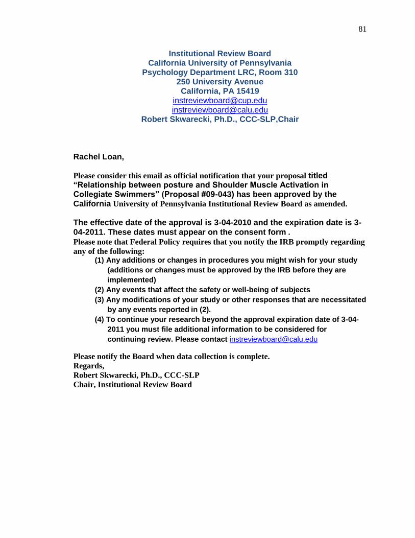
81
Institutional Review Board
California University of Pennsylvania
Psychology Department LRC, Room 310
250 University Avenue
California, PA 15419
Robert Skwarecki, Ph.D., CCC-SLP,Chair
Rachel Loan, Please consider this email as official notification that your proposal titled “Relationship between posture and Shoulder Muscle Activation in Collegiate Swimmers” (Proposal #09-043) has been approved by the California University of Pennsylvania Institutional Review Board as amended.
The effective date of the approval is 3-04-2010 and the expiration date is 3-
04-2011. These dates must appear on the consent form . Please note that Federal Policy requires that you notify the IRB promptly regarding
any of the following: (1) Any additions or changes in procedures you might wish for your study
(additions or changes must be approved by the IRB before they are
implemented)
(2) Any events that affect the safety or well-being of subjects
(3) Any modifications of your study or other responses that are necessitated
by any events reported in (2).
(4) To continue your research beyond the approval expiration date of 3-04-
2011 you must file additional information to be considered for
continuing review. Please contact [email protected]
Please notify the Board when data collection is complete.
Regards,
Robert Skwarecki, Ph.D., CCC-SLP
Chair, Institutional Review Board
82
Appendix C6
Electrode Placement and MVC
83
Muscle Electrode
Placement
MVC testing
Latissimus Dorsi Electrodes are
positioned obliquely
(approx 25 degrees
from horizontal in
the inferomedial
direction) 4cm below
the inferior angle of
the scapula. The
patient is asked to
cough while the
examiner palpates to
feel the latissimus
dorsi contract to
help locate placement
site.
Internal rotation
and extension of the
humerus with the
shoulder in 30
degrees of abduction
and the elbow is
extended. Resistance
is applied at the
distal forearm by
the researcher
Pectoralis Major Palpating along the
line of the clavicle
from the medial
clavicular head to
the acromion, one
third along and 3
cms down into the
sternal fibers of
the pectoralis major
Subject is placed
into humeral
adduction with the
shoulder in 90
degrees of flexion
and the palms are
facing each other.
The subject then
applies resistance
by pressing the
palms together
Serratus Anterior Electrodes place 2
cm apart just below
the axilla at the
level of the
inferior angle of
the scapula
Subjects arms are
forward flexed to
130 degrees. The
researcher‟s hands
are placed one over
the dorsal arm and
one on the lateral
scapula for
stability. Subject
then isometrically
flexes while the
researcher adds
resistance.
84
REFERENCES
1. Borsa PA, Laudner KG, Sauers EL. Mobility and
stability adaptations in the shoulder of the overhead
athlete: A theoretical and evidence-based perspective.
Sports Med. 2008; 38(1): 17-36
2. Pink M, Perry J, Browne A, Scovazzo ML, Kerrigan J.
The normal shoulder during freestyle swimming: An
electromyographic and cinematographic analysis of
twelve muscles. Am J Sports Med. 1991; 19(6): 569-576.
3. Weldon EJ, Richardson AB. Upper extremity overuse
injuries in swimming: A discussion of swimmer‟s
shoulder. Clinics in Sports Med. 2001; 20(3): 423-438
4. Levy O, Rath E. Traumatic soft tissue injuries of the
shoulder girdle. Trauma. 2002; 4: 223-235.
5. Starkey C, Ryan J. Eval of ortho and athletic
injuries. F.A. Davis Company. 2002; 2.
6. Sizer P, Gilbert K, Phelps V. Diagnosis and management
of the painful shoulder. Part 1. Clin anatomy and
pathomechanics. Pain Practice. 2003;3:39-57.
7. Kendall FP, McCreary EK, Provance PG, Rodger MM,
Romani WA. Muscles testing and function with posture
pain. Lippincott Williams & Williams. 2005; 5.
8. Decker MJ, Tokish JM, Ellis HB, Torry MR, Hawkins RJ.
Subscapularis muscle activity during selected
rehabilitation exercises. Am J Sports Med.
2003;31:126-134.
9. Swanik KA, Swanik CB. The effect of functional
training on the incidence of shoulder pain and
strength in intercollegiate swimmers. Sport
Rehabilitation. 2002;11:140-154.
85
10. Phadke V, Camago PR, Ludewig PM. Scapular and rotator
cuff muscle activity during arm elevation: A review of
normal function and alterations with shoulder
impingement. Brazilian J of Therapy. 2003;9:1-9
11. Wickham JB, Brown JM, McAndrew DJ. Muscles within
muscles: Anatomical and functional segmentation of
selected shoulder joint musculature. J Musculoskeletal
Research.2004; 8: 57-73.
12. Aujouannet YA, Bonifazi M, Hintzy F, Vuillerme N,
Rouard AH. Effects of a high-intensity swim test on
kinematic parameters in high-level athletes. Applied
Physiology, Nutrition, and Metabolism. 2006;31:150-
158.Maglischo, C.W.,
13. Maglischo EW, Higgins J, Hinricks R, Luedtke D,
Schleihauf RE, Thayer A. 1986. A biomechanical
analysis of the 1984 US Olympic swimming team: the
distance freestylers. J. Swim. Res. 2(3): 12–16.
14. Toussainti H, Truijens M. Biomechanical aspects of
peak performance in human swimming. Animal Biology.
2005;55:17-40.
15. Rackham GW. (1975) An analysis of arm propulsion in
swimming. In: J.P. Clarys & L. Lewillie
(Eds.), Swimming II, pp. 174-179. Baltimore, USA:
University Park Press.
16. Hollander AP, de Groot G, van Ingen Schenau GJ, Kahman
R, Toussaint HM. 1987. Contribution of the legs to
propulsion in front crawl swimming. In Swimming
science V. Edited by B.E. Ungerechts, K. Wilke, and K.
Reischle. University Park Press, Baltimore, Md. pp.
39–43.
17. Berger MAM, De Groot G, Hollander AP. 1995.
Hydrodynamic drag and lift forces on human hand/arm
models. J. Biomech. 28: 125–133.
86
18. Craig AB, Pendergast DR. 1979. Relationships of stroke
rate, distance per stroke, and velocity in competitive
swimming. Med. Sci. Sports, 11(3): 278–283.
19. East DJ. 1970. Swimming: an analysis of stroke
frequency, stroke length, and performance. N. Z. J.
Health Phys. Educ. Recr. 3: 16–27.
20. Brushoj C, Bak K, Johannsen HV, Faune P. Swimmers‟
painful shoulder; arthroscopic findings and return
rate to sports. Scandinavian J of Medicine and Science
in Sports. 2007;17:373-377.
21. Dominguez RH. Shoulder pain in swimmers. Physician
Sportsmed. 1980;8:35-42
22. Greipp JF. Swimmer's shoulder: the influence of
flexibility and weight training. Physician Sportsmed.
1985;13:92-105.
23. Kennedy DJ, Visco CJ, Press J. Current concepts for
shoulder training in the overhead athlete. Current
Sports Med Reports. 2009;8:154-160.
24. Zemek MJ. Comparison of glenohumeral joint laxity in
elite and recreational swimmers. Clin J Sport Med
1996; 6 (1): 40-47
25. Kleumper M, Uhl T, Hazelrigg H. Effect of stretching
and strengthening shoulder muscles on forward shoulder
posture in competitive swimmers. J of Sport Rehab.
2006; 15(1): 58-70.
26. McClure PW, Michener LA, Karduna AR. Shoulder function
and 3-dimensional scapular kinematics in people with
and without shoulder impingement syndrome. Physical
Therapy. 2006;86:1075-1090.
87
27. Scovazzo M, Browne A, Pink M, Jobe F, Kerrigan J. An
electromyographic cinematographic analysis of twelve
muscles. Am J Sports Med. 1991; 19(6): 577-582.
28. Perry M, Smith A, Straker L, Coleman J, O‟Sullivan P.
Reliability of sagital photographic spinal posture
assessment in adolescents. Advances in Physiotherapy.
2008; 10: 66-75.
29. Findlay S. Assessing posture: An essential tool. Sport
Ex. 2005; 6: 18-19.
30. Watson AWS, MacDonncha C. A reliable technique for the
assessment of posture: Assessment criteria for aspects
of posture. J of Sports Med and Phys Fitness. 2000; 9:
260-270.
31. Dark A, Ginn KA, Halaki M. Shoulder muscle recruitment
patterns during commonly used rotator cuff exercises:
An electromyographic study. Physical Therapy. 2007;
87(8): 1039-1045.
32. Kibler WB, Sciascia AD, Uhl TL, Tambay N, Cunningham
T. Electromyographic analysis of specific exercises
for scapular control in early phases of shoulder
rehabilitation. Am J Sports Med. 2008; 36(9): 1789-
1797.
33. Meldrum D, Cahalane E, Conroy R, Fitzgerald D,
Hardiman O. Maximum voluntary contraction: Reference
values and clinical application. Amytrophic Lateral
Sclerosis. 2008: 8: 47-55.
88
ABSTRACT
TITLE: Relationship between posture and peak
shoulder muscle activation in
collegiate swimmers
Researcher: Rachel Loan
Advisor: Dr. Robert Kane
Date: May 2010
Research Type: Master Thesis
Context: The purpose of the study was to examine
the relationship between posture and
muscle activity of three separate
muscles in NCAA Division III swimmers.
Objective: Few research studies have looked at the
relationship among different postures
and muscle activity in the major
muscles responsible for the freestyle
pull-through phase in swimming.
Design: This was a descriptive study using 16
volunteers from a NCAA Division III
swim team. Subjects came in for 30
minutes on one day.
Setting: Controlled laboratory setting
Participants: 16 NCAA Division III swimmers including
12 females and 4 males from Washington
and Jefferson College.
Interventions: Subjects came in on one day for 30
minutes. The WMPA photographs were
taken first. The subject was set up to
the EMG and performed MVIC of the three
muscles. The subject then performed the
functional shoulder extension exercise
that mimicked the pull-through phase of
the freestyle stroke.
89
Results: There was no significant relationship
among posture scores, using the Watson-
MacDonncha Posture Analysis (WMPA), and
percentage of peak muscle activity in
these shoulder muscles during a
resistive functional shoulder extension
exercise that mimicked the pull-through
phase of the freestyle swim stroke.
Conclusions: This descriptive study showed no
significant relationship among posture
and peak percentages of muscle activity
in three main swimming muscles It is
known though through previous studies,
that postural distortions can lead to
muscle imbalances, pain, and altered
biomechanics.
Word Count: 255