Embed Size (px)
Citation preview
Th
Phil. Trans. R. Soc. B (2005) 360, 903–911
doi:10.1098/rstb.2005.1640
Relating connectional architecture to grey matterfunction using diffusion imaging
Published online 29 May 2005
T. E. J. Behrens* and H. Johansen-Berg
One conof brain
*Autho
Oxford Centre for Functional Magnetic Resonance Imaging of the Brain (FMRIB), University of Oxford,John Radcliffe Hospital, Oxford OX3 9DU, UK
Understanding brain function in terms of connectional architecture is a major goal of neuroimaging.However, direct investigation of the influence of brain circuitry on function has been hindered by thelack of a technique for exploring anatomical connectivity in the in vivo brain. Recent advances inmagnetic resonance diffusion imaging have given scientists access to data relating to local whitematter architecture and, for the first time, have raised the possibility of in vivo investigations intobrain circuitry. This review investigates whether diffusion imaging may be used to identify regions ofgrey matter that are distinct in their connectional architecture, and whether these connectionaldifferences are reflected either in local cytoarchitecture or in local grey matter function.
Establishing a direct relationship between regional boundaries based on diffusion imaging andborders between regions that perform different functions would not only be of great significancewhen interpreting functional results, but would also provide a first step towards the validation ofdiffusion-based anatomical connectivity studies.
Keywords: grey matter parcellation; diffusion MRI; tractography; structure/function
1. INTRODUCTIONe relationship between brain structure and brainfunction is fundamental to neuroscience. Early studies
designed to parcellate human and non-human cortexinto regions that were distinct in their structure
(Brodman 1909; Vogt & Vogt 1919) acted under thehypothesis that these structurally defined regions also
differed in their function. Such studies parcellatecortical regions on the basis of microstructural featuressuch as cyto- or myeloarchitecture (Vogt et al. 1995;Roland & Zilles 1998; Zilles & Palomero-Gallagher2001), which have direct implications for local cellular
processing. Cortical regions, which may be delineatedon the basis of cellular microstructure, have also beenshown to differ in their connections to other brain
regions (e.g. Jones & Burton 1976; Vogt 1993).Independent of their respective relationships to
cellular microstructure, there is good reason to expecta strong correspondence between regional brain func-tion and connectional architecture. Anatomical
connections constrain the nature of the informationavailable to a region and the influence that it can exert
over other regions in a distributed network: localstructural organization can therefore be expected todetermine local functional specialization. However, this
relationship between circuitry and function has proveddifficult to test directly in the absence of a mechanism
for investigating connectivity in the functioning brain.Direct information relating to anatomical connectionsin the human brain is sparse and, until recently, only
available post-mortem. More direct assessment of
tribution of 21 to a Theme Issue ‘Multimodal neuroimagingconnectivity’.
r for correspondence ([email protected]).
903
connectivity is feasible using invasive tracer techniquesin non-human animals, but these approaches must befocused on specific regions, and assessment of functionin the same animals is technically demanding (althoughexcellent examples do exist; Yoshida et al. 2003).
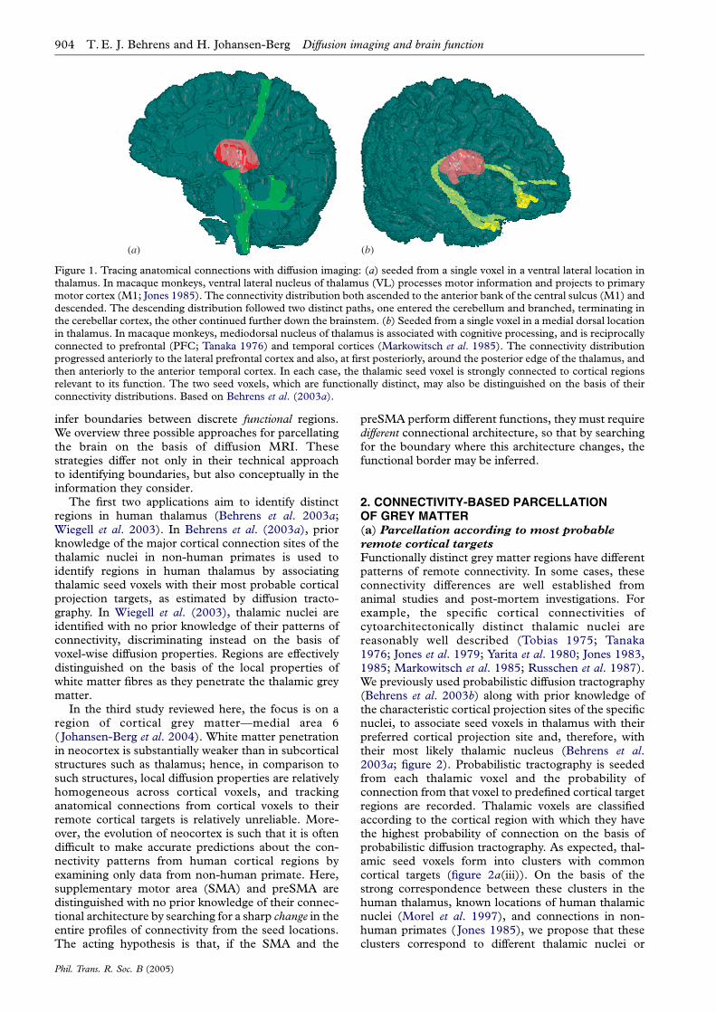
Magnetic resonance diffusion imaging is able tocharacterize the local diffusion properties of water intissue at a millimetre scale (Le Bihan 2003). In tissuewith a high degree of directional organization, the self-diffusion of water is hindered more in some directionsthan others. For example, water diffusion in brainwhite matter is more hindered perpendicular thanparallel to the major axis of a fibre bundle (Moseley etal. 1990), such that the direction of least resistance todiffusion (or principal diffusion direction) aligns wellwith the mean fibre orientation in an imaging voxel(Basser et al. 1994). Recent developments in diffusiontensor imaging techniques have enabled this diffusionanisotropy to be quantified, and for the principaldiffusion direction to be computed at each voxel.Using these local fibre orientations, early studies wereable to trace large white matter fibre tracts (Basser &Pierpaoli 1996; Mori et al. 1999; Catani et al. 2002),raising the possibility of explorations of anatomicalconnectivity in the living human brain. Using diffusiontractography, it has been possible to identify specificpatterns of anatomical connectivity associated withgrey matter ‘seed’ voxels (Conturo et al. 1999;Behrens et al. 2003a), and to demonstrate thatspatially near, but functionally distinct, grey matterseed voxels have very different connectivity patterns(figure 1).
In this paper, we investigate whether it is possibleto use diffusion-based connectivity information ina systematic fashion in order to parcellate grey matteraccording to its connectional architecture, and hence
q 2005 The Royal Society
(a) (b)
Figure 1. Tracing anatomical connections with diffusion imaging: (a) seeded from a single voxel in a ventral lateral location inthalamus. In macaque monkeys, ventral lateral nucleus of thalamus (VL) processes motor information and projects to primarymotor cortex (M1; Jones 1985). The connectivity distribution both ascended to the anterior bank of the central sulcus (M1) anddescended. The descending distribution followed two distinct paths, one entered the cerebellum and branched, terminating inthe cerebellar cortex, the other continued further down the brainstem. (b) Seeded from a single voxel in a medial dorsal locationin thalamus. In macaque monkeys, mediodorsal nucleus of thalamus is associated with cognitive processing, and is reciprocallyconnected to prefrontal (PFC; Tanaka 1976) and temporal cortices (Markowitsch et al. 1985). The connectivity distributionprogressed anteriorly to the lateral prefrontal cortex and also, at first posteriorly, around the posterior edge of the thalamus, andthen anteriorly to the anterior temporal cortex. In each case, the thalamic seed voxel is strongly connected to cortical regionsrelevant to its function. The two seed voxels, which are functionally distinct, may also be distinguished on the basis of theirconnectivity distributions. Based on Behrens et al. (2003a).
904 T. E. J. Behrens and H. Johansen-Berg Diffusion imaging and brain function
infer boundaries between discrete functional regions.We overview three possible approaches for parcellatingthe brain on the basis of diffusion MRI. Thesestrategies differ not only in their technical approachto identifying boundaries, but also conceptually in theinformation they consider.
The first two applications aim to identify distinctregions in human thalamus (Behrens et al. 2003a;Wiegell et al. 2003). In Behrens et al. (2003a), priorknowledge of the major cortical connection sites of thethalamic nuclei in non-human primates is used toidentify regions in human thalamus by associatingthalamic seed voxels with their most probable corticalprojection targets, as estimated by diffusion tracto-graphy. In Wiegell et al. (2003), thalamic nuclei areidentified with no prior knowledge of their patterns ofconnectivity, discriminating instead on the basis ofvoxel-wise diffusion properties. Regions are effectivelydistinguished on the basis of the local properties ofwhite matter fibres as they penetrate the thalamic greymatter.
In the third study reviewed here, the focus is on aregion of cortical grey matter—medial area 6( Johansen-Berg et al. 2004). White matter penetrationin neocortex is substantially weaker than in subcorticalstructures such as thalamus; hence, in comparison tosuch structures, local diffusion properties are relativelyhomogeneous across cortical voxels, and trackinganatomical connections from cortical voxels to theirremote cortical targets is relatively unreliable. More-over, the evolution of neocortex is such that it is oftendifficult to make accurate predictions about the con-nectivity patterns from human cortical regions byexamining only data from non-human primate. Here,supplementary motor area (SMA) and preSMA aredistinguished with no prior knowledge of their connec-tional architecture by searching for a sharp change in theentire profiles of connectivity from the seed locations.The acting hypothesis is that, if the SMA and the
Phil. Trans. R. Soc. B (2005)
preSMA perform different functions, they must requiredifferent connectional architecture, so that by searchingfor the boundary where this architecture changes, thefunctional border may be inferred.
2. CONNECTIVITY-BASED PARCELLATIONOF GREY MATTER(a) Parcellation according to most probable
remote cortical targets
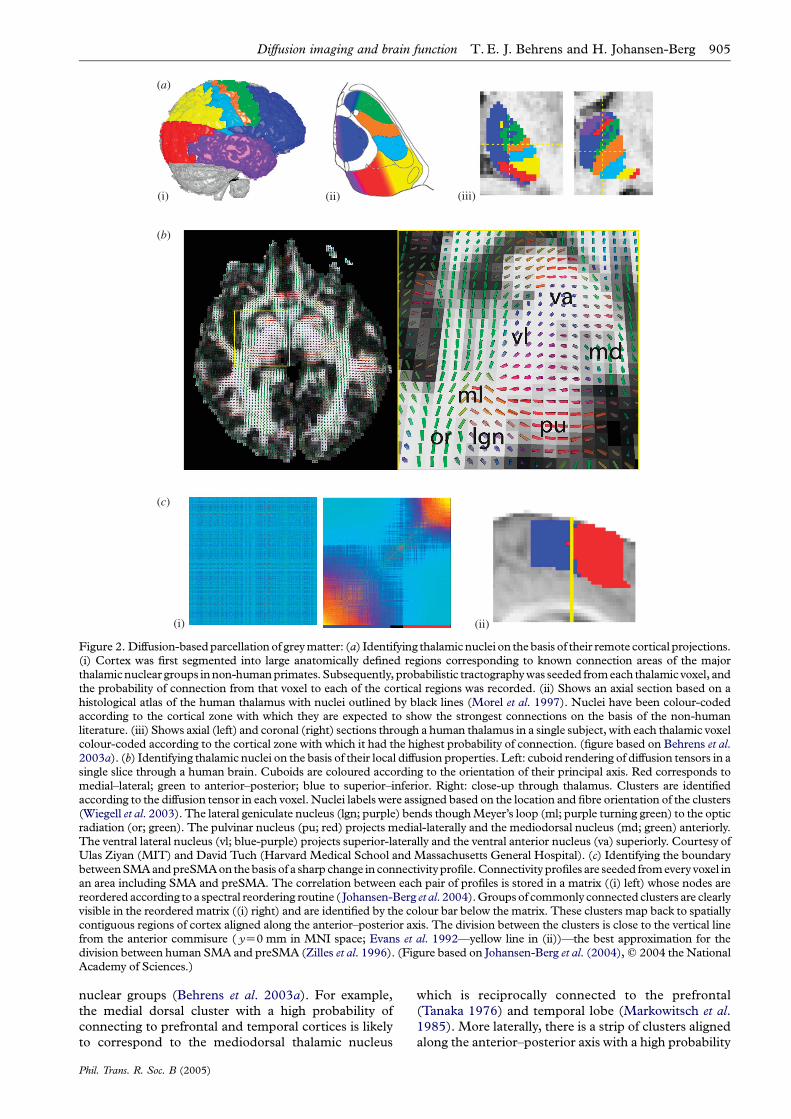
Functionally distinct grey matter regions have differentpatterns of remote connectivity. In some cases, theseconnectivity differences are well established fromanimal studies and post-mortem investigations. Forexample, the specific cortical connectivities ofcytoarchitectonically distinct thalamic nuclei arereasonably well described (Tobias 1975; Tanaka1976; Jones et al. 1979; Yarita et al. 1980; Jones 1983,1985; Markowitsch et al. 1985; Russchen et al. 1987).We previously used probabilistic diffusion tractography(Behrens et al. 2003b) along with prior knowledge ofthe characteristic cortical projection sites of the specificnuclei, to associate seed voxels in thalamus with theirpreferred cortical projection site and, therefore, withtheir most likely thalamic nucleus (Behrens et al.2003a; figure 2). Probabilistic tractography is seededfrom each thalamic voxel and the probability ofconnection from that voxel to predefined cortical targetregions are recorded. Thalamic voxels are classifiedaccording to the cortical region with which they havethe highest probability of connection on the basis ofprobabilistic diffusion tractography. As expected, thal-amic seed voxels form into clusters with commoncortical targets (figure 2a(iii)). On the basis of thestrong correspondence between these clusters in thehuman thalamus, known locations of human thalamicnuclei (Morel et al. 1997), and connections in non-human primates ( Jones 1985), we propose that theseclusters correspond to different thalamic nuclei or
(a)
(i)
(i) (ii)
(ii) (iii)
(b)
(c)
Figure 2. Diffusion-basedparcellationof greymatter: (a) Identifying thalamicnuclei on thebasis of their remote cortical projections.(i) Cortex was first segmented into large anatomically defined regions corresponding to known connection areas of the majorthalamicnuclear groups innon-humanprimates. Subsequently, probabilistic tractographywas seeded fromeach thalamic voxel, andthe probability of connection from that voxel to each of the cortical regions was recorded. (ii) Shows an axial section based on ahistological atlas of the human thalamus with nuclei outlined by black lines (Morel et al. 1997). Nuclei have been colour-codedaccording to the cortical zone with which they are expected to show the strongest connections on the basis of the non-humanliterature. (iii) Shows axial (left) and coronal (right) sections through a human thalamus in a single subject, with each thalamic voxelcolour-coded according to the cortical zone with which it had the highest probability of connection. (figure based on Behrens et al.2003a). (b) Identifying thalamic nuclei on the basis of their local diffusion properties. Left: cuboid rendering of diffusion tensors in asingle slice through a human brain. Cuboids are coloured according to the orientation of their principal axis. Red corresponds tomedial–lateral; green to anterior–posterior; blue to superior–inferior. Right: close-up through thalamus. Clusters are identifiedaccording to the diffusion tensor in each voxel. Nuclei labels were assigned based on the location and fibre orientation of the clusters(Wiegell et al. 2003). The lateral geniculate nucleus (lgn; purple) bends thoughMeyer’s loop (ml; purple turning green) to the opticradiation (or; green). The pulvinar nucleus (pu; red) projects medial-laterally and the mediodorsal nucleus (md; green) anteriorly.The ventral lateral nucleus (vl; blue-purple) projects superior-laterally and the ventral anterior nucleus (va) superiorly. Courtesy ofUlas Ziyan (MIT) and David Tuch (Harvard Medical School and Massachusetts General Hospital). (c) Identifying the boundarybetweenSMAandpreSMAon thebasis of a sharp change in connectivityprofile.Connectivity profiles are seeded fromevery voxel inan area including SMA and preSMA. The correlation between each pair of profiles is stored in a matrix ((i) left) whose nodes arereordered according to a spectral reordering routine ( Johansen-Berg et al. 2004).Groupsof commonly connected clusters are clearlyvisible in the reordered matrix ((i) right) and are identified by the colour bar below the matrix. These clusters map back to spatiallycontiguous regions of cortex aligned along the anterior–posterior axis. The division between the clusters is close to the vertical linefrom the anterior commisure ( yZ0 mm in MNI space; Evans et al. 1992—yellow line in (ii))—the best approximation for thedivision between human SMA and preSMA (Zilles et al. 1996). (Figure based on Johansen-Berg et al. (2004),q 2004 the NationalAcademy of Sciences.)
Diffusion imaging and brain function T.E. J. Behrens and H. Johansen-Berg 905
nuclear groups (Behrens et al. 2003a). For example,
the medial dorsal cluster with a high probability of
connecting to prefrontal and temporal cortices is likely
to correspond to the mediodorsal thalamic nucleus
Phil. Trans. R. Soc. B (2005)
which is reciprocally connected to the prefrontal
(Tanaka 1976) and temporal lobe (Markowitsch et al.1985). More laterally, there is a strip of clusters aligned
along the anterior–posterior axis with a high probability
906 T. E. J. Behrens and H. Johansen-Berg Diffusion imaging and brain function
of connecting to: premotor cortex (most anterior),primary motor cortex and somatosensory cortices(most posterior). These clusters are likely to corres-pond to the ventral anterior nucleus that connects topremotor cortex, the ventral lateral nucleus thatconnects to primary motor cortex and the ventralposterior nucleus that connects to somatosensorycortices ( Jones 1985).
Although the correspondence between the clustersidentified from connectivity data and histologicalsections of human thalamus is compelling(figure 2a(ii,iii)), the precise thalamic borders identifiedusing diffusion data in this way depend on the bordersthat are initially manually defined on the cortex. Thisdependence is not ideal for a number of reasons. First,the correspondence between gross anatomical land-marks underlying cortical cytoarchitecture is debatable(e.g. Amunts et al. 1999). Also, the parcellation dependson prior knowledge of what constitute meaningfulconnectivity target regions. This is reasonably wellachieved in the case of the thalamus, as thalamo-corticalrelationships are well described, but may be morechallenging for other regions.
(b) Parcellation according to local diffusion
properties
One solution to the problem of having to prespecifymeaningful target regions is to parcellate grey mattersimply on the basis of local diffusion properties. Thisapproach was taken by Wiegell et al. (2003), who alsoaimed to subdivide the human thalamus. Parcellation,according to local diffusion properties, does not rely ona priori anatomical knowledge, or on the explicitestimation of the connectivity patterns of thalamicvoxels. The authors hypothesize that differencesbetween nuclei in their remote anatomical connectivitypattern may be seen as differences between nuclei inthe characteristic orientation of the cortico-thalamic/thalamo-cortical striations within the nucleus. Hencevoxels belonging to different thalamic nuclei should bedistinguishable on the basis of their local diffusionproperties alone.
The diffusion tensor is reconstructed at each thal-amic voxel (figure 2b) and ametric is defined that allowsthe calculation of the dissimilarity or distance betweenany two diffusion tensors. The authors submit thesediffusion tensor data to an automated clustering routinethat attempts to partition the thalamus into groups ofvoxels that are close, both spatially and in terms of theirdiffusion tensors. Tensors are labelled as ‘close’ to oneanother if they are similar in properties such as diffusionanisotropy and mean diffusivity and if their principalorientations are well aligned. The resulting clusters(labelled in figure 2b) bear close resemblance to theconnectivity-defined regions in figure 2a, and topreviously reported histological segmentations of thethalamic nuclei. For comparison with the approachdescribed in the previous section, again note the medialdorsal and anterior clusters of voxels whose principalaxis of diffusion is in the anterior–posterior orientation(green), pointing towards prefrontal cortex, and notethe lateral cluster of voxels whose principal axis is in theinferior–superior orientation (blue/purple) pointingtowards sensorimotor cortices.
Phil. Trans. R. Soc. B (2005)
Parcellation with respect to local diffusion propertiescould provide a powerful means of subdividing greymatter. One strength of the technique is that it does notrequire prior specification of connectivity targets, anddoes not rely on the accuracy of diffusion-basedtractography routines. However, the approach doesrely on differences in remote connectivity being visibleas differences in local diffusion properties. Cases willexist in which voxels with similar local diffusionproperties (e.g. similar local fibre orientation) havedivergent patterns of remote connectivity. In suchcases, local information alone will not be enough toidentify the connectional differences.
(c) Parcellation on the basis of sharp changes
in connectivity
We have seen that by examining diffusion data relatingto either remote cortical connectivity or local fibreorientation it is possible to identify distinct regions inthe human thalamus that are proposed to correspondto functionally distinct thalamic nuclei. However, thetechniques used to identify these divisions may not beeasily generalizable to different regions in grey matter.Specifically, thalamus is a subcortical grey matterstructure in which there is a large degree of whitematter penetration. Local diffusion orientations withinthalamus are well defined and heterogeneous. Diffu-sion tractography seeded from thalamic voxels is robustand accurate. By comparison, cortical grey matterregions have a much lower degree of white matterpenetration, hence diffusion orientations are less welldefined and local diffusion properties in general aremore homogeneous. At least partly as a result of this,diffusion tractography can be less reliable when seededfrom cortical rather than from subcortical grey matter.Moreover, connectivity-based approaches relying onpredictions from non-human primate data cannot beapplied to human cortical regions with no unequivocalhomologue in other species.
In Johansen-Berg et al. (2004), we propose afundamentally different strategy for inferring structuralparcellation from diffusion data that allows ‘blind’discrimination of regions with different patterns ofconnection, and which benefits from the sensitivitygained by considering a seed point’s remote targetswithout having to rely on a complete, or even accurate,representation of its connectivity. Instead of classifying asingle seed point on the basis of its cortical target, weconsider the entire ‘profile’ of connectivity from all seedpoints simultaneously. This allows us to find clusters ofseed voxels with very similar patterns of connectivity or,equivalently, draw boundaries between adjacent brainareas whose patterns are markedly different.
The specific cortical focus in this paper is medialarea 6—an area which provides an excellent testcase. In macaque monkey, the medial part of thehomologue of Brodmann’s area 6 consists of twocytoarchitectonically distinct regions (Vogt & Vogt1919; Matelli et al. 1991), thought to correspond toSMA proper and preSMA. These two regions exhibitdifferent functional responses (Matsuzaka & Tanji1996), have distinct connections (Luppino et al.1993) and may be distinguished on the basis ofgross anatomy as lying on the medial surface, either
Diffusion imaging and brain function T.E. J. Behrens and H. Johansen-Berg 907
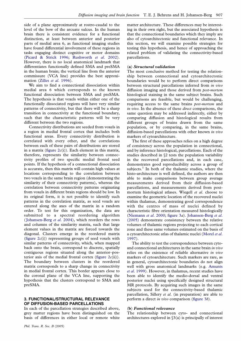
side of a plane approximately at rostro-caudal to thelevel of the bow of the arcuate sulcus. In the humanbrain there is consistent evidence for a functionaldistinction, at least between anterior and posteriorparts of medial area 6, as functional imaging studieshave found differential involvement of these regions intasks engaging distinct cognitive or motor domains(Picard & Strick 1996; Rushworth et al. 2002).However, there is no local anatomical landmark thatdifferentiates functionally defined SMA and preSMAin the human brain; the vertical line from the anteriorcommissure (VCA line) provides the best approxi-mation (Zilles et al. 1996).
We aim to find a connectional dissociation withinmedial area 6 which corresponds to the knownfunctional dissociation between SMA and preSMA.The hypothesis is that voxels within either one of thefunctionally dissociated regions will have very similarpatterns of connectivity, but that there will be a sharptransition in connectivity at the functional boundary,such that the characteristic patterns will be verydifferent between the two regions.
Connectivity distributions are seeded from voxels ina region in medial frontal cortex that includes bothfunctional areas. Every connectivity distribution iscorrelated with every other, and the correlationsbetween each of these pairs of distributions are storedin a matrix (figure 2c(i)). Each element in this matrix,therefore, represents the similarity between connec-tivity profiles of two specific medial frontal seedpoints. If the hypothesis of a connectional dissociationis accurate, then this matrix will contain high values atlocations corresponding to the correlation betweentwo voxels in the same brain region (demonstrating thesimilarity of their connectivity patterns), whereas thecorrelation between connectivity patterns originatingfrom voxels in different brain regions should be low. Inits original form, it is not possible to identify suchpatterns in the correlation matrix, as seed voxels areentered along the axes of the matrix in a randomorder. To test for this organization, the data aresubmitted to a spectral reordering algorithm( Johansen-Berg et al. 2004), which reorders the rowsand columns of the similarity matrix, such that largeelement values in the matrix are forced towards thediagonal. Clusters emerge in the reordered matrix(figure 2c(i)) representing groups of seed voxels withsimilar patterns of connectivity, which, when mappedback onto the brain, correspond to discrete, spatiallycontiguous regions situated along the anterior–pos-terior axis of the medial frontal cortex (figure 2c(ii)).The boundary between clusters in the reorderedmatrix corresponds to a sharp change in connectivityin medial frontal cortex. This border appears close tothe coronal plane of the VCA line, supporting thehypothesis that the clusters correspond to SMA andpreSMA.
3. FUNCTIONAL/STRUCTURAL RELEVANCEOF DIFFUSION-BASED PARCELLATIONSIn each of the parcellation strategies described above,grey matter regions have been distinguished on thebasis of differences in either local or remote white
Phil. Trans. R. Soc. B (2005)
matter architecture. These differences may be interest-ing in their own right, but the associated hypothesis isthat the connectional boundaries which they imply arealso of cytoarchitectonic and functional relevance. Inthis section, we will examine possible strategies fortesting this hypothesis, and hence of approaching therelated problem of validating the connectivity-basedparcellations.
(a) Structural validationThe most conclusive method for testing the relation-ship between connectional and cytoarchitectonicboundaries would be to perform direct comparisonsbetween structural parcellations inferred from in vivodiffusion imaging and those derived from post-mortemhistological staining in the same subject brains. Suchcomparisons are feasible, but would be challenging,requiring access to the same brains post-mortem andin vivo. In the absence of these direct comparisons, thesame question may be addressed indirectly, either bycomparing diffusion and histological results fromdifferent groups of brains drawn from the samepopulation, or by comparing, in the same brains,diffusion-based parcellations with other known in vivomarkers of cytoarchitecture.
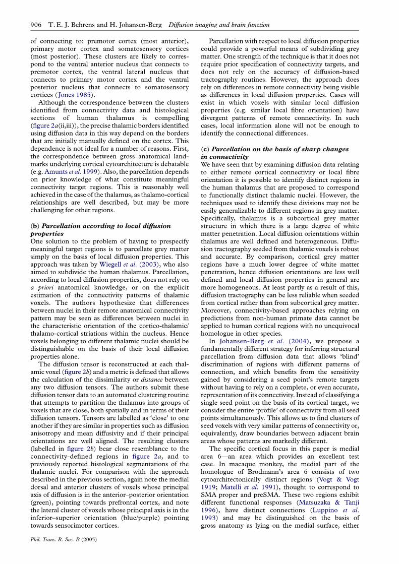
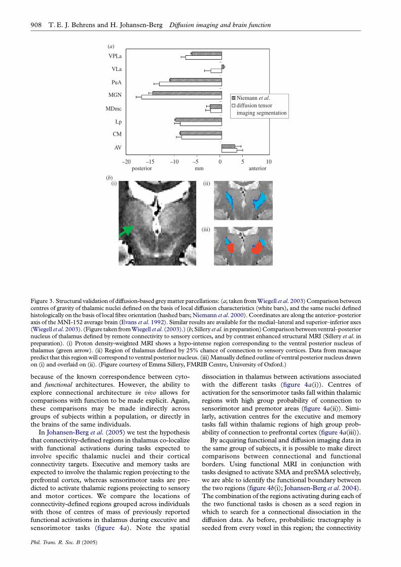
The first of these approaches relies on a high degreeof consistency across the population in connectional,and by inference histological, parcellations. Each of thestudies described in §2 tests the population variabilityin the recovered parcellations and, in each case,demonstrates good reproducibility across a group ofsubjects.1 In both of the thalamic studies, where thehisto-architecture is well defined, the authors are thenable to make comparisons between group averagemeasurements derived from their diffusion-basedparcellations, and measurements derived from post-mortem histological atlases. Wiegell et al. choose toexamine the geometric location of the recovered nucleiwithin thalamus, demonstrating good correspondencewith the centres of mass of nuclei defined bycharacteristic fibre orientation measured histologically(Niemann et al. 2000; figure 3a). Johansen-Berg et al.(2005) demonstrate consistency between the relativevolumes of thalamic regions projecting to each corticalzone and these same volumes estimated on the basis ofa cytoarchitectonic atlas of thalamic nuclei (Morel et al.1997).
The ability to test the correspondence between cyto-and connectional architectures in the same brain in vivorelies on the existence of reliable alternative in vivomarkers of cytoarchitecture. Such markers are rare, asin general, cytoarchitectonic boundaries do not alignwell with gross anatomical landmarks (e.g. Amuntset al. 1999). However, in thalamus, recent studies havebeen able to identify the medio-dorsal and ventralposterior nuclei using specifically designed structuralMR protocols. By acquiring such images in the samesubjects used for the connectivity-based thalamicparcellation, Sillery et al. (in preparation) are able toperform a direct in vivo comparison (figure 3b).
(b) Functional relevanceThe relationship between cyto- and connectionalarchitectures explored in §3(a) is principally of interest
(a)
(b)
VPLa
VLa
PuA
MGN
MDmc
Lp
CM
AV
(i) (ii)
(iii)
–20posterior mm
Niemann et al.diffusion tensor imaging segmentation
anterior–15 –10 –5 0 5 10
Figure 3. Structural validation of diffusion-based greymatter parcellations: (a; taken fromWiegell et al. 2003)Comparison betweencentres of gravity of thalamic nuclei defined on the basis of local diffusion characteristics (white bars), and the same nuclei definedhistologically on the basis of local fibre orientation (hashed bars;Niemann et al. 2000). Coordinates are along the anterior–posterioraxis of theMNI-152 average brain (Evans et al. 1992). Similar results are available for the medial–lateral and superior–inferior axes(Wiegell et al. 2003). (Figure taken fromWiegell et al. (2003).) (b; Sillery et al. inpreparation)Comparisonbetweenventral–posteriornucleus of thalamus defined by remote connectivity to sensory cortices, and by contrast enhanced structural MRI (Sillery et al. inpreparation). (i) Proton density-weighted MRI shows a hypo-intense region corresponding to the ventral posterior nucleus ofthalamus (green arrow). (ii) Region of thalamus defined by 25% chance of connection to sensory cortices. Data from macaquepredict that this regionwill correspond to ventral posterior nucleus. (iii)Manually definedoutline of ventral posterior nucleus drawnon (i) and overlaid on (ii). (Figure courtesy of Emma Sillery, FMRIB Centre, University of Oxford.)
908 T. E. J. Behrens and H. Johansen-Berg Diffusion imaging and brain function
because of the known correspondence between cyto-
and functional architectures. However, the ability to
explore connectional architecture in vivo allows for
comparisons with function to be made explicit. Again,
these comparisons may be made indirectly across
groups of subjects within a population, or directly in
the brains of the same individuals.
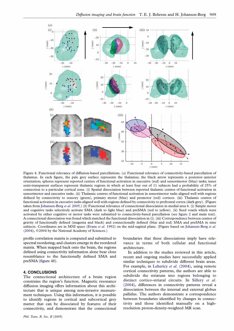
In Johansen-Berg et al. (2005) we test the hypothesisthat connectivity-defined regions in thalamus co-localize
with functional activations during tasks expected to
involve specific thalamic nuclei and their cortical
connectivity targets. Executive and memory tasks are
expected to involve the thalamic region projecting to the
prefrontal cortex, whereas sensorimotor tasks are pre-
dicted to activate thalamic regions projecting to sensory
and motor cortices. We compare the locations of
connectivity-defined regions grouped across individuals
with those of centres of mass of previously reported
functional activations in thalamus during executive and
sensorimotor tasks (figure 4a). Note the spatial
Phil. Trans. R. Soc. B (2005)
dissociation in thalamus between activations associated
with the different tasks (figure 4a(i)). Centres of
activation for the sensorimotor tasks fall within thalamic
regions with high group probability of connection to
sensorimotor and premotor areas (figure 4a(ii)). Simi-
larly, activation centres for the executive and memory
tasks fall within thalamic regions of high group prob-
ability of connection to prefrontal cortex (figure 4a(iii)).By acquiring functional and diffusion imaging data in
the same group of subjects, it is possible to make direct
comparisons between connectional and functional
borders. Using functional MRI in conjunction with
tasks designed to activate SMA and preSMA selectively,
we are able to identify the functional boundary between
the two regions (figure 4b(i); Johansen-Berg et al. 2004).
The combination of the regions activating during each of
the two functional tasks is chosen as a seed region in
which to search for a connectional dissociation in the
diffusion data. As before, probabilistic tractography is
seeded from every voxel in this region; the connectivity
(a)(i)
(b)
(i) (iii)
(ii)
0
–10
–20y (m
m)
–20x (mm)
–10
–30
20
10
0
z (m
m)
–20x (mm)
–10
20
10
0
–10
65
60
55
50
45–10 –5 0 5 10 15
z (m
m)
z (m
m)
–20–30x (mm)
–10 0
20
10
0
–10
z (m
m)
–20–30x (mm)
y (mm)
–10 100
–20
10
y (m
m)
–30y (mm)
–20 –10 0 10
0
–10
10
0
y (m
m)
–20–30x (mm)
–10 0 10
–30
–20
–10
(ii) (iii)
Figure 4. Functional relevance of diffusion-based parcellations. (a) Functional relevance of connectivity-based parcellation ofthalamus. In each figure, the pale grey surface represents the thalamus; the black arrow represents a posterior–anteriororientation; spheres represent reported centres of functional activation in executive (red) and sensorimotor (blue) tasks; innersemi-transparent surfaces represent thalamic regions in which at least four out of 11 subjects had a probability of 25% ofconnection to a particular cortical zone. (i) Spatial dissociation between reported thalamic centres of functional activation insensorimotor and executive tasks. (ii) Thalamic centres of functional activation in sensorimotor tasks aligned well with regionsdefined by connectivity to sensory (green), primary motor (blue) and premotor (red) cortices. (iii) Thalamic centres offunctional activation in executive tasks aligned well with regions defined by connectivity to prefrontal cortex (dark grey). (Figuretaken from Johansen-Berg et al. 2005.) (b) Functional relevance of connectional dissociation in medial area 6. (i) Simple motorand cognitive tasks selectively activate SMA (dark to light blue) and preSMA (red to yellow). (ii) Seed voxels which wereactivated by either cognitive or motor tasks were submitted to connectivity-based parcellation (see figure 2 and main text).A connectional dissociation was found which matched the functional dissociation in (i). (iii) Correspondence between centres ofgravity of functionally defined (magenta and black) and connectionally defined (blue and red) SMA and preSMA in ninesubjects. Coordinates are in MNI space (Evans et al. 1992) on the mid-sagittal plane. (Figure based on Johansen-Berg et al.(2004), q2004 by the National Academy of Sciences.)
Diffusion imaging and brain function T.E. J. Behrens and H. Johansen-Berg 909
profile correlation matrix is computed and submitted tospectral reordering; and clusters emerge in the reorderedmatrix. When mapped back onto the brain, the regionsdefined using connectivity information alone bear closeresemblance to the functionally defined SMA andpreSMA (figure 4b).
4. CONCLUSIONSThe connectional architecture of a brain regionconstrains the region’s function. Magnetic resonancediffusion imaging offers information about this archi-tecture that is unique among non-invasive measure-ment techniques. Using this information, it is possibleto identify regions in cortical and subcortical greymatter that can be dissociated by features of theirconnectivity, and demonstrate that the connectional
Phil. Trans. R. Soc. B (2005)
boundaries that these dissociations imply have rele-
vance in terms of both cellular and functional
architecture.
In addition to the studies reviewed in this article,
recent and ongoing studies have successfully applied
similar techniques to subdivide different brain areas.
For example, in Lehericy et al. (2004), using remote
cortical connectivity patterns, the authors are able to
subdivide the striatum into regions belonging to
distinct cortico-striatal circuits. In Sillery et al.
(2004), differences in connectivity patterns reveal a
dissociation between the internal and external globus
pallidus. The authors demonstrate a correspondence
between boundaries identified by changes in connec-
tivity and those identified manually on a high-
resolution proton-density-weighted MR scan.
910 T. E. J. Behrens and H. Johansen-Berg Diffusion imaging and brain function
A general relationship between boundaries in con-
nectional architecture visible to diffusion imaging andborders between cytoarchitectonically and functionally
distinct grey matter regions would be of greatsignificance to future neuroimaging studies. Such a
relationship would allow future functional imagingresults to be interpreted in the context of local
cytoarchitectonic borders. Functional data could becompared directly across studies with reference to the
cytoarchitectonic region of the activation site. Func-tionally relevant boundaries could be preserved when
aligning data from different subjects, allowing, forexample, for more accurate measurements of variability
in functional responses across a population. The ability
to infer cytoarchitectonic boundaries in vivowould alsohave significance for studies of brain structure.
Boundary locations, or regional volumes, could becompared across subject groups or brain hemispheres.
Lesion locations could be estimated accurately withrespect to the cytoarchitecture.
However, although the strategies reviewed in thisarticle have allowed for initial demonstrations of the
potential of diffusion data for inferring structural andfunctional parcellations, they are not yet in a position to
provide generally applicable tools. Any techniquedesigned to group data into dissociated clusters relies
on the ability to make hard decisions about, forexample, the number of clusters to choose and the
membership of each cluster. To date, diffusion-basedparcellation studies have relied on manual setting of
these parameters—Weigell et al. (2003) chose to searchfor 14 thalamic nuclei. Johansen-Berg et al. (2004)
identified clusters by eye in the reordered correlationmatrices. Before these techniques can be generally
applied to blind structural parcellation in grey matter,
statistical techniques should be developed to provide anautomated framework in which to make these
decisions. This problem is closely related to problemsfaced by scientists attempting to infer similar parcella-
tions from classical histology. Robust methods formaking decisions about the existence and location of
boundaries in cellular architecture are the subject ofongoing research (e.g. Schleicher et al. 2000).
The methods that we have described have all reliedon explicit models of local diffusion in biological tissue.
These models make simplifying assumptions, such asthe existence of only a single characteristic fibre
orientation within any voxel, which may precludethem from identifying boundaries in regions of the
brain where these assumptions break down. Recentresearch has allowed the representation of local
diffusion information within a voxel without the needfor these restrictive models (Alexander et al. 2002;
Tuch et al. 2003). This new technology will only
improve the sensitivity and accuracy of all diffusion-based connectivity studies.
The spatial resolution of the parcellations is alsolimited by current technology. A set of diffusion-
weighted images appropriate for tractography mighttake 40 min to acquire, and have spatial resolution of
only 2!2!2 mm3. As MR scanner technologyimproves, the spatial resolution of the images will
increase, allowing for finer anatomical parcellations.
Phil. Trans. R. Soc. B (2005)
Despite these limitations, we have shown thatdiffusion-based parcellations, carried out in controlledcircumstances in which functional or cytoarchitectonicboundaries are available, have identified connectionalborders which align well with the functional orstructural data. In the cases where the inferredparcellations depend on the accuracy of the underlyingdiffusion tractography, the functional/structural vali-dation of these parcellations may prove a first steptowards validating the tractography process. Even-tually, the validity of diffusion-based connectivitystudies should be established by direct comparisonwith invasive tracer studies in non-human primates.However, at present, the voxel resolution available todiffusion studies (of the order of 1 mm3) is such thatonly the largest fibre pathways are visible in a macaquebrain (Parker et al. 2002). The demonstration of goodcorrespondence between connectional, functional andcytoarchitectures inferred from diffusion imaging,functional imaging and histolology, respectively, notonly promises to provide powerful tools for studyingbrain structure and function in vivo, but lends weight tothe argument that diffusion imaging is providingmeasures relating to anatomical brain circuitry.
ENDNOTE1Note that group reproducibility of the thalamic parcellation
described in Behrens et al. (2003a) is examined in detail in
Johansen-Berg et al. (2004).
REFERENCESAlexander, D. C., Barker, G. J. & Arridge, S. R. 2002
Detection and modeling of non-Gaussian apparent diffu-sion coefficient profiles in human brain data.Magn. Reson.Med. 48, 331–340.
Amunts, K., Schleicher, A., Burgel, U., Mohlberg, H.,Uylings, H. B. & Zilles, K. 1999 Broca’s region revisited:cytoarchitecture and intersubject variability. J. Comp.Neurol. 412, 319–341.
Basser, P. & Pierpaoli, C. 1996 Microstructural and physio-logical features of tissues elucidated by quantitative-diffusion-tensor MRI. J. Magn. Reson. B 111, 209–219.
Basser, P. J., Matiello, J. & Le Bihan, D. 1994 Estimation ofthe effective self-diffusion tensor from the NMR spin echo.J. Magn. Reson. B 103, 247–254.
Behrens, T. E. J. et al. 2003a Non-invasive mapping ofconnections between human thalamus and cortex usingdiffusion imaging. Nat. Neurosci. 6, 750–757.
Behrens, T. E. J. et al. 2003b Characterization and propa-gation of uncertainty in diffusion-weighted MR imaging.Magn. Reson. Med. 50, 1077–1088.
Brodman, K. 1909 Lokalisationslehre der Grosshirnrinde inIhren Prinzipien Dargestellt auf Grund des Zellenbaues.Leipzig, Germany: Barth.
Catani, M., Howard, R. J., Pajevic, S. & Jones, D. K. 2002Virtual in vivo interactive dissection of white matterfasciculi in the human brain. NeuroImage 17, 77–94.
Conturo, T. E., Lori, N. F., Cull, T. S., Akbudak, E., Snyder,A. Z., Shimony, J. S., McKinstry, R. C., Burton, H. &Raichle, M. E. 1999 Tracking neuronal fiber pathways inthe living human brain. Proc. Natl Acad. Sci. USA 96,10 422–10 427.
Evans, A. C., Marrett, S., Neelin, P., Collins, L., Worsley, K.,Dai, W., Milot, S., Meyer, E. & Bub, D. 1992 Anatomical
Diffusion imaging and brain function T.E. J. Behrens and H. Johansen-Berg 911
mapping of functional activation in stereotactic coordinate
space. NeuroImage 1, 43–53.
Johansen-Berg, H., Behrens, T. E. J., Robson, M. D.,
Drobnjak, I., Rushworth, M. F. S., Brady, J. M., Smith,
S. M., Higham, D. J. & Matthews, P. M. 2004 Changes in
connectivity profiles define functionally distinct regions in
human medial frontal cortex. Proc. Natl Acad. Sci. USA
101, 13 335–13 340.
Johansen-Berg, H., Behrens, T. E., Sillery, E., Ciccarelli, O.,
Thompson, A. J., Smith, S. M. & Matthews, P. M. 2005
Functional–anatomical validation and individual variation
of diffusion tractography-based segmentation of the
human thalamus. Cereb. Cortex 15, 31–39.
Jones, E. G. 1983 Lack of collateral thalamocortical
projections to fields of the first somatic sensory cortex in
monkeys. Brain 52, 375–384.
Jones, E. G. 1985 The thalamus. New York: Plenum Press.
Jones, E. G. & Burton, H. 1976 Areal differences in the
laminar distribution of thalamic afferents in cortical fields
of the insular, parietal and temporal regions of primates.
J. Comp. Neurol. 168, 197–247.
Jones, E. G., Wise, S. P. & Coulter, J. D. 1979 Differential
thalamic relationships of sensory-motor and parietal
cortical fields in monkeys. J. Comp. Neurol. 183, 833–881.
Le Bihan, D. 2003 Looking into the functional architecture of
the brain with diffusion MRI. Nat. Rev. Neurosci. 4,
469–480.
Lehericy, S., Ducros,M., Van deMoortele, P.-F., Francois, C.,
Thivard, L., Poupon, C., Swindale, N., Ugurbil, K. & Kim,
D.-S. 2004 Diffusion tensor fiber tracking shows distinct
corticostriatal circuits in humans.Ann.Neurol.55, 522–529.
Luppino, G., Matelli, M., Camarda, R. & Rizzolatti, G. 1993
Corticocortical connections of area F3 (SMA-proper) and
area F6 (pre-SMA) in the macaque monkey. J. Comp.
Neurol. 338, 114–140.
Markowitsch, H. J., Emmans, D., Irle, E., Streicher, M. &
Preilowski, B. 1985 Cortical and subcortical afferent
connections of the primate’s temporal pole: a study of
rhesus monkeys, squirrel monkeys, and marmosets.
J. Comp. Neurol. 242, 425–458.
Matelli, M., Luppino, G. & Rizzolatti, G. 1991 Architecture of
superior andmesial area 6 and the adjacent cingulate cortex
in the macaque monkey. J. Comp. Neurol. 311, 445–462.
Matsuzaka, Y. & Tanji, J. 1996 Changing directions of
forthcoming arm movements: neuronal activity in the
presupplementary and supplementary motor area of
monkey cerebral cortex. J. Neurophysiol. 76, 2327–2342.
Morel, A., Magnin, M. & Jeanmonod, D. 1997
Multiarchitectonic and stereotactic atlas of the human
thalamus. J. Comp. Neurol. 387, 588–630.
Mori, S., Crain, B., Chacko, V. & Van Zijl, P. 1999 Three-
dimensional tracking of axonal projections in the brain by
magnetic resonance imaging. Ann. Neurol. 45, 265–269.
Moseley, M. E., Cohen, Y., Kucharaczyj, M. F.,
Mintorovitch, J., Asgari, H. S., Wendland, M. F.,
Tsuruda, J. & Norman, D. 1990 Diffusion-weighted mr
imaging of anisotropic water diffusion in cat central
nervous system. Radiology 176, 439–445.
Niemann, K., Mennicken, V. R., Jeanmonod, D. & Morel, A.
2000 The Morel stereotactic atlas of the human thalamus:
atlas-to-MR registration of internally consistent canonical
model. NeuroImage 12, 601–616.
Parker, G. J. M., et al. 2002 Initial demonstration of in vivo
tracing of axonal projections in the macaque brain and
Phil. Trans. R. Soc. B (2005)
comparison with the human brain using diffusion tensorimaging and fast marching tractography. NeuroImage 15,797–809.
Picard, N. & Strick, P. L. 1996 Motor areas of the medialwall: a review of their location and functional activation.Cereb. Cortex 6, 342–353.
Roland, P. E. & Zilles, K. 1998 Structural divisions andfunctional fields in the human cerebral cortex. Brain Res.Brain Res. Rev. 26, 87–105.
Rushworth, M. F. S., Hadland, K. A., Paus, T. & Sipila, P. K.2002 Role of the human medial frontal cortex in taskswitching: a combined fMRI and TMS study.J. Neurophysiol. 87, 2577–2592.
Russchen, F. T., Amaral, D. G. & Price, J. L. 1987 Theafferent input to the magnocellular division of themediodorsal thalamic nucleus in the monkey, Macacafascicularis. J. Comp. Neurol. 256, 175–210.
Schleicher, A., Amunts, K., Geyer, S., Kowalski, T.,Schormann, T., Palomero-Gallagher, N. & Zilles, K.2000 A stereological approach to human cortical archi-tecture: identification and delineation of cortical areas.J. Chem. Neuroanat. 20, 31–47.
Sillery, E., Hobden, P., Behrens, T. E., Nunes, R., Clare, S.,Matthews, P. M. & Johansen-Berg, H. 2004 Subdivisionof the human pallidum using diffusion tractography.Proceedings of the Tenth Annual Conference on FunctionalMapping of the Human Brain. Budapest: WileyInterscience.
Tanaka Jr, D. 1976 Thalamic projections of the dorsomedialprefrontal cortex in the rhesus monkey (Macaca mulatta).Brain Res. 110, 21–38.
Tobias, T. J. 1975 Afferents to prefrontal cortex from thethalamic mediodorsal nucleus in the rhesus monkey. BrainRes. 83, 191–212.
Tuch, D. S., Reese, T. G., Wiegell, M. R. & Wedeen, V. J.2003 Diffusion MRI of complex neural architecture.Neuron 40, 885–895.
Vogt, B. A. 1993 Structural organization of anterior cingulatecortex: areas, neurons and somatodendritic receptors. InNeurobiology of cingulate cortex and limbic thalamus (ed.B. A. Vogt & M. Gabriel), pp. 19–70. Boston: Birkhauser.
Vogt, O. & Vogt, C. 1919 Ergebnisse unserer Hirnforshung(Results of our neuroscience). J. Psychol. Neurol. 25,277–462.
Vogt, B. A., Nimchinsky, E. A., Vogt, L. J. & Hof, P. R. 1995Human cingulate cortex: surface features, flat maps, andcytoarchitecture. J. Comp. Neurol. 359, 490–506.
Wiegell, M. R., Tuch, D. S., Larsson, H. W. B. & Wedeen,V. J. 2003 Automatic segmentation of thalamic nuclei fromdiffusion tensor magnetic resonance imaging. NeuroImage19, 391–401.
Yarita, H., Iino, M., Tanabe, T., Kogure, S. & Takagi, S. F.1980 A transthalamic olfactory pathway to orbitofrontalcortex in the monkey. J. Neurophysiol. 43, 69–85.
Yoshida, M., Naya, Y. & Miyashita, Y. 2003 Anatomicalorganization of forward fiber projections from area TE toperirhinal neurons representing visual long-term memoryin monkeys. Proc. Natl Acad. Sci. USA 100, 4257–4262.
Zilles, K. & Palomero-Gallagher, N. 2001 Cyto-, myelo-, andreceptor architectonics of the human parietal cortex.NeuroImage 14, S8–S20.
Zilles, K., Schlaug, G., Geyer, S., Luppino, G., Matelli, M.,Qu, M., Schleicher, A. & Schormann, T. 1996 Anatomyand transmitter receptors of the supplementary motorareas in the human and nonhuman primate brain. Adv.Neurol. 70, 29–43.