Embed Size (px)
Citation preview
Available online at www.sciencedirect.com
gy 36 (2009) 323–334www.elsevier.com/locate/nucmedbio
Nuclear Medicine and Biolo
Reinvestigation of the synthesis and evaluation of [N-methyl-11C]vorozole, a radiotracer targeting cytochrome P450 aromataseSung Won Kima,⁎, Anat Biegona, Zachary E. Katsamanisa, Carolin W. Ehrlichb,Jacob M. Hookera, Colleen Sheaa, Lisa Muenchc, Youwen Xua, Payton Kinga,
Pauline Cartera, David L. Alexoff a, Joanna S. Fowlera,d,eaMedical Department, Brookhaven National Laboratory, Upton, NY 11973, USA
bJohannes-Gutenberg Universität Mainz, Institut für Organische Chemie, Duesbergweg 10-14, Mainz, GermanycNational Institute on Alcoholism and Alcohol Abuse, Bethesda, MD, USA
dDepartment of Psychiatry, Mount Sinai School of Medicine, New York, NY, USAeDepartment of Chemistry, State University of New York at Stony Brook, Stony Brook, NY, USA
Received 11 September 2008; received in revised form 1 December 2008; accepted 24 December 2008
Abstract
Introduction: We reinvestigated the synthesis of [N-methyl-11C]vorozole, a radiotracer for aromatase, and discovered the presence of anN-methyl isomer which was not removed in the original purification method. Herein we report the preparation and positron emissiontomography (PET) studies of pure [N-methyl-11C]vorozole.Methods: Norvorozole was alkylated with [11C]methyl iodide as previously described and also with unlabeled methyl iodide. A high-performance liquid chromatography (HPLC) method was developed to separate the regioisomers. Nuclear magnetic resonance (NMR)spectroscopy (13C and 2D-nuclear Overhauser effect spectroscopy NMR) was used to identify and assign structures to the N-methylatedproducts. Pure [N-methyl-11C]vorozole and the contaminating isomer were compared by PET imaging in the baboon.Results: Methylation of norvorozole resulted in a mixture of isomers (1:1:1 ratio) based on new HPLC analysis using apentafluorophenylpropyl bonded silica column, in which vorozole coeluted one of its isomers under the original HPLC conditions.Baseline separation of the three labeled isomers was achieved. The N-3 isomer was the contaminant of vorozole, thus correcting the originalassignment of isomers. PET studies of pure [N-methyl-11C]vorozole with and without the contaminating N-3 isomer revealed that only [N-methyl-11C]vorozole binds to aromatase. [N-methyl-11C]Vorozole accumulated in all brain regions with highest accumulation in thearomatase-rich amygdala and preoptic area. Accumulation was blocked with vorozole and letrozole consistent with reports of some level ofaromatase in many brain regions.Conclusions: The discovery of a contaminating labeled isomer and the development of a method for isolating pure [N-methyl-11C]vorozolecombine to provide a new scientific tool for PET studies of the biology of aromatase and for drug research and development.© 2009 Elsevier Inc. All rights reserved.
Keywords: [N-methyl-11C]vorozole; Positron emission tomography; Breast cancer; Steroid abuse; Estrogen
1. Introduction
Cytochrome P450 aromatase is the last enzyme inestrogen biosynthesis, catalyzing the conversion of andro-gens to estrogen [1]. It plays a major role in the sexualdifferentiation of the brain during development [2] and has
⁎ Corresponding author. Tel.: +1 631 344 4398(voice); fax: +1 631344 5815.
E-mail address: [email protected] (S.W. Kim).
0969-8051/$ – see front matter © 2009 Elsevier Inc. All rights reserved.doi:10.1016/j.nucmedbio.2008.12.013
been implicated in the brain response to injury [3] and in thepathophysiology of Alzheimer's disease [4,5]. The enzymeis highly expressed in liver, steroidogenic organs [6] andspecific regions of the brain including the amygdala, the bednucleus of the stria terminalis, and the preoptic area (POA,anterior hypothalamus) [7]. Moderate or lower levels havebeen observed in many other brain regions includingposterior and lateral hypothalamic nuclei, hippocampusand temporal cortex of rodents, nonhuman primates andman [8].
324 S.W. Kim et al. / Nuclear Medicine and Biology 36 (2009) 323–334
Aromatase activity can be inhibited reversibly orirreversibly by steroidal as well as nonsteroidal compounds[9]. Aromatase inhibitors are used clinically in the hormonaltherapy of breast cancer in women [10]. These drugs are alsoused by body builders and athletes who abuse anabolicsteroids [11-13] as a means of curtailing the estrogenic sideeffects of excess androgens.
(S)-Vorozole (6-[(S)-(4-chlorophenyl)-1H-1,2,4-triazol-1-ylmethyl]-1-methyl-1H-benzotriazole) (1) is a specificand potent nonsteroidal aromatase inhibitor [14,15] origin-ally developed as an antineoplastic agent [16]. It has beenlabeled with 11C (half-life=20.4 min) via the N-alkylationof (S)-norvorozole (5-[(S)-(4-chlorophenyl)-1H-1,2,4,-tria-zol-1-yl)methyl]-1H-benzotriazole) (4) with [11C]methyliodide for PET imaging [17,18] and in vitro studies as atranslational research tool for studies of aromatase in brainand peripheral organs for nearly a decade (Scheme 1) [19-22]. The previous study also reported that the N-3alkylation product (11C-3) is formed but that it is wellseparated from [N-methyl-11C]vorozole under the reportedHPLC conditions. The two isomers {[N-methyl-11C]vor-ozole (N-1) and the N-3 isomer} were reported to beproduced in a 2:1 ratio. Surprisingly, [N-methyl-11C]vorozole synthesized by the reported procedure showedlow target-to-background ratios (i.e., amygdala-to-cerebel-lum ratio of 1.25) and no reliable blockable uptake in the
Scheme 1. The radiosynthesis of [N-me
preoptic area, despite remarkable metabolic stability inplasma and reasonable brain penetration [18].
We recently reproduced the original synthesis andpurification conditions for 11C-labeled vorozole in ourlaboratory and also encountered low regional specificityand high nonspecific binding in baboon brain imagingstudies. After a detailed investigation of the alkylation ofnorvorozole (4) with both [12C] and [11C]methyl iodide(Scheme 1), we discovered that the original synthesisproduced three regioisomers, not two isomers, and thelabeled side product previously reported as the N-3 isomer(11C-3) was the N-2 isomer (11C-2). Moreover, under thereported HPLC conditions, the N-3 isomer coeluted with [N-methyl-11C]vorozole (N-1 isomer, 11C-1) and thus affectedPET imaging and analysis.
To overcome this, we developed a modified procedurethat provides reliable separation of all three methylatedproducts, thus affording pure [N-methyl-11C]vorozole (11C-1) for re-evaluation as an aromatase imaging radiotracer.Herein, we describe the investigation of the alkylation ofnorvorozole with [12C]methyl iodide and the developmentof rapid HPLC conditions which achieves baseline separa-tion of the three isomers. In addition, we clarify theprevious structural assignments using 13C nuclear magneticresonance (NMR) spectroscopy and 2D-nuclear Over-hauser effect spectroscopy (NOESY). We conclude with
thyl-11C]vorozole and its isomers.
325S.W. Kim et al. / Nuclear Medicine and Biology 36 (2009) 323–334
the first report of the brain distribution and pharmacoki-netics and pharmacological blockade of the pure[N-methyl-11C]vorozole (11C-1, N-1 isomer) with PET inthe female baboon.
2. Materials and methods
2.1. Chemistry
Vorozole and norvorozole and their enantiomers weregenerously given by Johnson & Johnson PharmaceuticalResearch and Development (Beerse, Belgium). All otherchemicals and solvents were purchased from AldrichChemical (Milwaukee, WI, USA) and were used withoutfurther purification. NMR spectra were recorded using aBruker Avance 400 MHz NMR spectrometer (BrukerInstruments, Billerica, MA, USA). Gas chromatography-mass spectroscopy (GC-MS) analyses were performedwith Agilent 6890/5973N GC/MSD system (AgilentTechnologies, Avondale, PA, USA) equipped with aDB-5 column (30 m length×0.250 mm inner diameter(ID), 0.25 μm film thickness; injector temperature; split;injection temperature, 280°C; oven temperature, 280°C,isothermal; carrier gas, He; flowrate 1 ml/min). Thetemperatures of source and quadrupole of mass spectro-meter were 280°C and 180°C, respectively.
[11C]Methyl iodide was produced by PETtrace MeIMicrolab (GE Medical Systems, Milwaukee, WI, USA)from [11C]carbon dioxide, which was generated from anitrogen/oxygen (1000 ppm) target [14N(p,α)11C] usingEBCO TR 19 cyclotron (Advanced Cyclotron Systems,Richmond, Canada). High-performance liquid chromatogra-phy (HPLC) purification was performed by a Knauer HPLCsystem (Sonntek, Woodcliff Lake, NJ, USA) with a modelK-5000 pump, a Rheodyne 7125 injector, a model 87variable wavelength monitor and NaI radioactivity detector.
During the radiosynthesis, 11C was measured by threep-type/intrinsic/n-type (PIN) diode detectors (3×3 mm Sidiode, Carroll Ramsey Associates, Berkeley, CA) equippedwith a triple-channel amplifier (model 101-HDC-3; CarrollRamsey Associates, Berkeley, CA, USA) and NI USB-6008 (National Instruments, Austin, TX, USA). Radio-activity of [N-methyl-11C]vorozole was measured by aCapintec CRC-712MV radioisotope calibrator (Capintec,Ramsey, NJ, USA). 11C Radioactivity of preparative HPLCsample was measured by a Packard MINAXI γ 5000automated gamma counter (Packard Instrument, Meriden,CT, USA). All radioactivity measurements were decaycorrected. Specific activity was measured by radioactivity/mass (Ci/μmol). For quality control, radiochemical puritieswere measured by analytical HPLC using aqueous formicacid solution [0.1% (v/v), pH=2.8]:methanol (1:2) at a flowrate 1 ml/min on a Luna PFP(2) (250×4.6 mm, 5 μm;Phenomenex, Torrance, CA, USA). For baboon studies, aformulation of [N-methyl-11C]vorozole in 4 ml of salinewas used.
2.1.1. Analysis of reaction products from the reaction ofnorvorozole with unlabeled methyl iodide
To a solution of (S)-norvorozole [6-[(S)-(4-chlorophenyl)-1H-1,2,4-triazol-1-ylmethyl]-1H-Benzotriazole (4), 1.3 mg,4 μmol] in anhydrous acetonitrile (1 ml) was addedpotassium carbonate (60 mg, 434 μmol) and methyl iodide(180 mg, 0.8 mmol). The reaction mixture was stirred atroom temperature for 7 min. After evaporation of acetonitrileand excess of methyl iodide under reduced pressure, thecrude product was extracted with ethyl acetate and dried withanhydrous sodium sulfate, filtered then evaporated todryness using the rotary evaporator under reduce pressure.This crude mixture was separated by HPLC using aqueousformic acid solution (pH=3.0):methanol (45:55) at a flowrate 1 ml/min on a Luna PFP(2) (250×4.6 mm, 5 μm;Phenomenex). The three major fractions of ultraviolet (UV)active peaks (3, TR=21 min; 1, TR=24 min; 2, TR=35 min)were collected separately (ratio, 1:1:1), and evaporated underreduced pressure. The residual water was extracted withethyl acetate, dried with anhydrous sodium sulfate, filteredand evaporated to give three analytical samples.
6-[(S)-(4-chlorophenyl)-1H-1,2,4-triazol-1-ylmethyl]-1-methyl-1H-benzotriazole [(S)-vorozole, 1], 1H-NMR(CDCl3) δ 8.08 (s, 1 H), 8.07 (s, 1 H), 8.06 (d, J=9.2, 1H), 7.39 (d, J=8.8 H), 7.19-7.21 (m, 2 H), 7.14 (d, J=8.8, 2H), 6.93 (s, 1 H), 4.26 (s, 3 H). 13C-NMR (CDCl3) 152.83,145.99, 143.82, 137.64, 136.01, 135.42, 133.86, 129.89.129.64, 124.26, 121.04, 108.91, 67.18. 34.61.
5-[(S)-(4-chlorophenyl)-1H-1,2,4-triazol-1-ylmethyl]-1-methyl-1H-benzotriazole (3) 1H-NMR (CDCl3) δ 8.07 (s, 1H), 8.01 (s, 1 H), 7.79 (s, 1 H), 7.68 (d, J=8.8, 1 H), 7.36-7.40 (m, 3 H), 7.11 (d, J=8.4, 1 H), 6.91 (s, 1 H), 4.33 (s, 3H). 13C-NMR (CDCl3) δ 152.51, 146.24, 143.67, 136.29,135.23, 133.87, 133.68, 129.60, 129.57, 127.76, 120.17,110.33, 67.18, 34.66.
5-[(S)-(4-chlorophenyl)-1H-1,2,4-triazol-1-ylmethyl]-2-methyl-2H-benzotriazole (2) 1H-NMR (CDCl3) δ 8.06 (s, 1H), 8.00 (s, 1 H), 7.87 (d, J=8.8 Hz, 1 H), 7.53 (s, 1 H), 7.38(d, J=8.5, 2 H), 7.21 (d, J=8.9, 1 H), 7.12 (d, J=8,5, 2 H),6.87 (s, 1 H), 4.52 (s, 3 H). 13C-NMR (CDCl3) δ 152.80,144.64, 144.51, 143.78, 136.26, 136.12, 135.17, 129.73,129.53, 126.62, 119.19, 117.96, 67.40, 43.64.
2.1.2. Reaction of nor-vorozole with [11C]methyl iodideTo (S)-norvorozole (4, 1 mg, 3.22 μmol) in dimethyl-
sulfoxide (300 μl) was added 5 M KOH (1 μl, 1.6 eq).After vortexing for 30 sec, the reaction mixture wastransferred into a 1.5-ml V-shape reaction vessel. [11C]methyl iodide was transferred in a helium stream from theGE Medical Systems methyl iodide system into thissolution at room temperature, and peak trapping wasobserved by a pin-diode detector. After the vessel washeated to 90°C for 3 min, the reaction mixture was cooledand diluted with HPLC eluent (1 ml). Preparative HPLCwas performed using a method simulating the originalpublication method for the synthesis of [N-methyl-11C]
326 S.W. Kim et al. / Nuclear Medicine and Biology 36 (2009) 323–334
vorozole (Method A) and another which we developed toseparate [N-methyl-11C]vorozole from 11C-2 and 11C-3(Method B).
2.1.2.1. Method A (partial separation). We slightlymodified the original HPLC method [17] based on columnavailability. Briefly, the reaction residue diluted with HPLCsolvent (1 ml) was subjected to HPLC chromatography on aSpherisorb ODS2 (Phenomenex, 250×10 mm, 5 μm) columnusing aqueous ammonium formate solution (50 mM,pH=3.5):acetonitrile (65:35), and a flow rate of 5 ml/min.The major 11C-labeled fraction (fraction X) was collected atthe expected retention time (TR=15 min) for vorozole and theother minor radioactive fraction was collected at 22 min. The15 and 22-min fractions were present in a ratio of 2:1,respectively. The solvent was removed from the 15-minfraction, and it was rechromatographed on a pentafluor-ophenylpropyl (PFPP) based column (Luna PFP(2), Phe-nomenex, 250×0.46 mm, 5 μm) column with aqueous formicacid (0.1%(v/v, pH=2.8):methanol (1:2) and flow rate of 1ml/min to reveal two peaks of equal radioactivity at 10 and11 min.
2.1.2.2. Method B (complete separation). The crudeproduct in DMSO was diluted with of HPLC solvent (1ml) and chromatographed using a solvent mixture ofaqueous formic acid solution (0.1% (v/v), pH=2.8):methanol (45:55) at a flow rate 5 ml/min on a Luna PFP(2) (Phenomenex, 250 mm×10 mm, 5 μm). [N-methyl-11C]vorozole (11C-1) eluted at 24.5 min and was collected. TheHPLC solvent was removed by azeotropic evaporation withacetonitrile using a rotary evaporator under the reducedpressure. The residue was taken up by saline (4 ml),filtered through a 0.2 μm HT tuffryn membrane filter(Acrodisc 25 mm Syringe Filter, Pall Life Sciences, AnnArbor, MI) into a sterile vial ready for the baboon study.The radiochemical purity for 11C-1 was N99% asdetermined by TLC (solvent, acetonitrile:water:NH4OH(conc.)=90:9:1) on Macherey-Nagel plastic back silicaplates (Rf: 0.6). In separate syntheses, fractions containing11C-2 and 11C-3 (which eluted at 21.2 and 39.5 minrespectively) were collected.
2.2. Measurement of log D
Log D7.4 for11C-1, 11C-2 and 11C-3 was measured using
a published method [23,24]. Briefly, into a mixture of 1-octanol (2.5 ml) and phosphate buffered saline (pH 7.4; 2.5ml) was added an aliquot (50 μl) of 11C solution. Themixture was vortexed for 2 min and then centrifuged at 7000rpm for 2 min. An aliquot (0.1 ml) of the clear octanol layerand 1.0 ml of the buffer layer were sampled separately intotwo empty vials and counted; 2 ml of the octanol layer wastransferred into a test tube containing 0.5 ml of fresh octanoland 2.5 ml of buffer. After vortexing and centrifuging in thesame way as the first measurement, counts of the second
batch were measured. In general, these processes wererepeated up to six times. Log D7.4 at pH=7.4 is as the log10 ofthe average of the ratios of the decay corrected counts in theoctanol: phosphate buffer (pH=7.4).
2.3. Measurement of free fraction in plasma
An aliquot of 11C-1, 11C-2 or 11C-3 was incubated atroom temperature for 10 min with baboon plasma (500 μl) asdescribed previously [24]. Briefly, aliquots (20-40 μl) of theincubated spike plasma were counted (unspun aliquots). Aportion of the incubation mixture (200–400 μl) wascentrifuged using a Centrifree tube (Amicon, Beverly, MA,USA; molecular weight cutoff, 30,000) for 10 min. The topportion of the Centrifree tube (the bound portion) wasremoved and precisely measured aliquots (20–40 μl) of theliquid in the cup (unbound fraction) were counted. The freefraction is the ratio of radioactivity of the unbound aliquots tothe radioactivity of the unspun aliquots. All counts weredecay-corrected.
2.4. PET studies in baboons
All animal studies were reviewed and approved by theBrookhaven Institutional Animal Use and Care committee.Four different baboons were studied in seven PET sessionswhere two or three radiotracers injections were administeredtwo hours apart to compare different isomers or to assessreproducibility of repeated measures or the effects of ablocking dose of vorozole or letrozole (0.1 mg/kg, 5 minprior). Baboons were anesthetized with ketamine (10 mg/kg)and then intubated and ventilated with a mixture of isoflurane(Forane, 1–4%) and nitrous oxide (1500 ml/min) and oxygen(800 ml/min). Animals were transported to and from the PETfacility in a temperature controlled transfer cage and amember of the staff attended them while they recovered fromanesthesia. Catheters were inserted in a popliteal artery andradial arm vein for arterial sampling a radiotracer injectionrespectively. Dynamic PET imaging was carried out on aSiemen's HR+high resolution, whole body PET scanner(4.5×4.5×4.8 mm at center of field of view) in 3D acquisitionmode, 63 planes. A transmission scan was obtained with a68Ge rotating rod source before the emission scan to correctfor attenuation before each radiotracer injection. The injecteddose of 11C-1, 11C-2 or 11C-3 ranged from 55.5 to 185MBq;the specific activity ranged from 16 to 19 Ci/μmol at the endof the bombardment, and the injected mass ranged from0.08–0.3 nmol. Dynamic scanning was carried out for 90 minwith the following time frames (1×10 s, 12×5 s, 1×20 s,1×30 s, 8×60 s, 4×300 s and 8×450 s). Arterial sampling wasperformed to obtain the time activity curve in plasma and toanalyze selected samples for the fraction of 11C present asunchanged parent compounds with the sample timesdescribed previously [24]. During the PET scan, baboonswere monitored for heart rate, respiration rate, PO2 andtemperature. Animals were allowed 4 weeks between studiesto recover from anesthesia and blood sampling.
327S.W. Kim et al. / Nuclear Medicine and Biology 36 (2009) 323–334
2.4.1. Plasma analysis for fraction of 11C-1, 11C-2and 11C-3
Plasma samples at 1, 5, 10, 30 and 60 min wereanalyzed by HPLC to determine the percent of residualparent tracer. Plasma samples from selected time pointswere added to 300 μl acetonitrile containing an appro-priate amount of standard, then subjected to cell disruptionusing a Polytron (Brinkmann Instruments), centrifuged for4 min and decanted into 300 μl water. HPLC conditionswere 60:40 0.1 M ammonium formate:acetontrile, Water-sμBondapak column (3.9×300 mm), flow 1.3 ml/min andUV detection at 254 nm. Supernatants were injected ontothe column, reserving a measured amount to determinerecovery of injected activity from the column. Retentiontimes were 7.5 min for vorozole itself and the N-3 isomerand 9.5 min for the N-2 isomer. Five fractions werecollected from each injection, three before the parent peak,the parent peak and, finally, the tail of the parentpeak. The percent unchanged tracer was determined bydividing the sum of the last two peaks by the sum of allHPLC fractions. Sample recoveries were determined to
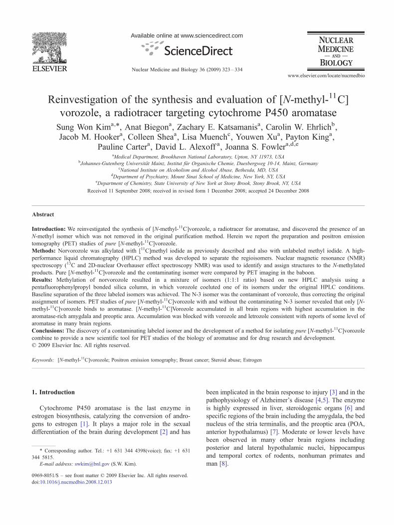
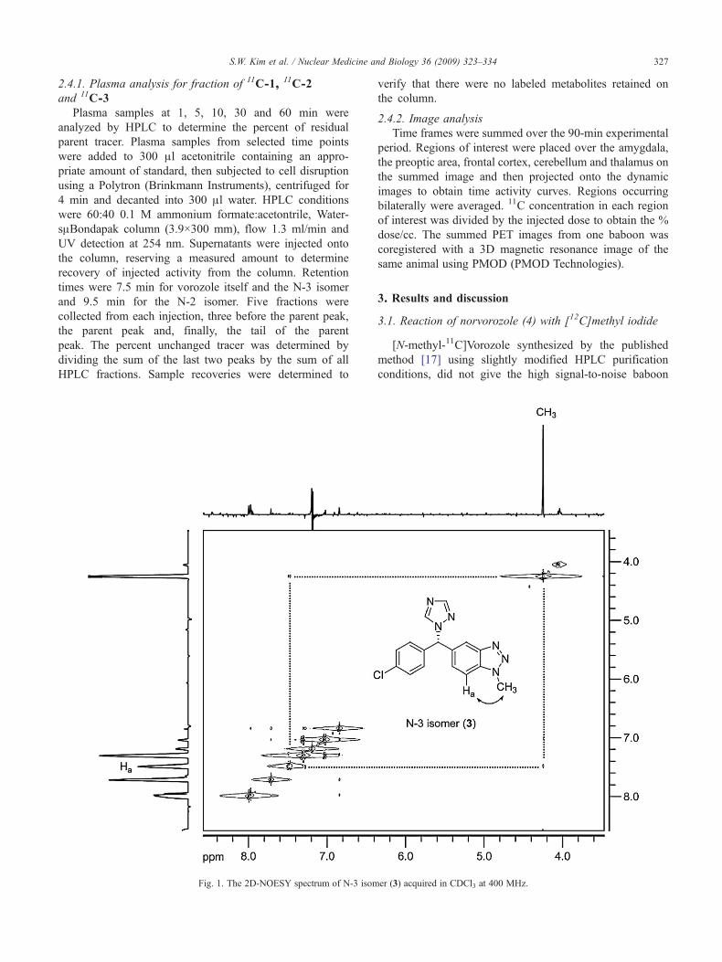
Fig. 1. The 2D-NOESY spectrum of N-3 isom
verify that there were no labeled metabolites retained onthe column.
2.4.2. Image analysisTime frames were summed over the 90-min experimental
period. Regions of interest were placed over the amygdala,the preoptic area, frontal cortex, cerebellum and thalamus onthe summed image and then projected onto the dynamicimages to obtain time activity curves. Regions occurringbilaterally were averaged. 11C concentration in each regionof interest was divided by the injected dose to obtain the %dose/cc. The summed PET images from one baboon wascoregistered with a 3D magnetic resonance image of thesame animal using PMOD (PMOD Technologies).
3. Results and discussion
3.1. Reaction of norvorozole (4) with [12C]methyl iodide
[N-methyl-11C]Vorozole synthesized by the publishedmethod [17] using slightly modified HPLC purificationconditions, did not give the high signal-to-noise baboon
er (3) acquired in CDCl3 at 400 MHz.
328 S.W. Kim et al. / Nuclear Medicine and Biology 36 (2009) 323–334
brain images and autoradiography results with rat brainsections which were described previously (data not shown).Given the alkaline reaction medium, we first considered thatthe nonspecific binding could be a result of racemization,thus forming the inactive (R)-vorozole [25], which wouldcoelute with the active form. However, chiral HPLC analysis(column, Chiralpak OJ-P, 4.6×15 mm; eluent, water:acetonitrile=75:35; flowrate, 0.5 ml/min) indicated that noracemization of either (S)-norvorozole or (S)-vorozoleoccurred under the reaction conditions (data not shown).
Serendipitously, during this investigation, we uncoveredthe presence of an unexpected reaction byproduct (thereforeobserving three N-methylated compounds in total) that wasformed in approximately equal yield to (S)-vorozole. Thisproduct was not separable under the HPLC conditionsoriginally reported, and thus, Lidström et al. [17] noted onlytwo major fractions “in a ratio of 5 to 2.” The authorsidentified the two fractions as {[N-methyl-11C]vorozole (N-1alkylation)} and an regioisomer, 11C-3 (N-3 alkylation).Their assignment was based on 13C NMR experiments, inwhich [13C]methyl iodide was added as a carrier in the 11Cmethylation reaction in order to have sufficient mass for a13C NMR. After HPLC isolation of the fraction with theexpected retention time of vorozole, the authors determinedthe chemical shift of the 13C-enriched product matched thatof vorozole (34.4 ppm). The minor isomer, which was wellseparated under their HPLC conditions, was also isolatedand analyzed by 13C NMR and assigned as the N-3
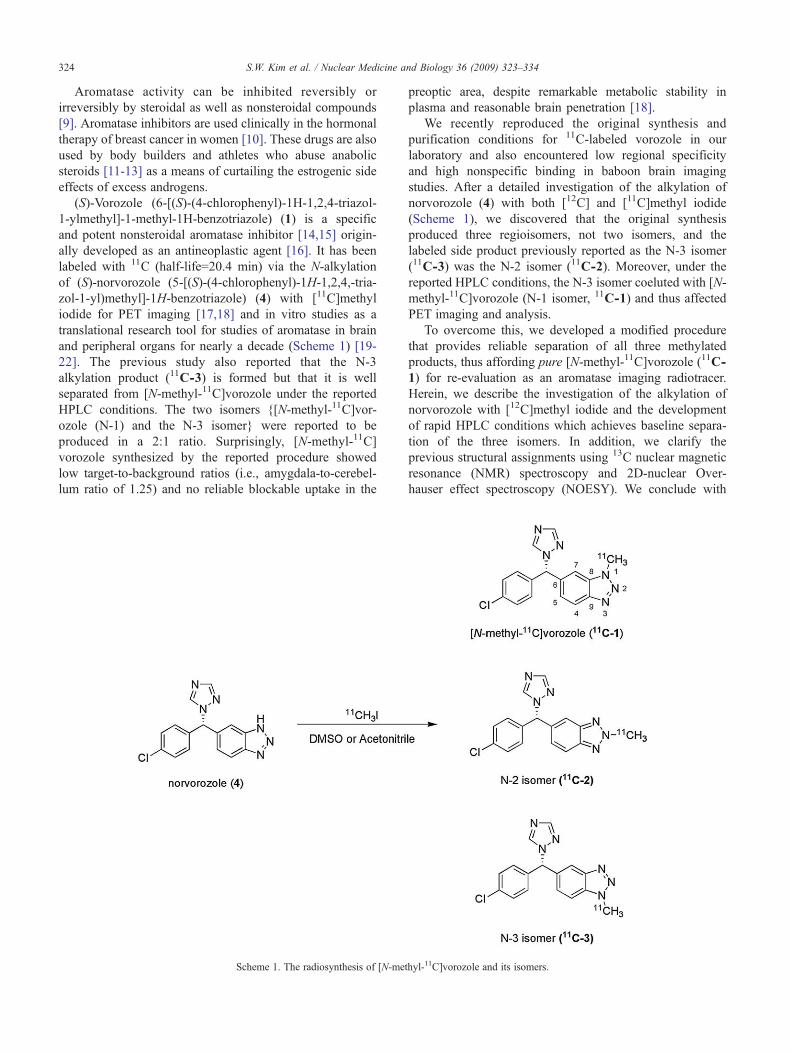
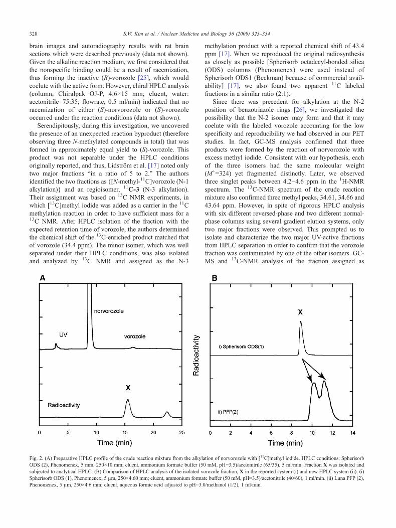
Fig. 2. (A) Preparative HPLC profile of the crude reaction mixture from the alkylODS (2), Phenomenex, 5 mm, 250×10 mm; eluent, ammonium formate buffer (5subjected to analytical HPLC. (B) Comparison of HPLC analysis of the isolated vSpherisorb ODS (1), Phenomenex, 5 μm, 250×4.60 mm; eluent, ammonium formaPhenomenex, 5 μm, 250×4.6 mm; eluent, aqueous formic acid adjusted to pH=3.
methylation product with a reported chemical shift of 43.4ppm [17]. When we reproduced the original radiosynthesisas closely as possible [Spherisorb octadecyl-bonded silica(ODS) columns (Phenomenex) were used instead ofSpherisorb ODS1 (Beckman) because of commercial avail-ability] [17], we also found two apparent 11C labeledfractions in a similar ratio (2:1).
Since there was precedent for alkylation at the N-2position of benzotriazole rings [26], we investigated thepossibility that the N-2 isomer may form and that it maycoelute with the labeled vorozole accounting for the lowspecificity and reproducibility we had observed in our PETstudies. In fact, GC-MS analysis confirmed that threeproducts were formed by the reaction of norvorozole withexcess methyl iodide. Consistent with our hypothesis, eachof the three isomers had the same molecular weight(M+=324) yet fragmented distinctly. Later, we observedthree singlet peaks between 4.2–4.6 ppm in the 1H-NMRspectrum. The 13C-NMR spectrum of the crude reactionmixture also confirmed three methyl peaks, 34.61, 34.66 and43.64 ppm. However, in spite of rigorous HPLC analysiswith six different reversed-phase and two different normal-phase columns using several gradient elution systems, onlytwo major fractions were observed. This prompted us toisolate and characterize the two major UV-active fractionsfrom HPLC separation in order to confirm that the vorozolefraction was contaminated by one of the other isomers. GC-MS and 13C-NMR analysis of the fraction assigned as
ation of norvorozole with [11C]methyl iodide. HPLC conditions: Spherisorb0 mM, pH=3.5)/acetonitrile (65/35), 5 ml/min. Fraction X was isolated andorozole fraction, X in the reported system (i) and new HPLC system (ii). (i)te buffer (50 mM, pH=3.5)/acetonitrile (40/60), 1 ml/min. (ii) Luna PFP (2),0/methanol (1/2), 1 ml/min.
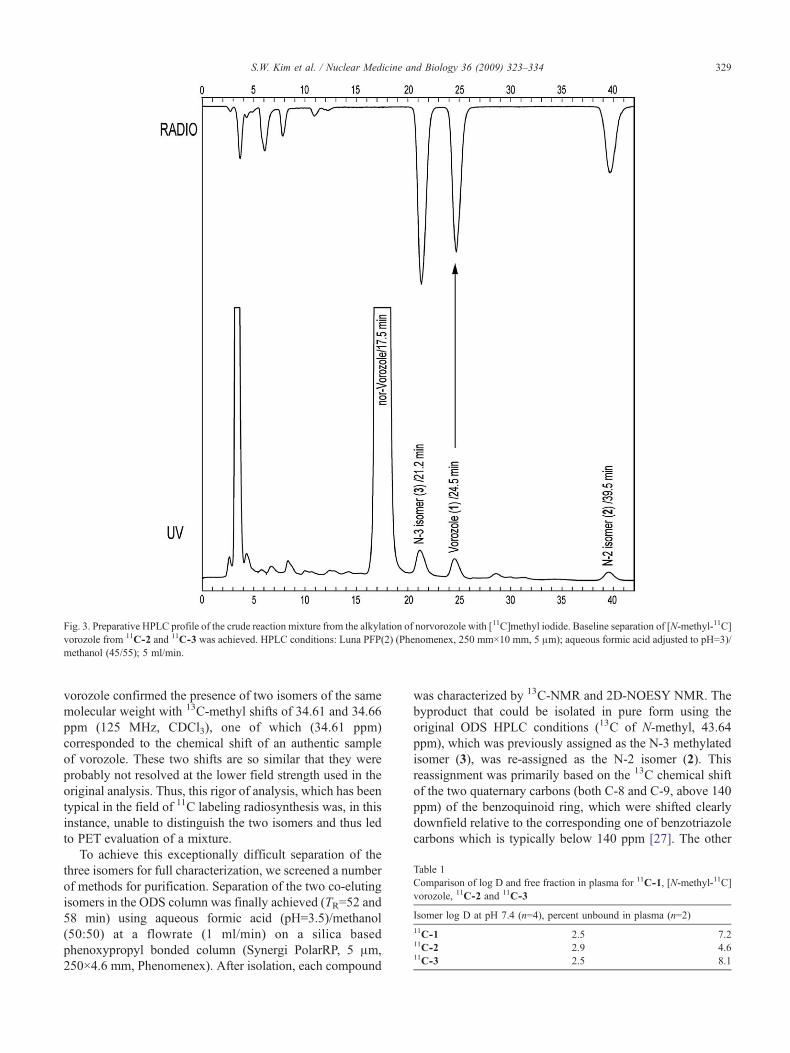
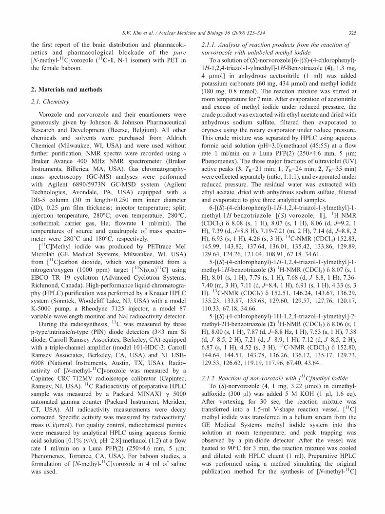
Fig. 3. Preparative HPLC profile of the crude reaction mixture from the alkylation of norvorozole with [11C]methyl iodide. Baseline separation of [N-methyl-11C]vorozole from 11C-2 and 11C-3 was achieved. HPLC conditions: Luna PFP(2) (Phenomenex, 250 mm×10 mm, 5 μm); aqueous formic acid adjusted to pH=3)/methanol (45/55); 5 ml/min.
able 1omparison of log D and free fraction in plasma for 11C-1, [N-methyl-11C]orozole, 11C-2 and 11C-3
omer log D at pH 7.4 (n=4), percent unbound in plasma (n=2)
C-1 2.5 7.2C-2 2.9 4.6C-3 2.5 8.1
329S.W. Kim et al. / Nuclear Medicine and Biology 36 (2009) 323–334
vorozole confirmed the presence of two isomers of the samemolecular weight with 13C-methyl shifts of 34.61 and 34.66ppm (125 MHz, CDCl3), one of which (34.61 ppm)corresponded to the chemical shift of an authentic sampleof vorozole. These two shifts are so similar that they wereprobably not resolved at the lower field strength used in theoriginal analysis. Thus, this rigor of analysis, which has beentypical in the field of 11C labeling radiosynthesis was, in thisinstance, unable to distinguish the two isomers and thus ledto PET evaluation of a mixture.
To achieve this exceptionally difficult separation of thethree isomers for full characterization, we screened a numberof methods for purification. Separation of the two co-elutingisomers in the ODS column was finally achieved (TR=52 and58 min) using aqueous formic acid (pH=3.5)/methanol(50:50) at a flowrate (1 ml/min) on a silica basedphenoxypropyl bonded column (Synergi PolarRP, 5 μm,250×4.6 mm, Phenomenex). After isolation, each compound
was characterized by 13C-NMR and 2D-NOESY NMR. Thebyproduct that could be isolated in pure form using theoriginal ODS HPLC conditions (13C of N-methyl, 43.64ppm), which was previously assigned as the N-3 methylatedisomer (3), was re-assigned as the N-2 isomer (2). Thisreassignment was primarily based on the 13C chemical shiftof the two quaternary carbons (both C-8 and C-9, above 140ppm) of the benzoquinoid ring, which were shifted clearlydownfield relative to the corresponding one of benzotriazolecarbons which is typically below 140 ppm [27]. The other
TCv
Is11
11
11
330 S.W. Kim et al. / Nuclear Medicine and Biology 36 (2009) 323–334
two isomers, which co-eluted under the original HPLCconditions but which were separated by the PFPP column,were identified as vorozole (N-1 isomer) and the N-3 isomer(3). Their structural assignments were largely based on thenuclear Overhauser effect correlation of the H-7 or H-4 to themethyl group (Fig. 1).
3.2. Alkylation of norvorozole with [11C]methyliodide: reproduction of the previous synthesis [17,18] andseparation of [N-methyl-11C]vorozole from 11C-3 and11C-2
Initially, we set out (1) to reproduce the previousradiosynthesis and HPLC conditions (observation of tworadioactive fractions) and (2) to separate the “vorozole”fraction if it was actually a mixture of vorozole and the N-3isomer as our 12C synthesis results suggested. Since theHPLC column (Spherisorb ODS1, Beckman, 250×10 mm)used previously was not commercially available, wereplaced it with a similar HPLC column [Spherisorb ODS(2), Phenomenex, 5 μm, 250×10 mm). Similar to theprevious report, we obtained only two radioactive fractionsin a ratio of 2:1. The major radioactive 15-min fraction X(Fig. 2A), which has the same retention time as vorozole,was isolated and subjected to further analysis (Fig. 2B) usingan analytical HPLC system. We chose the same solventsystem and a similar HPLC column [Spherisorb ODS (1),Phenomenex, 5 μm, 250×4.60 mm) to that reportedpreviously [17,18]. The radioactive fraction, X, showedonly one peak in this system (Fig. 2B). Other conventionalODS columns gave the same results using various solvent
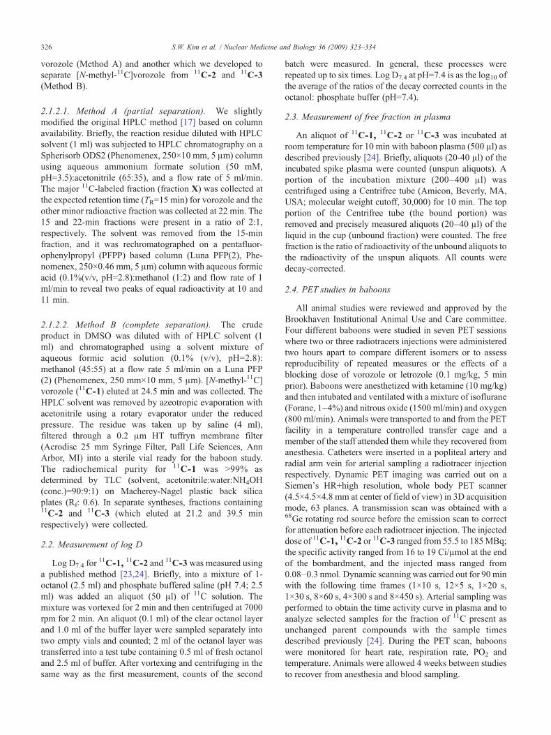
Fig. 4. PET images in the (transaxial (top row) and coronal (bottom row) planes ifraction (X), [11C-1+11C-3, 176.49 MBq (4.77 mCi)], (B) [N-methyl-11C]vorozo(3.33 mCi)] and (D) N-2 isomer [11C-2, 44.77 MBq (1.21 mCi)]. All PET images
systems (data not shown). However, a PFPP bonded-phaseHPLC column resolved the 15-min fraction X into twopeaks, which have the same retention time as “cold” N-3isomer (3) and vorozole (1), respectively (Fig. 2B). Later, allthree regioisomers of the crude reaction mixture were wellseparated using a semipreparative scale PFPP column(Fig. 3). The radiochemical yields of the three regioisomerswere approximately 30% each. The radiochemical puritywas N97%, and the specific activity ranged 10–19 Ci/μmol(0.3–0.7 GBq/μmol). The total radiosynthesis time was 65min after the end of the cyclotron bombardment.
3.3. Comparison of 11C labeled fraction A (11C-1+11C-3),[N-methyl-11C]vorozole (11C-1), 11C-3 and 11C-2 in thebaboon brain
We determined that all three regioisomers have alipophilicity (log D7.4) considered suitable for blood–brainbarrier penetration. Notably, the log D's of 11C-1 and 11C-3were almost identical (Table 1), which is consistent with theircoelution on HPLC using the ODS column. The free fractionof 11C-1 and 11C-3 in baboon plasma was also similar.
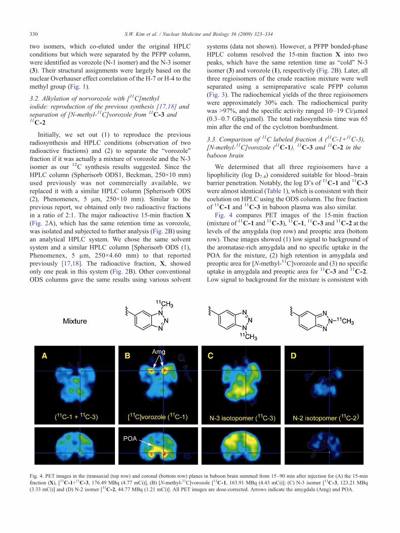
Fig. 4 compares PET images of the 15-min fraction(mixture of 11C-1 and 11C-3), 11C-1, 11C-3 and 11C-2 at thelevels of the amygdala (top row) and preoptic area (bottomrow). These images showed (1) low signal to background ofthe aromatase-rich amygdala and no specific uptake in thePOA for the mixture, (2) high retention in amygdala andpreoptic area for [N-methyl-11C]vorozole and (3) no specificuptake in amygdala and preoptic area for 11C-3 and 11C-2.Low signal to background for the mixture is consistent with
n baboon brain summed from 15–90 min after injection for (A) the 15-minle [11C-1, 163.91 MBq (4.43 mCi)]; (C) N-3 isomer [11C-3, 123.21 MBqare dose-corrected. Arrows indicate the amygdala (Amg) and POA.
331S.W. Kim et al. / Nuclear Medicine and Biology 36 (2009) 323–334
contamination with the N-3 isomer (11C-3). The high signalto noise in the amygdala and preoptic areas for pure 11C-1was consistent with the high affinity of vorozole foraromatase. We note that affinity values for the twononvorozole isomers (2 and 3) for aromatase are notavailable, though we would predict low affinities relativeto vorozole (IC50=2.7 nM) [28] based on our imaging results.
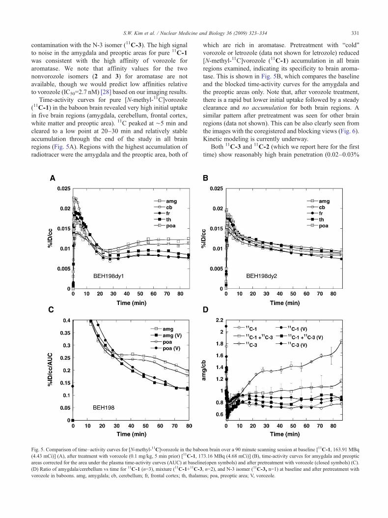
Time-activity curves for pure [N-methyl-11C]vorozole(11C-1) in the baboon brain revealed very high initial uptakein five brain regions (amygdala, cerebellum, frontal cortex,white matter and preoptic area). 11C peaked at ∼5 min andcleared to a low point at 20–30 min and relatively stableaccumulation through the end of the study in all brainregions (Fig. 5A). Regions with the highest accumulation ofradiotracer were the amygdala and the preoptic area, both of
Fig. 5. Comparison of time–activity curves for [N-methyl-11C]vorozole in the babo(4.43 mCi)] (A), after treatment with vorozole (0.1 mg/kg, 5 min prior) [11C-1, 17areas corrected for the area under the plasma time-activity curves (AUC) at baselin(D) Ratio of amygdala/cerebellum vs time for 11C-1 (n=3), mixture (11C-1+11C-3vorozole in baboons. amg, amygdala; cb, cerebellum; fr, frontal cortex; th, thalam
which are rich in aromatase. Pretreatment with “cold”vorozole or letrozole (data not shown for letrozole) reduced[N-methyl-11C]vorozole (11C-1) accumulation in all brainregions examined, indicating its specificity to brain aroma-tase. This is shown in Fig. 5B, which compares the baselineand the blocked time-activity curves for the amygdala andthe preoptic areas only. Note that, after vorozole treatment,there is a rapid but lower initial uptake followed by a steadyclearance and no accumulation for both brain regions. Asimilar pattern after pretreatment was seen for other brainregions (data not shown). This can be also clearly seen fromthe images with the coregistered and blocking views (Fig. 6).Kinetic modeling is currently underway.
Both 11C-3 and 11C-2 (which we report here for the firsttime) show reasonably high brain penetration (0.02–0.03%
on brain over a 90 minute scanning session at baseline [11C-1, 163.91 MBq3.16 MBq (4.68 mCi)] (B), time-activity curves for amygdala and preoptice(open symbols) and after pretreatment with vorozole (closed symbols) (C)., n=2), and N-3 isomer (11C-3, n=1) at baseline and after pretreatment withus; poa, preoptic area; V, vorozole.
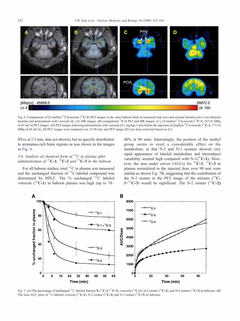
Fig. 6. Comparisons of [N-methyl-11C]vorozole (11C-1) PET images in the same baboon brain in transaxial (top row) and coronal (bottom row) view betweenbaseline and pretreatment with vorozole (1). (A) MR images. (B) coregistered 11C-1 PET and MR images. (C), [N-methyl-11C]vorozole [11C-1, 163.91 MBq(4.43 mCi)] PET images. (D) PET images following pretreatment with vorozole (0.1 mg/kg) 5 min before the injection of [methyl-11C]vorozole [11C-1, 173.16MBq (4.68 mCi)]. All PET images were summed over 15-90 min and PET image (D) was dose-corrected based on (C).
332 S.W. Kim et al. / Nuclear Medicine and Biology 36 (2009) 323–334
ID/cc at 2.5 min, data not shown), but no specific distributionto aromatase-rich brain regions as was shown in the imagesin Fig. 4.
3.4. Analysis of chemical form of 11C in plasma afteradministration of 11C-1, 11C-2 and 11C-3 in the baboon
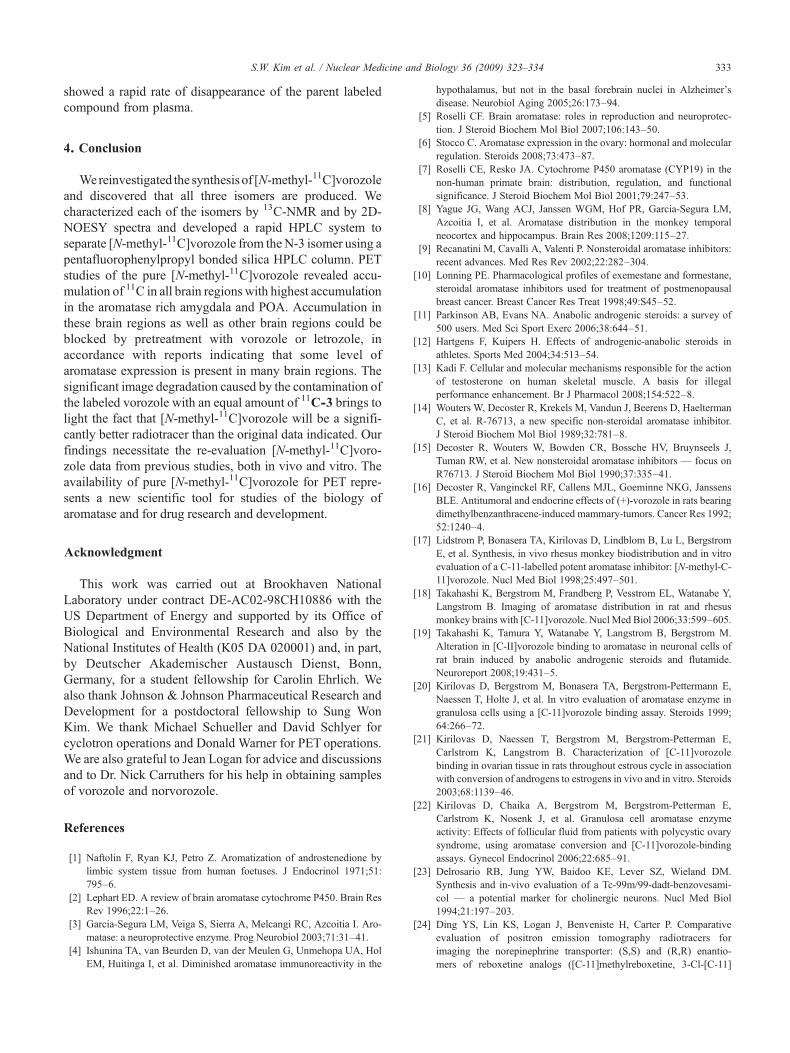
For all baboon studies, total 11C in plasma was measuredand the unchanged fraction of 11C-labeled compound wasdetermined by HPLC. The % unchanged 11C labeledvorozole (11C-1) in baboon plasma was high (up to 78–
Fig. 7. (A) The percentage of unchanged 11C labeled fraction X (11C-1+11C-3), voThe time-AUC plots of 11C-labeled vorozole (11C-1), N-2 isomer (11C-2) and N-
80% at 90 min). Interestingly, the position of the methylgroup seems to exert a considerable effect on themetabolism, in that N-2 and N-3 isomers showed veryrapid appearance of labeled metabolites and intersubjectvariability seemed high compared with N-1(11C-1). How-ever, the area under curves (AUCs) for 11C-1, 11C-3 inplasma normalized to the injected dose over 90 min weresimilar as shown Fig. 7B, suggesting that the contribution ofthe N-3 isomer in the PET image of the mixture (11C-1+11C-3) would be significant. The N-2 isomer (11C-2)
rozole (11C-1), N-2 isomer (11C-2), and N-3 isomer (11C-3) in baboons. (B)3 isomer (11C-3) in baboons.
333S.W. Kim et al. / Nuclear Medicine and Biology 36 (2009) 323–334
showed a rapid rate of disappearance of the parent labeledcompound from plasma.
4. Conclusion
Wereinvestigated the synthesis of [N-methyl-11C]vorozoleand discovered that all three isomers are produced. Wecharacterized each of the isomers by 13C-NMR and by 2D-NOESY spectra and developed a rapid HPLC system toseparate [N-methyl-11C]vorozole from the N-3 isomer using apentafluorophenylpropyl bonded silica HPLC column. PETstudies of the pure [N-methyl-11C]vorozole revealed accu-mulation of 11C in all brain regions with highest accumulationin the aromatase rich amygdala and POA. Accumulation inthese brain regions as well as other brain regions could beblocked by pretreatment with vorozole or letrozole, inaccordance with reports indicating that some level ofaromatase expression is present in many brain regions. Thesignificant image degradation caused by the contamination ofthe labeled vorozole with an equal amount of 11C-3 brings tolight the fact that [N-methyl-11C]vorozole will be a signifi-cantly better radiotracer than the original data indicated. Ourfindings necessitate the re-evaluation [N-methyl-11C]voro-zole data from previous studies, both in vivo and vitro. Theavailability of pure [N-methyl-11C]vorozole for PET repre-sents a new scientific tool for studies of the biology ofaromatase and for drug research and development.
Acknowledgment
This work was carried out at Brookhaven NationalLaboratory under contract DE-AC02-98CH10886 with theUS Department of Energy and supported by its Office ofBiological and Environmental Research and also by theNational Institutes of Health (K05 DA 020001) and, in part,by Deutscher Akademischer Austausch Dienst, Bonn,Germany, for a student fellowship for Carolin Ehrlich. Wealso thank Johnson & Johnson Pharmaceutical Research andDevelopment for a postdoctoral fellowship to Sung WonKim. We thank Michael Schueller and David Schlyer forcyclotron operations and Donald Warner for PET operations.We are also grateful to Jean Logan for advice and discussionsand to Dr. Nick Carruthers for his help in obtaining samplesof vorozole and norvorozole.
References
[1] Naftolin F, Ryan KJ, Petro Z. Aromatization of androstenedione bylimbic system tissue from human foetuses. J Endocrinol 1971;51:795–6.
[2] Lephart ED. A review of brain aromatase cytochrome P450. Brain ResRev 1996;22:1–26.
[3] Garcia-Segura LM, Veiga S, Sierra A, Melcangi RC, Azcoitia I. Aro-matase: a neuroprotective enzyme. Prog Neurobiol 2003;71:31–41.
[4] Ishunina TA, van Beurden D, van der Meulen G, Unmehopa UA, HolEM, Huitinga I, et al. Diminished aromatase immunoreactivity in the
hypothalamus, but not in the basal forebrain nuclei in Alzheimer'sdisease. Neurobiol Aging 2005;26:173–94.
[5] Roselli CF. Brain aromatase: roles in reproduction and neuroprotec-tion. J Steroid Biochem Mol Biol 2007;106:143–50.
[6] Stocco C. Aromatase expression in the ovary: hormonal and molecularregulation. Steroids 2008;73:473–87.
[7] Roselli CE, Resko JA. Cytochrome P450 aromatase (CYP19) in thenon-human primate brain: distribution, regulation, and functionalsignificance. J Steroid Biochem Mol Biol 2001;79:247–53.
[8] Yague JG, Wang ACJ, Janssen WGM, Hof PR, Garcia-Segura LM,Azcoitia I, et al. Aromatase distribution in the monkey temporalneocortex and hippocampus. Brain Res 2008;1209:115–27.
[9] Recanatini M, Cavalli A, Valenti P. Nonsteroidal aromatase inhibitors:recent advances. Med Res Rev 2002;22:282–304.
[10] Lonning PE. Pharmacological profiles of exemestane and formestane,steroidal aromatase inhibitors used for treatment of postmenopausalbreast cancer. Breast Cancer Res Treat 1998;49:S45–52.
[11] Parkinson AB, Evans NA. Anabolic androgenic steroids: a survey of500 users. Med Sci Sport Exerc 2006;38:644–51.
[12] Hartgens F, Kuipers H. Effects of androgenic-anabolic steroids inathletes. Sports Med 2004;34:513–54.
[13] Kadi F. Cellular and molecular mechanisms responsible for the actionof testosterone on human skeletal muscle. A basis for illegalperformance enhancement. Br J Pharmacol 2008;154:522–8.
[14] Wouters W, Decoster R, Krekels M, Vandun J, Beerens D, HaeltermanC, et al. R-76713, a new specific non-steroidal aromatase inhibitor.J Steroid Biochem Mol Biol 1989;32:781–8.
[15] Decoster R, Wouters W, Bowden CR, Bossche HV, Bruynseels J,Tuman RW, et al. New nonsteroidal aromatase inhibitors — focus onR76713. J Steroid Biochem Mol Biol 1990;37:335–41.
[16] Decoster R, Vanginckel RF, Callens MJL, Goeminne NKG, JanssensBLE. Antitumoral and endocrine effects of (+)-vorozole in rats bearingdimethylbenzanthracene-induced mammary-tumors. Cancer Res 1992;52:1240–4.
[17] Lidstrom P, Bonasera TA, Kirilovas D, Lindblom B, Lu L, BergstromE, et al. Synthesis, in vivo rhesus monkey biodistribution and in vitroevaluation of a C-11-labelled potent aromatase inhibitor: [N-methyl-C-11]vorozole. Nucl Med Biol 1998;25:497–501.
[18] Takahashi K, Bergstrom M, Frandberg P, Vesstrom EL, Watanabe Y,Langstrom B. Imaging of aromatase distribution in rat and rhesusmonkey brains with [C-11]vorozole. Nucl Med Biol 2006;33:599–605.
[19] Takahashi K, Tamura Y, Watanabe Y, Langstrom B, Bergstrom M.Alteration in [C-II]vorozole binding to aromatase in neuronal cells ofrat brain induced by anabolic androgenic steroids and flutamide.Neuroreport 2008;19:431–5.
[20] Kirilovas D, Bergstrom M, Bonasera TA, Bergstrom-Pettermann E,Naessen T, Holte J, et al. In vitro evaluation of aromatase enzyme ingranulosa cells using a [C-11]vorozole binding assay. Steroids 1999;64:266–72.
[21] Kirilovas D, Naessen T, Bergstrom M, Bergstrom-Petterman E,Carlstrom K, Langstrom B. Characterization of [C-11]vorozolebinding in ovarian tissue in rats throughout estrous cycle in associationwith conversion of androgens to estrogens in vivo and in vitro. Steroids2003;68:1139–46.
[22] Kirilovas D, Chaika A, Bergstrom M, Bergstrom-Petterman E,Carlstrom K, Nosenk J, et al. Granulosa cell aromatase enzymeactivity: Effects of follicular fluid from patients with polycystic ovarysyndrome, using aromatase conversion and [C-11]vorozole-bindingassays. Gynecol Endocrinol 2006;22:685–91.
[23] Delrosario RB, Jung YW, Baidoo KE, Lever SZ, Wieland DM.Synthesis and in-vivo evaluation of a Tc-99m/99-dadt-benzovesami-col — a potential marker for cholinergic neurons. Nucl Med Biol1994;21:197–203.
[24] Ding YS, Lin KS, Logan J, Benveniste H, Carter P. Comparativeevaluation of positron emission tomography radiotracers forimaging the norepinephrine transporter: (S,S) and (R,R) enantio-mers of reboxetine analogs ([C-11]methylreboxetine, 3-Cl-[C-11]

334 S.W. Kim et al. / Nuclear Medicine and Biology 36 (2009) 323–334
methylreboxetine and [F-18]fluororeboxetine), (R)-[C-11]nisoxetine,[C-11]oxaprotiline and [C-11]lortalamine. J Neurochem 2005;94:337–51.
[25] Wouters W, de Coster R, van Dun J, Krekels MDWG, Dillen A,Raeymaekers A, et al. Comparative effects of the aromatase inhibitorR76713 and of its enantiomers R83839 and R83842 on steroidbiosynthesis in vitro and in vivo. J Steroid BiochemMol Biol 1990;37:1049–54.
[26] Kopanska K, Najda A, Zebrowska J, Chomicz L, Piekarczyk J, MyjakP, et al. Synthesis and activity of 1H-benzimidazole and 1H-
benzotriazole derivatives as inhibitors of Acanthamoeba castellanii.Bioorgan Med Chem 2004;12:2617–24.
[27] Carta A, Piras S, Boatto G, Paglietti G. 1H,6H-triazolo[4,5-e]benzotriazole-3-oxides and 5,5 '-(Z)diazene-1,2-diylbis(2-methyl-2H-1,2,3-benzotriazole) derived from chloronitrobenzotriazoles andhydrazine. Heterocycles 2005;65:2471–81.
[28] Wouters W, Decoster R, Tuman RW, Bowden CR, Bruynseels J,Vanderpas H, et al. Aromatase inhibition by R-76713 -— experimentaland clinical-pharmacology. J Steroid Biochem Mol Biol 1989;34:427–30.