Embed Size (px)
Citation preview
Review ArticleRegulation of Redox Homeostasis by Nonthermal BiocompatiblePlasma Discharge in Stem Cell Differentiation
Ying Li,1,2 Eun Ha Choi ,1,2,3 and Ihn Han 2
1Department of Plasma-Bio Display, Kwangwoon University, 447-1, Seoul 01897, Republic of Korea2Plasma Bioscience Research Center, Applied PlasmaMedicine Center, Kwangwoon University, 447-1, Seoul 01897, Republic of Korea3Department of Electronic and Biological Physics, Kwangwoon University, 447-1, Seoul 01897, Republic of Korea
Correspondence should be addressed to Ihn Han; [email protected]
Received 21 December 2018; Accepted 24 February 2019; Published 31 March 2019
Guest Editor: Paola Venditti
Copyright © 2019 Ying Li et al. This is an open access article distributed under the Creative Commons Attribution License, whichpermits unrestricted use, distribution, and reproduction in any medium, provided the original work is properly cited.
Recently, a growing body of evidence has shown the role of reactive species as secondary messengers in cell proliferation anddifferentiation, as opposed to the harmful metabolism byproducts that they were previously solely recognized as. Thus, thebalance of intracellular reduction-oxidation (redox) homeostasis plays a vital role in the regulation of stem cell self-renewal anddifferentiation. Nonthermal biocompatible plasma (NBP) has emerged as a novel tool in biomedical applications. Recently, NBPhas also emerged as a powerful tool in the tissue engineering field for the surface modification of biomaterial and the promotionof stem cell differentiation by the regulation of intracellular redox biology. NBP can generate various kinds of reactive oxygenspecies (ROS) and reactive nitrogen species (RNS), which may play the role of the second passenger in the cell signalingnetwork and active antioxidant system in cells. Herein, we review the current knowledge on mechanisms by which NBPregulates cell proliferation and differentiation through redox modification. Considering the importance of redox homeostasis inthe regulation of stem cell differentiation, understanding the underlying molecular mechanisms involved will provide importantnew insights into NBP-induced stem cell differentiation for tissue engineering.
1. Redox Homeostasis in StemCell Differentiation
The focus of tissue engineering is regenerating damagedtissues through the restoration, maintenance, and improve-ment of tissue function [1]. For example, in bone tissue, thecritical size of bone defects, which lies beyond the spontane-ous regeneration capacity of a patient and thus requiressurgical invention, has guided research into bone tissueengineering-based therapeutics [2]. Stem cells are the criticalcell sources in tissue engineering that possess the characteris-tics of self-renewal and potential to differentiate into multiplecell types for the repair and/or regeneration of defectivetissues and organs, such as the bone, cartilage, heart, neurons,and spinal cord [3–7]. To induce stem cell differentiation,growth factors are the most commonly used technique.Other techniques are also being studied, such as the electro-magnetic field, vibration, radiation, heat shock, and oxidativestress [8–17]. Scaffolds provide a framework for stem cells to
migrate to, attach to, and specialize on [2]. However, the lowefficiency of expansion and differentiation of stem cellsin vitro is resulting in attempts to develop new methods toimprove their characteristics.
Since stem cells are an essential part of tissue regenera-tion, extensive research has been conducted on the factorsregulating stem cell self-renewal and differentiation. Reactiveoxygen species (ROS), the highly chemically reactive bypro-ducts of aerobic metabolism, are important mediators instem cell biology [18, 19]. Changes in ROS levels can be usedto monitor the balance of stem cell self-renewal and differ-entiation. Although high levels of ROS have long beensuggested to be detrimental to mediating oxidative stress,mounting experimental evidence indicates that the physio-logical levels of ROS are involved in the maintenance ofintracellular reduction-oxidation (redox) homeostasis andvarious cellular signaling pathways [20]. ROS in redoxhomeostasis plays a pivotal role in the maintenance ofstem cell self-renewal with low levels of ROS, whereas in
HindawiOxidative Medicine and Cellular LongevityVolume 2019, Article ID 2318680, 15 pageshttps://doi.org/10.1155/2019/2318680

differentiated stem cells, ROS is accumulated [21]. For exam-ple, a quantitative study comparing human embryonic stemcells (ESC) with their differentiated descendants has shownthat ESC are characterized by a lower ROS level, whiledifferentiated cells contain more oxidative species. However,biochemical normalization of the ROS level to cell volume/protein indicates that all cell types maintain a similar intra-cellular redox of the ROS level as a measure of intracellularredox balance [22]. ROS are also involved in signal transduc-tion cascades in enhancing the differentiation of ESC towardthe cardiomyogenic and vascular cell lineage [23]. Thesefindings imply that redox signaling plays a crucial role inmodulating the fate of stem cells. Therefore, it is possible thatmanipulating the exogenous “ROS donor” tool could activateintracellular redox-dependent signaling to maintain stemcell differentiation.
2. Nonthermal Biocompatible Plasma (NBP)
Nonthermal biocompatible plasma (NBP) (or plasma) isproduced by applying a sufficiently high-voltage electric fieldacross the discharge gap to initiate a breakdown of gas atatmospheric pressure [24]. When NBP is generated, themajor components of charged particles, neutral gas species,reactive species, electric field, and radiation are produced.NBP was first employed in antimicrobial applications,because it produces a variety of biotoxic agents that includereactive species, UV radiation, and charged particles. Sincethen, NBP has come to be extensively studied in otherapplications in the biomedical field, including in sterilization,cancer cell apoptosis, wound healing, blood coagulation, andteeth whitening [25–31], which has made NBP a promisingtool for biomedical use. An increasing number of studieshave shown the role of NBP in tissue engineering on thesurface modification of biomaterials [32–34] and as anexogenous stimulator that directly induces stem cell prolif-eration and differentiation [35–40]. In this section, NBPdevices and their characteristics will be summarized andanalyzed so as to provide a more detailed concept ofNBP production and composition.
2.1. Classification of NBP Devices. NBP devices for stem celldifferentiation can be broadly classified into two major
categories: plasma jet and dielectric barrier discharge(DBD) plasma. Figure 1 shows an example schematic of aplasma jet and DBD device produced in our research center.The basic structure of the plasma jet type consists of an innerhigh-voltage electrode, which is coupled with the powersource and covered with a dielectric barrier and a groundedouter electrode. Then, either a mixture of noble gas withreactive gas or just pure gas is fed into the annular spacebetween the two electrodes. The plasma ionization degreein the jet is very low (at 10-6–10-7) (gas density is at 1 atmo-spheric pressure and 300K is around 2 × 1019 cm−3). In theDBD plasma device, the high-voltage and grounded elec-trodes with an electrode gap of around 400μm are coatedon glass and covered with a dielectric barrier with a thick-ness of 50μm. Normally, high-voltage current is alternatedbetween the two electrodes in order to prevent high tem-perature buildup and transition to arc discharge [41].
2.2. RONS Generated by NBP. Several researchers haveapplied NBP to stem cell differentiation. However, thedevices that have been used to generate NBP in these studieshave various characteristics in each research group, with thegeneration of different RONS concentration. Typically, opti-cal emission spectroscopy (OES) is used to show the excitedelements in NBP generation, due to the unique emissionspectra specific to each element as it transitions from theexcited to group state [42]. One of the main factors affectingthe NBP-generated RONS species is feeding gas. Accordingto feeding gas, the inert gases, such as argon (Ar) and helium(He), are initially used, because they have relatively lowerbreakdown voltages. The mixture of inert gas with differentpercentages of oxygen (O2) can generate more ROS [39, 43].Recently, reactive pure gas, such as nitrogen (N2) and air,has come to be mostly used to generate various ROS andRNS. Particularly, when using N2 gas, OES shows an RNS-dominant signal, including N2 second positive species(SPS), N2
∗, and nitric oxide (NOγ) (Table 1).
2.3. Interaction of NBP with Living Cells. Initially, NBP wasgenerated by ionizing neutral gases with high voltage. Asshown in Figure 2(a), the surface of culture media was thenbombarded by RONS generated during NBP discharge andthe reaction of species could be explained by plasma-
Gas
High‑voltageelectrode
Dielectricbarrier
Groundelectrode
AC Powersupply
Plasma
(a)
Plasma
High‑voltagepower supply
Glass
Gas Gas
Hydration preventionlayer
Electrodes
Dielectric barrier
Aluminium oxide(Al2O3) layer
(b)
Figure 1: Schematic of the (a) jet type and (b) DBD type of plasma devices to produce NBP.
2 Oxidative Medicine and Cellular Longevity

Table1:Summaryof
NBPcharacteristics,extra-
andintracellularROS/RNSspecies,andinvolvem
entof
molecules
andsignalingpathwaysin
variou
sbiologicalsamples
ofin
vitroand
invivo
NBPapplications.
Devicetype
Gas
OES
Extracellu
lar
RONS
Intracellular
RONS
Scavenger
Cell
Effect
NBP-activated
pathway
ormolecules
NBP-reduced
effects
Ref.
Jet
N2
NO• ,N2∗
NO
NO
cPTIO
Osteoprogenitor
celllin
eOsteogenesis
ALP
,COL-1
—[36]
Jet
He
OI,NO• ,
N2∗,O
3—
NO
—hP
DLMSC
sOsteogenesis
ALP
enzyme
[37]
Jet
N2
NO• ,N2∗,
O2+
——
—Animal
Myogenesis
p38,MYH3,MHC,
MypG,M
yoD
—[38]
Jet
He+
1%O 2
• OH,O
2∗,O
,NO
NO
NO
Hgb
Murine-im
mortalized
neuralstem
celllin
eC17.2
Primaryratneuralstem
cells
(isolatedfrom
thehipp
ocam
pus)
Neurogenesis
β-Tub
linIII,APC,
Nestin
[39]
Jet
He
• OH,•O2-
H2O
2—
—Osteoprogenitor
cells
(MC3T
3-E1celllin
e)Osteogenesis
ALP
enzyme,
ALP
,OCN
—[40]
DBD
Air
—H
2O2
H2O
2,O2•-
NAC,
TEMPOL
Preosteocyticcelllin
e(M
LO-A
5);N
1511
chon
drocytecelllin
e
Osteogenesis&
chon
drogenesis
——
[35]
DBD
N2
NO• ,
N2∗,• O
HH
2O2,NO
——
Osteoprogenitor
celllin
eOsteogenesis
p38,Fo
xO1
—[46]
DBD
N2andair
Excited
OI
NO
O2•-
(mitocho
ndrial),
H2O
2(cytosolic)
MitoT
EMPO-
Trolox,NAC-
Mou
seneuroblastom
aNeuro
2ANeurogenesis
ERK,T
rk,R
as—
[55]
DBD
He
ROSRNS
—ROSNO
BHA
Hum
anadiposetissue-
derivedstem
cells
(ASC
s)Proliferation
AKT,E
RK,N
F-κB
—[47]
Jet
Air
• OH,O
I,N2∗
O2- ,
• OH,
H2O
2
• OH,H
2O2
Mannitol,
catalase
and
sodium
pyruvate
Brain
andlung
cancer
celllin
esApo
ptosis
ERKBak,bax,
caspase,H2A
X,
bcl-2
[82]
DBD
He
——
——
Cervicalcancercelllin
eInhibition
ofmigration
—MMP9ERK,JNK
(cellm
igration
)[52]
Jet
He+
O 2• O
H,O
∗,
O+(~10
13/m
3
ROSdensity)
—TotalROS
NAC
Headandneck
cancer
cells
Apo
ptosis
JNK,p
38,
caspase3
Migration
invasion
[78]
Jet
He+
O 2• O
H,O
∗,O
+—
TotalROS
NAC
Colorectalcancercell
Apo
ptosis
p38,JN
K,E
RK,
β-catenin
Cyclin
D1
[79]
Jet
He+
O 2O
I,NO• ,
O+FN
S,N2∗,•OH
—TotalROS
NAC
Thyroid
cancer
cell
Apo
ptosis
JNK,p
38,
caspase3
—[43]
3Oxidative Medicine and Cellular Longevity

Table1:Con
tinu
ed.
Devicetype
Gas
OES
Extracellu
lar
RONS
Intracellular
RONS
Scavenger
Cell
Effect
NBP-activated
pathway
ormolecules
NBP-reduced
effects
Ref.
Jet
Air
H2O
2,Ox,
OH
- ,• O
2,NOx
H2O
2,NO
H2O
2,NO
NACcPTIO
Cervicalcancercells
Apo
ptosis
JNK,p
38—
[80]
Jet
He
• OH,N
2∗,O
I—
TotalROS,NO
NAC
Melanom
acancer
cells
Apo
ptosis
TNF,
ASK
-1,JNK,
p38r-H2A
X,
casp3/7
—[81]
Jet
Ar
• OH,O
I,NO,
N2∗
H2O
2—
NAC
Ovarian
cancer
cell,
human
prim
ary
mesothelialcells,
BALB
/Cmice
Cancercell
migration
—MMP9inhibits
JNKandp38
[53]
Jet
Ar
—H
2O2
——
Immun
ecelllin
es—
ERK,p
38,JNK
HSP
27(THP-1)
—[83]
Jet
Air
——
TotalROS
NACcatalase
Breastcarcinom
aApo
ptosis
PTEN
pAKT,STAT3
pathway,IL-6R
pathway
[92]
DBD
Air
• OH,O
I,N2∗
—TotalROS
NAC
Glio
blastomaandlung
adenocarcino
macell
lines,B
ALB
/cmice
Migration
,grow
thE-cadherin
(epithelialm
aker)
PI3K/A
kt,
N-cadherin,
Slug,Z
eb-1
[102]
Jet
HeandO2
ROSdensity
with
~101
3 /m
3—
TotalROS
NAC
Headandneck
cancer
celllin
esGrowth
inhibition
—Akt
degradation
[94]
4 Oxidative Medicine and Cellular Longevity
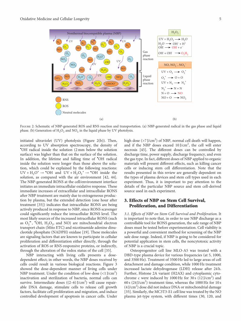
initiated ultraviolet (UV) photolysis (Figure 2(b)). Then,according to UV absorption spectroscopy, the density of•OH radical inside the solution (2mm below the solutionsurface) was higher than that on the surface of the solution.In addition, the lifetime and falling time of •OH radicalinside the solution were longer than those above the solu-tion, which could be explained by the following reactions:UV +H2O∗ → •OH and UV +H2O2
∗ → •OH inside thesolution, as compared with the air environment [42, 44].The NBP-generated RONS at the cell/environment interfaceinitiates an immediate intracellular oxidative response. Theseimmediate increases of extracellular and intracellular RONSafter NBP treatment are mainly due to extrageneous produc-tion by plasma, but the extended detection (one hour aftertreatment [35]) indicates that intracellular RONS are beingactively produced in response to NBP, since RONS scavengercould significantly reduce the intracellular RONS level. Themost likely sources of the increased intracellular RONS (suchas O2
•-, •OH, H2O2, and NO) are mitochondrial electrontransport chain (Mito ETC) and nicotinamide adenine dinu-cleotide phosphate (NADPH) oxidase [19]. These moleculesare signaling factors that are known to participate in cellularproliferation and differentiation either directly, through theactivation of ROS or RNS-responsive proteins, or indirectly,through the alteration of the redox status of the cell [35].
NBP interacting with living cells presents a dose-dependent effect; in other words, the NBP doses received bycells could result in various biological reactions. Figure 3showed the dose-dependent manner of living cells underNBP treatment. Under the condition of low-dose (<1 J/cm2)inactivation and sterilization of bacteria, normal cells cansurvive. Intermediate doses ((2–6) J/cm2) will cause repair-able DNA damage, stimulate cells to release cell growthfactors, facilitate cell proliferation and migration, and inducecontrolled development of apoptosis in cancer cells. Under
high dose (>7 J/cm2) of NBP, normal cell death will happen,and if the NBP doses exceed 10 J/cm2, the cell will enternecrosis [45]. The different doses can be controlled bydischarge time, power supply, discharge frequency, and eventhe gas type. In fact, different doses of NBP applied to organicmaterials will present different effects, such as killing cancercells or inducing stem cell differentiation. Note that theresults presented in this review are generally dependent onthe types of plasma devices and stem cell types used in eachexperiment. Thus, it is important to pay attention to thedetails of the particular NBP source and stem cell-derivedsource used in each experiment.
3. Effects of NBP on Stem Cell Survival,Proliferation, and Differentiation
3.1. Effects of NBP on Stem Cell Survival and Proliferation. Itis important to note that, in order to use NBP discharge as acontrollable tool for RONS generation, the safe range of NBPdoses must be tested before experimentation. Cell viability isa powerful and convenient method for screening of the NBPsafe dose range. Indeed, if NBP is going to be considered forpotential application in stem cells, the noncytotoxic activityof NBP is a crucial topic.
Osteoprogenitor cell line MLO-A5 was treated with aDBD-type plasma device for various frequencies (at 5, 1000,and 3500Hz). Treatment of 3500Hz led to large areas of celldetachment and damage condition, while 1000Hz treatmentincreased lactate dehydrogenase (LDH) release after 24 h.Further, Histone 2A variant (H2AX) and cytoplasmic cyto-chrome c were induced by 1000Hz for 30 s (12 J/cm2) and60 s (24 J/cm2) treatment time, whereas the 1000Hz for 10 s(4 J/cm2) dose did not induce DNA or mitochondrial damage[35]. Similarly, the MC3T3-E1 cell line was treated by the NOplasma jet-type system, with different times (30, 120, and
Nonthermal biocompatible plasma (NBP)
UVUV
NO·
RNS
ROS
Neutral molecules
NO· NO·NO·
·OH
NO2-
NO3-
NO2-
ONOO−
·OH
NO·
NO· NO·
NO·
Gasphasereaction
Liquidphasereaction
UVUV
UV
N2⁎
NO2
NO3
N2⁎
N2⁎
N2⁎N2
⁎
O2⁎
O2⁎ O2
⁎
O2⁎
O2
O2O2
O2
O3
H2O⁎
H2O2H2O2
H2O2
O2
H2O2
H2O⁎
(a)
UV + H2O2
H2O2
H2O·
OH·
OH· + OH·
NO, NO2−, NO3
−
OH· + e−
O2H2
H2O· OH− + H+
UV + O2
O + O
O2⁎
O2⁎
UV + N2 N2⁎
N + NN2⁎
N + O NONO + O NO2
(b)
Figure 2: Schematic of NBP-generated ROS and RNS reaction and transportation. (a) NBP-generated radical in the gas phase and liquidphase. (b) Generation of H2O2 and NOx in the liquid phase by UV photolysis.
5Oxidative Medicine and Cellular Longevity

180 s). There was no significant presence of dead cells, evenwith prolonged NO-NBP treatment durations of up to180 s, as compared to control cells. Even when increasingthe treatment time (to 360 and 480 s), only 2 and 4%, respec-tively, cell toxicity was observed [36]. Very recently, in aprevious study of ours, we found that the viability ofMC3T3-E1 cells treated with a DBD plasma device showeda NBP treatment time-dependent decrease [46].
Aside from progenitor cell lines, the effect of NBP onprimary mesenchymal stem cell derived from human peri-odontal ligament stem cells (hPDLSCs) was studied as well.NBP showed no significant toxicity to cells, even whentreated with up to 1.6W for 120 s with 1 slm helium gas flow[37]. The use of a helium-based DBD device increased theproliferation of adipose tissue-derived stem cells (ASCs),and the cells still maintained their stemness and capabilityto differentiate into adipocytes; however, no cellular senes-cence was observed. The proliferation-enhancing ability ofNBP treatment was significantly decreased when NO scaven-ger was added, while no significant change was found withthe presence of the ROS scavenger. These results suggest thatNBP-generated NO, rather than ROS, is responsible for theenhanced proliferation of ASCs [47]. A recent study indi-cated that a proper dose of NBP treatment could facilitateneural stem cell differentiation into neurons, which wasregulated by the NBP-induced NO production [39]. LowNO concentration was shown to promote cell survival andproliferation in various cells, including stem cells [47], whilehigh NO concentration was shown to lead to cell cycle arrestand cell death [48]. More recently, NBP was found to exhibita protective role over the neuroblastoma-derived cell lineunder oxidative stress, suggesting the therapeutic potentialof NBP as a novel “NO donor” in neuroprotection andneurodegenerative diseases [49].
3.2. Effects of NBP on Stem Cell Migration. Cell migration iscentral to many biological and pathological processes,including embryogenesis, tissue repair, and regeneration, aswell as cancer and the inflammatory response. Research hasaddressed how low-intensity helium NBP affects cells, suchas fibroblasts and endothelial and smooth muscle cells, byinducing the disruption of cell-to-cell adhesion, as well assubsequent cell detachment from the substrates, acting in adose-dependent manner [50, 51]. More importantly, the
observed effects were reversible, since after NBP treatment,the detached cells remained viable and reattached to the platesurface and then, after a short incubation time, continuedproliferating. The adhesion assay data showed that treatmentwith NBP did not significantly affect hPDLSC adhesion,although some detachment of the cells was observed whenthe higher powers of plasma were combined with 0.5 lpmflow plasma treatment, which considerably decreased themigration capacity of the hPDLSCs. In the study on NBPinteraction with cancer cells, invasion and adhesion weresuppressed by either NBP directly or NBP-activated mediaat a certain dilution ratio. In addition, NBP treatment inhib-ited the matrix metallopeptidase 9 (MMP9) enzyme, which isinvolved in the degradation of the extracellular matrix duringcancer cell migration [52, 53].
3.3. Effects of NBP on Stem Cell Differentiation. Osteogenesisis the process consisting of the formation and development ofthe bone. This process begins at the eighth week of embryodevelopment of bone formation and happens continuouslywith bone growth until early adulthood; in addition, all ofthe developments of bone thickness, remodeling, and repair-ing are connected with osteogenesis. During this process,the osteogenic genes, including alkaline phosphate (ALP),type I collagen (COL-1), Runx-related transcription factor2 (Runx 2), osteocalcin (OCN), osteopontin (OPN), andosterix, are expressed. These genes are related to bonemineralization and calcium ion homeostasis during thedifferentiation of stem cells into osteoblasts. One researcheffort initially suggested that NBP does not significantlypromote osteogenesis; however, when inducing stem cellswere entering the differentiation stage, NBP could promotemore differentiation-specific protein expression [35]. Interms of ALP enzyme activity, it has been shown that theNBP-treated group was significantly higher than the onlyhelium-treated or nontreated group [37]. Later, the NBPeffect was studied without differentiation media. Since ALPand COL-1 are early expression genes of osteogenesis, OPNandOCN are expressed in late differentiation andmineraliza-tion, which indicated that NBP could induce early osteogenicdifferentiation of progenitor/stem cells [36].
In order to determine whether the differentiation effect ofNBP was osteoblast specific, the N1511 chondrocyte cell linewas treated by NBP with the presence of bone morphogenetic
ROS level
Low dose(< 1 J/cm2)Inactivation andsterilization ofbacteria
(i) (i) (i)(ii)
(iii)
(iv)
(ii) Normal cell can bealive
Intermediate dose (2~6 J/cm2)Reparation of DNA damageStimulate cells to release growthfactorsFacilitate cell proliferation andmigrationInduce controlled development ofapoptosis in cancer cells
High dose (< 7 J/cm2)Normal cell death
Figure 3: Different effects of NBP interaction with living cells according to NBP dose levels.
6 Oxidative Medicine and Cellular Longevity
protein 2 (BMP2), a known inducer of chondrocyte differ-entiation [54]. Twenty-four hours after treatment, chondro-cyte differentiation markers Runx2 and ALP were increased3- to 6-fold above BMP2-treated controls. By 56 hours afterNBP treatment, collagen type X (Col X) and another latemarker, matrix metalloprotease 13 (MMP13), were increased(20- and 4-fold, respectively) above BMP-treated control.The results show that once the chondrogenic differentiationis started, NBP works as a synergic function to the N1511chondrocyte cell line [35].
Neuronal cells have attracted substantial interest for themedical treatment of neurodegenerative diseases and trau-matic injuries of the central nervous system (CNS), butefforts to produce these cells have thus far only been met withmodest success. In an attempt to find new approaches, Xiongand his colleague [39] treated neural stem cells (NSCs) withNBP to differentiate them into the neuronal lineage. NBP-treated cells exhibited rapid proliferation and differentiationwith longer neurites and cell bodies, eventually formingneuronal networks. The treated cells showed increasedexpression of different cell lineage markers such as β-tubulinIII (for neurons) and oligodendrocyte marker, O4, whilethe expression of glial fibrillary acidic protein (GFAP)(for astrocytes) remained unchanged.
Taken together, there are several advantages to NBP-induced stem cell differentiation. First, the differentiationprocess with NBP treatment is faster. Second, the differentia-tion efficiency dramatically increased with upregulated-specific genes and differentiation signs by NBP treatment.Finally, NBP treatment with/without other chemical inducerscould reach a higher percentage of differentiated cells [55].In vivo research has also indicated that NBP promotesneural differentiation into mature neurons in transgeniczebrafish. Specifically, GFP+ mature neurons in developingzebrafish were observed in the central nervous system after6 h with 1min NBP treatment and these were maintainedthrough 33 h [55].
4. Potential Mechanism of NBPInteraction with Living Cells
4.1. NBP Facilitates Intracellular RONS Accumulation andAlters the Antioxidant System. Mechanistically, research hasdirectly linked NBP interaction with living cells via ROS orRNS generation. During NBP discharge, the working gas isionized into charged particles and chemical species, whichthen collide with the molecules present in air (O2, N2, H2O,and CO2), resulting in the direct formation of numerousRONS [56, 57]. RONS are small, short-lived reactive mole-cules that display high chemical reactivity toward multipleproteins involved in signaling pathways that regulate cellfunction. However, the excessive generation of ROS andimbalance between ROS and antioxidant proteins can causeoxidative stress to cells. For example, H2O2 plays an impor-tant physiological role as an intracellular signaling molecule,regulating a wide variety of biological processes, dependingon its intracellular concentration (<100 nM) [58]. In osteo-blast progenitors, the continuous production of low levelsof H2O2 stimulates proliferation and also augments their
potential to differentiate into mature osteoblasts throughthe upregulation of Runx2 and osterix [35, 59]. Thus, theproper level of ROS, that is, a physiologically sufficientamount, acts as a secondary signaling messenger for stimu-lating stem cell proliferation and maintains intracellularredox balance for cell survival.
Table 1 shows a summary of studies that have revealedthe extracellular ROS level (in culture media) and intracellu-lar ROS. Based on these, we can observe that the mostcommonly existing chemical species in culture media follow-ing NBP treatment are NO• and H2O2, while intracellularROS or RNS accumulation varies from NO to H2O2 and alsoincludes mitochondria O2
•-. These chemical species areresponsible for stem cell lineage commitment to osteoblasts,neurons, myocytes, and chondrocytes. One study reportedthe role of O2
•- in neurogenesis: in normal condition, O2•-
is produced through an electron reduction of oxygen byblocking normal electron transfer in the electron transportchain (ETC). Mitochondria O2
•- is accumulated via anincrease of extracellular NO• concentration, because of NO•
competing with oxygen to bind to the active site of themitochondrial ETC complex IV, cytochrome c oxidase(COX), and reversibly inhibits its activity; therefore, O2
•-
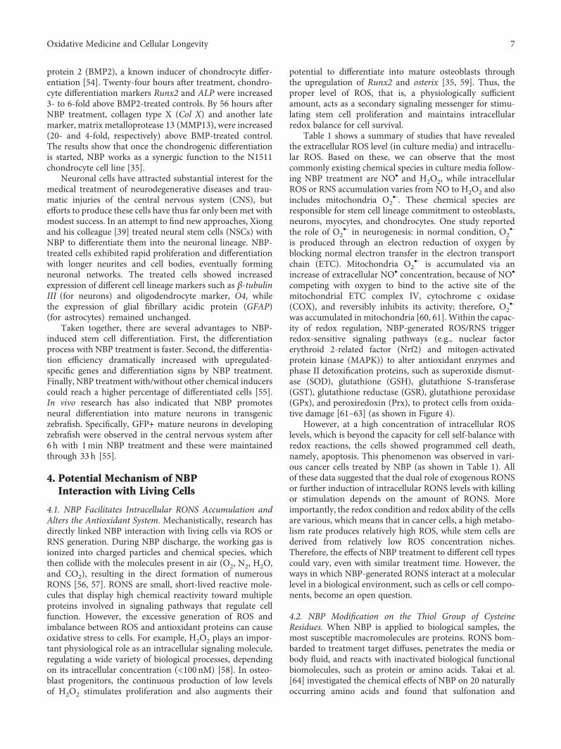
was accumulated in mitochondria [60, 61]. Within the capac-ity of redox regulation, NBP-generated ROS/RNS triggerredox-sensitive signaling pathways (e.g., nuclear factorerythroid 2-related factor (Nrf2) and mitogen-activatedprotein kinase (MAPK)) to alter antioxidant enzymes andphase II detoxification proteins, such as superoxide dismut-ase (SOD), glutathione (GSH), glutathione S-transferase(GST), glutathione reductase (GSR), glutathione peroxidase(GPx), and peroxiredoxin (Prx), to protect cells from oxida-tive damage [61–63] (as shown in Figure 4).
However, at a high concentration of intracellular ROSlevels, which is beyond the capacity for cell self-balance withredox reactions, the cells showed programmed cell death,namely, apoptosis. This phenomenon was observed in vari-ous cancer cells treated by NBP (as shown in Table 1). Allof these data suggested that the dual role of exogenous RONSor further induction of intracellular RONS levels with killingor stimulation depends on the amount of RONS. Moreimportantly, the redox condition and redox ability of the cellsare various, which means that in cancer cells, a high metabo-lism rate produces relatively high ROS, while stem cells arederived from relatively low ROS concentration niches.Therefore, the effects of NBP treatment to different cell typescould vary, even with similar treatment time. However, theways in which NBP-generated RONS interact at a molecularlevel in a biological environment, such as cells or cell compo-nents, become an open question.
4.2. NBP Modification on the Thiol Group of CysteineResidues. When NBP is applied to biological samples, themost susceptible macromolecules are proteins. RONS bom-barded to treatment target diffuses, penetrates the media orbody fluid, and reacts with inactivated biological functionalbiomolecules, such as protein or amino acids. Takai et al.[64] investigated the chemical effects of NBP on 20 naturallyoccurring amino acids and found that sulfonation and
7Oxidative Medicine and Cellular Longevity
disulfide linkage were formed out of thiol groups in cysteineby NBP treatment. In addition, the formation of aromaticrings by hydroxylation and nitration was found in tyro-sines, phenylalanine, and tryptophan, while sulfoxidationwas found in methionine and amidation of the ring openingof the five-membrane rings was found in histidine and pro-line. Another study also reported the NBP modification ofphenylalanine by hydroxylation. More recently, the samegroup confirmed the formation of disulfide linkages betweenthe thiol groups of cysteines by NBP [65, 66], suggesting thatredox modifications by NBP-generated RONS of the redox-sensitive cysteine residues are a pivotal mechanism for thefunctional regulation of a variety of proteins.
Cysteine residues that exist as thiolate anions (Cys-S-) aremore susceptible to oxidation by NBP-generated RONS [67].In response to oxidation by H2O2, the thiol group can revers-ibly form sulfenic acid (Cys-SOH) as well as intramolecularor intermolecular disulfide. The sulfenic form can be reducedto its original state by the disulfide reductases of glutaredoxin(Grx) and thioredoxin (Trx) [68]. However, with the persis-tent presence of high-concentration H2O2, sulfenic acid canbe irreversibly oxidized into sulfinic acid and, further, intosulfonic acid. For example, the presence of RNS by NO• leadsto an S-nitrosothiol bond (SNO), while that by ONOO- leadsto an S-nitrothiol group (SNO2) [18, 69, 70] (as shown inFigure 5). The reversible reaction between thiol groups andRONS is the way in which NBP interacts with redox-sensitive proteins to further activate or inactivate them.
4.3. Regulation of Redox Sensors by NBP-GeneratedROS/RNS. Most of the key regulators for cell survival anddifferentiation, including transcriptional factors and kinases,
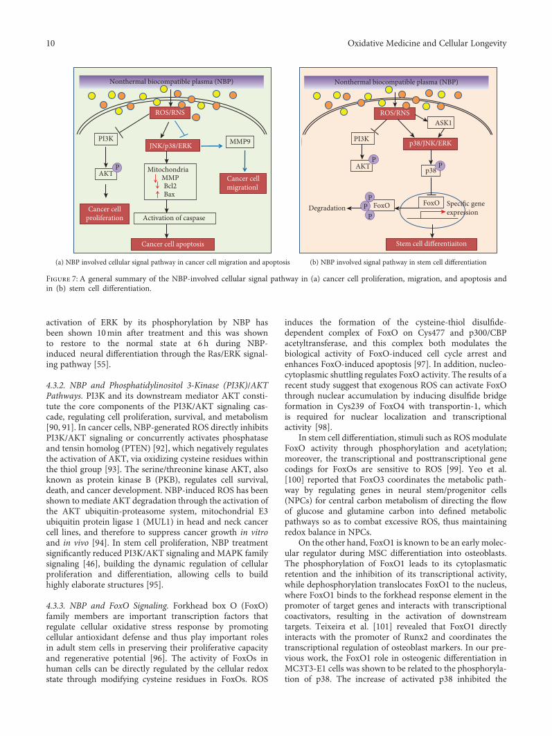
are susceptible to redox modification and recognized asredox sensors [18] (Figure 6). Some of the redox sensorsare involved in the regulation of stem cell self-renewal anddifferentiation, including transcriptional factors and kinasesinvolved in ROS signaling and cell cycle regulation, such ashypoxia-inducible factor-1alpha (HIF-1α), forkhead box O(FoxO), p38, c-Jun N-terminal kinases (JNK), phosphoinosi-tide 3-kinase (PI3K), nuclear factor (erythroid-derived 2-)like 2 (Nrf2), and octamer-binding transcription factor 4(Oct4) [19]. The NBP modifications of these redox sensorsare the initial and direct executors of ROS signaling bythe activation or inactivation of these proteins. Structuralchanges during posttranslation modification allow for pro-tein activity to be modified, which can result in altered cellu-lar function [71]. In particular, redox couples are thosecontaining reactive thiol groups, including cysteine, glutathi-one, and thioredoxin-1. The ROS oxidative modification ofthese thiol groups initiates signals and promotes downstreamcellular responses by affecting the activity and expression ofspecific transcription factors [72–74]. The cell first perceivesextracellular signals and then responds to intracellular ROSthrough the activation of a variety of signaling pathways,including the mitogen-activated protein kinase (MAPK)and PI3K-Akt pathways. Figure 7 showed the summaryof the NBP-induced signal pathway in cancer cells(Figure 7(a)) and stem cell differentiation (Figure 7(b)).
4.3.1. NBP and MAPK Signaling Pathways. The ROS-responsive MAPK family, consisting of p38, JNK, andextracellular signal-regulated protein kinase (ERK) subfamilyfactors [75], is known to control a wide range of cellular pro-cesses, including cellular differentiation, cell survival, geneexpression, cell cycle control, cytokine and growth factorsignaling, cell survival, and apoptosis [76, 77]. Survival andproliferation signaling pathways are important cellular sig-naling circuits which are strongly involved in carcinogenesis.
The MAPK signaling pathway has been well studied inNBP-induced cancer apoptosis. Based on the publishedliterature, JNK and p38 are the most frequently activatedMAPKs in response to NBP-induced cancer apoptosis, witheffects having been reported in head and neck cancer [78],colorectal cancer [79], thyroid cancer [43], cervical cancer[80], and melanoma [81], among which colorectal cancer cellERK was also activated. In addition, only the activation ofERK signal was reported in brain and lung cancer cells byH2O2 and
•OH radical-mediated DNA damage [82]. How-ever, adding these different ROS or RNS scavengers, such asROS scavenger of N-acetylcysteine (NAC), sodium pyruvate,catalase (specific for H2O2), mannitol and 4-hydroxy-2,2,6,6-tetramethylpiperidinyloxy (specific for O2
•-), and RONSscavenger of butylated hydroxyanisole (BHA), Trolox,and 2-phenyl-4,4,5,5-tetramethylimidazoline-1-oxyl 3-oxide(cPTIO, specific for NO•), inhibited the NBP-induced apo-ptosis of cancer cell, indicating the roles of ROS and RNSin apoptotic signal activation in cancer cells (Table 1).
The MAPKs play various roles in different cell types, andin cancer cell apoptosis, they are mostly activated by NBPtreatment; however, in other cell types, MAPKs serve a pro-tective role from apoptosis or even inactivation to inhibit
SOD
RONS
RONS
Nonthermal biocompatible plasma (NBP)
SOD GPx GSTGSRGSHPrx
Nrf2 MAPK pathway
·NO O2·–
ONOO–H2O2
H2O GSSH
GSR
GSHPrx
·OH
GPx
ETC
Figure 4: NBP triggers nuclear translocation of nuclear factorerythroid 2-related factor (Nrf2) and mitogen-activated proteinkinase (MAPK) signaling pathways to modulate redox homeostasisby altering the antioxidant system and phase II detoxificationenzymes and proteins (e.g., superoxide dismutase (SOD),glutathione (GSH), glutathione S-transferase (GST), glutathionereductase (GSR), glutathione peroxidase (GPx), and peroxiredoxin(Prx)).
8 Oxidative Medicine and Cellular Longevity

the migration of cancer cells. Bundscherer et al. [83] reportedthe activation of the MAPK family, including proapoptoticsignaling proteins as p38 and JNK and proproliferationsignaling protein as ERK in immune cell lines by plasmajet. The main ROS species could be H2O2, due to the additionof exogenous H2O2 and plasma treatment having a similareffect. More recently, Li et al. [52] reported the inhibition ofcervical cancer cell migration by NBP through the sup-pression of ERK and JNK of the MAPK family, but notof p38, as well as the downregulation of matrix metallo-proteinase- (MMP-) 9 enzyme.
In addition, MAPKs play a vital role in the regulation ofstem cell self-renewal and differentiation. The kinase p38αas a redox sensor has two cysteine residues, Cys-119 andCys-162, and was activated by the formation of a disulfidebond [84]; it was therefore considered to be involved in stemcell differentiation. Ito et al. [85] reported that p38 MAPKwas activated by increasing the level of ROS. The inhibitionof p38 MAPK was also shown to rescue the ROS-induced
defects in HSC repopulating capacity and in the maintenanceof HSC quiescence, indicating that the ROS-p38 MAPKpathway contributes to exhaustion of the stem cell popula-tion and also protects HSCs against loss of self-renewalcapacity. Protective roles of p38 MAPK from intracellularoxidative stress have also been found in ESC [86] and NSC[87]. These data indicate that p38 MAPK is a redox regulatorthat is always activated by ROS in the modulation of stem cellself-renewal and differentiation. Aside from the directactivation of p38 by ROS, the MAP kinase apoptosis signal-regulating kinase 1 (ASK1) is particularly sensitive to ROS,as its activity is tightly regulated by ROS-sensitive proteins,such as thioredoxin and glutaredoxin [77]. ROS-activatedASK1 phosphorylates and activates both p38 and JNK, whichplay key roles in cellular differentiation [41] as well as theregulation of apoptosis [72]. The activation of ASK1, p38,and/or JNK promotes the differentiation of several celllineages, including chondrocytes, osteoblasts, neuronal,myoblasts, and keratinocytes [76, 77, 88, 89]. Further, the
SH
R
Thiol
S
R
Sulfenic acid Sulfinic acid Sulfonic acid
S
R
OH
O
S-Nitrosothiol
S
R
NO
O
S-Nitrothiol
S S
Intramoleculardisulfide
Intermoleculardisulfide
S
R
S
R R
SH
R
R
S NO
S
R
OH
OOONOO−
NO·
H2O2H2O2
S
R
S
G
S-Glutathionylation
OH H2O2
Figure 5: The modification of a redox sensor with a thiol group by NBP-generated RONS.
Activated
ROS/RNS
Functionalcomplex
Cell proliferationor differentiation
Antioxidants
Redoxsensor
Reversible
Figure 6: ROS/RNS-mediated intracellular signaling. The physical amount of ROS/RNS can posttranscriptionally modulate the redox sensorprotein structure to activate protein and make a functional complex to conduct the downstream signaling pathway. Meanwhile, theantioxidant system in cell can be activated in response to the amount of ROS/RNS in order to maintain intracellular redox homeostasis.
9Oxidative Medicine and Cellular Longevity
activation of ERK by its phosphorylation by NBP hasbeen shown 10min after treatment and this was shownto restore to the normal state at 6 h during NBP-induced neural differentiation through the Ras/ERK signal-ing pathway [55].
4.3.2. NBP and Phosphatidylinositol 3-Kinase (PI3K)/AKTPathways. PI3K and its downstream mediator AKT consti-tute the core components of the PI3K/AKT signaling cas-cade, regulating cell proliferation, survival, and metabolism[90, 91]. In cancer cells, NBP-generated ROS directly inhibitsPI3K/AKT signaling or concurrently activates phosphataseand tensin homolog (PTEN) [92], which negatively regulatesthe activation of AKT, via oxidizing cysteine residues withinthe thiol group [93]. The serine/threonine kinase AKT, alsoknown as protein kinase B (PKB), regulates cell survival,death, and cancer development. NBP-induced ROS has beenshown to mediate AKT degradation through the activation ofthe AKT ubiquitin-proteasome system, mitochondrial E3ubiquitin protein ligase 1 (MUL1) in head and neck cancercell lines, and therefore to suppress cancer growth in vitroand in vivo [94]. In stem cell proliferation, NBP treatmentsignificantly reduced PI3K/AKT signaling and MAPK familysignaling [46], building the dynamic regulation of cellularproliferation and differentiation, allowing cells to buildhighly elaborate structures [95].
4.3.3. NBP and FoxO Signaling. Forkhead box O (FoxO)family members are important transcription factors thatregulate cellular oxidative stress response by promotingcellular antioxidant defense and thus play important rolesin adult stem cells in preserving their proliferative capacityand regenerative potential [96]. The activity of FoxOs inhuman cells can be directly regulated by the cellular redoxstate through modifying cysteine residues in FoxOs. ROS
induces the formation of the cysteine-thiol disulfide-dependent complex of FoxO on Cys477 and p300/CBPacetyltransferase, and this complex both modulates thebiological activity of FoxO-induced cell cycle arrest andenhances FoxO-induced apoptosis [97]. In addition, nucleo-cytoplasmic shuttling regulates FoxO activity. The results of arecent study suggest that exogenous ROS can activate FoxOthrough nuclear accumulation by inducing disulfide bridgeformation in Cys239 of FoxO4 with transportin-1, whichis required for nuclear localization and transcriptionalactivity [98].
In stem cell differentiation, stimuli such as ROSmodulateFoxO activity through phosphorylation and acetylation;moreover, the transcriptional and posttranscriptional genecodings for FoxOs are sensitive to ROS [99]. Yeo et al.[100] reported that FoxO3 coordinates the metabolic path-way by regulating genes in neural stem/progenitor cells(NPCs) for central carbon metabolism of directing the flowof glucose and glutamine carbon into defined metabolicpathways so as to combat excessive ROS, thus maintainingredox balance in NPCs.
On the other hand, FoxO1 is known to be an early molec-ular regulator during MSC differentiation into osteoblasts.The phosphorylation of FoxO1 leads to its cytoplasmaticretention and the inhibition of its transcriptional activity,while dephosphorylation translocates FoxO1 to the nucleus,where FoxO1 binds to the forkhead response element in thepromoter of target genes and interacts with transcriptionalcoactivators, resulting in the activation of downstreamtargets. Teixeira et al. [101] revealed that FoxO1 directlyinteracts with the promoter of Runx2 and coordinates thetranscriptional regulation of osteoblast markers. In our pre-vious work, the FoxO1 role in osteogenic differentiation inMC3T3-E1 cells was shown to be related to the phosphoryla-tion of p38. The increase of activated p38 inhibited the
ROS/RNS
JNK/p38/ERK
Mitochondria MMP Bcl2Bax
Cancer cell apoptosis
Activation of caspase
Nonthermal biocompatible plasma (NBP)
MMP9
Cancer cellmigrationl
PI3K
AKT
Cancer cellproliferation
P
(a) NBP involved cellular signal pathway in cancer cell migration and apoptosis
ROS/RNS
p38/JNK/ERK
Stem cell differentiaiton
ASK1
Specific geneexpression
FoxO
p38
FoxODegradation
PI3K
AKT PP
PP
P
Nonthermal biocompatible plasma (NBP)
(b) NBP involved signal pathway in stem cell differentiation
Figure 7: A general summary of the NBP-involved cellular signal pathway in (a) cancer cell proliferation, migration, and apoptosis andin (b) stem cell differentiation.
10 Oxidative Medicine and Cellular Longevity

phosphorylation of FoxO1, thereby increasing the nuclearaccumulation of FoxO1 and transcript osteogenic-relatedgenes [46].
5. Conclusion
The groundbreaking studies of NBP for novel regenerativemedicine have been expected to lead to a nonlethal oxidativecellular burst that promotes progenitor/stem cell differentia-tion by the manipulation of intracellular redox homeostasis.Furthermore, the physical levels of ROS and RNS producedin response to NBP influence signaling pathways that areresponsible for cellular proliferation and differentiation andmimic the natural intracellular signaling pathway, ratherthan having deleterious effect. However, it is important tonote that there are several advantages and disadvantages ofNBP in inducing stem cell differentiation as compared withother strategies, i.e., chemical, biological, and physical stimuli(shown in Table 2). Additionally, further investigation is alsoneeded to clarify the other factors that affect establishingeffective differentiation using NBP. Considering NBP cangenerate controllable amount and mixture ratio of ROS/RNS,which can be benefit to stem cell differentiation process, itwould be reasonable to conclude that NBP will play animportant role in regenerative therapies with the potentialto advance the treatment and management of incurabledisease.
Conflicts of Interest
The authors declare that they have no conflicts of interest.
Acknowledgments
This work was supported by the Leading ForeignResearch Institute Recruitment Program through a grantfrom the National Research Foundation of Korea (NRF-2016K1A4A3914113), funded by the Ministry of Science,
ICT, and Future Planning (MSIP) of the Korean governmentfor EH Choi and I Han, and through a grant from theBasic Science Research Program through the NRF ofKorea, funded by the Ministry of Education of the Koreagovernment (NRF-2015R1C1A2A01054137) for I Han.
References
[1] F. J. O'Brien, “Biomaterials & scaffolds for tissue engineer-ing,” Materials Today, vol. 14, no. 3, pp. 88–95, 2011.
[2] A. R. Shrivats, M. C. McDermott, and J. O. Hollinger, “Bonetissue engineering: state of the union,” Drug Discovery Today,vol. 19, no. 6, pp. 781–786, 2014.
[3] E. M. Horwitz, P. L. Gordon, W. K. K. Koo et al., “Isolatedallogeneic bone marrow-derived mesenchymal cells engraftand stimulate growth in children with osteogenesis imper-fecta: implications for cell therapy of bone,” Proceedings ofthe National Academy of Sciences of the United States ofAmerica, vol. 99, no. 13, pp. 8932–8937, 2002.
[4] J.M.Murphy,D. J. Fink, E. B.Hunziker, and F. P. Barry, “Stemcell therapy in a caprine model of osteoarthritis,” Arthritisand Rheumatism, vol. 48, no. 12, pp. 3464–3474, 2003.
[5] C. Stamm, B. Westphal, H. D. Kleine et al., “Autologousbone-marrow stem-cell transplantation for myocardialregeneration,” The Lancet, vol. 361, no. 9351, pp. 45-46, 2003.
[6] E. Mezey, S. Key, G. Vogelsang, I. Szalayova, G. D. Lange, andB. Crain, “Transplanted bone marrow generates new neuronsin human brains,” Proceedings of the National Academy ofSciences of the United States of America, vol. 100, no. 3,pp. 1364–1369, 2003.
[7] I. Han, B. S. Kwon, H. K. Park, and K. S. Kim, “Differentiationpotential of mesenchymal stem cells is related to theirintrinsic mechanical properties,” International NeurourologyJournal, vol. 21, Supplement 1, pp. S24–S31, 2017.
[8] M. O. Kim, H. Jung, S. C. Kim, J. K. Park, and Y. K. Seo,“Electromagnetic fields and nanomagnetic particles increasethe osteogenic differentiation of human bone marrow-derived mesenchymal stem cells,” International Journal ofMolecular Medicine, vol. 35, no. 1, pp. 153–160, 2015.
Table 2: Comparison of advantages and disadvantages between NBP and other strategies in inducing stem cell differentiation.
Advantages Disadvantages Reference
Chemical and biologicalstimuli
High efficiencyEasy applicationWell established
High rates of proliferation and differentiationCan be produced in large quantities
High costVery labile
May cause side effectsActive multiple signaling pathways and lack
of specificity
[103, 104]
Physical stimuli
Manipulation of extracellular and physicalenvironment for cells
Proper electric field is benefit forcardiomyogenic differentiation
Heat effectElectrical effect
Hard to control the proper amountStandardization required
[105–108]
NBP
Low costConvenient and portable
High availabilityThe main role of exogenous free radical and ROSEasy control of the amount of ROS and RNSMimic natural biological pathways and have
minimum side effects
Mostly research based and very few clinical trialsMechanically not understood well yet
Standardization requiredLow efficiency to differentiate
[35, 36, 46]
11Oxidative Medicine and Cellular Longevity
[9] M. Song, D. Zhao, S. Wei et al., “The effect of electromag-netic fields on the proliferation and the osteogenic or adipo-genic differentiation of mesenchymal stem cells modulatedby dexamethasone,” Bioelectromagnetics, vol. 35, no. 7,pp. 479–490, 2014.
[10] M. Y. Song, J. Z. Yu, D. M. Zhao et al., “The time-dependentmanner of sinusoidal electromagnetic fields on rat bonemarrow mesenchymal stem cells proliferation, differentia-tion, and mineralization,” Cell Biochemistry and Biophysics,vol. 69, no. 1, pp. 47–54, 2014.
[11] Q. Ma, P. Deng, G. Zhu et al., “Extremely low-frequencyelectromagnetic fields affect transcript levels of neuronaldifferentiation-related genes in embryonic neural stem cells,”PLoS One, vol. 9, no. 3, article e90041, 2014.
[12] Y. K. Choi, D. H. Lee, Y. K. Seo, H. Jung, J. K. Park, andH. Cho, “Stimulation of neural differentiation in human bonemarrow mesenchymal stem cells by extremely low-frequencyelectromagnetic fields incorporated with MNPs,” AppliedBiochemistry and Biotechnology, vol. 174, no. 4, pp. 1233–1245, 2014.
[13] J. E. Park, Y. K. Seo, H. H. Yoon, C. W. Kim, J. K. Park, andS. Jeon, “Electromagnetic fields induce neural differentiationof human bone marrow derived mesenchymal stem cells viaROS mediated EGFR activation,” Neurochemistry Interna-tional, vol. 62, no. 4, pp. 418–424, 2013.
[14] M. Esposito, A. Lucariello, C. Costanzo et al., “Differentiationof human umbilical cord-derived mesenchymal stem cells,WJ-MSCs, into chondrogenic cells in the presence of pulsedelectromagnetic fields,” In Vivo, vol. 27, no. 4, pp. 495–500,2013.
[15] J. K. Park, J. E. Park, and S. Jeon, “Electromagnetic fields(EMFs) can promote neuronal differentiation of humanbone marrow-derived mesenchymal stem cells,” Journal ofTissue Engineering and Regenerative Medicine, vol. 6,pp. 409-410, 2012.
[16] J. H. W. Jansen, O. P. van der Jagt, B. J. Punt et al., “Stimula-tion of osteogenic differentiation in human osteoprogenitorcells by pulsed electromagnetic fields: an in vitro study,”BMC Musculoskeletal Disorders, vol. 11, no. 1, 2010.
[17] Z. Schwartz, B. J. Simon, M. A. Duran, G. Barabino,R. Chaudhri, and B. D. Boyan, “Pulsed electromagnetic fieldsenhance BMP-2 dependent osteoblastic differentiation ofhuman mesenchymal stem cells,” Journal of OrthopaedicResearch, vol. 26, no. 9, pp. 1250–1255, 2008.
[18] F. Ren, K. Wang, T. Zhang, J. Jiang, E. C. Nice, and C. Huang,“New insights into redox regulation of stem cell self-renewaland differentiation,” Biochimica et Biophysica Acta (BBA) -General Subjects, vol. 1850, no. 8, pp. 1518–1526, 2015.
[19] K. Wang, T. Zhang, Q. Dong, E. C. Nice, C. Huang, andY. Wei, “Redox homeostasis: the linchpin in stem cell self-renewal and differentiation,” Cell Death & Disease, vol. 4,no. 3, article e537, 2013.
[20] Y. S. Bae, H. Oh, S. G. Rhee, and Y. D. Yoo, “Regulation ofreactive oxygen species generation in cell signaling,” Mole-cules and Cells, vol. 32, no. 6, pp. 491–509, 2011.
[21] C. I. Kobayashi and T. Suda, “Regulation of reactive oxygenspecies in stem cells and cancer stem cells,” Journal of CellularPhysiology, vol. 227, no. 2, pp. 421–430, 2012.
[22] O. G. Lyublinskaya, J. S. Ivanova, N. A. Pugovkina et al.,“Redox environment in stem and differentiated cells: a quan-titative approach,” Redox Biology, vol. 12, pp. 758–769, 2017.
[23] H. Sauer and M. Wartenberg, “Reactive oxygen species assignaling molecules in cardiovascular differentiation ofembryonic stem cells and tumor-induced angiogenesis,”Antioxidants & Redox Signaling, vol. 7, no. 11-12, pp. 1423–1434, 2005.
[24] E. E. Kunhardt, “Generation of large-volume, atmospheric-pressure, nonequilibrium plasmas,” IEEE Transactions onPlasma Science, vol. 28, no. 1, pp. 189–200, 2000.
[25] H. Ayan, G. Fridman, D. Staack et al., “Heating effect ofdielectric barrier discharges for direct medical treatment,”IEEE Transactions on Plasma Science, vol. 37, no. 1,pp. 113–120, 2009.
[26] W. Murphy, C. Carroll, and M. Keidar, “Simulation of theeffect of plasma species on tumor growth and apoptosis,”Journal of Physics D: Applied Physics, vol. 47, no. 47, article472001, 2014.
[27] G. Daeschlein, S. Scholz, R. Ahmed et al., “Skin decontamina-tion by low-temperature atmospheric pressure plasma jet anddielectric barrier discharge plasma,” The Journal of HospitalInfection, vol. 81, no. 3, pp. 177–183, 2012.
[28] Y. Li, M. Ho Kang, H. Sup Uhm, G. Joon Lee, E. Ha Choi,and I. Han, “Effects of atmospheric-pressure non-thermalbio-compatible plasma and plasma activated nitric oxidewater on cervical cancer cells,” Scientific Reports, vol. 7,no. 1, article 45781, 2017.
[29] Z. Ke and Q. Huang, “Haem-assisted dityrosine-cross-linkingof fibrinogen under non-thermal plasma exposure: oneimportant mechanism of facilitated blood coagulation,”Scientific Reports, vol. 6, no. 1, article 26982, 2016.
[30] B. Haertel, T. . Woedtke, K. D. Weltmann, and U. Lindequist,“Non-thermal atmospheric-pressure plasma possible appli-cation in wound healing,” Biomolecules & Therapeutics,vol. 22, no. 6, pp. 477–490, 2014.
[31] S. H. Nam, H. W. Lee, S. H. Cho, J. K. Lee, Y. C. Jeon, andG. C. Kim, “High-efficiency tooth bleaching using non-thermal atmospheric pressure plasma with low concentrationof hydrogen peroxide,” Journal of Applied Oral Science,vol. 21, no. 3, pp. 265–270, 2013.
[32] M.Wang, X. Cheng, W. Zhu, B. Holmes, M. Keidar, and L. G.Zhang, “Design of biomimetic and bioactive cold plasma-modified nanostructured scaffolds for enhanced osteogenicdifferentiation of bone marrow-derived mesenchymal stemcells,” Tissue Engineering. Part A, vol. 20, no. 5-6, pp. 1060–1071, 2014.
[33] M. Wang, P. Favi, X. Cheng et al., “Cold atmospheric plasma(CAP) surface nanomodified 3D printed polylactic acid(PLA) scaffolds for bone regeneration,” Acta Biomaterialia,vol. 46, pp. 256–265, 2016.
[34] W. Zhu, N. J. Castro, X. Cheng, M. Keidar, and L. G. Zhang,“Cold atmospheric plasma modified electrospun scaffoldswith embedded microspheres for improved cartilage regener-ation,” PLoS One, vol. 10, no. 7, article e0134729, 2015.
[35] M. J. Steinbeck, N. Chernets, J. Zhang et al., “Skeletal celldifferentiation is enhanced by atmospheric dielectric barrierdischarge plasma treatment,” PLoS One, vol. 8, no. 12, articlee82143, 2013.
[36] M. Elsaadany, G. Subramanian, H. Ayan, and E. Yildirim-Ayan, “Exogenous nitric oxide (NO) generated by NO-plasma treatment modulates osteoprogenitor cells earlydifferentiation,” Journal of Physics D: Applied Physics,vol. 48, no. 34, 2015.
12 Oxidative Medicine and Cellular Longevity

[37] M. Miletić, S. Mojsilović, I. Okić Đorđević et al., “Effects ofnon-thermal atmospheric plasma on human periodontalligament mesenchymal stem cells,” Journal of Physics D:Applied Physics, vol. 46, no. 34, article 345401, 2013.
[38] J. W. Choi, S. U. Kang, Y. E. Kim et al., “Novel therapeuticeffects of non-thermal atmospheric pressure plasma formuscle regeneration and differentiation,” Scientific Reports,vol. 6, no. 1, 2016.
[39] Z. Xiong, S. Zhao, X. Mao et al., “Selective neuronal differ-entiation of neural stem cells induced by nanosecondmicroplasma agitation,” Stem Cell Research, vol. 12, no. 2,pp. 387–399, 2014.
[40] K. Tominami, H. Kanetaka, S. Sasaki, T. Mokudai, T. Kaneko,and Y. Niwano, “Cold atmospheric plasma enhances osteo-blast differentiation,” PLoS One, vol. 12, no. 7, articlee0180507, 2017.
[41] M. Keidar, “Plasma for cancer treatment,” Plasma SourcesScience and Technology, vol. 24, no. 3, 2015.
[42] Y. H. Kim, Y. J. Hong, K. Y. Baik et al., “Measurement of reac-tive hydroxyl radical species inside the biosolutions duringnon-thermal atmospheric pressure plasma jet bombardmentonto the solution,” Plasma Chemistry and Plasma Processing,vol. 34, no. 3, pp. 457–472, 2014.
[43] S. Y. Lee, S. U. Kang, K. I. Kim et al., “Nonthermal plasmainduces apoptosis in ATC cells: involvement of JNKand p38 MAPK-dependent ROS,” Yonsei Medical Journal,vol. 55, no. 6, pp. 1640–1647, 2014.
[44] P. Attri, Y. H. Kim, D. H. Park et al., “Generation mechanismof hydroxyl radical species and its lifetime prediction duringthe plasma-initiated ultraviolet (UV) photolysis,” ScientificReports, vol. 5, no. 1, 2015.
[45] D. Dobrynin, G. Fridman, G. Friedman, and A. Fridman,“Physical and biological mechanisms of direct plasma inter-action with living tissue,” New Journal of Physics, vol. 11,no. 11, 2009.
[46] I. Han and E. H. Choi, “The role of non-thermal atmosphericpressure biocompatible plasma in the differentiation of oste-oblastic precursor cells, MC3T3-E1,” Oncotarget, vol. 8,no. 22, pp. 36399–36409, 2017.
[47] J. Park, H. Lee, H. J. Lee et al., “Non-thermal atmosphericpressure plasma efficiently promotes the proliferation ofadipose tissue-derived stem cells by activating no-responsepathways,” Scientific Reports, vol. 6, no. 1, 2016.
[48] M. C. Carreras and J. J. Poderoso, “Mitochondrial nitricoxide in the signaling of cell integrated responses,” AmericanJournal of Physiology-Cell Physiology, vol. 292, no. 5,pp. C1569–C1580, 2007.
[49] X. Yan, Y. Qiao, J. Ouyang, M. Jia, J. Li, and F. Yuan,“Protective effect of atmospheric pressure plasma on oxida-tive stress-induced neuronal injuries: an in vitro study,” Jour-nal of Physics D: Applied Physics, vol. 50, no. 9, 2017.
[50] M. Arai, Y. Shibata, K. Pugdee, Y. Abiko, and Y. Ogata,“Effects of reactive oxygen species (ROS) on antioxidantsystem and osteoblastic differentiation in MC3T3-E1 cells,”IUBMB Life, vol. 59, no. 1, pp. 27–33, 2007.
[51] Y. Kanamaru, T. Takada, R. Saura, and K. Mizuno, “Effect ofnitric oxide on mouse clonal osteogenic cell, MC3T3-E1,proliferation in vitro,” The Kobe Journal of Medical Sciences,vol. 47, no. 1, pp. 1–11, 2001.
[52] W. Li, K. N. Yu, L. Bao, J. Shen, C. Cheng, and W. Han,“Non-thermal plasma inhibits human cervical cancer HeLa
cells invasiveness by suppressing the MAPK pathway anddecreasing matrix metalloproteinase-9 expression,” ScientificReports, vol. 6, no. 1, article 19720, 2016.
[53] K. Nakamura, Y. Peng, F. Utsumi et al., “Novel intraperito-neal treatment with non-thermal plasma-activated mediuminhibits metastatic potential of ovarian cancer cells,” ScientificReports, vol. 7, no. 1, p. 6085, 2017.
[54] N. Kamiya, A. Jikko, K. Kimata, C. Damsky, K. Shimizu, andH. Watanabe, “Establishment of a novel chondrocytic cellline N1511 derived from p53-null mice,” Journal of Boneand Mineral Research, vol. 17, no. 10, pp. 1832–1842, 2002.
[55] J. Y. Jang, Y. J. Hong, J. Lim et al., “Cold atmospheric plasma(CAP), a novel physicochemical source, induces neuraldifferentiation through cross-talk between the specific RONScascade and Trk/Ras/ERK signaling pathway,” Biomaterials,vol. 156, pp. 258–273, 2018.
[56] A. Fridman, A. Chirokov, and A. Gutsol, “Non-thermalatmospheric pressure discharges,” Journal of Physics D:Applied Physics, vol. 38, no. 2, pp. R1–R24, 2005.
[57] G. Fridman, G. Friedman, A. Gutsol, A. B. Shekhter, V. N.Vasilets, and A. Fridman, “Applied plasma medicine,”Plasma Processes and Polymers, vol. 5, no. 6, pp. 503–533, 2008.
[58] L. B. Valdez, S. Lores Arnaiz, J. Bustamante, S. Alvarez, L. E.Costa, and A. Boveris, “Free radical chemistry in biologicalsystems,” Biological Research, vol. 33, no. 2, 2000.
[59] Y. Choe, J. Y. Yu, Y. O. Son et al., “Continuously gener-ated H2O2 stimulates the proliferation and osteoblasticdifferentiation of human periodontal ligament fibroblasts,”Journal of Cellular Biochemistry, vol. 113, no. 4, pp. 1426–1436, 2012.
[60] J. J. Poderoso, M..́ C. Carreras, C. Lisdero, N. Riobó,F. Schöpfer, and A. Boveris, “Nitric oxide inhibits electrontransfer and increases superoxide radical production inrat heart mitochondria and submitochondrial particles,”Archives of Biochemistry and Biophysics, vol. 328, no. 1,pp. 85–92, 1996.
[61] B. D'Autreaux and M. B. Toledano, “ROS as signalling mole-cules: mechanisms that generate specificity in ROS homeo-stasis,” Nature Reviews Molecular Cell Biology, vol. 8, no. 10,pp. 813–824, 2007.
[62] S. Bekeschus, T. von Woedtke, A. Kramer, K. D. Weltmann,and K. Masur, “Cold physical plasma treatment alters redoxbalance in human immune cells,” Plasma Medicine, vol. 3,no. 4, pp. 267–278, 2013.
[63] M. Weiss, D. Gümbel, E. M. Hanschmann et al., “Cold atmo-spheric plasma treatment induces anti-proliferative effects inprostate cancer cells by redox and apoptotic signaling path-ways,” PLoS One, vol. 10, no. 7, article e0130350, 2015.
[64] E. Takai, T. Kitamura, J. Kuwabara et al., “Chemical modifi-cation of amino acids by atmospheric-pressure cold plasmain aqueous solution,” Journal of Physics D: Applied Physics,vol. 47, no. 28, article 285403, 2014.
[65] Z. Ke, Z. Yu, and Q. Huang, “Assessment of damage ofglutathione by glow discharge plasma at the gas-solutioninterface through Raman spectroscopy,” Plasma Processesand Polymers, vol. 10, no. 2, pp. 181–188, 2013.
[66] Z. Ke, Q. Huang, B. Dang et al., “A study of low-energyion induced radiolysis of thiol-containing amino acid cys-teine in the solid and aqueous solution states,” NuclearInstruments and Methods in Physics Research Section B:
13Oxidative Medicine and Cellular Longevity
Beam Interactions with Materials and Atoms, vol. 268,no. 17-18, pp. 2729–2734, 2010.
[67] T. Finkel, “From sulfenylation to sulfhydration: what athiolate needs to tolerate,” Science Signaling, vol. 5, no. 215,article pe10, 2012.
[68] C. C. Winterbourn and M. B. Hampton, “Thiol chemistryand specificity in redox signaling,” Free Radical Biology andMedicine, vol. 45, no. 5, pp. 549–561, 2008.
[69] S. Hoffman, J. Nolin, D. McMillan, E. Wouters, Y. Janssen-Heininger, and N. Reynaert, “Thiol redox chemistry: role ofprotein cysteine oxidation and altered redox homeostasisin allergic inflammation and asthma,” Journal of CellularBiochemistry, vol. 116, no. 6, pp. 884–892, 2015.
[70] M. Schieber and N. S. Chandel, “ROS function in redoxsignaling and oxidative stress,” Current Biology, vol. 24,no. 10, pp. R453–R462, 2014.
[71] D. Shao, S. Oka, C. D. Brady, J. Haendeler, P. Eaton, andJ. Sadoshima, “Redox modification of cell signaling in thecardiovascular system,” Journal of Molecular and CellularCardiology, vol. 52, no. 3, pp. 550–558, 2012.
[72] A. Matsuzawa and H. Ichijo, “Redox control of cell fateby MAP kinase: physiological roles of ASK1-MAP kinasepathway in stress signaling,” Biochimica et BiophysicaActa (BBA) - General Subjects, vol. 1780, no. 11, pp. 1325–1336, 2008.
[73] Y. Hou, P. Xue, Y. Bai et al., “Nuclear factor erythroid-derived factor 2-related factor 2 regulates transcription ofCCAAT/enhancer-binding protein β during adipogenesis,”Free Radical Biology and Medicine, vol. 52, no. 2, pp. 462–472, 2012.
[74] H. Motohashi, M. Kimura, R. Fujita et al., “NF-E2 domina-tion over Nrf2 promotes ROS accumulation and megakaryo-cytic maturation,” Blood, vol. 115, no. 3, pp. 677–686, 2010.
[75] T. Tsakiridis, J. Wright, E. Tsiani et al., “Mitogen-activatedprotein kinase (MAPK) expression and activation in non-small cell lung cancer (NSCLC): investigating their prognos-tic significance and role in radiation responses,” Radiotherapyand Oncology, vol. 72, pp. S40–S41, 2004.
[76] J. Li, Z. Zhao, J. Liu et al., “MEK/ERK and p38 MAPK regu-late chondrogenesis of rat bone marrow mesenchymal stemcells through delicate interaction with TGF-β1/Smads path-way,” Cell Proliferation, vol. 43, no. 4, pp. 333–343, 2010.
[77] P. Tran, S. M. Ho, B. G. Kim et al., “TGF-β-activated kinase 1(TAK1) and apoptosis signal-regulating kinase 1 (ASK1)interact with the promyogenic receptor Cdo to promote myo-genic differentiation via activation of p38MAPK pathway,”Journal of Biological Chemistry, vol. 287, no. 15, pp. 11602–11615, 2012.
[78] S. U. Kang, J. H. Cho, J. W. Chang et al., “Nonthermal plasmainduces head and neck cancer cell death: the potentialinvolvement of mitogen-activated protein kinase-dependentmitochondrial reactive oxygen species,” Cell Death & Disease,vol. 5, no. 2, article e1056, 2014.
[79] C. H. Kim, J. H. Bahn, S. H. Lee et al., “Induction of cellgrowth arrest by atmospheric non-thermal plasma in colo-rectal cancer cells,” Journal of Biotechnology, vol. 150, no. 4,pp. 530–538, 2010.
[80] H. J. Ahn, K. I. Kim, N. N. Hoan et al., “Targeting cancer cellswith reactive oxygen and nitrogen species generated byatmospheric-pressure air plasma,” PLoS One, vol. 9, no. 1,article e86173, 2014.
[81] M. Ishaq, S. Kumar, H. Varinli et al., “Atmospheric gasplasma-induced ROS production activates TNF-ASK1 path-way for the induction of melanoma cancer cell apoptosis,”Molecular Biology of the Cell, vol. 25, no. 9, pp. 1523–1531, 2014.
[82] N. Kaushik, N. Uddin, G. B. Sim et al., “Responses of solidtumor cells in dmem to reactive oxygen species generatedby non-thermal plasma and chemically induced ros systems,”Scientific Reports, vol. 5, article 8587, 2015.
[83] L. Bundscherer, K. Wende, K. Ottmüller et al., “Impact ofnon-thermal plasma treatment onMAPK signaling pathwaysof human immune cell lines,” Immunobiology, vol. 218,no. 10, pp. 1248–1255, 2013.
[84] R. Bassi, J. R. Burgoyne, G. F. DeNicola et al., “Redox-dependent dimerization of p38α mitogen-activated proteinkinase with mitogen-activated protein kinase kinase 3,”Journal of Biological Chemistry, vol. 292, no. 39, pp. 16161–16173, 2017.
[85] K. Ito, A. Hirao, F. Arai et al., “Reactive oxygen species actthrough p38 MAPK to limit the lifespan of hematopoieticstem cells,”NatureMedicine, vol. 12, no. 4, pp. 446–451, 2006.
[86] L. Ding, X. G. Liang, Y. Hu, D. Y. Zhu, and Y. J. Lou,“Involvement of p38MAPK and reactive oxygen species inicariin-induced cardiomyocyte differentiation of murineembryonic stem cells in vitro,” Stem Cells and Development,vol. 17, no. 4, pp. 751–760, 2008.
[87] J. Kim and P. K. Y. Wong, “Loss of ATM impairs prolifera-tion of neural stem cells through oxidative stress-mediatedp38 MAPK signaling,” Stem Cells, vol. 27, no. 8, pp. 1987–1998, 2009.
[88] A. Saito, K. Ochiai, S. Kondo et al., “Endoplasmic reticulumstress response mediated by the PERK-eIF2α-ATF4 pathwayis involved in osteoblast differentiation induced by BMP2,”Journal of Biological Chemistry, vol. 286, no. 6, pp. 4809–4818, 2011.
[89] R. Faigle, A. Brederlau, M. Elmi et al., “ASK1 inhibitsastroglial development via p38 mitogen-activated proteinkinase and promotes neuronal differentiation in adulthippocampus-derived progenitor cells,”Molecular and Cellu-lar Biology, vol. 24, no. 1, pp. 280–293, 2004.
[90] J. S. L. Yu and W. Cui, “Proliferation, survival and metabo-lism: the role of PI3K/AKT/mTOR signalling in pluripotencyand cell fate determination,” Development, vol. 143, no. 17,pp. 3050–3060, 2016.
[91] I. Han, M. Yun, E. O. Kim, B. Kim,M. H. Jung, and S. H. Kim,“RETRACTED ARTICLE: Umbilical cord tissue-derivedmesenchymal stem cells induce apoptosis in PC-3 prostatecancer cells through activation of JNK and downregulationof PI3K/AKT signaling,” Stem Cell Research & Therapy,vol. 5, no. 2, p. 54, 2014.
[92] Y. Liu, S. Tan, H. Zhang et al., “Selective effects of non-thermal atmospheric plasma on triple-negative breast normaland carcinoma cells through different cell signaling path-ways,” Scientific Reports, vol. 7, no. 1, article 7980, 2017.
[93] N. R. Leslie and C. P. Downes, “PTEN: the down side ofPI 3-kinase signalling,” Cellular Signalling, vol. 14, no. 4,pp. 285–295, 2002.
[94] S. Y. Kim, H. J. Kim, S. U. Kang et al., “Non-thermal plasmainduces AKT degradation through turn-on the MUL1 E3ligase in head and neck cancer,” Oncotarget, vol. 6, no. 32,pp. 33382–33396, 2015.
14 Oxidative Medicine and Cellular Longevity

[95] P. Rué and A. M. Arias, “Cell dynamics and gene expressioncontrol in tissue homeostasis and development,” MolecularSystems Biology, vol. 11, no. 2, p. 792, 2015.
[96] Z. Tothova and D. G. Gilliland, “FoxO transcription factorsand stem cell homeostasis: insights from the hematopoieticsystem,” Cell Stem Cell, vol. 1, no. 2, pp. 140–152, 2007.
[97] T. B. Dansen, L. M. M. Smits, M. H. van Triest et al., “Redox-sensitive cysteines bridge p300/CBP-mediated acetylationand FoxO4 activity,” Nature Chemical Biology, vol. 5, no. 9,pp. 664–672, 2009.
[98] M. Putker, T. Madl, H. R. Vos et al., “Redox-dependentcontrol of FOXO/DAF-16 by transportin-1,” Molecular Cell,vol. 49, no. 4, pp. 730–742, 2013.
[99] L. O. Klotz, C. Sánchez-Ramos, I. Prieto-Arroyo, P. Urbánek,H. Steinbrenner, and M. Monsalve, “Redox regulation ofFoxO transcription factors,” Redox Biology, vol. 6, pp. 51–72, 2015.
[100] H. Yeo, C. A. Lyssiotis, Y. Zhang et al., “FoxO3 coordinatesmetabolic pathways to maintain redox balance in neural stemcells,” EMBO Journal, vol. 32, no. 19, pp. 2589–2602, 2013.
[101] C. C. Teixeira, Y. Liu, L. M. Thant, J. Pang, G. Palmer, andM. Alikhani, “Foxo1, a novel regulator of osteoblast differen-tiation and skeletogenesis,” Journal of Biological Chemistry,vol. 285, no. 40, pp. 31055–31065, 2010.
[102] N. K. Kaushik, N. Kaushik, K. C. Yoo et al., “Low doses ofPEG-coated gold nanoparticles sensitize solid tumors to coldplasma by blocking the PI3K/AKT-driven signaling axis tosuppress cellular transformation by inhibiting growth andEMT,” Biomaterials, vol. 87, pp. 118–130, 2016.
[103] S. Shojaei, M. Tafazzoli-Shahdpour, M. A. Shokrgozar, andN. Haghighipour, “Effects of mechanical and chemical stim-uli on differentiation of human adipose-derived stem cellsinto endothelial cells,” The International Journal of ArtificialOrgans, vol. 36, no. 9, pp. 663–673, 2013.
[104] Q. Chen, P. Shou, C. Zheng et al., “Fate decision ofmesenchymal stem cells: adipocytes or osteoblasts?,” CellDeath and Differentiation, vol. 23, no. 7, pp. 1128–1139, 2016.
[105] B. C. Heng, Haider HKh, E. K. Sim, T. Cao, and S. C. Ng,“Strategies for directing the differentiation of stem cellsinto the cardiomyogenic lineage in vitro,” CardiovascularResearch, vol. 62, no. 1, pp. 34–42, 2004.
[106] K. C. Clause, L. J. Liu, and K. Tobita, “Directed stem cell dif-ferentiation: the role of physical forces,” Cell Communication& Adhesion, vol. 17, no. 2, pp. 48–54, 2010.
[107] A. Ibrahim, N. W. Bulstrode, I. S. Whitaker, D. M. Eastwood,D. Dunaway, and P. Ferretti, “Nanotechnology for stimulat-ing osteoprogenitor differentiation,” The Open OrthopaedicsJournal, vol. 10, pp. 849–861, 2016.
[108] A. Maziarz, B. Kocan, M. Bester et al., “How electromag-netic fields can influence adult stem cells: positive andnegative impacts,” Stem Cell Research & Therapy, vol. 7,no. 1, p. 54, 2016.
15Oxidative Medicine and Cellular Longevity

Stem Cells International
Hindawiwww.hindawi.com Volume 2018
Hindawiwww.hindawi.com Volume 2018
MEDIATORSINFLAMMATION
of
EndocrinologyInternational Journal of
Hindawiwww.hindawi.com Volume 2018
Hindawiwww.hindawi.com Volume 2018
Disease Markers
Hindawiwww.hindawi.com Volume 2018
BioMed Research International
OncologyJournal of
Hindawiwww.hindawi.com Volume 2013
Hindawiwww.hindawi.com Volume 2018
Oxidative Medicine and Cellular Longevity
Hindawiwww.hindawi.com Volume 2018
PPAR Research
Hindawi Publishing Corporation http://www.hindawi.com Volume 2013Hindawiwww.hindawi.com
The Scientific World Journal
Volume 2018
Immunology ResearchHindawiwww.hindawi.com Volume 2018
Journal of
ObesityJournal of
Hindawiwww.hindawi.com Volume 2018
Hindawiwww.hindawi.com Volume 2018
Computational and Mathematical Methods in Medicine
Hindawiwww.hindawi.com Volume 2018
Behavioural Neurology
OphthalmologyJournal of
Hindawiwww.hindawi.com Volume 2018
Diabetes ResearchJournal of
Hindawiwww.hindawi.com Volume 2018
Hindawiwww.hindawi.com Volume 2018
Research and TreatmentAIDS
Hindawiwww.hindawi.com Volume 2018
Gastroenterology Research and Practice
Hindawiwww.hindawi.com Volume 2018
Parkinson’s Disease
Evidence-Based Complementary andAlternative Medicine
Volume 2018Hindawiwww.hindawi.com
Submit your manuscripts atwww.hindawi.com


![Homemade an Atmospheric Pressure Cold Plasma Sterilization ... · plasma reactor shown in Fig. 1 [2]. By our DBD plasma at atmospheric pressure system, that can succeed in . E. coli](https://img.pdfslide.us/doc/110x75/6087616ab358a048a252f115/homemade-an-atmospheric-pressure-cold-plasma-sterilization-plasma-reactor-shown.jpg)

![FLOW CONTROL USING A DBD PLASMA ACTUATOR FOR …congress.cimne.com/.../admin/files/filePaper/p2935.pdf · 2014-03-15 · DBD plasma actuator for the wind turbine[5, 6, 7] have been](https://img.pdfslide.us/doc/110x75/5e469a7775e7b142fd6ba544/flow-control-using-a-dbd-plasma-actuator-for-2014-03-15-dbd-plasma-actuator-for.jpg)


![Plasma Reactor Coupled with Catalyst...temperature. Nader et al. [25] treated a mixed waste gas of benzene, toluene, and xylene with a synthesized catalyst and nonthermal plasma, e](https://img.pdfslide.us/doc/110x75/60e9e84b1a9293432b401905/plasma-reactor-coupled-with-catalyst-temperature-nader-et-al-25-treated.jpg)