Embed Size (px)
Citation preview
Regulation of Ovule Development
Debra J. Skinner,a,b,1 Theresa A. Hill,a,1 and Charles S. Gasser a,2
a Section of Molecular and Cellular Biology, University of California, Davis, California 95616bGenetics Graduate Group, University of California, Davis, California 95616
INTRODUCTION
Ovule development in Arabidopsis and other plants has been
the focus of classic and molecular genetic analyses in recent
years. This has been an exciting time to be involved in plant
developmental biology, because many genetic pathways and
regulatory mechanisms have been elucidated. Continued stud-
ies of ovule mutants have contributed to and benefited from
these advances. Ovule development involves the same basic
processes necessary for the formation of other plant organs,
such as primordium initiation and specification, directed cell
division and expansion, and asymmetric growth and differenti-
ation. However, ovules differ from other plant structures in their
reproductive function and apparent evolutionary origin from
sporangiophores (Herr, 1995), allowing the basic developmental
processes to be studied in a unique context. Researchers today
are using both the wealth of botanical knowledge and new
molecular insights to achieve a more comprehensive under-
standing of ovule morphogenesis and evolution. Recent reviews
by Grossniklaus and Schneitz (1998) and Gasser et al. (1998)
describe progress in the analysis of several ovule mutants. Here,
we extend these earlier reviews with the latest information on
the roles and nature of genes involved in the formation of the
placenta as the site of ovule initiation, ovule identity, patterning of
the ovule primordium, and control of integument morphogene-
sis. We focus primarily on recent results in Arabidopsis and
indicate when results from other species are being discussed.
Ovules are the site of processes essential for sexual plant
reproduction, including the formation of the megagametophyte,
fertilization, embryogenesis, and finally, the formation of the
persistent propagule—the seed. Arabidopsis ovules are initiated
as small, finger-like primordia from regions (the placentas) of the
internal surface of the carpels (Robinson-Beers et al., 1992). The
inner and outer integuments arise from the surface of each ovule
primordium, with their region of origin defining the chalaza, which
separates the apical nucellus from the funiculus (Figures 1A
and 1B). The two integuments grow to cover and enclose the
nucellus, leaving a small opening, the micropyle (Figure 1C).
The funiculus, or stalk, provides a conduit for nutrients to the
developing ovule and embryo and partly determines the position
of the micropyle. The integuments are required to house the
embryo sac, contribute to ovule positioning, and later, form the
protective seed coat. The nucellus provides the cellular initial for
the differentiation of amegasporocyte, which undergoesmeiosis
and mitosis to produce a seven-celled megagametophyte, the
embryo sac (Figure 1D) (Webb and Gunning, 1990; Mansfield
et al., 1991).
Molecular genetic analyses with genes important for ovule
development that have contributed to recent advances revealing
the genetic pathways and regulatory mechanisms involved in
plant development are the focus of this review.
SPECIFICATION AND FORMATION OF THE PLACENTA
Ovules derive from specialized meristematic regions within the
carpels referred to as the placentas. In Arabidopsis, the mature
gynoecium consists of two congenitally fused carpels whose
locules are separated by a central septum. During gynoecium
development, longitudinal medial ridges, several cells wide, pro-
trude from opposite sides into the center of the cylinder formed
by the elongation of the fused carpel primordia (Figure 2A)
(Bowman et al., 1999). The ridges of cells fuse and give rise to
the septum. Placental tissue differentiates along the length of
the septum adjacent to the lateral walls (Figure 2A). The ovule
primordia emerge from the placental regions during stage 9 of
flower development (Smyth et al., 1990). Despite the placenta’s
importance in ovule development, themolecular events that lead
to placental development are not yet well understood.
To understand the relationship of the placenta to the rest of the
gynoecium, it is useful to consider the possible evolutionary
origins of the carpels. The apparentmonophyly of the seed plants
(Chaw et al., 2000) and the extensive gymnosperm fossil record
show that carpels arose after ovules (Stewart, 1983). Carpels are
proposed to have evolved from ancestral foliar organs, either
leaf-like sporophylls that folded to enclose the ovules (Cronquist,
1988) or bract-like structures that subtend shoot-like ovules
(Taylor, 1991). The evolutionary origin of the placenta and the
chronology of its enclosure within the angiosperm carpel are
ambiguous. Several lines of evidence, reviewedbyBowmanet al.
(1999), support the theory that themedial ridge is an outgrowth of
part of a marginal domain that would correspond to the edge
of the ancestral leaf-like structure and that the placenta is part
of, or has been fused to, this margin. Extensive redundancies
between the pathways and genes involved in the formation of the
medial region and the placenta have made genetic analysis of
this region problematic.
The carpel represents a complex scenario for the patterning
and maintenance of meristematic cells. The elongating and dif-
ferentiating carpel primordia must maintain existing or produce
1 These authors contributed equally to this work.2 To whom correspondence should be addressed. E-mail [email protected]; fax 530-752-3085.Article, publication date, and citation information can be found atwww.plantcell.org/cgi/doi/10.1105/tpc.015933.
The Plant Cell, Vol. 16, S32–S45, Supplement 2004, www.plantcell.orgª 2004 American Society of Plant Biologists
new meristematic regions—the placentas—that will subse-
quently generate ovule primordia. The medial domain of the
gynoecium is thought to remain in a relatively undifferentiated
state, compared with the lateral domains, while the cylinder
elongates. This state is required to allow the later development
of marginal tissues, including the meristematic activity of
the placenta. SHOOT MERISTEMLESS (STM), CUP-SHAPED
COTYLEDONS1 (CUC1), and CUC2 are expressed between
incipient primordia in meristems and are known to be involved
in meristematic cell maintenance (Long et al., 1996; Long and
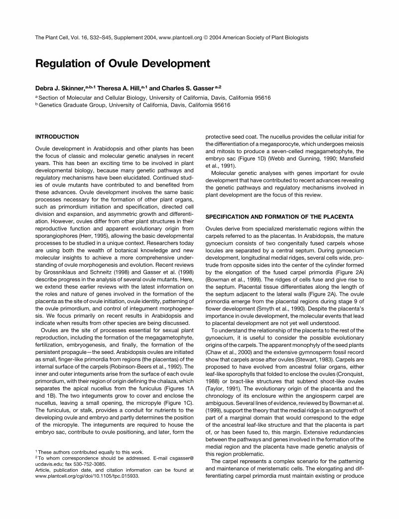
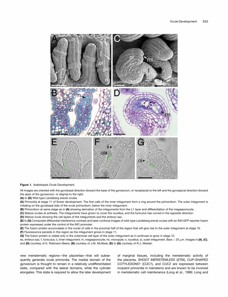
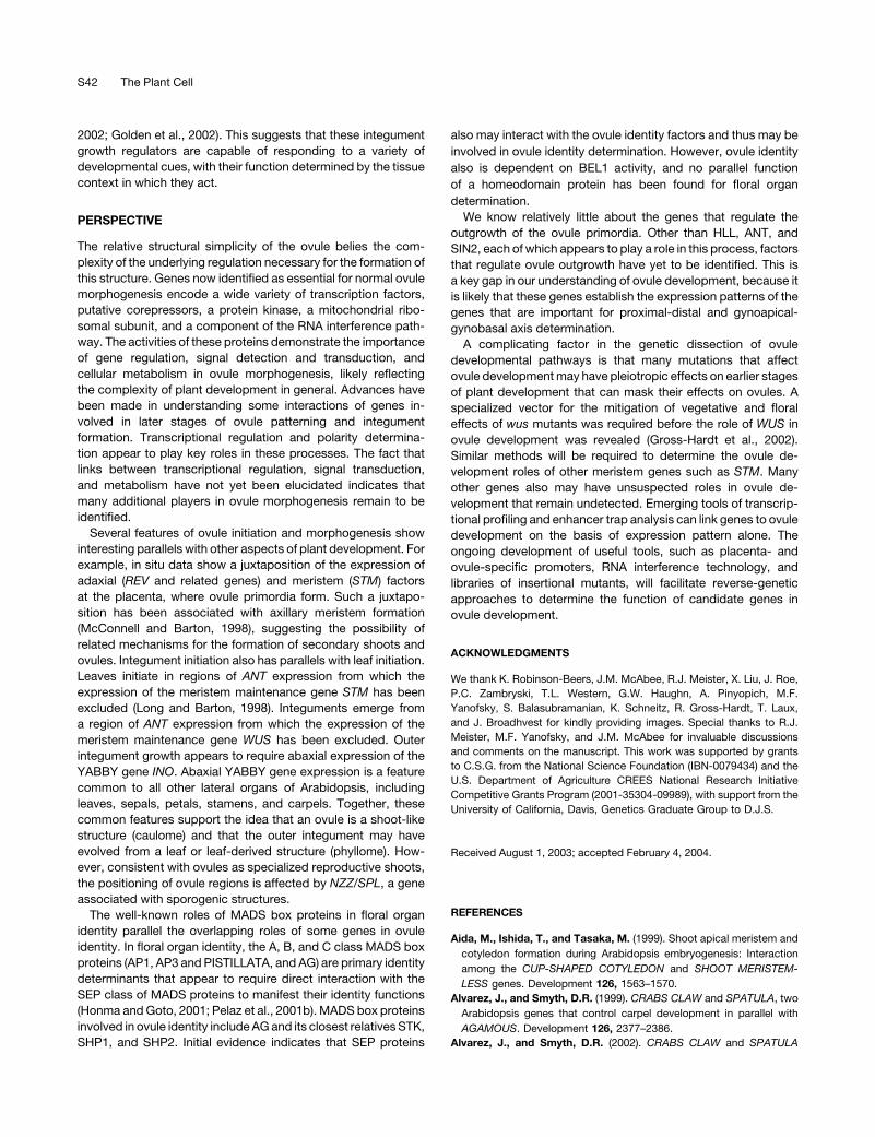
Figure 1. Arabidopsis Ovule Development.
All images are oriented with the gynobasal direction (toward the base of the gynoecium, or receptacle) to the left and the gynoapical direction (toward
the apex of the gynoecium, or stigma) to the right.
(A) to (D) Wild-type Landsberg erecta ovules.
(A) Primordia at stage 11 of flower development. The first cells of the inner integument form a ring around the primordium. The outer integument is
initiating on the gynobasal side of the ovule primordium, below the inner integument.
(B) Primordium at same stage as in (A) showing derivation of the integuments from the L1 layer and differentiation of the megasporocyte.
(C) Mature ovules at anthesis. The integuments have grown to cover the nucellus, and the funiculus has curved in the opposite direction.
(D) Mature ovule showing the cell layers of the integuments and the embryo sac.
(E) to (G) Composite differential interference contrast and laser confocal images of wild-type Landsberg erecta ovules with an INO:GFP reporter fusion
protein expressed under the control of the INO promoter.
(E) The fusion protein accumulates in the nuclei of cells in the proximal half of the region that will give rise to the outer integument at stage 10.
(F) Fluorescence persists in this region as the integument grows in stage 11.
(G) The fusion protein is visible only in the outermost cell layer of the outer integument as it continues to grow in stage 12.
es, embryo sac; f, funiculus; ii, inner integument; m, megasporocyte; mi, micropyle; n, nucellus; oi, outer integument. Bars ¼ 25 mm. Images in (A), (C),
and (D) courtesy of K. Robinson-Beers; (B) courtesy of J.M. McAbee; (E) to (G) courtesy of R.J. Meister.
Ovule Development S33
Barton, 1998; Aida et al., 1999; Takada et al., 2001), a process
recently reviewed by Clark (2001) and Lenhard et al. (2002). In
the carpel, they are restricted to expression in the medial
domain and are likely to maintain a meristematic state there.
AINTEGUMENTA (ANT) encodes an APETALA2 (AP2) domain
transcriptional regulator whose main function is to promote the
outgrowth of determinate structures through cell proliferation
(Elliott et al., 1996; Klucher et al., 1996; Krizek, 1999). As
expected by their antagonistic roles, ANT expression does not
overlap with STM expression in meristems (Elliott et al., 1996;
Long and Barton, 2000). However, in the carpel, ANT is
expressed in the medial domain with STM. This could be
attributable to an ongoing requirement for ANT in the outgrowth
of the carpel primordia, which is balanced with the negative
effects on differentiation of STM in the medial domain.
In addition, ANT is likely to play a larger role in medial domain
development, as shown by double mutant analysis with LEUNIG
(LUG), a putative transcriptional corepressor (Conner and Liu,
2000). lug and ant mutants both have weak effects on marginal
tissue formation, but double mutants show a strong synergistic
phenotype: total loss of septum, placentas, and ovules (Figure
2B) (Liu et al., 2000). Although both of these genes are known
to repress AGAMOUS (AG) genetically, this loss of the medial
domain is not an effect of AG overexpression. Rather, ANT and
LUG seem to share a vital role in promoting cell proliferation in the
marginal tissues of the pistil, specifically the medial ridge. In
addition, both genes show high expression in placentas and
ovules that persists after carpel expression decreases (Elliott
et al., 1996;Conner and Liu, 2000). Thus, in addition to other roles
within the ovule, ANT and LUG also may be required specifically
to promote the growth of the placenta to allow ovule primordium
formation.
Other genes that may influence medial domain formation and
affect the production of ovules include CRABS CLAW (CRC),
SPATULA (SPT), and TOUSLED (TSL) (Roe et al., 1993, 1997a,
1997b; Alvarez and Smyth, 1999, 2002). The reduction in ovule
formation shown by mutants of these and other genes can be
attributed to defects in marginal regions. CRC also may play an
additional role, however, because there is a longitudinal strand of
expression in the internal region of the carpel, adjacent to the
placenta, early in carpel development (Bowman and Smyth,
1999). The lossof thispart ofCRCexpression ispartly responsible
for the reduced number of ovules observed in crc mutants
(Bowman et al., 1999; Bowman and Smyth, 1999; Alvarez and
Smyth, 2002). Thus, CRC may contribute more directly to pla-
centa and primordium initiation, perhaps through abaxial sig-
naling in the region or by limiting the expression of STM or other
KNOX genes, which are known functions of the YABBY domain
protein family (Bowman et al., 2002; Kumaran et al., 2002).
The interaction of meristem factors in the medial ridge and
adaxial factors in the differentiating valve also could provide the
impetus for the initiation of placental growth in a manner that is
similar to the positive effect on meristem formation of adaxial
identity in lateral organs (McConnell and Barton, 1998; Alvarez
and Smyth, 2002). STM, expressed in medial domains, and
REVOLUTA (REV) (and other related genes), expressed laterally,
could be these factors (Figure 2A). However, it is unlikely that
these factors contribute directly to identity in this region.
A double mutant of the ettin and tsl genes produces structures
that could be interpreted as naked placentas: there is no
evidence of carpel structures other than placenta and ovules
(Figure 2C) (Roe et al., 1997b). Thus, signals provided by carpel
walls may not be necessary for placental formation. This result,
and the fact that placental position is particularly plastic across
angiosperm species, suggests that the placenta may have
evolved from an originally separate fertile structure that was
recruited later onto carpel walls. However, fossil evidence corro-
borating this model is lacking.
Identity of Ovule Structures
Placentas and ovules are formed onlywithin the context of carpel
identity. AG is a potent promoter of carpel identity and, as such,
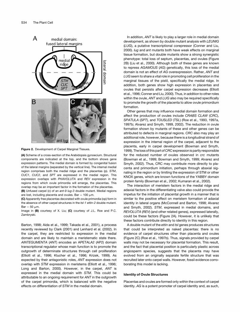
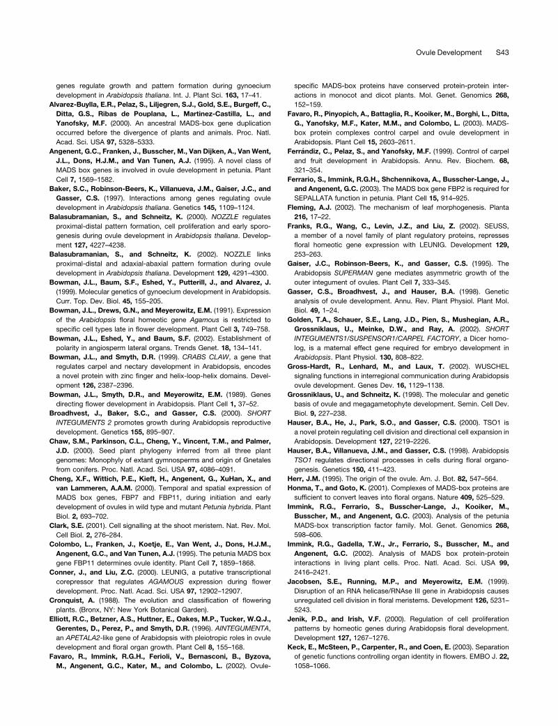
Figure 2. Development of Carpel Marginal Tissues.
(A) Scheme of a cross-section of the Arabidopsis gynoecium. Structural
components are indicated at the top, and the bottom shows gene
expression patterns. The medial domain is formed by congenital fusion
of the lateral margins (separated by the vertical line). The internal medial
region comprises both the medial ridge and the placentas (p). STM,
CUC1, CUC2, and SPT are expressed in the medial region. This
expression overlaps with PHAVOLUTA and REV expression in the
regions from which ovule primordia will emerge, the placentas. This
overlap may be an important factor in the formation of the placentas.
(B) Unfused carpel (c) of an ant-9 lug-3 double mutant. Medial regions
are lost, including placenta and ovules. Bar ¼ 100 mm.
(C) Apparently free placentas decorated with ovule primordia (op) form in
the absence of other carpel structures in the tsl-1 ettin-2 double mutant.
Bar ¼ 50 mm.
Image in (B) courtesy of X. Liu; (C) courtesy of J.L. Roe and P.C.
Zambryski.
S34 The Plant Cell

possibly promotes the formation of all structures within the
gynoecium, including ovules (Bowman et al., 1989; Yanofsky
et al., 1990; Ferrandiz et al., 1999). AG expression, initially seen
throughout the gynoecium, remains high in ovule primordia and
integuments (Bowman et al., 1991; Reiser et al., 1995). Although
the ovules formed on the carpelloid sepals of ap2 agmutants are
evidence that AG is not absolutely required for ovule formation,
the fact that there is an increase in the number of undifferentiated
primordia relative to ap2 single mutants indicates that AG is
a contributor to ovule development. However, overexpression of
AG homologs in tobacco ovules led to the conversion of ovules
to carpelloid structures (Mandel et al., 1992). In Arabidopsis,
ectopic expression of AG had little effect on ovules, whereas
expression of BAG, the Brassica ortholog of AG, caused trans-
formation of the integuments into carpelloid structures (Ray et al.,
1994). Therefore, the designation of the primordium as an ovule
requires discrimination between carpel and ovule identity. Mu-
tants have been observed that have phenotypes similar to
the overexpression of AG, with conversion of some ovule struc-
tures to carpel structures. These phenotypes suggest that these
geneswork to create ormaintain the necessary balance between
AG activity and ovule identity that allows ovule structures to
develop.
BEL1 encodes a homeodomain protein known to be required
for integument morphogenesis, andmutations in this gene affect
ovule identity (Robinson-Beers et al., 1992; Modrusan et al.,
1994; Ray et al., 1994; Reiser et al., 1995). bel1 mutants initiate
a single amorphous structure that grows as a collar around the
nucellus in place of the integuments (Figure 3A). This structure
has been shown to accumulate AG transcript at anthesis, when
AG expression decreases in the wild type (Modrusan et al., 1994;
Ray et al., 1994). In some ovules, this aberrant outgrowth
continues to proliferate after anthesis and becomes carpelloid,
displaying features of the ovary, style, and stigma (Figure 3B) and
even producing a set of secondary ovules (Modrusan et al., 1994;
Ray et al., 1994). In addition, the funiculi of bel1mutants continue
to divide and lose their characteristic organized appearance.
Because the expression of AG in the integuments increases only
aftermanifestation of thebel1 integument phenotype and at least
someAG activity contributes to ovule identity, themechanism for
BEL1 repression of the AG carpel promotion function remains
unclear. It appears that the loss of integument or chalazal identity
allows development to proceed along a poorly defined pathway
that can sometimes default to the background pathway of carpel
development.
Analysis of the phenotypes observed in a triple mutant of ag,
bel1, and ap2 expands our understanding of the function ofBEL1
and may reveal why there is a delay in the acquisition of carpel
identity in the bel1mutant. In 1% of triple mutant ovules, there is
a restoration of normal integument growth, which never occurred
in bel1 single or bel1 ap2 double mutants (Western and Haughn,
1999). Therefore, other genes are capable of providing in-
tegument identity in the absence of BEL1. These genes also
could act to repress carpel identity during the earlier stages,
delaying the onset of the carpel phenotype in bel1. However,
because normal integument growth is seen only when AG also is
removed, it seems that the integument promotion functions of
these genes cannot compete effectively with the carpel identity
provided by AG. Thus, one model for the function of BEL1 is that
its primary function is to control AG activity, which in turn allows
other genes to provide integument identity. BEL1 also is likely to
have some contribution to ovule identity, because there is an
increase in the number of undifferentiated ovule primordia in the
triple mutant ag bel1 ap2 relative to ag ap2 (Figure 3C) (Western
and Haughn, 1999). These functions could be causally related:
the strong promotion of chalazal identity by BEL1 may override
the effect of AG in this region. BEL1 and other factors play
a positive role in integument identity, and the absence of that role
could allow AG to eventually predominate, causing the integu-
ment to become carpelloid in a self-reinforcing manner.
Recent results demonstrate that three MADS box genes that
form amonophyletic cladewithAG—SHATTERPROOF1 (SHP1),
SHP2, and SEEDSTICK (STK, also known as AGL11)—share
a common function in promoting aspects of ovule identity (Ma
et al., 1991; Rounsley et al., 1995; Alvarez-Buylla et al., 2000;
Liljegren et al., 2000; Pinyopich et al., 2003). These genes, which
likely arose through gene duplication, have retained probable
ancestral roles in reproductive development, including redun-
dant roles in carpel identity, and have gained other roles in the
current derived carpel structure. For example, SHP1 and SHP2
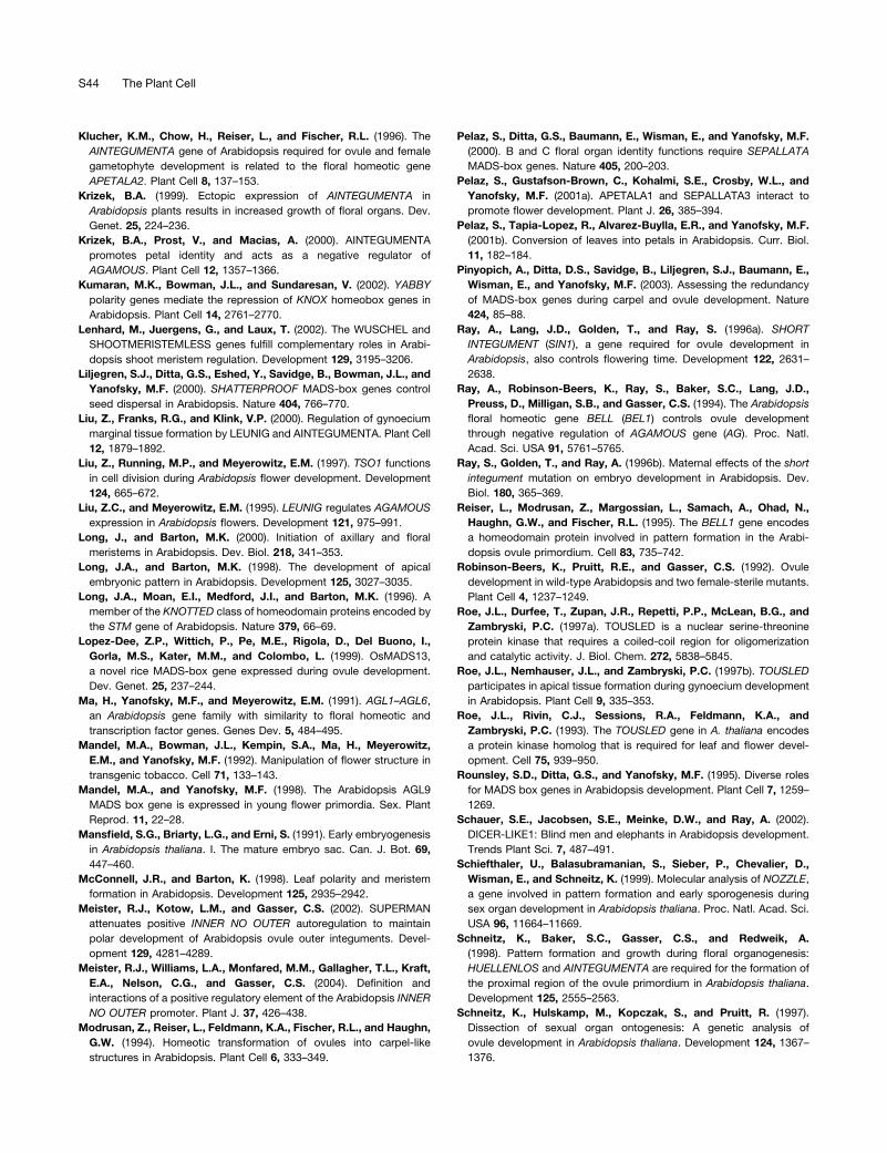
Figure 3. Ovule Identity Mutant Interactions.
(A) bel1-1 ovules at stage 12 of flower development have an amorphous
collar of cells in place of the two integuments (arrowheads).
(B) After anthesis, the collar of cells can continue to grow and become
carpelloid. Stigmatic papillae (arrowhead) are visible on this secondary
carpel. However, some ovules arrest growth after anthesis and de-
generate (arrow).
(C) In the ap2-6 bel1-3 ag-1 triple mutant, ovules are formed on the
carpelloid sepals. These ovules usually are undifferentiated structures
(u), although normal bel1-like (b) and carpelloid (c) ovules also form.
(D) An ovule of the stk shp1 shp2 triple mutant after anthesis. A funiculus
supports valve-like (v) and style-like (s) structures that form in the distal
portion of the mutant ovule. An arrowhead marks the distal end of the
funiculus.
Bars ¼ 100 mm in (A) and (B) and 50 mm in (C) and (D). Images in (A) and
(B) courtesy of K. Robinson-Beers; (C) courtesy of T.L. Western and
G.W. Haughn; (D) courtesy of A. Pinyopich and M.F. Yanofsky.
Ovule Development S35
have redundant functions in the promotion of valve margin
specification (Liljegren et al., 2000) and can substitute for AG
function in carpel specification (Pinyopich et al., 2003). All four
AG-related genes (AG, SHP1, SHP2, and STK) have overlapping
expression patterns in the placenta and developing ovule
primordia (Rounsley et al., 1995; Ferrandiz et al., 1999). Although
the double mutant shp1 shp2 does not exhibit altered ovule
development, the stkmutant does have ovule defects, but only in
funiculus differentiation, in which each funiculus has more cells
than the wild type and fails to develop an abscission zone
(Pinyopich et al., 2003). By contrast, when the functions of all
three genes are lost in the triple mutant stk shp1 shp2, fewer
ovules are initiated and ovule development is severely disrupted
(Pinyopich et al., 2003). Examination of the triple mutant pheno-
type seen in Figure 3D shows that the funiculus still is present,
with the features expected for the stkmutant, whereas the distal
portions of the ovules show conversion to carpel-like structures.
This dramatic phenotype suggests that these genes are major
identity determinants of at least the distal ovule structures.
The triple mutant phenotype has similarities with the bel1
phenotype, because carpel structures can form from the region
that produces integuments in both mutants. The phenotype of
the triple mutant at early ovule stages has not been reported,
so the timing of the manifestation of the phenotype is unknown.
The loss of STK, SHP1, and SHP2 function produces a more
consistent homeotic change than that seen in bel1 mutants, in
that 95% of ovules show this conversion of the integuments
compared with <15% in bel1 (Ray et al., 1994; Pinyopich et al.,
2003). If carpel identity is suppressed by the presence of
integument identity, the triple mutant phenotype shows that
BEL1 cannot provide this identity without one of the three
redundant genes, STK, SHP1, or SHP2. This could mean that
BEL1 is not active in a stk shp1 shp2 background and may even
be genetically downstream of these genes. An alternative model
would be that BEL1 is upstream of STK, SHP1, and SHP2 and
that the integument identity seen occasionally in the ap2 ag bel1
mutant is provided by other unknown genes. Examination of
quadruple mutants of bel1 with stk shp1 and shp2, as well as
observation of the change in expression pattern of bel1 in the
triple mutant, will help to discriminate between these models.
In summary, BEL1 and the AG clade all contribute to the
identity of ovule structures.Without the identity provided bySTK,
SHP1, SHP2, and BEL1, the integuments acquire carpel fea-
tures. Because the primary floral C class geneAG is expressed in
the region, the primordia may revert to an underlying pathway of
carpel development when the ovule identity factors are absent.
No mutants or mutant combinations have been observed that
result in the loss of the defining ovule features, funiculus,
integuments, and nucellus, indicating that factors important for
ovule identity remain to be discovered.
The importance of theAG clade in ovule development also has
been demonstrated in other species. The Petunia hybrida genes
FLORALBINDINGPROTEIN7 (FBP7) and FBP11, which are 90%
identical at the amino acid level, fall within the petunia AG clade
(Angenent et al., 1995). Phylogenetic analyses indicate that FBP7
and FBP11 share the greatest similarity with STK (Theissen et al.,
1996, 2000; Immink et al., 2003). The two petunia genes are
first expressed in the placenta before ovule primordia emerge.
There is some expression in young primordia, and expression
increases in the funiculus and integuments (Angenent et al.,
1995; Cheng et al., 2000). This expression pattern is similar to
that of STK, SHP1, and SHP2. FBP11 and FBP7 have been
reported to cause occasional formation of ovules on sepals,
petals, and carpels when expressed ectopically (Colombo et al.,
1995; Cheng et al., 2000). However, both the sepals and petals in
these plants showed other phenotypes correlated with the
acquisition of carpel identity. These ovules could be an indirect
effect of the expression of AG homologs that promote car-
pel development. Cosuppression experiments with FBP7 and
FBP11 showed a partial loss in ovule identity in a subset (7.5%) of
transformants. In these plants, many ovules were converted to
carpel- or style-like structures. Because the funiculi of petunia
ovules are very short (Angenent et al., 1995), it is not possible to
determine from the available data whether the integument or an
entire primordium becomes carpelloid. The presence of some
normal ovules even in those plants with undetectable FBP7 and
FBP11 mRNA levels shows that ovule identity still was present.
This finding indicates that cosuppression was incomplete or that
other genes are capable of providing ovule identity.
In Arabidopsis, interactions between SEPALLATA (SEP)
MADS box proteins and floral B and C function MADS box pro-
teins, such as AP3 and AG, are necessary for the determination
of floral organ identity (Pelaz et al., 2000, 2001a; Honma and
Goto, 2001). Recent evidence suggests that the SEP proteins
could be required for the determination of ovule identity in
a manner similar to that of floral organ identity. The Arabidopsis
geneSEP3 (AGL9) is expressed in placentas and ovule primordia
until embryogenesis (Mandel and Yanofsky, 1998). SEP1 and
SEP2 are expressed in ovules beginning at floral stage 10, the
time of integument emergence (Ma et al., 1991). Favaro et al.
(2003) recently showed thatSEP1/sep1 sep2 sep3mutant ovules
have tissue transformations resembling those in stk shp1 shp2
ovules. In addition, STK, AG, SHP1, and SHP2 were shown to
interact with SEP proteins to formmultimeric complexes (Favaro
et al., 2003). Together, these results make a strong case for the
importance of large MADS complexes in ovule identity. Recent
reports in other species further support this hypothesis. In
petunia, FBP7 and FBP11 (apparent STK orthologs) interact in
vitro with FBP2 and FBP5, which are putative orthologs to the
SEP proteins of Arabidopsis (Pelaz et al., 2000; Immink et al.,
2002, 2003; Ferrario et al., 2003). In addition, the rice STK
homolog OsMADS13 also interacts with SEP-related proteins
from rice: OsMADS24 and OsMADS45 (Lopez-Dee et al., 1999;
Favaro et al., 2002). These results from Arabidopsis, rice, and
petunia support themodel that the SEP proteins form complexes
with STK, SHP1, SHP2, and possibly AG to maintain ovule
identity in divergent species.
Certain Arabidopsis ap2 alleles exhibit some conversion of
ovules to carpelloid structures in the gynoecium (Modrusan et al.,
1994), suggesting thatAP2may suppressAG in ovules as well as
in the outer whorls of the flower. Interestingly, double mutants of
the ap2-like genes LIPLESS (LIP1) and LIP2 in Antirrhinummajus
show a similar phenotype (Keck et al., 2003). However, this
carpelloidy is not caused by an increase in PLENA expression
(theAG ortholog) in ovules, suggesting a different mechanism for
C function regulation in A. majus.
S36 The Plant Cell
PRIMORDIUM OUTGROWTH
The initiation of ovule primordia is not disrupted significantly by
defects in ovule identity genes, as illustrated by the relatively
normal appearance of the primordia that later form the carpelloid
and undifferentiated mutant ovules discussed above. The re-
cently described pattern ofCUC3 expression, which is observed
in a ring surrounding thebaseof eachprimordium (Vroemenet al.,
2003), indicates that the CUC genes may play a role in the
location of primordium initiation. Outgrowth of the ovule pri-
mordium requires an unknown signal to direct cells in the pla-
centa to expand and divide out of the plane of the developing
septum. These processes appear dependent on metabolic
energy to fuel growth and cell divisions. Mutations that show
effects on the outgrowth of ovule primordia provide an in-
teresting view of these basal requirements for organ outgrowth.
An apparent requirement for metabolic energy in primordium
outgrowth and cellular health is shown by the huellenlos (hll)
mutant (Schneitz et al., 1998), which has lost a vital mitochondrial
ribosomal protein (Skinner et al., 2001). The mutant is thought to
have impaired mitochondrial function in ovule primordia, and the
phenotype is short, degenerating primordia. Because of re-
dundancy with the HUELLENLOS PARALOG (HLP) gene, the
mitochondrial defect is manifest primarily in the gynoecia and
ovules. Presumably, the cells of the primordia cannot perform
normal cellular function and thus fail to undergo division and
subsequently die. Interestingly, this loss of mitochondrial func-
tion reveals the action of other genes in promoting primordium
growth. ant mutants have normal primordia that lack integu-
ments. However, the ant hll double mutant has shortened
primordia that lack obvious funiculi (Schneitz et al., 1998). The
hll mutant also shows synergistic interactions with the short
integuments2 (sin2) mutant (Broadhvest et al., 2000). The double
mutant ovule primordia are reduced severely to small outgrowths
of only a few cells. Thus, ANT is clearly active in promoting cell
proliferation here, as it is in shoot and floral meristems. This
finding also sheds light on the possible role of the SIN2 protein,
which appears to be involved in aspects of cell division in a way
that is particularly sensitive to the reduction in metabolic health.
PATTERNING THE OVULE PRIMORDIUM
After the emergence of an ovule primordium, positional in-
formation along the proximal-distal axis must be interpreted
so that the different regions of the primordium take on their
characteristic fates. Three zones of differentiation are easily
recognized: the distal zone becomes the nucellus, which gives
rise to the megaspores and embryo sac; the central region is
referred to as the chalaza and is the location of integument
formation; and the proximal zone becomes the funiculus, a sup-
porting stalk. There also are differences along the sides of the
ovule facing the apical (stigma) and basal (receptacle) ends
of the carpel. We use the terms ‘‘gynoapical’’ and ‘‘gynobasal’’
to represent these two directions along this axis, which is
perpendicular to the proximal distal axis of the ovule. We use
these terms in place of earlier terms such as anterior/posterior,
dorsal/ventral, and abaxial/adaxial that have been used to refer
to this axis (abaxial/adaxial will be reserved to designate the two
faces of lateral organs). Differential growth of the funiculus and
outer integument along the gynoapical/gynobasal axis results in
the final S shape of the Arabidopsis ovule (Figure 1C). The
mechanisms required for the establishment of these domains are
largely unclear. However, we are beginning to identify compo-
nents of the pathways that define the position and extent of
integument growth along the proximal/distal and gynoapical/
gynobasal axes.
Two genes are essential for integument initiation and also have
important roles in proximal/distal patterning. The AP2 domain
transcription factor ANT appears to have a role in promoting
primordium outgrowth at a number of steps in reproductive
development (Elliott et al., 1996; Krizek et al., 2000) and is
required for integument initiation and growth (Elliott et al., 1996;
Klucher et al., 1996; Baker et al., 1997). Severe ant alleles, such
as ant-4 and ant-5, lead to a complete absence of integuments,
with the resulting ovules having only a rudimentary bulge in the
chalazal region as a result of cell expansion (Figure 4D) (Baker
et al., 1997). ANT is expressed throughout the young ovule
primordia. However, by early stage 2, before integument initia-
tion, its expression becomes restricted to the distal funiculus
and chalaza (Elliott et al., 1996; Balasubramanian and Schneitz,
2000). Once integument cell divisions have begun, ANT expres-
sion becomes limited to the integument primordia and distal
funiculus.
The ant mutant ovules are similar in appearance to those
produced by wuschel (wus) mutants when the earlier effects of
wus are mitigated (Figures 4D and 4E) (Gross-Hardt et al., 2002).
TheWUS homeodomain protein is required tomaintain the shoot
meristem, and wusmutants normally lack flowers.WUS expres-
sion was detected in the nucellar region of the ovule primordium
(Gross-Hardt et al., 2002). Therefore, to determine the role of
WUS during ovule development, Gross-Hardt et al. (2002)
created wus mutants carrying a CLAVATA1 promoter:WUS
cDNA fusion transgene (P-CLV1:WUS). Because P-CLV1 pro-
duces expression inmeristems and not ovules, these plantswere
able to overcome the wus meristem and floral defects to reveal
ovule effects of the mutation. The flowers of these plants con-
tained ovules completely lacking integuments (Figure 4E).
The position of integument initiation along the proximal-distal
axis of the ovule primordia is influenced by the novel nuclear
protein NOZZLE/SPOROCYTELESS (NZZ/SPL) (Figures 4A and
4B) (Schiefthaler et al., 1999; Yang et al., 1999; Balasubramanian
and Schneitz, 2000). One effect of nzz/splmutations is to reduce
the length of the nucellar region of the primordium. This could
result from reduced nucellar growth attributable to a failure to
produce amegasporocyte, consistent with a similar effect of this
mutation on anther and pollen development (Yang et al., 1999).
Alternatively, it could represent a direct distal shift in the position
of integument formation along the proximal-distal axis, con-
sistent with the observed increased length of the funiculus
(Figures 4B and 4C) (Schiefthaler et al., 1999; Balasubramanian
and Schneitz, 2000). This change in the location of integument
initiation is associated with a distal expansion of the ANT ex-
pression domain (Balasubramanian and Schneitz, 2000). The
ectopic ANT expression appears to be required for the nzz
phenotype. nzz ant double mutants are ant-like, with no obvious
reduction in nucellar tissue, indicating that a major role of NZZ is
Ovule Development S37
the spatial limitation of ANT. Therefore, it appears that the
chalazal region is defined in part by the distal extent of ANT
expression at stage 2-I and that NZZ influences this through
negative regulation of ANT in the nucellus. This implies a close
link between the pattern of ANT expression and the location of
integument initiation. Later in ovule development, nzz mutants
show an overproliferation of the nucellus and the gynoapical
region of the outer integument (Figure 4C). Because NZZ/SPL
has been reported to be expressed throughout the ovule
primordium (Balasubramanian and Schneitz, 2000) or only in
the nucellar region (Yang et al., 1999), the mechanisms by which
NZZ/SPL regulates the ANT expression domain are unclear.
The link betweenANT expression and chalazal positioning can
be tested by altering the expression of ANT. Unfortunately, the
effects on ovule development of ectopic ANT expression from
transgenes have been difficult to interpret. The 35S promoter of
Cauliflower mosaic virus (35S) is used commonly in transgenic
overexpression/ectopic expression studies. However, the activ-
ity of this promoter is very low in the ovule primordia at inception
and increases to a higher, but still variable, level throughout the
ovule at later stages (Jenik and Irish, 2000). 35S:ANT transgenes
result in variable ovule phenotypes with reduction of inner
integument growth, overproliferation of the nucellus, and ectopic
growth of the outer integument on the gynoapical side of
the ovule (Krizek, 1999). In this case, the relatively stronger
endogenous ANT expression may provide the location for inner
integument initiation, with this boundary somewhat blurred by
the weaker but more widespread expression from the 35S
promoter. This could lead to a less efficient initiation followed by
retarded growth. Alternatively, expression of ANT in tissues
underlying the inner integument may be inhibitory to its growth.
The phenotypic variability observed in the 35S:ANT ovules also is
likely to be attributable to the weak and variable activity of the
35S promoter. The late overproliferation of the nucellus and
gynoapical outer integument is similar to that in nzz mutants,
which also have ectopic ANT expression in the nucellus (Krizek,
1999; Balasubramanian and Schneitz, 2000).
An appropriate pattern ofWUS expression also is important to
define the region of an ovule primordium capable of giving rise to
integuments. Surprisingly, theWUS expression pattern does not
appear to include the cells from which the integuments derive.
Even though the effects of the wus mutation on ovule de-
velopment (the absence of integuments; Figure 4E) appear
specific to the chalazal region, WUS is restricted to the nucellus
(Gross-Hardt et al., 2002). This fact suggests thatWUS promotes
integument growth through a non-cell-autonomous mechanism.
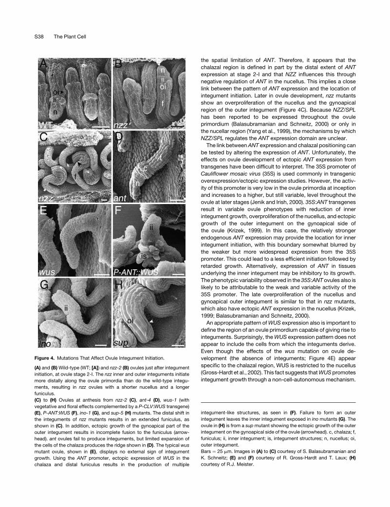
Figure 4. Mutations That Affect Ovule Integument Initiation.
(A) and (B) Wild-type (WT; [A]) and nzz-2 (B) ovules just after integument
initiation, at ovule stage 2-I. The nzz inner and outer integuments initiate
more distally along the ovule primordia than do the wild-type integu-
ments, resulting in nzz ovules with a shorter nucellus and a longer
funiculus.
(C) to (H) Ovules at anthesis from nzz-2 (C), ant-4 (D), wus-1 (with
vegetative and floral effects complemented by a P-CLV:WUS transgene)
(E), P-ANT:WUS (F), ino-1 (G), and sup-5 (H) mutants. The distal shift in
the integuments of nzz mutants results in an extended funiculus, as
shown in (C). In addition, ectopic growth of the gynoapical part of the
outer integument results in incomplete fusion to the funiculus (arrow-
head). ant ovules fail to produce integuments, but limited expansion of
the cells of the chalaza produces the ridge shown in (D). The typical wus
mutant ovule, shown in (E), displays no external sign of integument
growth. Using the ANT promoter, ectopic expression of WUS in the
chalaza and distal funiculus results in the production of multiple
integument-like structures, as seen in (F). Failure to form an outer
integument leaves the inner integument exposed in ino mutants (G). The
ovule in (H) is from a supmutant showing the ectopic growth of the outer
integument on the gynoapical side of the ovule (arrowhead). c, chalaza; f,
funiculus; ii, inner integument; is, integument structures; n, nucellus; oi,
outer integument.
Bars ¼ 25 mm. Images in (A) to (C) courtesy of S. Balasubramanian and
K. Schneitz; (E) and (F) courtesy of R. Gross-Hardt and T. Laux; (H)
courtesy of R.J. Meister.
S38 The Plant Cell
WUS is expressed normally in the nucellar region of the ovule
primordia from very early stages, with the highest level of
expression at stage 2-II to 2-III, coincident with integument
initiation (Gross-Hardt et al., 2002). The importance of this
expression pattern is demonstrated by the phenotype of plants
ectopically expressing WUS in the chalazal region under the
control of the ANT promoter. The P-ANT:WUS plants produce
what appear to be ectopic integuments that form reiteratively
toward the base of the ovule (Figure 4F). The first integuments
produced by these ovules appear similar to those in thewild type,
which demonstrates that WUS does not need to be excluded
from the cells giving rise to the inner integument. However, the
ectopic integuments that do form are highly irregular. These
irregular outgrowths appear to have some integument identity,
because they exhibit ANT expression in an integument-like
pattern. However, whether they have outer or inner integument
characteristics is undetermined. TheP-ANT:WUS plants indicate
that WUS is capable of inducing ectopic integument growth in
the context of the ovule.
Why are multiple integument-like structures formed in
P-ANT:WUS plants? This is likely attributable to P-ANT:WUS
expression in the distal funiculus and chalaza. This region then
would includemultiple new boundaries ofANT andWUS activity,
created by the upregulation of endogenousWUSbyP-ANT:WUS
in the existing central chalazal region and the downregulation of
ANT in this central chalazal region upon integument outgrowth
(Gross-Hardt et al., 2002) (Figure 5). ANT expression is main-
tained in the distal funiculus, whereas WUS is expressed in the
distal funiculus and the chalaza. Therefore, a new expression
boundary is created at the junction of the distal funiculus and the
chalaza. In wild-type plants, when ANT is downregulated in the
chalazal region, the domains ofANT andWUS expression are not
in close proximity. Because only two integuments are formed in
wild-type plants, it is likely that ANT needs to be within a certain
distance of WUS to induce integument initiation. These data are
consistent with a model whereby, in wild-type plants, the spatial
relationship between WUS expression in the nucellus and ANT
expression in the chalaza determines the site of inner integument
growth and NZZ is required to establish the distal extent of ANT
expression (Figure 5). The mechanisms that establish the WUS
expression domain also are unknown.
OUTER INTEGUMENT INITIATION AND ORIENTATION
Although ANT and WUS are essential for the formation of both
integuments, outgrowth of the outer integument also requires the
activity of a third transcription factor, the YABBY protein INNER
NO OUTER (INO) (Baker et al., 1997). Strong ino mutants com-
pletely lack an outer integument but otherwise are phenotypically
wild type (Figure 4G) (Gaiser et al., 1995; Baker et al., 1997;
Schneitz et al., 1997). Therefore, INO is required specifically
for outer integument formation (Bowman and Smyth, 1999;
Villanueva et al., 1999).
INO expression is first observed at stage 2-I just before the
initiation of either integument. Expression is only on the gyno-
basal side of the ovule, in the proximal half of the region of cells
that will give rise to the outer integument (Figure 1E) (Villanueva
et al., 1999; Meister et al., 2002). The sequential initiation of
integument growth,with the inner integument emerging first,may
indicate that the initiation of outer integument growth, and hence
the position of the initial expression of INO, may be dependent on
the position of the inner integument. This notion is supported by
the fact that the absence of the inner integument is always
accompanied by the absence of the outer integument in known
mutants. Because the position of the inner integument is linked
closely to the location of ANT expression, the model described
above implies a link between ANT, INO, and the location of the
outer integument. Experimental results support this link. In the
nzz mutant, both INO expression and the site of initiation of
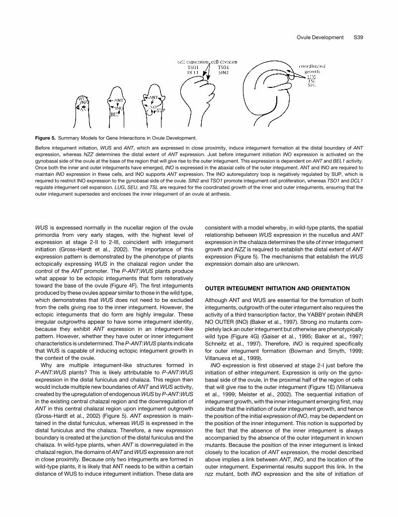
Figure 5. Summary Models for Gene Interactions in Ovule Development.
Before integument initiation, WUS and ANT, which are expressed in close proximity, induce integument formation at the distal boundary of ANT
expression, whereas NZZ determines the distal extent of ANT expression. Just before integument initiation INO expression is activated on the
gynobasal side of the ovule at the base of the region that will give rise to the outer integument. This expression is dependent on ANT and BEL1 activity.
Once both the inner and outer integuments have emerged, INO is expressed in the abaxial cells of the outer integument. ANT and INO are required to
maintain INO expression in these cells, and INO supports ANT expression. The INO autoregulatory loop is negatively regulated by SUP, which is
required to restrict INO expression to the gynobasal side of the ovule. SIN2 and TSO1 promote integument cell proliferation, whereas TSO1 and DCL1
regulate integument cell expansion. LUG, SEU, and TSL are required for the coordinated growth of the inner and outer integuments, ensuring that the
outer integument supersedes and encloses the inner integument of an ovule at anthesis.
Ovule Development S39
the outer integument are closer to the distal end of the ovule
primordium than in the wild type, as would be expected from the
shift in the pattern of ANT expression (Figures 4A and 4B)
(Balasubramanian and Schneitz, 2000). In addition, in strong ant
mutants, such as ant-72F5 and ant-5, INO expression is delayed
and at a lower level than in the wild type (Balasubramanian and
Schneitz, 2000; Meister et al., 2004) and may be absent in ant-4
(Meister et al., 2004). Thus, ANT may promote outer integument
development by facilitating inner integument development and
through positive regulation of INO (Figure 5).
The interaction of ANT with INO may be bidirectional. In the
strong ino-2 mutant, ANT expression is at normal levels in the
inner integument but is reduced in the region that normally would
produce the outer integument (Balasubramanian and Schneitz,
2002). This implies that INO positively affects ANT expression.
Thus, ANT and INO may constitute an autoregulatory loop in
which each reinforces the expression of the other (Figure 5). INO
has been shown to positively affect its own expression; both INO
transcript accumulation and aP-INO:GUS reporter gene showed
reduced expression in an ino mutant background (Villanueva
et al., 1999; Meister et al., 2002). The autoregulation of INO may
be direct, or the autoactivation of INO could be a secondary
effect of the INO activation of ANT expression. Transgenic
studies show that regulation by both factors is transcriptional
because it is mediated by the INO promoter sequence (Meister
et al., 2004). The positive regulation of ANT by INO may provide
an explanation for the restoration of the normal length of the
nucellar region in ino nzz double mutants relative to nzz single
mutants (Balasubramanian and Schneitz, 2002). The distal shift
of ANT expression hypothesized to contribute to the reduction in
the size of the nucellar regionmay not occur in the absence of the
positive influence of INO on ANT expression.
The expression of INO in ant-72F5 ovules suggests that factors
in addition to ANTmust contribute to the INO expression pattern.
A likely candidate is the homeodomain proteinBEL1 (Reiser et al.,
1995). BEL1 is expressed in the chalazal region from stage 1,
independent of ANT function. Mutations in this gene result in
delayed or reduced INO expression (Balasubramanian and
Schneitz, 2000; Meister et al., 2004). Because both BEL1 and
ANT expression domains occur throughout the chalazal region,
additional factors must be required to define the more limited
INO expression pattern. Our model suggests that at least some
of these factors would be associated with the induction of the
inner integument.
INO expression is regulated not only along the proximal-distal
axis but also along the gynoapical-gynobasal axis. INO expres-
sion initiates only toward the gynobasal side of the ovule, where
the outer integument initiates and exhibits greatest growth
(Figures 1E to 1G). Throughout ovule development, this pattern
of expression is maintained (Villanueva et al., 1999). The
maintenance of the expression pattern is dependent on the
putative zinc finger transcription factor SUPERMAN (SUP)
(Meister et al., 2002). By stage 2-V, sup-5 mutants exhibit INO
expression on both the gynobasal and gynoapical sides of the
ovule primordium, resulting in ectopic growth of the outer
integument on the gynoapical side of the ovule. The SUP-
mediated negative regulation requires INO function, because
replacement of the INO coding sequence with that of the
paralogous CRABS CLAW gene also results in ectopic growth
and INO promoter activity on the gynoapical side of the ovule
(Meister et al., 2002). The simplest model to explain the
involvement of INO function in the negative regulation by SUP
is for SUP to interfere with the INO positive autoregulation (Figure
5). Although the interplay between positive regulation by INO and
negative regulation by SUP can explain the maintenance of the
asymmetrical pattern of INO expression, the factors for the initial
induction of this pattern remain unknown. The overproliferation
of the gynoapical outer integument of nzzmutants is similar to the
sup ovule phenotype (Balasubramanian and Schneitz, 2000). As
seen in sup mutants, the gynoapical overproliferation is accom-
panied by the extension of INO expression to the gynoapical side
of the ovule (Balasubramanian and Schneitz, 2000, 2002). This
suggests thatNZZ is important for the negative regulation of INO
by SUP. This effect may be indirect. A distal shift in the chalazal
domain without an accompanying shift in SUP activity would
result in INO expression distal to the range of SUP activity, and
the influence of SUP would be reduced. This would allow
a gynoapical expansion of the INO expression domain in nzz
mutants.
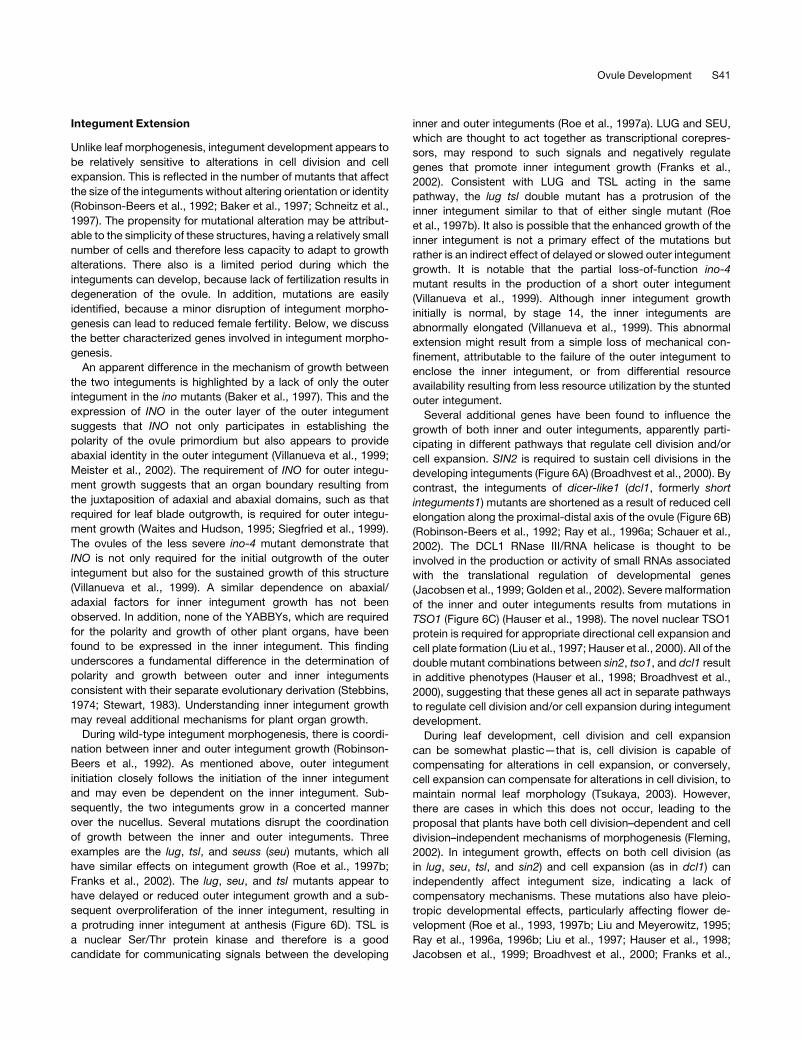
Figure 6. Ovules of Integument Cell Proliferation and Expansion
Mutants at Anthesis.
(A) sin2-1 integument cells stop dividing prematurely.
(B) dcl1-7 mutants have normal numbers of cells in the integuments, but
the cells fail to expand.
(C) tso1-3 integument cells undergo aberrant cell expansion and cell
division.
(D) tsl-1 mutants have reduced proliferation of the outer integument and
an overproliferation of the inner integument, resulting in its protrusion
beyond the outer integument.
i, integument; ii, inner integument; oi, outer integument. Bars ¼ 20 mm.
Images in (B) and (C) courtesy of J. Broadhvest; (D) courtesy of J.L. Roe
and P.C. Zambryski.
S40 The Plant Cell
Integument Extension
Unlike leaf morphogenesis, integument development appears to
be relatively sensitive to alterations in cell division and cell
expansion. This is reflected in the number of mutants that affect
the size of the integuments without altering orientation or identity
(Robinson-Beers et al., 1992; Baker et al., 1997; Schneitz et al.,
1997). The propensity for mutational alteration may be attribut-
able to the simplicity of these structures, having a relatively small
number of cells and therefore less capacity to adapt to growth
alterations. There also is a limited period during which the
integuments can develop, because lack of fertilization results in
degeneration of the ovule. In addition, mutations are easily
identified, because a minor disruption of integument morpho-
genesis can lead to reduced female fertility. Below, we discuss
the better characterized genes involved in integument morpho-
genesis.
An apparent difference in the mechanism of growth between
the two integuments is highlighted by a lack of only the outer
integument in the ino mutants (Baker et al., 1997). This and the
expression of INO in the outer layer of the outer integument
suggests that INO not only participates in establishing the
polarity of the ovule primordium but also appears to provide
abaxial identity in the outer integument (Villanueva et al., 1999;
Meister et al., 2002). The requirement of INO for outer integu-
ment growth suggests that an organ boundary resulting from
the juxtaposition of adaxial and abaxial domains, such as that
required for leaf blade outgrowth, is required for outer integu-
ment growth (Waites and Hudson, 1995; Siegfried et al., 1999).
The ovules of the less severe ino-4 mutant demonstrate that
INO is not only required for the initial outgrowth of the outer
integument but also for the sustained growth of this structure
(Villanueva et al., 1999). A similar dependence on abaxial/
adaxial factors for inner integument growth has not been
observed. In addition, none of the YABBYs, which are required
for the polarity and growth of other plant organs, have been
found to be expressed in the inner integument. This finding
underscores a fundamental difference in the determination of
polarity and growth between outer and inner integuments
consistent with their separate evolutionary derivation (Stebbins,
1974; Stewart, 1983). Understanding inner integument growth
may reveal additional mechanisms for plant organ growth.
During wild-type integument morphogenesis, there is coordi-
nation between inner and outer integument growth (Robinson-
Beers et al., 1992). As mentioned above, outer integument
initiation closely follows the initiation of the inner integument
and may even be dependent on the inner integument. Sub-
sequently, the two integuments grow in a concerted manner
over the nucellus. Several mutations disrupt the coordination
of growth between the inner and outer integuments. Three
examples are the lug, tsl, and seuss (seu) mutants, which all
have similar effects on integument growth (Roe et al., 1997b;
Franks et al., 2002). The lug, seu, and tsl mutants appear to
have delayed or reduced outer integument growth and a sub-
sequent overproliferation of the inner integument, resulting in
a protruding inner integument at anthesis (Figure 6D). TSL is
a nuclear Ser/Thr protein kinase and therefore is a good
candidate for communicating signals between the developing
inner and outer integuments (Roe et al., 1997a). LUG and SEU,
which are thought to act together as transcriptional corepres-
sors, may respond to such signals and negatively regulate
genes that promote inner integument growth (Franks et al.,
2002). Consistent with LUG and TSL acting in the same
pathway, the lug tsl double mutant has a protrusion of the
inner integument similar to that of either single mutant (Roe
et al., 1997b). It also is possible that the enhanced growth of the
inner integument is not a primary effect of the mutations but
rather is an indirect effect of delayed or slowed outer integument
growth. It is notable that the partial loss-of-function ino-4
mutant results in the production of a short outer integument
(Villanueva et al., 1999). Although inner integument growth
initially is normal, by stage 14, the inner integuments are
abnormally elongated (Villanueva et al., 1999). This abnormal
extension might result from a simple loss of mechanical con-
finement, attributable to the failure of the outer integument to
enclose the inner integument, or from differential resource
availability resulting from less resource utilization by the stunted
outer integument.
Several additional genes have been found to influence the
growth of both inner and outer integuments, apparently parti-
cipating in different pathways that regulate cell division and/or
cell expansion. SIN2 is required to sustain cell divisions in the
developing integuments (Figure 6A) (Broadhvest et al., 2000). By
contrast, the integuments of dicer-like1 (dcl1, formerly short
integuments1) mutants are shortened as a result of reduced cell
elongation along the proximal-distal axis of the ovule (Figure 6B)
(Robinson-Beers et al., 1992; Ray et al., 1996a; Schauer et al.,
2002). The DCL1 RNase III/RNA helicase is thought to be
involved in the production or activity of small RNAs associated
with the translational regulation of developmental genes
(Jacobsen et al., 1999; Golden et al., 2002). Severe malformation
of the inner and outer integuments results from mutations in
TSO1 (Figure 6C) (Hauser et al., 1998). The novel nuclear TSO1
protein is required for appropriate directional cell expansion and
cell plate formation (Liu et al., 1997; Hauser et al., 2000). All of the
double mutant combinations between sin2, tso1, and dcl1 result
in additive phenotypes (Hauser et al., 1998; Broadhvest et al.,
2000), suggesting that these genes all act in separate pathways
to regulate cell division and/or cell expansion during integument
development.
During leaf development, cell division and cell expansion
can be somewhat plastic—that is, cell division is capable of
compensating for alterations in cell expansion, or conversely,
cell expansion can compensate for alterations in cell division, to
maintain normal leaf morphology (Tsukaya, 2003). However,
there are cases in which this does not occur, leading to the
proposal that plants have both cell division–dependent and cell
division–independent mechanisms of morphogenesis (Fleming,
2002). In integument growth, effects on both cell division (as
in lug, seu, tsl, and sin2) and cell expansion (as in dcl1) can
independently affect integument size, indicating a lack of
compensatory mechanisms. These mutations also have pleio-
tropic developmental effects, particularly affecting flower de-
velopment (Roe et al., 1993, 1997b; Liu and Meyerowitz, 1995;
Ray et al., 1996a, 1996b; Liu et al., 1997; Hauser et al., 1998;
Jacobsen et al., 1999; Broadhvest et al., 2000; Franks et al.,
Ovule Development S41
2002; Golden et al., 2002). This suggests that these integument
growth regulators are capable of responding to a variety of
developmental cues, with their function determined by the tissue
context in which they act.
PERSPECTIVE
The relative structural simplicity of the ovule belies the com-
plexity of the underlying regulation necessary for the formation of
this structure. Genes now identified as essential for normal ovule
morphogenesis encode a wide variety of transcription factors,
putative corepressors, a protein kinase, a mitochondrial ribo-
somal subunit, and a component of the RNA interference path-
way. The activities of these proteins demonstrate the importance
of gene regulation, signal detection and transduction, and
cellular metabolism in ovule morphogenesis, likely reflecting
the complexity of plant development in general. Advances have
been made in understanding some interactions of genes in-
volved in later stages of ovule patterning and integument
formation. Transcriptional regulation and polarity determina-
tion appear to play key roles in these processes. The fact that
links between transcriptional regulation, signal transduction,
and metabolism have not yet been elucidated indicates that
many additional players in ovule morphogenesis remain to be
identified.
Several features of ovule initiation and morphogenesis show
interesting parallels with other aspects of plant development. For
example, in situ data show a juxtaposition of the expression of
adaxial (REV and related genes) and meristem (STM) factors
at the placenta, where ovule primordia form. Such a juxtapo-
sition has been associated with axillary meristem formation
(McConnell and Barton, 1998), suggesting the possibility of
related mechanisms for the formation of secondary shoots and
ovules. Integument initiation also has parallels with leaf initiation.
Leaves initiate in regions of ANT expression from which the
expression of the meristem maintenance gene STM has been
excluded (Long and Barton, 1998). Integuments emerge from
a region of ANT expression from which the expression of the
meristem maintenance gene WUS has been excluded. Outer
integument growth appears to require abaxial expression of the
YABBY gene INO. Abaxial YABBY gene expression is a feature
common to all other lateral organs of Arabidopsis, including
leaves, sepals, petals, stamens, and carpels. Together, these
common features support the idea that an ovule is a shoot-like
structure (caulome) and that the outer integument may have
evolved from a leaf or leaf-derived structure (phyllome). How-
ever, consistent with ovules as specialized reproductive shoots,
the positioning of ovule regions is affected by NZZ/SPL, a gene
associated with sporogenic structures.
The well-known roles of MADS box proteins in floral organ
identity parallel the overlapping roles of some genes in ovule
identity. In floral organ identity, the A, B, and C class MADS box
proteins (AP1, AP3 and PISTILLATA, and AG) are primary identity
determinants that appear to require direct interaction with the
SEP class of MADS proteins to manifest their identity functions
(Honma andGoto, 2001; Pelaz et al., 2001b). MADS box proteins
involved in ovule identity includeAGand its closest relatives STK,
SHP1, and SHP2. Initial evidence indicates that SEP proteins
also may interact with the ovule identity factors and thus may be
involved in ovule identity determination. However, ovule identity
also is dependent on BEL1 activity, and no parallel function
of a homeodomain protein has been found for floral organ
determination.
We know relatively little about the genes that regulate the
outgrowth of the ovule primordia. Other than HLL, ANT, and
SIN2, each of which appears to play a role in this process, factors
that regulate ovule outgrowth have yet to be identified. This is
a key gap in our understanding of ovule development, because it
is likely that these genes establish the expression patterns of the
genes that are important for proximal-distal and gynoapical-
gynobasal axis determination.
A complicating factor in the genetic dissection of ovule
developmental pathways is that many mutations that affect
ovule developmentmay have pleiotropic effects on earlier stages
of plant development that can mask their effects on ovules. A
specialized vector for the mitigation of vegetative and floral
effects of wus mutants was required before the role of WUS in
ovule development was revealed (Gross-Hardt et al., 2002).
Similar methods will be required to determine the ovule de-
velopment roles of other meristem genes such as STM. Many
other genes also may have unsuspected roles in ovule de-
velopment that remain undetected. Emerging tools of transcrip-
tional profiling and enhancer trap analysis can link genes to ovule
development on the basis of expression pattern alone. The
ongoing development of useful tools, such as placenta- and
ovule-specific promoters, RNA interference technology, and
libraries of insertional mutants, will facilitate reverse-genetic
approaches to determine the function of candidate genes in
ovule development.
ACKNOWLEDGMENTS
We thank K. Robinson-Beers, J.M. McAbee, R.J. Meister, X. Liu, J. Roe,
P.C. Zambryski, T.L. Western, G.W. Haughn, A. Pinyopich, M.F.
Yanofsky, S. Balasubramanian, K. Schneitz, R. Gross-Hardt, T. Laux,
and J. Broadhvest for kindly providing images. Special thanks to R.J.
Meister, M.F. Yanofsky, and J.M. McAbee for invaluable discussions
and comments on the manuscript. This work was supported by grants
to C.S.G. from the National Science Foundation (IBN-0079434) and the
U.S. Department of Agriculture CREES National Research Initiative
Competitive Grants Program (2001-35304-09989), with support from the
University of California, Davis, Genetics Graduate Group to D.J.S.
Received August 1, 2003; accepted February 4, 2004.
REFERENCES
Aida, M., Ishida, T., and Tasaka, M. (1999). Shoot apical meristem and
cotyledon formation during Arabidopsis embryogenesis: Interaction
among the CUP-SHAPED COTYLEDON and SHOOT MERISTEM-
LESS genes. Development 126, 1563–1570.
Alvarez, J., and Smyth, D.R. (1999). CRABS CLAW and SPATULA, two
Arabidopsis genes that control carpel development in parallel with
AGAMOUS. Development 126, 2377–2386.
Alvarez, J., and Smyth, D.R. (2002). CRABS CLAW and SPATULA
S42 The Plant Cell
genes regulate growth and pattern formation during gynoecium
development in Arabidopsis thaliana. Int. J. Plant Sci. 163, 17–41.
Alvarez-Buylla, E.R., Pelaz, S., Liljegren, S.J., Gold, S.E., Burgeff, C.,
Ditta, G.S., Ribas de Pouplana, L., Martinez-Castilla, L., and
Yanofsky, M.F. (2000). An ancestral MADS-box gene duplication
occurred before the divergence of plants and animals. Proc. Natl.
Acad. Sci. USA 97, 5328–5333.
Angenent, G.C., Franken, J., Busscher, M., Van Dijken, A., Van Went,
J.L., Dons, H.J.M., and Van Tunen, A.J. (1995). A novel class of
MADS box genes is involved in ovule development in petunia. Plant
Cell 7, 1569–1582.
Baker, S.C., Robinson-Beers, K., Villanueva, J.M., Gaiser, J.C., and
Gasser, C.S. (1997). Interactions among genes regulating ovule
development in Arabidopsis thaliana. Genetics 145, 1109–1124.
Balasubramanian, S., and Schneitz, K. (2000). NOZZLE regulates
proximal-distal pattern formation, cell proliferation and early sporo-
genesis during ovule development in Arabidopsis thaliana. Develop-
ment 127, 4227–4238.
Balasubramanian, S., and Schneitz, K. (2002). NOZZLE links
proximal-distal and adaxial-abaxial pattern formation during ovule
development in Arabidopsis thaliana. Development 129, 4291–4300.
Bowman, J.L., Baum, S.F., Eshed, Y., Putterill, J., and Alvarez, J.
(1999). Molecular genetics of gynoecium development in Arabidopsis.
Curr. Top. Dev. Biol. 45, 155–205.
Bowman, J.L., Drews, G.N., and Meyerowitz, E.M. (1991). Expression
of the Arabidopsis floral homeotic gene Agamous is restricted to
specific cell types late in flower development. Plant Cell 3, 749–758.
Bowman, J.L., Eshed, Y., and Baum, S.F. (2002). Establishment of
polarity in angiosperm lateral organs. Trends Genet. 18, 134–141.
Bowman, J.L., and Smyth, D.R. (1999). CRABS CLAW, a gene that
regulates carpel and nectary development in Arabidopsis, encodes
a novel protein with zinc finger and helix-loop-helix domains. Devel-
opment 126, 2387–2396.
Bowman, J.L., Smyth, D.R., and Meyerowitz, E.M. (1989). Genes
directing flower development in Arabidopsis. Plant Cell 1, 37–52.
Broadhvest, J., Baker, S.C., and Gasser, C.S. (2000). SHORT
INTEGUMENTS 2 promotes growth during Arabidopsis reproductive
development. Genetics 155, 895–907.
Chaw, S.M., Parkinson, C.L., Cheng, Y., Vincent, T.M., and Palmer,
J.D. (2000). Seed plant phylogeny inferred from all three plant
genomes: Monophyly of extant gymnosperms and origin of Gnetales
from conifers. Proc. Natl. Acad. Sci. USA 97, 4086–4091.
Cheng, X.F., Wittich, P.E., Kieft, H., Angenent, G., XuHan, X., and
van Lammeren, A.A.M. (2000). Temporal and spatial expression of
MADS box genes, FBP7 and FBP11, during initiation and early
development of ovules in wild type and mutant Petunia hybrida. Plant
Biol. 2, 693–702.
Clark, S.E. (2001). Cell signalling at the shoot meristem. Nat. Rev. Mol.
Cell Biol. 2, 276–284.
Colombo, L., Franken, J., Koetje, E., Van Went, J., Dons, H.J.M.,
Angenent, G.C., and Van Tunen, A.J. (1995). The petunia MADS box
gene FBP11 determines ovule identity. Plant Cell 7, 1859–1868.
Conner, J., and Liu, Z.C. (2000). LEUNIG, a putative transcriptional
corepressor that regulates AGAMOUS expression during flower
development. Proc. Natl. Acad. Sci. USA 97, 12902–12907.
Cronquist, A. (1988). The evolution and classification of flowering
plants. (Bronx, NY: New York Botanical Garden).
Elliott, R.C., Betzner, A.S., Huttner, E., Oakes, M.P., Tucker, W.Q.J.,
Gerentes, D., Perez, P., and Smyth, D.R. (1996). AINTEGUMENTA,
an APETALA2-like gene of Arabidopsis with pleiotropic roles in ovule
development and floral organ growth. Plant Cell 8, 155–168.
Favaro, R., Immink, R.G.H., Ferioli, V., Bernasconi, B., Byzova,
M., Angenent, G.C., Kater, M., and Colombo, L. (2002). Ovule-
specific MADS-box proteins have conserved protein-protein inter-
actions in monocot and dicot plants. Mol. Genet. Genomics 268,
152–159.
Favaro, R., Pinyopich, A., Battaglia, R., Kooiker, M., Borghi, L., Ditta,
G., Yanofsky, M.F., Kater, M.M., and Colombo, L. (2003). MADS-
box protein complexes control carpel and ovule development in
Arabidopsis. Plant Cell 15, 2603–2611.
Ferrandiz, C., Pelaz, S., and Yanofsky, M.F. (1999). Control of carpel
and fruit development in Arabidopsis. Annu. Rev. Biochem. 68,
321–354.
Ferrario, S., Immink, R.G.H., Shchennikova, A., Busscher-Lange, J.,
and Angenent, G.C. (2003). The MADS box gene FBP2 is required for
SEPALLATA function in petunia. Plant Cell 15, 914–925.
Fleming, A.J. (2002). The mechanism of leaf morphogenesis. Planta
216, 17–22.
Franks, R.G., Wang, C., Levin, J.Z., and Liu, Z. (2002). SEUSS,
a member of a novel family of plant regulatory proteins, represses
floral homeotic gene expression with LEUNIG. Development 129,
253–263.
Gaiser, J.C., Robinson-Beers, K., and Gasser, C.S. (1995). The
Arabidopsis SUPERMAN gene mediates asymmetric growth of the
outer integument of ovules. Plant Cell 7, 333–345.
Gasser, C.S., Broadhvest, J., and Hauser, B.A. (1998). Genetic
analysis of ovule development. Annu. Rev. Plant Physiol. Plant Mol.
Biol. 49, 1–24.
Golden, T.A., Schauer, S.E., Lang, J.D., Pien, S., Mushegian, A.R.,
Grossniklaus, U., Meinke, D.W., and Ray, A. (2002). SHORT
INTEGUMENTS1/SUSPENSOR1/CARPEL FACTORY, a Dicer homo-
log, is a maternal effect gene required for embryo development in
Arabidopsis. Plant Physiol. 130, 808–822.
Gross-Hardt, R., Lenhard, M., and Laux, T. (2002). WUSCHEL
signaling functions in interregional communication during Arabidopsis
ovule development. Genes Dev. 16, 1129–1138.
Grossniklaus, U., and Schneitz, K. (1998). The molecular and genetic
basis of ovule and megagametophyte development. Semin. Cell Dev.
Biol. 9, 227–238.
Hauser, B.A., He, J., Park, S.O., and Gasser, C.S. (2000). TSO1 is
a novel protein regulating cell division and directional cell expansion in
Arabidopsis. Development 127, 2219–2226.
Hauser, B.A., Villanueva, J.M., and Gasser, C.S. (1998). Arabidopsis
TSO1 regulates directional processes in cells during floral organo-
genesis. Genetics 150, 411–423.
Herr, J.M. (1995). The origin of the ovule. Am. J. Bot. 82, 547–564.
Honma, T., and Goto, K. (2001). Complexes of MADS-box proteins are
sufficient to convert leaves into floral organs. Nature 409, 525–529.
Immink, R.G., Ferrario, S., Busscher-Lange, J., Kooiker, M.,
Busscher, M., and Angenent, G.C. (2003). Analysis of the petunia
MADS-box transcription factor family. Mol. Genet. Genomics 268,
598–606.
Immink, R.G., Gadella, T.W., Jr., Ferrario, S., Busscher, M., and
Angenent, G.C. (2002). Analysis of MADS box protein-protein
interactions in living plant cells. Proc. Natl. Acad. Sci. USA 99,
2416–2421.
Jacobsen, S.E., Running, M.P., and Meyerowitz, E.M. (1999).
Disruption of an RNA helicase/RNAse III gene in Arabidopsis causes
unregulated cell division in floral meristems. Development 126, 5231–
5243.
Jenik, P.D., and Irish, V.F. (2000). Regulation of cell proliferation
patterns by homeotic genes during Arabidopsis floral development.
Development 127, 1267–1276.
Keck, E., McSteen, P., Carpenter, R., and Coen, E. (2003). Separation
of genetic functions controlling organ identity in flowers. EMBO J. 22,
1058–1066.
Ovule Development S43
Klucher, K.M., Chow, H., Reiser, L., and Fischer, R.L. (1996). The
AINTEGUMENTA gene of Arabidopsis required for ovule and female
gametophyte development is related to the floral homeotic gene
APETALA2. Plant Cell 8, 137–153.
Krizek, B.A. (1999). Ectopic expression of AINTEGUMENTA in
Arabidopsis plants results in increased growth of floral organs. Dev.
Genet. 25, 224–236.
Krizek, B.A., Prost, V., and Macias, A. (2000). AINTEGUMENTA
promotes petal identity and acts as a negative regulator of
AGAMOUS. Plant Cell 12, 1357–1366.
Kumaran, M.K., Bowman, J.L., and Sundaresan, V. (2002). YABBY
polarity genes mediate the repression of KNOX homeobox genes in
Arabidopsis. Plant Cell 14, 2761–2770.
Lenhard, M., Juergens, G., and Laux, T. (2002). The WUSCHEL and
SHOOTMERISTEMLESS genes fulfill complementary roles in Arabi-
dopsis shoot meristem regulation. Development 129, 3195–3206.
Liljegren, S.J., Ditta, G.S., Eshed, Y., Savidge, B., Bowman, J.L., and
Yanofsky, M.F. (2000). SHATTERPROOF MADS-box genes control
seed dispersal in Arabidopsis. Nature 404, 766–770.
Liu, Z., Franks, R.G., and Klink, V.P. (2000). Regulation of gynoecium
marginal tissue formation by LEUNIG and AINTEGUMENTA. Plant Cell
12, 1879–1892.
Liu, Z., Running, M.P., and Meyerowitz, E.M. (1997). TSO1 functions
in cell division during Arabidopsis flower development. Development
124, 665–672.
Liu, Z.C., and Meyerowitz, E.M. (1995). LEUNIG regulates AGAMOUS
expression in Arabidopsis flowers. Development 121, 975–991.
Long, J., and Barton, M.K. (2000). Initiation of axillary and floral
meristems in Arabidopsis. Dev. Biol. 218, 341–353.
Long, J.A., and Barton, M.K. (1998). The development of apical
embryonic pattern in Arabidopsis. Development 125, 3027–3035.
Long, J.A., Moan, E.I., Medford, J.I., and Barton, M.K. (1996). A
member of the KNOTTED class of homeodomain proteins encoded by
the STM gene of Arabidopsis. Nature 379, 66–69.
Lopez-Dee, Z.P., Wittich, P., Pe, M.E., Rigola, D., Del Buono, I.,
Gorla, M.S., Kater, M.M., and Colombo, L. (1999). OsMADS13,
a novel rice MADS-box gene expressed during ovule development.
Dev. Genet. 25, 237–244.
Ma, H., Yanofsky, M.F., and Meyerowitz, E.M. (1991). AGL1–AGL6,
an Arabidopsis gene family with similarity to floral homeotic and
transcription factor genes. Genes Dev. 5, 484–495.
Mandel, M.A., Bowman, J.L., Kempin, S.A., Ma, H., Meyerowitz,
E.M., and Yanofsky, M.F. (1992). Manipulation of flower structure in
transgenic tobacco. Cell 71, 133–143.
Mandel, M.A., and Yanofsky, M.F. (1998). The Arabidopsis AGL9
MADS box gene is expressed in young flower primordia. Sex. Plant
Reprod. 11, 22–28.
Mansfield, S.G., Briarty, L.G., and Erni, S. (1991). Early embryogenesis
in Arabidopsis thaliana. I. The mature embryo sac. Can. J. Bot. 69,
447–460.
McConnell, J.R., and Barton, K. (1998). Leaf polarity and meristem
formation in Arabidopsis. Development 125, 2935–2942.
Meister, R.J., Kotow, L.M., and Gasser, C.S. (2002). SUPERMAN
attenuates positive INNER NO OUTER autoregulation to maintain
polar development of Arabidopsis ovule outer integuments. Devel-
opment 129, 4281–4289.
Meister, R.J., Williams, L.A., Monfared, M.M., Gallagher, T.L., Kraft,
E.A., Nelson, C.G., and Gasser, C.S. (2004). Definition and
interactions of a positive regulatory element of the Arabidopsis INNER
NO OUTER promoter. Plant J. 37, 426–438.
Modrusan, Z., Reiser, L., Feldmann, K.A., Fischer, R.L., and Haughn,
G.W. (1994). Homeotic transformation of ovules into carpel-like
structures in Arabidopsis. Plant Cell 6, 333–349.
Pelaz, S., Ditta, G.S., Baumann, E., Wisman, E., and Yanofsky, M.F.
(2000). B and C floral organ identity functions require SEPALLATA
MADS-box genes. Nature 405, 200–203.
Pelaz, S., Gustafson-Brown, C., Kohalmi, S.E., Crosby, W.L., and
Yanofsky, M.F. (2001a). APETALA1 and SEPALLATA3 interact to
promote flower development. Plant J. 26, 385–394.
Pelaz, S., Tapia-Lopez, R., Alvarez-Buylla, E.R., and Yanofsky, M.F.
(2001b). Conversion of leaves into petals in Arabidopsis. Curr. Biol.
11, 182–184.
Pinyopich, A., Ditta, D.S., Savidge, B., Liljegren, S.J., Baumann, E.,
Wisman, E., and Yanofsky, M.F. (2003). Assessing the redundancy
of MADS-box genes during carpel and ovule development. Nature
424, 85–88.
Ray, A., Lang, J.D., Golden, T., and Ray, S. (1996a). SHORT
INTEGUMENT (SIN1), a gene required for ovule development in
Arabidopsis, also controls flowering time. Development 122, 2631–
2638.
Ray, A., Robinson-Beers, K., Ray, S., Baker, S.C., Lang, J.D.,
Preuss, D., Milligan, S.B., and Gasser, C.S. (1994). The Arabidopsis
floral homeotic gene BELL (BEL1) controls ovule development
through negative regulation of AGAMOUS gene (AG). Proc. Natl.
Acad. Sci. USA 91, 5761–5765.
Ray, S., Golden, T., and Ray, A. (1996b). Maternal effects of the short
integument mutation on embryo development in Arabidopsis. Dev.
Biol. 180, 365–369.
Reiser, L., Modrusan, Z., Margossian, L., Samach, A., Ohad, N.,
Haughn, G.W., and Fischer, R.L. (1995). The BELL1 gene encodes
a homeodomain protein involved in pattern formation in the Arabi-
dopsis ovule primordium. Cell 83, 735–742.
Robinson-Beers, K., Pruitt, R.E., and Gasser, C.S. (1992). Ovule
development in wild-type Arabidopsis and two female-sterile mutants.
Plant Cell 4, 1237–1249.
Roe, J.L., Durfee, T., Zupan, J.R., Repetti, P.P., McLean, B.G., and
Zambryski, P.C. (1997a). TOUSLED is a nuclear serine-threonine
protein kinase that requires a coiled-coil region for oligomerization
and catalytic activity. J. Biol. Chem. 272, 5838–5845.
Roe, J.L., Nemhauser, J.L., and Zambryski, P.C. (1997b). TOUSLED
participates in apical tissue formation during gynoecium development
in Arabidopsis. Plant Cell 9, 335–353.
Roe, J.L., Rivin, C.J., Sessions, R.A., Feldmann, K.A., and
Zambryski, P.C. (1993). The TOUSLED gene in A. thaliana encodes
a protein kinase homolog that is required for leaf and flower devel-
opment. Cell 75, 939–950.
Rounsley, S.D., Ditta, G.S., and Yanofsky, M.F. (1995). Diverse roles
for MADS box genes in Arabidopsis development. Plant Cell 7, 1259–
1269.
Schauer, S.E., Jacobsen, S.E., Meinke, D.W., and Ray, A. (2002).
DICER-LIKE1: Blind men and elephants in Arabidopsis development.
Trends Plant Sci. 7, 487–491.
Schiefthaler, U., Balasubramanian, S., Sieber, P., Chevalier, D.,
Wisman, E., and Schneitz, K. (1999). Molecular analysis of NOZZLE,
a gene involved in pattern formation and early sporogenesis during
sex organ development in Arabidopsis thaliana. Proc. Natl. Acad. Sci.
USA 96, 11664–11669.
Schneitz, K., Baker, S.C., Gasser, C.S., and Redweik, A.
(1998). Pattern formation and growth during floral organogenesis:
HUELLENLOS and AINTEGUMENTA are required for the formation of
the proximal region of the ovule primordium in Arabidopsis thaliana.
Development 125, 2555–2563.
Schneitz, K., Hulskamp, M., Kopczak, S., and Pruitt, R. (1997).
Dissection of sexual organ ontogenesis: A genetic analysis of
ovule development in Arabidopsis thaliana. Development 124, 1367–
1376.
S44 The Plant Cell
Siegfried, K.R., Eshed, Y., Baum, S.F., Otsuga, D., Drews, D.N., and
Bowman, J.L. (1999). Members of the YABBY gene family specify
abaxial cell fate in Arabidopsis. Development 128, 4117–4128.
Skinner, D.J., Baker, S.C., Meister, R.J., Broadhvest, J., Schneitz,
K., and Gasser, C.S. (2001). The Arabidopsis HUELLENLOS gene,
which is essential for normal ovule development, encodes a mito-
chondrial ribosomal protein. Plant Cell 13, 2719–2730.
Smyth, D.R., Bowman, J.L., and Meyerowitz, E.M. (1990). Early flower
development in Arabidopsis. Plant Cell 2, 755–767.
Stebbins, G.L. (1974). Flowering Plants: Evolution above the Species
Level. (Cambridge, MA: Belknap Press of Harvard University Press).
Stewart, W.N. (1983). Paleobotany and the Evolution of Plants. (New
York: Cambridge University Press).
Takada, S., Hibara, K., Ishida, T., and Tasaka, M. (2001). The CUP-
SHAPED COTYLEDON1 gene of Arabidopsis regulates shoot apical
meristem formation. Development 128, 1127–1135.
Taylor, D.W. (1991). Angiosperm ovules and carpels: Their characters
and polarities, distribution in basal clades, and structural evolution.
Postilla 208, 1–40.
Theissen, G., Becker, A., Di Rosa, A., Kanno, A., Kim, J.T., Muenster,
T., Winter, K.-U., and Saedler, H. (2000). A short history of MADS-
box genes in plants. Plant Mol. Biol. 42, 115–149.
Theissen, G., Kim, J.T., and Saedler, H. (1996). Classification and
phylogeny of the MADS-box multigene family suggest defined roles
of MADS-box gene subfamilies in the morphological evolution of
eukaryotes. J. Mol. Evol. 43, 484–516.
Tsukaya, H. (2003). Organ shape and size: A lesson from studies of leaf
morphogenesis. Curr. Opin. Plant Biol. 6, 57–62.
Villanueva, J.M., Broadhvest, J., Hauser, B.A., Meister, R.J.,
Schneitz, K., and Gasser, C.S. (1999). INNER NO OUTER regulates
abaxial-adaxial patterning in Arabidopsis ovules. Genes Dev. 13,
3160–3169.
Vroemen, C.W., Mordhorst, A.P., Albrecht, C., Kwaaitaal, M.A., and
De Vries, S.C. (2003). The CUP-SHAPED COTYLEDON3 gene is
required for boundary and shoot meristem formation in Arabidopsis.
Plant Cell 15, 1563–1577.
Waites, R., and Hudson, A. (1995). phantastica: A gene required for
dorsoventrality of leaves in Antirrhinum majus. Development 121,
2143–2154.
Webb, M.C., and Gunning, B.E.S. (1990). Embryo sac development in
Arabidopsis thaliana. 1. Megasporogenesis, including the microtubu-
lar cytoskeleton. Sex. Plant Reprod. 3, 244–256.
Western, T.L., and Haughn, G.W. (1999). BELL1 and AGAMOUS
genes promote ovule identity in Arabidopsis thaliana. Plant J. 18,
329–336.
Yang, W.-C., Ye, D., Xu, J., and Sundaresan, V. (1999). The
SPOROCYTELESS gene of Arabidopsis is required for initiation of
sporogenesis and encodes a novel nuclear protein. Genes Dev. 13,
2108–2117.
Yanofsky, M.F., Ma, H., Bowman, J.L., Drews, G.N., Feldmann, K.A.,
and Meyerowitz, E.M. (1990). The protein encoded by the Arabi-
dopsis homeotic gene agamous resembles transcription factors.
Nature 346, 35–39.
Ovule Development S45

DOI 10.1105/tpc.015933; originally published online May 6, 2004; 2004;16;S32-S45Plant Cell
Debra J. Skinner, Theresa A. Hill and Charles S. GasserRegulation of Ovule Development
This information is current as of July 7, 2018
References /content/16/suppl_1/S32.full.html#ref-list-1
This article cites 93 articles, 50 of which can be accessed free at:
Permissions https://www.copyright.com/ccc/openurl.do?sid=pd_hw1532298X&issn=1532298X&WT.mc_id=pd_hw1532298X
eTOCs http://www.plantcell.org/cgi/alerts/ctmain
Sign up for eTOCs at:
CiteTrack Alerts http://www.plantcell.org/cgi/alerts/ctmain
Sign up for CiteTrack Alerts at:
Subscription Information http://www.aspb.org/publications/subscriptions.cfm
is available at:Plant Physiology and The Plant CellSubscription Information for
ADVANCING THE SCIENCE OF PLANT BIOLOGY © American Society of Plant Biologists