Embed Size (px)
Citation preview

TREPAR-1239; No. of Pages 10
Regulation of immunopathogenesisduring Plasmodium and Toxoplasmainfections: more parallels thandistinctions?Noah S. Butler1*, Tajie H. Harris2*, and Ira J. Blader3
1 Department of Microbiology and Immunology, University of Oklahoma Health Sciences Center, Oklahoma City, OK 73104, USA2 Department of Neuroscience, University of Virginia School of Medicine, Charlottesville, VA 22908, USA3 Department of Microbiology and Immunology, The University at Buffalo, The State University of New York, Buffalo,
NY 14214, USA
Review
Toxoplasma and Plasmodium parasites exact a signifi-cant toll on public health. Host immunity required forefficient control of infection by these Apicomplexansinvolves the induction of potent T cell responses, whichsometimes results in immunopathological damage.Thus, protective immune responses must be balancedby regulatory networks that limit immunopathology. Wereview several key cellular and molecular immunoregu-latory networks operational during Toxoplasma andPlasmodium infections. Accumulating data show thatdespite differences in how the immune response con-trols these parasites, many host immunoregulatorypathways and cellular networks are common to both.Thus, understanding the cellular and molecular circuitsthat prevent or regulate immunopathological responsesagainst one parasite is likely to inform our understand-ing of the host response to the other parasite.
Protective immunity and immunopathology afterPlasmodium or Toxoplasma infectionPlasmodium and Toxoplasma represent two of the mostprevalent and successful parasites. Reasons for this in-clude the complex life cycle of each parasite and a limitedunderstanding of the interplay between the parasites andhost immune response. Although these organisms infectdifferent tissues and cause distinct patterns of disease(Boxes 1 and 2), one feature common to both parasites isthat some disease manifestations are directly linked to thehighly inflammatory nature of the host immune response(Box 3). Moreover, hosts that lack key immunoregulatorymolecules, cell types, or pathways cannot control parasitegrowth and succumb to lethal immunopathology [1–3].
1471-4922/$ – see front matter
� 2013 Elsevier Ltd. All rights reserved. http://dx.doi.org/10.1016/j.pt.2013.10.002
Corresponding authors: Butler, N.S. ([email protected]);Harris, T.H. ([email protected]).Keywords: Plasmodium; Toxoplasma; immunopathology; IL-10; IL-27; TGF-b.
* These authors contributed equally to this work.
Thus, several manifestations of malaria and toxoplasmosisare likely to be a consequence of the highly inflammatorynature of the innate and T cell mediated immune responsestriggered during the acute phases of infection that developto limit parasite replication.
Clinical and experimental study of these two Apicom-plexan parasites has provided critical insight into basiccellular and molecular circuits that regulate immuno-pathogenesis. We highlight the parallel pathways of immu-noregulation that are operational after Plasmodium andToxoplasma gondii infections. We focus on inhibitoryreceptors, regulatory cytokines, and functionally distinctimmune cell subsets (Figure 1). Insight gained from thestudy of one parasite infection is likely to shed light onmechanisms of immunoregulation during infection by theother, as well as reveal fundamental insight into thebiology of immunoregulation. A better understanding ofthe molecular and cellular factors that regulate immuno-pathogenesis should aid in the identification of novelopportunities to intervene and improve health outcomesafter these or other microbial infections.
Immunoregulation by cell surface inhibitory receptorsAfter acute microbial infection or vaccination, professionalantigen presenting cells (APCs) capture and present antigento naı̈ve T cells in secondary lymphoid tissues. Appropriate-ly activated T cells undergo clonal expansion and transientlyexpress an array of inhibitory receptors includingcytotoxic T lymphocyte antigen-4 (CTLA-4) and pro-grammed death-1 (PD-1). Inhibitory receptors counterbal-ance exuberant T cell activation and are essential forpreventing immunopathologies. The ligands for inhibitoryreceptors and the mechanisms by which these receptorssuppress T cell activity vary. For example, CTLA-4 out-competes CD28 (an activating receptor) for binding B7family member ligands (CD80 or CD86) expressed by APCs.Thus, sequential expression of CD28 followed by CTLA-4results in a shift from activating to inhibitory signals ineffector T cells. PD-1 is a second inhibitory receptor, itsligation by CD80, programmed death ligand 1 (PD-L1), orPD-L2 on APCs, or PD-L1 on non-hematopoietic cells
Trends in Parasitology xx (2013) 1–10 1
Glossary
Bradyzoites: slow dividing form of Toxoplasma that encysts in tissues of
mammalian hosts.
CCR5: cell surface expressed receptor for chemokines CCL4 and CCL5.
Chemokine: secreted factor that mediates chemotaxis of cells expressing
appropriate receptors.
CXCR3: cell surface receptor for chemokines CXCL9, CXCL10, and CXCL11.
Cytotoxic T cells: T cell subsets characterized by expression of the CD8
coreceptor, restricted to association with antigen/MHC class I complexes, and
critical for protective immunity against intracellular pathogens via induction of
apoptosis in pathogen-infected cell targets.
Dendritic cells (DCs): key antigen presenting phagocytic cells responsible for
activating naı̈ve T cells and initiating cellular adaptive immunity.
Effector T cells: recently activated T cells that have acquired the capacity to
traffic to sites of infection and exhibit potent antimicrobial activity via cytokine
secretion or cytolysis of infected cells.
Foxp3: transcription factor essential for the differentiation and maintenance of
natural and subsets of peripherally induced T regulatory cells.
gd T cells: a small subset of T cells that expresses a T cell receptor composed of
g and d chains. gd T cells are found in mucosal tissues and contribute to innate
and adaptive responses.
Helper T cells: T cell subsets characterized by expression of the CD4 coreceptor
and restricted to association with antigen/MHC class II complexes. These cells
are critical for the activation of macrophages, orchestration of antibody-
secreting B cell responses, and regulation of immunity via their differentiation
into one of several functionally distinct subsets (e.g., Th1, Th2, Th17, Treg, Tr1,
etc.).
Interferon-g (IFN-g): inflammatory cytokine mainly expressed by T cells that
promotes parasite clearance through activation of phagocytes and antibody
isotype switching in B cells.
Interleukin-2 (IL-2): an essential mitogen and T cell growth factor.
Interleukin-6 (IL-6): inflammatory cytokine and pyrogen that promotes IL-17
expression by T cells, B cell activation and differentiation, and the antimicrobial
properties of phagocytes.
Interleukin-10 (IL-10): regulatory cytokine that directly suppresses professional
APC activation, which indirectly suppresses the induction of potent T cell
responses.
Interleukin-17 (IL-17): inflammatory cytokine that promotes recruitment,
activation, and differentiation of phagocytes and neutrophils.
Interleukin-27 (IL-27): regulatory cytokine that suppresses the activation and
proliferation of IL-17-secreting T cells and promotes the generation of IL-10-
secreting T cells.
Macrophages: tissue resident phagocytes essential for control of Plasmodium
and Toxoplasma via direct engulfment of parasite or parasite-infected host
cells.
Merozoites: non-motile, asexually reproducing form of the Plasmodium
parasite responsible for infection and destruction of host red blood cells.
Monocytes: circulating cells of the myeloid lineage that are rapidly recruited to
sites of inflammation where they differentiate into macrophages or DCs.
Naturally occurring T regulatory cells (nTregs): regulatory Foxp3+ CD4 T cell
that develops in the thymus, exhibits specificity for self-antigens, and exerts
suppressive function through both contact-dependent mechanisms and/or
secretion of IL-10 and/or TGF-b.
Peripherally induced T regulatory cells (iTregs): regulatory Foxp3+ CD4 T cell
that develops in the periphery after infection or immune insult, exhibits
specificity for either self- or non-self-antigens, and exerts suppressive function
through both contact-dependent mechanisms and secretion of IL-10 and/or
TGF-b.
Polarized T cell: a T cell that is activated and expresses molecules associated
with a particular T helper cell subtype.
Pyrogens: proteins produced by phagocytes that induce fever.
Secondary lymphoid tissue: an essential immune tissue (draining lymph nodes
and spleen) that serves as the site of naı̈ve T cell activation and induction of
adaptive immune responses.
Sporozoites: form of Plasmodium deposited during mosquito blood meal
feeding. Plasmodium sporozoites infect hepatocytes to establish infection in
mammalian hosts. Also an infectious form of Toxoplasma excreted by felids
while encased in oocysts. Sporozoites are released from ingested oocysts and
develop into bradyzoites or tachyzoites.
Tachyzoites: rapidly replicating form of Toxoplasma responsible for direct cell
death, tissue pathology, and systemic dissemination in mammalian hosts.
T-bet: transcription factor functionally linked to the differentiation and activity
of Th1 cells and inhibition of Th2 and Th17 cell differentiation.
Tr1, peripherally induced T regulatory cells: regulatory population of CD4 T cell
that develops in the periphery after infection or immune insult, exhibits
specificity for non-self-antigens and exerts suppressive function through
secretion of IL-10 and/or TGF-b. Unlike iTregs, Tr1 cells do not express Foxp3.
Transforming growth factor-b (TGF-b): pleiotropic anti-inflammatory cytokine
that suppresses T helper cell activity and promotes the differentiation and
maintenance of Tregs.
Tumor necrosis factor-a (TNF-a): inflammatory cytokine and pyrogen ex-
pressed primarily by macrophages but also by T cells and B cells. TNF-a
promotes apoptotic cell death and contributes to pathogen control through
activation, proliferation, and differentiation of leukocytes.
Type 1 T helper cell (Th1) cells: functionally distinct subset of CD4 T helper cells
associated with IFN-g and TNF-a expression and resistance to intracellular
pathogens.
Type 2 T helper (Th2) cells: functionally distinct subset of CD4 T helper cells
associated with IL-4, IL-5, and IL-13 expression and the induction of allergic
responses.
Type 17 T helper (Th17) cells: functionally distinct subset of CD4 T helper cells
associated with IL-17 expression and resistance to bacterial and fungal
pathogens.
Review Trends in Parasitology xxx xxxx, Vol. xxx, No. x
TREPAR-1239; No. of Pages 10
2
attenuates T cell receptor signaling. Inhibitory receptors aregenerally downregulated during the transition from effectorto memory T cells, whereas sustained expression can lead totheir functional impairment or ‘exhaustion’ [4]. Functional Tcell exhaustion has been reported during both prolongedPlasmodium [5] and chronic T. gondii [6] infection in mice(reviewed in [7]).
Multiple reports highlight the critical roles for CTLA-4and PD-1 in preventing immunopathology during acutePlasmodium and Toxoplasma infection. In Plasmodiumberghei-infected mice, blockade of CTLA-4 enhances cen-tral nervous system (CNS) and liver immunopathology inexperimental cerebral malaria (ECM)-susceptible C57BL/6 mice [8], and blockade of CTLA-4 and PD-1 triggers thedevelopment of interferon-gamma (IFN-g; see Glossary)and T cell mediated, ECM-like disease in normally resis-tant BALB/c mice [9–11]. Furthermore, CTLA-4 expres-sion by regulatory CD4 T cells (Tregs) can also limiteffector T cell activation and pathology in ECM-susceptiblemice [12]. During experimental ocular toxoplasmosis,intravitreal delivery of Toxoplasma tachyzoites triggeredIFN-g-dependent upregulation of major histocompatibilitycomplex (MHC) class II and PD-L1 on infiltrating hemato-poietic and resident retinal cells. Importantly, infiltratingCD4 T cells from Toxoplasma-infected retinas expressedhigh levels of PD-1, and retinal cells suppressed CD4 T cellactivation in a PD-L1-dependent manner [13]. Thesereports illustrate the important roles that CTLA-4 andPD-1 play in T cell activation and immunopathology intoxoplasmosis and malaria. Future work should addressthe factors that control the expression of CTLA-4, PD-1,and other inhibitory receptors during parasitic infections.It is also of interest to determine which factors influencethe cellular, temporal, or spatial patterns of inhibitoryreceptor–ligand expression, and ultimately whether thisinformation can be exploited to regulate T cell mediatedinflammation.
Immunoregulation by secreted factorsInterleukin-10 (IL-10)
IL-10 is a pleotropic cytokine that acts to suppress theactivity of several immune cell types, including APCs, Bcells, and T cells (Table 1). Initially characterized as acytokine expressed by Type 2 T helper cells (Th2), it is nowclear that most lymphocyte subsets [natural killer (NK)cells, CD8 T cells, and Th1, Th17, and Tregs] and severalmyeloid-derived cells [including macrophages, dendriticcells (DCs), and neutrophils] express IL-10 [14]. The find-ing that IL-10-deficient mice develop spontaneous immu-nopathology due to T cell responses directed against
Box 1. Induction of cell mediated immunity after
Plasmodium infection
Plasmodium infection begins with mosquito deposition of spor-
ozoites in the mammalian dermis. Motile sporozoites enter the
circulation, passively transit to the liver, and initiate an asympto-
matic period of differentiation in hepatocytes. Plasmodium mer-
ozoites are released from hepatocytes and subsequently infect host
erythrocytes. The blood stage of Plasmodium infection is respon-
sible for all clinical symptoms of malaria. During this phase, asexual
replication of merozoites in erythrocytes stimulates potent, highly
inflammatory immune responses [76]. Early activation of host
immunity is associated with accumulation of parasite-infected
erythrocytes in the spleen. There, innate immune cells including
inflammatory monocytes, macrophages, DCs, NK cells, and gd T
cells release several proinflammatory cytokines and pyrogens,
including LT-a, TNF-a, IL-1, IFN-g, and IL-6 (See Figure 1 in main
text) [77]. IL-12-mediated induction of highly activated, parasite-
specific CD4 T cells expressing IFN-g (Th1) is also central to
protection against blood stage Plasmodium infection [78–81].
Box 2. Induction of cell mediated immunity after
Toxoplasma gondii infection
Human infection with T. gondii results from the ingestion of oocysts
from the environment, the ingestion of tissue cysts from infected
animals, or through vertical transmission of parasites from infected
mothers to their fetus [82]. Once digested, parasites rupture from
the cyst, infect intestinal cells where they transform into tachyzoites,
and trigger the recruitment of numerous leukocytes including
monocytes and DCs [52]. The parasite can also infect phagocytes
and use them to initiate their dissemination to a wide variety of
tissues including immune-privileged sites such as the brain or retina
[83]. In the tissue, the parasite converts from the tachyzoite form to
the slowly replicating bradyzoite form that resides within tissue
cysts. Bradyzoites periodically reactivate to rapidly replicating
tachyzoites, and an immune response must be mounted to control
the reactivated infection [82]. Resistance to T. gondii in both the gut
and CNS involves innate immune activation coupled with the
development of highly polarized T cell responses necessary to limit
parasite survival and persistence [84]. Initial recognition of parasites
by APCs triggers the expression of chemokines and inflammatory
cytokines including IL-12, IL-6, and TNF-a. Recent studies have
shown that CD8+ DCs are the critical source of IL-12 during T. gondii
infection [85]. IL-12 polarizes CD4 helper cells towards the Th1
lineage [86] and along with other inflammatory cytokines, such as
IL-18 and IL-1, can further amplify inflammation by stimulating the
release of IFN-g by NK cells [87,88].
Review Trends in Parasitology xxx xxxx, Vol. xxx, No. x
TREPAR-1239; No. of Pages 10
endogenous gut flora revealed the essential role for IL-10in maintaining immune homeostasis [15]. Subsequently,IL-10 was shown to prevent immunopathology after infec-tion with several pathogens, including Toxoplasma andPlasmodium [2,3,16–21].
The earliest and clearest example of the importance ofIL-10 in limiting immunopathogenesis after microbial in-fection was shown by infection with Toxoplasma in IL-10-deficient mice. Compared with wild type (WT) mice, IL-10-deficient mice had greater Th1 responsiveness, elevatedIFN-g levels, and superior control of Toxoplasma growth.Despite better parasite control, these mice had more severedisease and died from fulminant immunopathology. Im-portantly, depletion of CD4 T cells rescued Toxoplasma-infected IL-10-deficient mice demonstrating that CD4 Tcells are responsible for immunopathological responses [2].Although an initial report suggested that follicular B cellswere an important source of IL-10 in Toxoplasma-infectedmice [22], subsequent work demonstrated that IFN-g-expressing Th1 effector cells were the major source ofIL-10 during infection and that IL-10 acted by antagoniz-ing the expression co-stimulatory molecules and IL-12 byAPCs [18,19]. This was an important shift in the view ofimmunoregulation by IL-10, because the long-standingmodel suggested that effector T cells responsible for immu-nopathology or parasite clearance did not express regula-tory cytokines. Identifying additional cellular sources ofIL-10 and determining whether IL-10 expression by Th1cells is regulated by APCs or other cytokines remain areasof intense investigation.
In mice acutely infected with Plasmodium chabaudi,Th1 effectors also appear to be a critical source of IL-10that limits immunopathology [20,21]. Studies using specif-ic ablation of IL-10 expression in B cells, myeloid cells, or Tcells in combination with adoptive transfer of WT or IL-10-deficient Tregs elegantly showed that highly activated Th1effectors, but not Tregs, were essential sources of IL-10that minimized weight loss, hypothermia, and anemia inmice [23]. Notably, the absence of IL-10 did not improveparasite clearance in these animals, suggesting that theeffects of IL-10 do not include suppression of protectiveimmunity against P. chabaudi. By contrast, IL-10 in
Plasmodium yoelii-infected mice acted to both limit immu-nopathology and to impede the induction and maintenanceof highly activated T cells necessary for efficient parasiteclearance [17]. In that report, the majority of IL-10 wasproduced by an infection-induced population of Foxp3-negative Tregs, termed Tr1 cells, which are distinct fromFoxp3-expressing natural and peripherally induced Tregs.Thus, depending on the Plasmodium species and rodenthost, both the source of IL-10 and its impact on immuno-pathogenesis and parasite control can be fairly distinct.
Consistent with data from experimental rodent modelstudies, data from studies on humans infected with Plas-modium falciparum indicate that IL-10 may have variableroles. For example, significant correlations between in-creased serum IL-10 levels and reduced risk of developingsevere malarial anemia have been established [24], where-as other studies have either failed to identify links [25] ordemonstrated that high IL-10 levels correlate with higherparasitemia or worse clinical outcomes [26–28]. Recentdata also showed that sustained circulating levels of IL-10 in P. falciparum- or Plasmodium vivax-infected individ-uals correlated with reduced frequencies of mature DCs.Decreased numbers of DCs were due to DC apoptosis, andin vitro blockade of IL-10 inhibited DC apoptosis [29]. IL-10-mediated DC apoptosis represents a novel pathway ofimmunoregulation during Plasmodium infection; earlierdata from experimental Plasmodium infection suggestedthat reduced DC survival was associated with direct inter-actions between DCs and infected erythrocytes [30]. Thus,these new data show that the loss of circulating matureDCs can largely be a consequence of the immune responsedirected against the parasite, rather than a direct effect ofthe parasite on the survival or maintenance of mature DCs.
Transforming growth factor-b (TGF-b)
TGF-b is expressed by and acts upon a diverse range ofcells and tissues (Table 1). In the immune system, TGF-b
3

Box 3. Inflammation and immunopathology during
toxoplasmosis and malaria
T. gondii and Plasmodium parasites activate innate phagocytic cells
via interactions between parasite-expressed pathogen-associated
molecular patterns (PAMPs) and pathogen recognition receptors
(PRRs) on monocytes, macrophages, and DCs. Appropriately
activated phagocytes respond by secreting proinflammatory cyto-
kines (e.g., IL-12, TNF-a, IL-6, and IL-1), engulfing parasites or
parasite-infected host cells and migrating to regional draining
lymph nodes or the spleen [88–92]. Local cytokine production also
activates other innate immune cells, such as NK cells and gd T cells
[93,94]. In secondary lymphoid tissues, proinflammatory cytokine
expression continues, and antigens from the parasites are pro-
cessed and presented to CD4 and CD8 T cells, which triggers T cell
activation and proliferation [81,95,96]. Specific cytokines, notably IL-
12, induce activated and proliferating CD4 T cells to differentiate into
potent Th1 effector cells that secrete copious amounts of IFN-g
[86,88,97,98]. High levels of IFN-g potentiate macrophage activation
by driving the production of TNF-a, reactive oxygen species (NO–,
O2–), and chemokine expression [99,100]. However, sustained high
levels of IFN-g are also associated with acute illness via the further
induction and release of TNF-a, IL-6, and other pyrogenic factors
responsible for fever, suppression of hematopoiesis, and anemia
[27,101]. Th1 cells and proinflammatory cytokines are additionally
associated with activation of brain endothelial cells and subsequent
recruitment and activation of pathological CD8 T cells in rodent
models of ECM [37,56,77,102]. During toxoplasmosis, local proin-
flammatory cytokine release can trigger apoptosis or necrosis of
normal tissue. Systemically, proinflammatory cytokines trigger
fever and alter hematopoiesis. Immunopathology is clinically
evident during acute toxoplasmosis when parasites infect the gut
and in cases of toxoplasmic retinochoroiditis, where unresolved
inflammation threatens vision [103–105].
Review Trends in Parasitology xxx xxxx, Vol. xxx, No. x
TREPAR-1239; No. of Pages 10
primarily acts to suppress effector T cells and promote Tregmaintenance and differentiation [31,32]. In this context,TGF-b has proven essential for regulating T cell mediatedinflammation and maintaining immune homeostasis andtolerance. Given its critical role in regulating inflammatoryTh1 cells and cytotoxic CD8 T cells, it is not surprising thatTGF-b is linked to regulating the immunopathogenesis ofboth Plasmodium and Toxoplasma infections.
TGF-b was initially implicated as a regulator of Plas-modium infection-induced inflammation in studies com-paring P. berghei infection in ECM-resistant and -susceptible strains of mice [33]. In these studies, analysesof cytokine profiles revealed that ECM-susceptible micehad markedly reduced expression of TGF-b, suggestingthat it might be critical for maintaining the balance be-tween T cell mediated protection and immunopathology.Circulating TGF-b is also a key correlate of improvedoutcomes during severe malarial anemia and cerebralmalaria (CM) in P. falciparum-infected humans [34,35].Although the precise pathways are unknown, developmentof CM is associated with proinflammatory cytokine stormsthat include release of lymphotoxin-a (LT-a), IL-6, andtumor necrosis factor-a (TNF-a), as well as the expansionof cytotoxic CD8 T cells and inflammatory Th1 cells [36–38]. Thus, suppression of effector T cell activation is prob-ably one way that TGF-b regulates and/or reduces theseverity of CM. A recent report demonstrated that TGF-b limits effector T cell survival via suppression of theantiapoptotic molecule Bcl-2 [39]. Future studies aimedat determining the precise contribution of TGF-b to regu-lating T cell mediated immunopathology during malaria
4
could reveal potential avenues for therapeutic interventionduring severe Plasmodium infections.
During Toxoplasma infection of the CNS, neuronal cellsdirectly respond to IL-6-driven inflammation by expressingthe regulatory cytokines TGF-b and IL-27. When IL-6signaling was specifically ablated in neurons by deletinga component of the IL-6 receptor (gp130), mice failed toexpress either TGF-b or IL-27 at the site of infection anddied as a result of an encephalitis associated with Th1 andTh17 cell accumulation in the CNS [39,40]. Because gp130is also a component of a functional IL-27 receptor, the roleof IL-27 in this study remains a query. An essentialimmunoregulatory role for TGF-b has also been describedduring intestinal Toxoplasma infection. Resident gutintraepithelial CD8 T cells were the major source ofTGF-b, the expression of which was essential for reducingToxoplasma-induced inflammation and mucosal immuno-pathology, and for maintaining homeostasis in the intes-tine [41].
These examples highlight the similar and importantfunctions of TGF-b in regulating immunopathogenesisafter Plasmodium or Toxoplasma infection. Moreover,these studies reveal that the cellular sources of TGF-bduring these infections can be diverse. In addition tounderstanding how TGF-b expression is regulated, it isimportant to identify the cellular targets of TGF-b as wellas define how the inflammatory environment influencesthe activity and effects of TGF-b. For example, studieshave shown that in the presence of IL-6, TGF-b triggersinflammatory Th17 differentiation instead of suppressingeffector T cell activity or promoting Treg differentiation[42]. Thus, TGF-b can function via complicated circuits tointegrate environmental cues to both suppress T cell activ-ity and potentiate inflammation.
Interleukin-27
IL-27 is a member of a family of cytokines that includes IL-6and IL-12, two factors associated with inducing proinflam-matory immune responses during infection with eitherPlasmodium or Toxoplasma. Early studies demonstratedthat IL-27 could skew the differentiation of naı̈ve CD4 T cellstowards a Th1 functional profile via the induction of the keyTh1 transcription factor, T-bet [43]. Thus, IL-27 was origi-nally thought to function solely as a proinflammatory cyto-kine. More recently, IL-27 has been linked to functionalsuppression of effector CD4 T cells, including Th1, Th2, andTh17 cell populations [44]. The suppression of Th2 and Th17cell activity is linked to the ability of IL-27 to antagonize IL-2production [45], a cytokine necessary for proliferation andsurvival of antigen-specific T cells during an immuneresponse.
The specific mechanisms by which IL-27 suppressesTh1 responses are also becoming clear; in mice, Th1-driven intestinal immunopathology that develops in theabsence of IL-27 appears linked to reductions in uniqueTreg populations [46]. Oral high-dose Toxoplasma infec-tions were associated with expansions of phenotypicallyand functionally distinct Tregs that also expressed mole-cules associated with effector Th1 cell trafficking andfunction, including CXCR3 and T-bet. After adoptivetransfer, these T-bet+ CXCR3+ Treg populations appeared
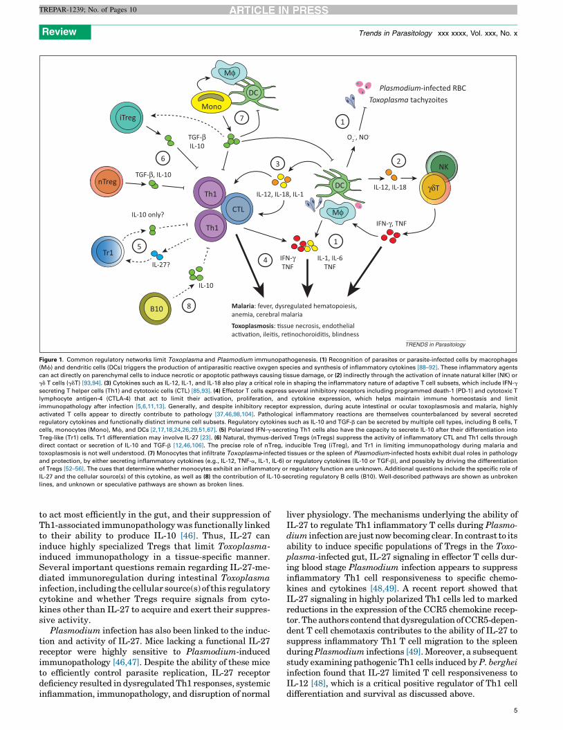
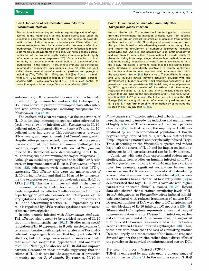
Mφ
Plasmodium-infected RBC
NK
γδT
Toxoplasma tachyzoites
Th1
CTL
B10
DC IL-12, IL-18IL-12, IL-18, IL-1
IL-10
IL-27?
Toxoplasmosis: �ssue necrosis, endothelial ac�va�on, ilei�s, re�nochoroidi�s, blindness
O2-, NO-
IFN-γ, TNF
Malaria: fever, dysregulated hematopoiesis, anemia, cerebral malaria
IFN-γTNF
IL-1, IL-6TNF
iTreg
Tr1
IL-10 only?
TGF-βIL-10
nTregTh1
TGF-β, IL-10
DC
Mφ
Mono
1
3
7
6
5
4
8
1
2
TRENDS in Parasitology
Figure 1. Common regulatory networks limit Toxoplasma and Plasmodium immunopathogenesis. (1) Recognition of parasites or parasite-infected cells by macrophages
(Mf) and dendritic cells (DCs) triggers the production of antiparasitic reactive oxygen species and synthesis of inflammatory cytokines [88–92]. These inflammatory agents
can act directly on parenchymal cells to induce necrotic or apoptotic pathways causing tissue damage, or (2) indirectly through the activation of innate natural killer (NK) or
gd T cells (gdT) [93,94]. (3) Cytokines such as IL-12, IL-1, and IL-18 also play a critical role in shaping the inflammatory nature of adaptive T cell subsets, which include IFN-g
secreting T helper cells (Th1) and cytotoxic cells (CTL) [85,93]. (4) Effector T cells express several inhibitory receptors including programmed death-1 (PD-1) and cytotoxic T
lymphocyte antigen-4 (CTLA-4) that act to limit their activation, proliferation, and cytokine expression, which helps maintain immune homeostasis and limit
immunopathology after infection [5,6,11,13]. Generally, and despite inhibitory receptor expression, during acute intestinal or ocular toxoplasmosis and malaria, highly
activated T cells appear to directly contribute to pathology [37,46,98,104]. Pathological inflammatory reactions are themselves counterbalanced by several secreted
regulatory cytokines and functionally distinct immune cell subsets. Regulatory cytokines such as IL-10 and TGF-b can be secreted by multiple cell types, including B cells, T
cells, monocytes (Mono), Mf, and DCs [2,17,18,24,26,29,51,67]. (5) Polarized IFN-g-secreting Th1 cells also have the capacity to secrete IL-10 after their differentiation into
Treg-like (Tr1) cells. Tr1 differentiation may involve IL-27 [23]. (6) Natural, thymus-derived Tregs (nTregs) suppress the activity of inflammatory CTL and Th1 cells through
direct contact or secretion of IL-10 and TGF-b [12,46,106]. The precise role of nTreg, inducible Treg (iTreg), and Tr1 in limiting immunopathology during malaria and
toxoplasmosis is not well understood. (7) Monocytes that infiltrate Toxoplasma-infected tissues or the spleen of Plasmodium-infected hosts exhibit dual roles in pathology
and protection, by either secreting inflammatory cytokines (e.g., IL-12, TNF-a, IL-1, IL-6) or regulatory cytokines (IL-10 or TGF-b), and possibly by driving the differentiation
of Tregs [52–56]. The cues that determine whether monocytes exhibit an inflammatory or regulatory function are unknown. Additional questions include the specific role of
IL-27 and the cellular source(s) of this cytokine, as well as (8) the contribution of IL-10-secreting regulatory B cells (B10). Well-described pathways are shown as unbroken
lines, and unknown or speculative pathways are shown as broken lines.
Review Trends in Parasitology xxx xxxx, Vol. xxx, No. x
TREPAR-1239; No. of Pages 10
to act most efficiently in the gut, and their suppression ofTh1-associated immunopathology was functionally linkedto their ability to produce IL-10 [46]. Thus, IL-27 caninduce highly specialized Tregs that limit Toxoplasma-induced immunopathology in a tissue-specific manner.Several important questions remain regarding IL-27-me-diated immunoregulation during intestinal Toxoplasmainfection, including the cellular source(s) of this regulatorycytokine and whether Tregs require signals from cyto-kines other than IL-27 to acquire and exert their suppres-sive activity.
Plasmodium infection has also been linked to the induc-tion and activity of IL-27. Mice lacking a functional IL-27receptor were highly sensitive to Plasmodium-inducedimmunopathology [46,47]. Despite the ability of these miceto efficiently control parasite replication, IL-27 receptordeficiency resulted in dysregulated Th1 responses, systemicinflammation, immunopathology, and disruption of normal
liver physiology. The mechanisms underlying the ability ofIL-27 to regulate Th1 inflammatory T cells during Plasmo-dium infection are just now becoming clear. In contrast to itsability to induce specific populations of Tregs in the Toxo-plasma-infected gut, IL-27 signaling in effector T cells dur-ing blood stage Plasmodium infection appears to suppressinflammatory Th1 cell responsiveness to specific chemo-kines and cytokines [48,49]. A recent report showed thatIL-27 signaling in highly polarized Th1 cells led to markedreductions in the expression of the CCR5 chemokine recep-tor. The authors contend that dysregulation of CCR5-depen-dent T cell chemotaxis contributes to the ability of IL-27 tosuppress inflammatory Th1 T cell migration to the spleenduring Plasmodium infections [49]. Moreover, a subsequentstudy examining pathogenic Th1 cells induced by P. bergheiinfection found that IL-27 limited T cell responsiveness toIL-12 [48], which is a critical positive regulator of Th1 celldifferentiation and survival as discussed above.
5
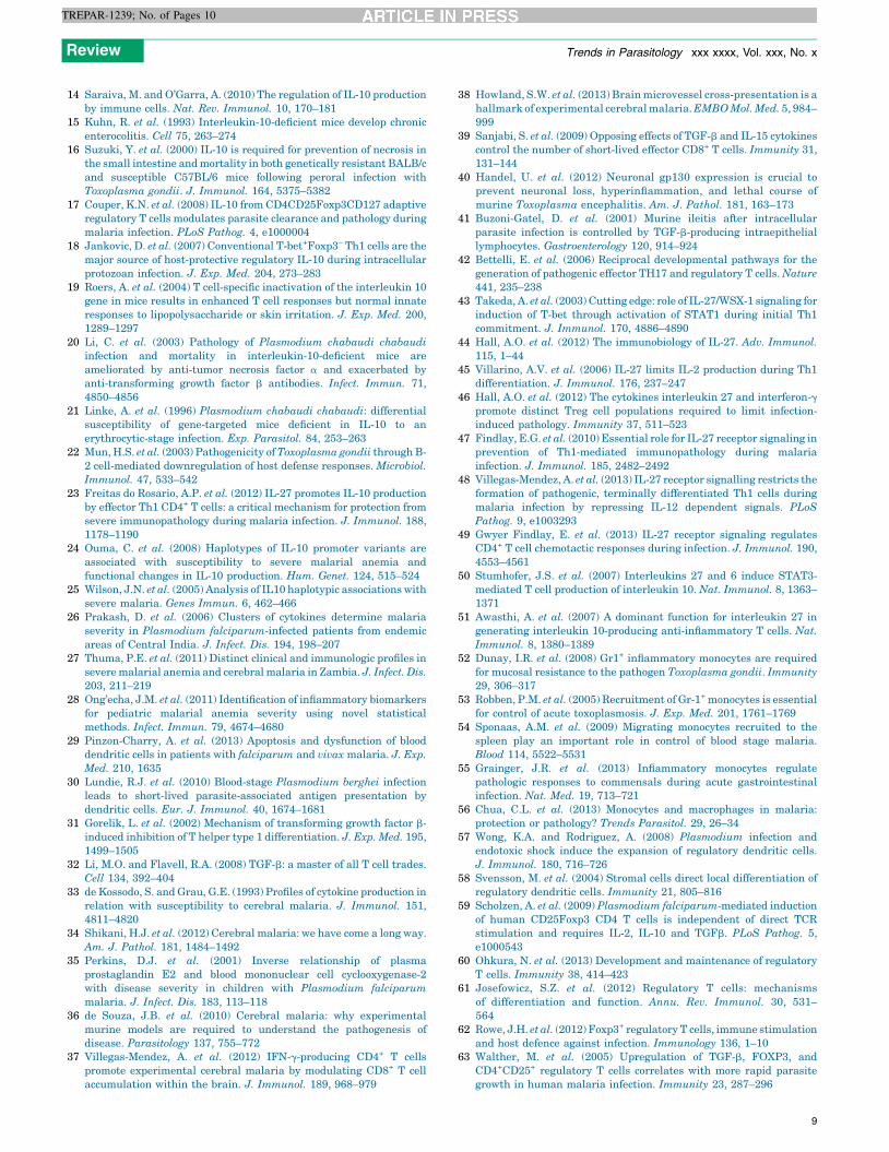
Table 1. Regulatory cytokines that limit immunopathology during toxoplasmosis and malaria
Cytokine Key cellular sources Cellular targets Predominant effect in vivo Refs
IL-10 T cells, macrophages, DCs, B cells DCs, B cells, macrophages,
epithelial cells, endothelial cells
Suppress activation and
antigen presentation by DCs,
B cells,
and macrophages
[2,17,18,24,26,29,51,67]
TGF-b T cells, B cells, NK cells, macrophages,
epithelial cells, endothelial cells,
platelets, neurons
T cells, DCs, macrophages, B cells Induction of iTreg and Th17
differentiation, trigger effector
T cell apoptosis
[26,32,33,40,63,107–109]
IL-27 Unknown CD4 T cells, DCs, macrophages Induction of Tr1 cell
differentiation
[23,40,46–49,110]
Review Trends in Parasitology xxx xxxx, Vol. xxx, No. x
TREPAR-1239; No. of Pages 10
Through a variety of experimental models, these resultshighlight the importance of IL-27 in regulating Th1-medi-ated immunopathogenesis during infections with eitherToxoplasma or Plasmodium and influencing the outcomeof disease. In addition to inducing specific populations ofTregs or limiting responsiveness to cytokines or chemo-kines, IL-27 is also a well-known trigger of IL-10 expres-sion by T cells [50,51]. For example, the generation of IL-10-expressing effector T cells during P. chabaudi infectionrequires IL-27 [23]. By contrast, a recent report showedthat IL-27 is also important for IL-10-independent protec-tion against immunopathology in P. berghei-infected mice[47]. The heterogeneous patterns of expression and respon-siveness to IL-27 in vivo underscore the complexity ofimmunoregulation by this cytokine.
Immunoregulation by myeloid cellsBone marrow-derived myeloid precursors differentiate in-to several functionally distinct innate immune cells includ-ing neutrophils, eosinophils, basophils, and monocytes.Significant interest in understanding the function andregulation of monocytes has grown from early reportsshowing that these cells are essential for control of bothToxoplasma and Plasmodium [52–54]. After infection,monocytes traffic from the blood to inflamed tissues wherethey can further differentiate into phagocytic macrophagesand DCs. Thus, monocytes contribute to resistance byclearing pathogens through phagocytosis, releasing proin-flammatory cytokines, and providing a pool of APCs to aidin promoting T and B cell responses.
In addition to these protective inflammatory responses,infiltrating monocytes and bone marrow-derived myeloidcells play immunoregulatory roles during both toxoplas-mosis and malaria. For instance, intestinal infection byspecific strains of Toxoplasma is associated with an influxof inflammatory monocytes that adopt regulatory proper-ties characterized by IL-10 and prostaglandin E2 (PGE2)expression. Unexpectedly, IL-10 and PGE2 were expressedin response to infection with commensal bacteria but notother inflammatory cues such as Toxoplasma lysates, andin the absence of these secreted regulatory factors micedevelop neutrophil-dependent intestinal pathology [55].Thus, monocytes respond to diverse signals to regulateToxoplasma-induced immunopathology.
Similarly, Plasmodium infections are associated withantimalarial, monocyte-driven inflammatory cascades[54]. These protective activities, which include phagocyto-sis of infected erythrocytes and secretion of cytokines, havebeen recently reviewed [56]. By contrast, Plasmodium
6
blood stage infection has been linked to the accumulationof distinct subsets of myeloid cells that exhibit potentregulatory functions. In rodents infected with blood stagePlasmodium parasites, a striking inversion occurs in theratio of proinflammatory DCs and regulatory DCs thatexpress IL-10 themselves and also induce IL-10 expressionin CD4 T cells [57]. Finally, in vitro studies have shownthat interactions between circulating monocytes and P.falciparum-infected erythrocytes can trigger Treg differ-entiation. Notably, the in vitro induction of these Tregs wasassociated with the capacity of monocytes to secrete IL-10and TGF-b [58,59]. These latter observations are consis-tent with data showing that splenic inflammatory mono-cytes express IL-10 during acute P. chabaudi infection inmice [54]. Further study is required to determine whethersuch cellular interactions and regulatory pathways occurin P. falciparum-infected individuals. Given the criticalrole of IL-10 in regulating immunity during Plasmodiuminfection, these data suggest that infiltrating monocytesand resident myeloid cells can also function within regula-tory networks that act to prevent or limit immunopatholo-gy.
The precise origins and relationships between parasite-induced myeloid cells exhibiting inflammatory or regula-tory functions are not entirely clear, and in each of theexamples cited above the regulatory function of monocyteswas dependent on local cytokine, chemokine, or tissuemicroenvironments. Thus, it will be necessary to not onlydetermine developmental relationships between inflam-matory and regulatory myeloid cells but to also understandhow specific microenvironments shape their differentia-tion or function. Such information could reveal novel strat-egies for regulating pathological inflammatory responsesduring malaria or toxoplasmosis.
Immunoregulation by lymphoid cellsT regulatory cells
Multiple subsets of CD4 T cells exhibiting regulatoryfunction have been described, including Foxp3+ naturalTregs (nTreg) that arise from the thymus, Foxp3+ activa-tion-induced Tregs (iTreg) that develop in the peripheryduring immune activation, and Foxp3-negative Th1 cellsexpressing IL-10 (Tr1) that also develop after peripheralactivation [60]. In addition to Foxp3 expression, Tregs arealso typified by expression of the high-affinity IL-2 recep-tor, CD25. nTreg and iTreg function via release of solublefactors, such as IL-10 and TGF-b, and via direct cell–cellcontact involving inhibitory molecules such as CTLA-4 orthe glucocorticoid-induced TNF receptor (TNFR) family

Review Trends in Parasitology xxx xxxx, Vol. xxx, No. x
TREPAR-1239; No. of Pages 10
related gene (GITR) [60,61], whereas the suppressive func-tion of Tr1 cells appears limited to IL-10 and TGF-bsecretion. In addition to their critical role in maintainingimmune homeostasis and preventing autoimmunity, Tregshave become an important contributor in controllingimmunopathogenesis during microbial infections [62].
The appearance and expansion of Tregs has been de-scribed during infection with either Toxoplasma or Plas-modium. For instance, malaria has been linked to a higherratio of circulating Tregs relative to effector T cells [63];however, their relative contribution to parasite control anddisease pathogenesis is not fully understood and remainscontroversial. Some studies have shown that Tregs para-doxically correlate with enhanced immune-mediated clear-ance of parasites and protection against pathologicalinflammatory responses [64,65]. By contrast, other studieshave reported that Treg suppression of protective immu-nity correlated to higher parasite burdens [65]. Similarly,conflicting results have been observed in rodent models(primarily murine ECM models), in which mechanisticdissection of the role of Tregs is possible (reviewed in[66,67]). Thus, the biological relevance of Treg expansionsin humans and other species remains an unansweredquestion.
Inconsistencies surrounding the role of Tregs in modu-lating immunity against Toxoplasma are also apparent.Multiple studies reported that the depletion of Tregs usingthe anti-CD25 monoclonal antibody (clone PC61) inrodents revealed limited roles for these cells during thechronic phases of infection [68]. Conversely, adoptivetransfer of FoxP3+ Tregs was shown to potently limitTh1-driven immunopathology in mice acutely infectedwith Toxoplasma [46]. Yet, DCs and effector Th1 cells wereshown to simultaneously impair Treg suppressive functionand induce Treg differentiation into Th1-like effector cellsthat contribute to immunopathogenesis via secretion ofIFN-g [69]. Further adding to this debate, recent data alsosuggest a harmful role for Tregs via their ability to con-strain protective immunity; rapid proliferative expansionof Th1 effector cells after Toxoplasma infection is report-edly linked to a transient decrease in Tregs [70]. Mecha-nistically, this effect was attributed to Treg deprivation ofIL-2, because exogenous IL-2 could restore Treg numbers,which suppressed Th1 effector responses and resulted inloss of parasite control. Thus, a transient reduction inTregs appears necessary for optimal Th1 effector responsesagainst Toxoplasma [70]. This latter study demonstratesthat the numerical expansion and suppressive activity ofTregs, if not restrained, can significantly limit protectiveimmunity after acute infection. Furthermore, the treat-ment of chronically infected mice with IL-2 complexesleads to increased cyst burdens in the CNS, suggestingthat increased numbers of Tregs can limit protection in theCNS [69,70].
Tregs appear to play a role in either limiting immuno-pathogenesis or controlling antiparasitic immunity duringToxoplasma and Plasmodium infections. Of key impor-tance will be defining how or whether local secretion ofregulatory cytokines in specific tissues and microenviron-ments influences disease outcomes. For example, it is ofinterest to determine the role of Tregs and the contribution
of their suppressive effector molecules in the CNS of Toxo-plasma- and Plasmodium-infected hosts. Toxoplasma canbreach the gut and disseminate throughout the host, includ-ing the eyes and brain. Within these immune-privilegedtissues, highly activated T cells and IFN-g are required tolimit parasite replication. Similarly, accumulation of Th1cells in the microvasculature of the CNS after Plasmodiuminfection is linked to the development of ECM. Thus, it isessential to tightly regulate these polarized cellular reac-tions in these critical tissues to prevent immune-mediatedpathology. Tregs could act in numerous ways to balanceprotection and pathology; it is possible that Tregs suppressantigen presentation in Toxoplasma-infected neural tis-sues, the vasculature of the CNS during CM, or limit thelocal activity of effector T cells. Dissecting these mechanismsin vivo will require the use of powerful technologies, includ-ing intravital microscopy, to identify and study cellularinteractions that may prove critical for understanding theregulation of immunopathogenesis during Toxoplasmainfections. Finally, it is also of interest to define the rolesof nTregs and iTregs, and determine whether true parasite-specific iTregs are expanded after Toxoplasma or Plasmo-dium infection. Given the critical roles for Tregs to both limitimmunopathology and constrain protective immunity afterinfection, modulation of the number, localization, or func-tion of Tregs may hold promise as interventional immune-based strategies for toxoplasmosis or malaria.
B regulatory cells
Another regulatory immune cell subset that has been thefocus of attention recently is the B regulatory (B10) cell, so-called because of its propensity to regulate effector CD4 Tcell activity through the secretion of IL-10. B10 cells arephenotypically defined as CD19+CD1dhiCD5+. CD1d is anon-classical MHC class I molecule necessary for presen-tation of lipid antigens, and CD5 has been functionallylinked to antagonizing both T cell and B cell receptorsignaling [71,72]. Originally defined as being critical reg-ulators of Th1-driven inflammation in autoimmune dis-ease [73], B10 cells are now a focus of attention asimportant regulators of inflammation during infection.Recent data show that B10 cells numerically expand andmodulate immunity during parasitic infections, includingbabesiosis and schistosomiasis [74,75]. Follicular (B-2) Bcell derived IL-10 was reported as a potent inhibitor of hostimmunity during Toxoplasma infection [22], although theprecise ontogeny of B10 cell development is unknown.Even though no formal reports of B10 cell expansionduring either Plasmodium or Toxoplasma infection exist,given the critical mutual counterbalance between inflam-matory Th1 cells and IL-10-mediated immunoregulationreported for these two Apicomplexan infections, it wouldnot be surprising if B10 cells play a functional role.
Concluding remarksDuring Toxoplasma and Plasmodium infections, severaloverlapping immunoregulatory pathways maintain thebalance between health and disease. Indeed, the potentimmune responses that serve to limit parasite persistencewithin the host can also be responsible for local or systemicpathologies associated with these infections. Multiple
7

Review Trends in Parasitology xxx xxxx, Vol. xxx, No. x
TREPAR-1239; No. of Pages 10
regulatory factors can independently and coordinately actto limit immunopathology during the acute stages of toxo-plasmosis and malaria. The striking regulatory potential ofmonocytes is now appreciated, and the developmentalrelationships, similarities, and functional distinctions be-tween naturally occurring and peripherally induced Tregsis growing clearer. Secreted factors, such as IL-10, TGF-b,and IL-27, are known potent regulators of immunity, anddata from experimental models and human clinical studieshighlight the critical and complex role that these cytokinesplay in regulating immunity against Toxoplasma andPlasmodium. Finally, several cell surface-expressed inhib-itory receptors are critical for limiting T cell mediatedimmunopathology during ocular toxoplasmosis and ECM.
Although much information exists regarding the abilityof these cellular and secreted factors to regulate immuno-pathology after infection, many questions remain (Box 4).Regulatory cytokines and inhibitory receptor ligands areexpressed by and act on a diverse range of cell types,underscoring the complexity of these regulatory circuits.TGF-b can both promote and prevent immunopathology,depending on whether IL-6 and specific T cell subsets arepresent, and IL-27 has the capacity to either potentiate orinhibit Th1 responses. Whether or how Tregs modulateinflammatory effector T cells in local tissue environments,such as the eye or brain, during toxoplasmosis, remainsunknown. Understanding how these cytokines are tempo-rally regulated after infection, identifying the specific cel-lular sources, and determining whether the anatomy ofinteractions between inflammatory and regulatory cellsubsets determines disease outcomes remain importantand unanswered questions.
Rodent models of Toxoplasma and Plasmodium infec-tion have been extensively used to dissect host–pathogeninteractions and pathways of immunoregulation duringacute and chronic protozoan infections. The power andutility of these models relate to the numerous Toxoplasmagenotypes and rodent-specific species of Plasmodium para-sites available for study, as well as the vast number of
Box 4. Outstanding questions
� Much information has been learned about these regulatory cells,
circuits, and pathways from experimental models, but which
features of immunoregulation are operational during clinical
malaria or acute ocular or intestinal toxoplasmosis?
� What are the key cellular sources for secreted regulatory factors
such as IL-27, IL-10, and TGF-b, and do the key cellular sources of
regulatory cytokines temporally shift as infection progresses?
� How does the tissue microenvironment influence the expression
of regulatory cytokines and inhibitory receptor ligands, or the
manner in which they exert suppressive effects on target cells?
� Are parasite-specific Tregs induced during toxoplasmosis or
malaria, and what are the critical signals driving their activation,
differentiation, or proliferation?
� Do monocytes exhibit specific regulatory functions before differ-
entiating into macrophages or DCs in Toxoplasma-infected
tissues or the spleen of Plasmodium-infected hosts, and what
are the signals that stimulate the acquisition of suppressive
function by infiltrating monocytes?
� Given that both IL-6 and TGF-b are coexpressed after infection
with either Toxoplasma or Plasmodium, why is there so little
evidence for an immunopathological role for Th17 cells during
toxoplasmosis or malaria?
8
strains and genotypes of susceptible and resistant labora-tory rodents. The use of multiple independent models hasrevealed important information about immunoregulatorypathways during these infections. Depending on the modelsystem, the contribution of a given immunoregulatoryfactor can be different. For example, the immunoregulato-ry biology of IL-27 may be fairly distinct when expressed inthe Toxoplasma-infected intestine and the spleen of Plas-modium-infected mice. Although unique experimental sys-tems can reveal seemingly disparate mechanisms of actionfor these regulatory cytokines and cellular subsets, themass of accumulating data will ultimately translate into amore comprehensive understanding of the biology ofimmunoregulation. A more complete understanding ofthe contribution of regulatory pathways to both limitingimmunopathology and impeding potent immunity againstToxoplasma and Plasmodium infections will help shapefuture treatment and therapy against these and otherinfections.
AcknowledgmentsThe authors acknowledge members of their laboratories for their helpfuldiscussions and Dr. Kristina Wasson-Blader for editorial assistance. Wealso offer apologies to the many investigators whose contributions wewere unable to discuss owing to space limitations. Work in the Butlerlaboratory is supported by grants from the National Institutes ofHealth (NIH, AI099070) and the American Heart Association(13BGIA17140002). Work in the Blader laboratory is supported by grantsfrom the NIH (AI069986 and EY021259).
References1 Stumhofer, J.S. et al. (2006) Interleukin 27 negatively regulates the
development of interleukin 17-producing T helper cells during chronicinflammation of the central nervous system. Nat. Immunol. 7, 937–945
2 Gazzinelli, R.T. et al. (1996) In the absence of endogenous IL-10, miceacutely infected with Toxoplasma gondii succumb to a lethal immuneresponse dependent on CD4+ T cells and accompanied byoverproduction of IL-12, IFN-g and TNF-a. J. Immunol. 157, 798–805
3 Wilson, E.H. et al. (2005) A critical role for IL-10 in limitinginflammation during toxoplasmic encephalitis. J. Neuroimmunol.165, 63–74
4 Wherry, E.J. (2011) T cell exhaustion. Nat. Immunol. 12, 492–4995 Butler, N.S. et al. (2012) Therapeutic blockade of PD-L1 and LAG-3
rapidly clears established blood-stage Plasmodium infection. Nat.Immunol. 13, 188–195
6 Bhadra, R. et al. (2011) Control of Toxoplasma reactivation by rescueof dysfunctional CD8+ T-cell response via PD-1–PDL-1 blockade. Proc.Natl. Acad. Sci. U.S.A. 108, 9196–9201
7 Gigley, J.P. et al. (2012) T cell exhaustion in protozoan disease. TrendsParasitol. 28, 377–384
8 Jacobs, T. et al. (2002) Murine malaria is exacerbated by CTLA-4blockade. J. Immunol. 169, 2323–2329
9 Jacobs, T. et al. (2004) CTLA-4-dependent mechanisms prevent T cellinduced-liver pathology during the erythrocyte stage of Plasmodiumberghei malaria. Eur. J. Immunol. 34, 972–980
10 Lepenies, B. et al. (2007) CTLA-4 blockade differentially influencesthe outcome of non-lethal and lethal Plasmodium yoelii infections.Microbes Infect. 9, 687–694
11 Hafalla, J.C. et al. (2012) The CTLA-4 and PD-1/PD-L1 inhibitorypathways independently regulate host resistance to Plasmodium-induced acute immune pathology. PLoS Pathog. 8, e1002504
12 Haque, A. et al. (2010) CD4+ natural regulatory T cells preventexperimental cerebral malaria via CTLA-4 when expanded in vivo.PLoS Pathog. 6, e1001221
13 Charles, E. et al. (2010) CD4 T-cell suppression by cells fromToxoplasma gondii-infected retinas is mediated by surface proteinPD-L1. Infect. Immun. 78, 3484–3492
Review Trends in Parasitology xxx xxxx, Vol. xxx, No. x
TREPAR-1239; No. of Pages 10
14 Saraiva, M. and O’Garra, A. (2010) The regulation of IL-10 productionby immune cells. Nat. Rev. Immunol. 10, 170–181
15 Kuhn, R. et al. (1993) Interleukin-10-deficient mice develop chronicenterocolitis. Cell 75, 263–274
16 Suzuki, Y. et al. (2000) IL-10 is required for prevention of necrosis inthe small intestine and mortality in both genetically resistant BALB/cand susceptible C57BL/6 mice following peroral infection withToxoplasma gondii. J. Immunol. 164, 5375–5382
17 Couper, K.N. et al. (2008) IL-10 from CD4CD25Foxp3CD127 adaptiveregulatory T cells modulates parasite clearance and pathology duringmalaria infection. PLoS Pathog. 4, e1000004
18 Jankovic, D. et al. (2007) Conventional T-bet+Foxp3– Th1 cells are themajor source of host-protective regulatory IL-10 during intracellularprotozoan infection. J. Exp. Med. 204, 273–283
19 Roers, A. et al. (2004) T cell-specific inactivation of the interleukin 10gene in mice results in enhanced T cell responses but normal innateresponses to lipopolysaccharide or skin irritation. J. Exp. Med. 200,1289–1297
20 Li, C. et al. (2003) Pathology of Plasmodium chabaudi chabaudiinfection and mortality in interleukin-10-deficient mice areameliorated by anti-tumor necrosis factor a and exacerbated byanti-transforming growth factor b antibodies. Infect. Immun. 71,4850–4856
21 Linke, A. et al. (1996) Plasmodium chabaudi chabaudi: differentialsusceptibility of gene-targeted mice deficient in IL-10 to anerythrocytic-stage infection. Exp. Parasitol. 84, 253–263
22 Mun, H.S. et al. (2003) Pathogenicity of Toxoplasma gondii through B-2 cell-mediated downregulation of host defense responses. Microbiol.Immunol. 47, 533–542
23 Freitas do Rosario, A.P. et al. (2012) IL-27 promotes IL-10 productionby effector Th1 CD4+ T cells: a critical mechanism for protection fromsevere immunopathology during malaria infection. J. Immunol. 188,1178–1190
24 Ouma, C. et al. (2008) Haplotypes of IL-10 promoter variants areassociated with susceptibility to severe malarial anemia andfunctional changes in IL-10 production. Hum. Genet. 124, 515–524
25 Wilson, J.N. et al. (2005) Analysis of IL10 haplotypic associations withsevere malaria. Genes Immun. 6, 462–466
26 Prakash, D. et al. (2006) Clusters of cytokines determine malariaseverity in Plasmodium falciparum-infected patients from endemicareas of Central India. J. Infect. Dis. 194, 198–207
27 Thuma, P.E. et al. (2011) Distinct clinical and immunologic profiles insevere malarial anemia and cerebral malaria in Zambia. J. Infect. Dis.203, 211–219
28 Ong’echa, J.M. et al. (2011) Identification of inflammatory biomarkersfor pediatric malarial anemia severity using novel statisticalmethods. Infect. Immun. 79, 4674–4680
29 Pinzon-Charry, A. et al. (2013) Apoptosis and dysfunction of blooddendritic cells in patients with falciparum and vivax malaria. J. Exp.Med. 210, 1635
30 Lundie, R.J. et al. (2010) Blood-stage Plasmodium berghei infectionleads to short-lived parasite-associated antigen presentation bydendritic cells. Eur. J. Immunol. 40, 1674–1681
31 Gorelik, L. et al. (2002) Mechanism of transforming growth factor b-induced inhibition of T helper type 1 differentiation. J. Exp. Med. 195,1499–1505
32 Li, M.O. and Flavell, R.A. (2008) TGF-b: a master of all T cell trades.Cell 134, 392–404
33 de Kossodo, S. and Grau, G.E. (1993) Profiles of cytokine production inrelation with susceptibility to cerebral malaria. J. Immunol. 151,4811–4820
34 Shikani, H.J. et al. (2012) Cerebral malaria: we have come a long way.Am. J. Pathol. 181, 1484–1492
35 Perkins, D.J. et al. (2001) Inverse relationship of plasmaprostaglandin E2 and blood mononuclear cell cyclooxygenase-2with disease severity in children with Plasmodium falciparummalaria. J. Infect. Dis. 183, 113–118
36 de Souza, J.B. et al. (2010) Cerebral malaria: why experimentalmurine models are required to understand the pathogenesis ofdisease. Parasitology 137, 755–772
37 Villegas-Mendez, A. et al. (2012) IFN-g-producing CD4+ T cellspromote experimental cerebral malaria by modulating CD8+ T cellaccumulation within the brain. J. Immunol. 189, 968–979
38 Howland, S.W. et al. (2013) Brain microvessel cross-presentation is ahallmark of experimental cerebral malaria. EMBO Mol. Med. 5, 984–999
39 Sanjabi, S. et al. (2009) Opposing effects of TGF-b and IL-15 cytokinescontrol the number of short-lived effector CD8+ T cells. Immunity 31,131–144
40 Handel, U. et al. (2012) Neuronal gp130 expression is crucial toprevent neuronal loss, hyperinflammation, and lethal course ofmurine Toxoplasma encephalitis. Am. J. Pathol. 181, 163–173
41 Buzoni-Gatel, D. et al. (2001) Murine ileitis after intracellularparasite infection is controlled by TGF-b-producing intraepitheliallymphocytes. Gastroenterology 120, 914–924
42 Bettelli, E. et al. (2006) Reciprocal developmental pathways for thegeneration of pathogenic effector TH17 and regulatory T cells. Nature441, 235–238
43 Takeda, A. et al. (2003) Cutting edge: role of IL-27/WSX-1 signaling forinduction of T-bet through activation of STAT1 during initial Th1commitment. J. Immunol. 170, 4886–4890
44 Hall, A.O. et al. (2012) The immunobiology of IL-27. Adv. Immunol.115, 1–44
45 Villarino, A.V. et al. (2006) IL-27 limits IL-2 production during Th1differentiation. J. Immunol. 176, 237–247
46 Hall, A.O. et al. (2012) The cytokines interleukin 27 and interferon-gpromote distinct Treg cell populations required to limit infection-induced pathology. Immunity 37, 511–523
47 Findlay, E.G. et al. (2010) Essential role for IL-27 receptor signaling inprevention of Th1-mediated immunopathology during malariainfection. J. Immunol. 185, 2482–2492
48 Villegas-Mendez, A. et al. (2013) IL-27 receptor signalling restricts theformation of pathogenic, terminally differentiated Th1 cells duringmalaria infection by repressing IL-12 dependent signals. PLoSPathog. 9, e1003293
49 Gwyer Findlay, E. et al. (2013) IL-27 receptor signaling regulatesCD4+ T cell chemotactic responses during infection. J. Immunol. 190,4553–4561
50 Stumhofer, J.S. et al. (2007) Interleukins 27 and 6 induce STAT3-mediated T cell production of interleukin 10. Nat. Immunol. 8, 1363–1371
51 Awasthi, A. et al. (2007) A dominant function for interleukin 27 ingenerating interleukin 10-producing anti-inflammatory T cells. Nat.Immunol. 8, 1380–1389
52 Dunay, I.R. et al. (2008) Gr1+ inflammatory monocytes are requiredfor mucosal resistance to the pathogen Toxoplasma gondii. Immunity29, 306–317
53 Robben, P.M. et al. (2005) Recruitment of Gr-1+ monocytes is essentialfor control of acute toxoplasmosis. J. Exp. Med. 201, 1761–1769
54 Sponaas, A.M. et al. (2009) Migrating monocytes recruited to thespleen play an important role in control of blood stage malaria.Blood 114, 5522–5531
55 Grainger, J.R. et al. (2013) Inflammatory monocytes regulatepathologic responses to commensals during acute gastrointestinalinfection. Nat. Med. 19, 713–721
56 Chua, C.L. et al. (2013) Monocytes and macrophages in malaria:protection or pathology? Trends Parasitol. 29, 26–34
57 Wong, K.A. and Rodriguez, A. (2008) Plasmodium infection andendotoxic shock induce the expansion of regulatory dendritic cells.J. Immunol. 180, 716–726
58 Svensson, M. et al. (2004) Stromal cells direct local differentiation ofregulatory dendritic cells. Immunity 21, 805–816
59 Scholzen, A. et al. (2009) Plasmodium falciparum-mediated inductionof human CD25Foxp3 CD4 T cells is independent of direct TCRstimulation and requires IL-2, IL-10 and TGFb. PLoS Pathog. 5,e1000543
60 Ohkura, N. et al. (2013) Development and maintenance of regulatoryT cells. Immunity 38, 414–423
61 Josefowicz, S.Z. et al. (2012) Regulatory T cells: mechanismsof differentiation and function. Annu. Rev. Immunol. 30, 531–564
62 Rowe, J.H. et al. (2012) Foxp3+ regulatory T cells, immune stimulationand host defence against infection. Immunology 136, 1–10
63 Walther, M. et al. (2005) Upregulation of TGF-b, FOXP3, andCD4+CD25+ regulatory T cells correlates with more rapid parasitegrowth in human malaria infection. Immunity 23, 287–296
9
Review Trends in Parasitology xxx xxxx, Vol. xxx, No. x
TREPAR-1239; No. of Pages 10
64 Walther, M. et al. (2009) Distinct roles for FOXP3 and FOXP3 CD4 Tcells in regulating cellular immunity to uncomplicated and severePlasmodium falciparum malaria. PLoS Pathog. 5, e1000364
65 Minigo, G. et al. (2009) Parasite-dependent expansion of TNF receptorII-positive regulatory T cells with enhanced suppressive activity inadults with severe malaria. PLoS Pathog. 5, e1000402
66 Scholzen, A. et al. (2010) Heroes or villains? T regulatory cells inmalaria infection. Trends Parasitol. 26, 16–25
67 Freitas do Rosario, A.P. and Langhorne, J. (2012) T cell-derived IL-10and its impact on the regulation of host responses during malaria. Int.J. Parasitol. 42, 549–555
68 Couper, K.N. et al. (2009) Anti-CD25 antibody-mediated depletion ofeffector T cell populations enhances susceptibility of mice to acute butnot chronic Toxoplasma gondii infection. J. Immunol. 182, 3985–3994
69 Oldenhove, G. et al. (2009) Decrease of Foxp3+ Treg cell number andacquisition of effector cell phenotype during lethal infection.Immunity 31, 772–786
70 Benson, A. et al. (2012) Microbial infection-induced expansion ofeffector T cells overcomes the suppressive effects of regulatory Tcells via an IL-2 deprivation mechanism. J. Immunol. 188, 800–810
71 Perez-Villar, J.J. et al. (1999) CD5 negatively regulates the T-cellantigen receptor signal transduction pathway: involvement of SH2-containing phosphotyrosine phosphatase SHP-1. Mol. Cell. Biol. 19,2903–2912
72 Gary-Gouy, H. et al. (2002) CD5-negative regulation of B cell receptorsignaling pathways originates from tyrosine residue Y429 outside animmunoreceptor tyrosine-based inhibitory motif. J. Immunol. 168,232–239
73 Kalampokis, I. et al. (2013) IL-10-producing regulatory B cells (B10cells) in autoimmune disease. Arthritis Res. Ther. 15 (Suppl. 1), S1
74 Jeong, Y.I. et al. (2012) Induction of IL-10-producing CD1dhighCD5+
regulatory B cells following Babesia microti-infection. PLoS ONE 7,e46553
75 Tang, H. et al. (2013) Development of adult worms and granulomatouspathology are collectively regulated by T- and B-cells in mice infectedwith Schistosoma japonicum. PLoS ONE 8, e54432
76 Riley, E.M. et al. (2006) Regulating immunity to malaria. ParasiteImmunol. 28, 35–49
77 Hunt, N.H. and Grau, G.E. (2003) Cytokines: accelerators and brakesin the pathogenesis of cerebral malaria. Trends Immunol. 24, 491–499
78 Langhorne, J. et al. (1990) The role of CD4+ T cells in the protectiveimmune response to Plasmodium chabaudi in vivo. Immunol. Lett. 25,101–107
79 Meding, S.J. et al. (1990) Role of g interferon during infection withPlasmodium chabaudi chabaudi. Infect. Immun. 58, 3671–3678
80 Stephens, R. et al. (2005) Malaria-specific transgenic CD4+ T cellsprotect immunodeficient mice from lethal infection and demonstraterequirement for a protective threshold of antibody production forparasite clearance. Blood 106, 1676–1684
81 Spence, P.J. and Langhorne, J. (2012) T cell control of malariapathogenesis. Curr. Opin. Immunol. 24, 444–448
82 Hill, D.E. et al. (2005) Biology and epidemiology of Toxoplasma gondiiin man and animals. Anim. Health Res. Rev. 6, 41–61
83 Bierly, A.L. et al. (2008) Dendritic cells expressing plasmacytoidmarker PDCA-1 are Trojan horses during Toxoplasma gondiiinfection. J. Immunol. 181, 8485–8491
84 Suzuki, Y. et al. (1988) Interferon-g: the major mediator of resistanceagainst Toxoplasma gondii. Science 240, 516–518
85 Mashayekhi, M. et al. (2011) CD8a+ dendritic cells are the criticalsource of interleukin-12 that controls acute infection by Toxoplasmagondii tachyzoites. Immunity 35, 249–259
86 Gazzinelli, R.T. et al. (1994) Parasite-induced IL-12 stimulates earlyIFN-g synthesis and resistance during acute infection withToxoplasma gondii. J. Immunol. 153, 2533–2543
87 Hunter, C.A. et al. (1994) Production of g interferon by natural killercells from Toxoplasma gondii-infected SCID mice: regulation byinterleukin-10, interleukin-12, and tumor necrosis factor a. Infect.Immun. 62, 2818–2824
88 Scanga, C.A. et al. (2002) Cutting edge: MyD88 is requiredfor resistance to Toxoplasma gondii infection and regulates
10
parasite-induced IL-12 production by dendritic cells. J. Immunol.168, 5997–6001
89 Koblansky, A.A. et al. (2013) Recognition of profilin by Toll-likereceptor 12 is critical for host resistance to Toxoplasma gondii.Immunity 38, 119–130
90 Yarovinsky, F. et al. (2005) TLR11 activation of dendritic cells by aprotozoan profilin-like protein. Science 308, 1626–1629
91 Baccarella, A. et al. (2013) Toll-like receptor 7 mediates early innateimmune responses to malaria. Infect. Immun. http://dx.doi.org/10.1128/IAI.00923-13
92 Coban, C. et al. (2005) Toll-like receptor 9 mediates innate immuneactivation by the malaria pigment hemozoin. J. Exp. Med. 201, 19–25
93 Doolan, D.L. and Hoffman, S.L. (1999) IL-12 and NK cells are requiredfor antigen-specific adaptive immunity against malaria initiated byCD8+ T cells in the Plasmodium yoelii model. J. Immunol. 163,884–892
94 Goldszmid, R.S. et al. (2012) NK cell-derived interferon-g orchestratescellular dynamics and the differentiation of monocytes into dendriticcells at the site of infection. Immunity 36, 1047–1059
95 Dzierszinski, F. et al. (2007) Presentation of Toxoplasma gondiiantigens via the endogenous major histocompatibility complexclass I pathway in nonprofessional and professional antigen-presenting cells. Infect. Immun. 75, 5200–5209
96 Jung, S. et al. (2002) In vivo depletion of CD11c+ dendritic cellsabrogates priming of CD8+ T cells by exogenous cell-associatedantigens. Immunity 17, 211–220
97 Del Rio, L. et al. (2004) Toxoplasma gondii triggers myeloiddifferentiation factor 88-dependent IL-12 and chemokine ligand 2(monocyte chemoattractant protein 1) responses using distinctparasite molecules and host receptors. J. Immunol. 172, 6954–6960
98 Vossenkamper, A. et al. (2004) Both IL-12 and IL-18 contribute tosmall intestinal Th1-type immunopathology following oral infectionwith Toxoplasma gondii, but IL-12 is dominant over IL-18 in parasitecontrol. Eur. J. Immunol. 34, 3197–3207
99 Yap, G.S. and Sher, A. (1999) Effector cells of both nonhemopoieticand hemopoietic origin are required for interferon (IFN)-g- and tumornecrosis factor (TNF)-a-dependent host resistance to the intracellularpathogen, Toxoplasma gondii. J. Exp. Med. 189, 1083–1092
100 Khan, I.A. et al. (2000) IP-10 is critical for effector T cell traffickingand host survival in Toxoplasma gondii infection. Immunity 12, 483–494
101 Lamikanra, A.A. et al. (2007) Malarial anemia: of mice and men.Blood 110, 18–28
102 Amante, F.H. et al. (2010) Immune-mediated mechanisms of parasitetissue sequestration during experimental cerebral malaria. J.Immunol. 185, 3632–3642
103 Liesenfeld, O. et al. (1996) Association of CD4+ T cell-dependent,interferon-g-mediated necrosis of the small intestine with geneticsusceptibility of mice to peroral infection with Toxoplasma gondii. J.Exp. Med. 184, 597–607
104 Garweg, J.G. and Candolfi, E. (2009) Immunopathology in oculartoxoplasmosis: facts and clues. Mem. Inst. Oswaldo Cruz 104, 211–220
105 Weiss, L.M. and Dubey, J.P. (2009) Toxoplasmosis: a history of clinicalobservations. Int. J. Parasitol. 39, 895–901
106 Wu, J.J. et al. (2010) Natural regulatory T cells mediate thedevelopment of cerebral malaria by modifying the pro-inflammatory response. Parasitol. Int. 59, 232–241
107 Wahl, S.M. (1994) Transforming growth factor b: the good, the bad,and the ugly. J. Exp. Med. 180, 1587–1590
108 Longenecker, G. et al. (2002) Endocrine expression of the active formof TGF-b1 in the TGF-b1 null mice fails to ameliorate lethalphenotype. Cytokine 18, 43–50
109 Nagineni, C.N. et al. (2002) Transforming growth factor-b expression inhuman retinal pigment epithelial cells is enhanced by Toxoplasmagondii: a possible role in the immunopathogenesis of retinochoroiditis.Clin. Exp. Immunol. 128, 372–378
110 Pot, C. et al. (2009) Cutting edge: IL-27 induces the transcriptionfactor c-Maf, cytokine IL-21, and the costimulatory receptor ICOSthat coordinately act together to promote differentiation of IL-10-producing Tr1 cells. J. Immunol. 183, 797–801