Embed Size (px)
Citation preview

Copyright © 2003 by the American Society for Biochemistry and Molecular Biology, Inc.
1322 Journal of Lipid Research
Volume 44, 2003
This article is available online at http://www.jlr.org
Regulation of human CETP gene expression: role of SP1
and SP3 transcription factors at promoter sites
�
690,
�
629, and
�
37
Wilfried Le Goff, Maryse Guerin, Laure Petit, M. John Chapman,
1
and Joëlle Thillet
Institut National de la Santé et de la Recherche Médicale (INSERM) Unité 551
��
Dyslipoproteinemias and Atherosclerosis: Genetics, Metabolism and Therapy
��
, Hôpital de la Pitié, 83 boulevard de l’Hôpital 75651 Paris, France
Abstract Cholesteryl ester transfer protein (CETP) is a keyfactor in plasma reverse cholesterol transport and is implicatedin the pathophysiology of atherogenic dyslipidemia. Variationsobserved in plasma CETP mass and activity in both normolipi-demic and dyslipidemic individuals may reflect differences inCETP gene expression. We evaluated the respective roles ofthe Sp1 and Sp3 transcription factors on the promoter activityof the human CETP gene at a new Sp1/Sp3 site identified at
position
�
690, and at two previously described Sp1/Sp3 sitesat positions
�
37 and
�
629. In transient transfection in HepG2cells, site-directed mutagenesis using luciferase reporter con-structs containing a promoter fragment from
�
32 to
�
745 in-dicated that the new
�
690 site acts as a repressive element in
reducing CETP promoter activity (
�
22%;
P
�
0.05); equally,this site exerts an additive effect with the
�
629 site, inducingmarked repression (
�
42%;
P
�
0.005). In contrast, in NCTCcells that display a 16-fold lower level of Sp3, the repressive ef-fect at the
�
690 site was enhanced 2-fold (
�
45%;
P
�
0.05),whereas the
�
629 site exerted no effect. Cotransfection ofSp1 and/or Sp3 in SL2 insect cells lacking endogenous Spfactors demonstrated that Sp1 and Sp3 act as activators at
the
�
690 and
�
37 sites, whereas Sp3 acts as a repressor atthe
�
629 site. Taken together, our data demonstrate thatSp1 and Sp3 regulate human CETP promoter activity throughthree Sp1/Sp3 binding sites in a distinct manner, and that theSp1/Sp3 ratio is a key factor in determining the relative contri-bution of these sites to total promoter activity.
—Le Goff,
W., M. Guerin, L. Petit, M. J. Chapman, and J. Thillet.
Regula-tion of human CETP gene expression: role of SP1 and SP3transcription factors at promoter sites
�
690,
�
629, and
�
37.
J.
Lipid Res.
2003.
44:
1322–1331.
Supplementary key words
reverse cholesterol transport • atheroscle-rosis • transcriptional repression
Plasma cholesteryl ester transfer protein (CETP) plays akey role in reverse cholesterol transport by mediating thetransfer of cholesteryl esters (CEs) from HDL to athero-
genic apolipoprotein B-containing lipoproteins, includingVLDL, VLDL remnants, IDL, and LDL (1). The relation-ship between plasma CETP mass and activity on the onehand and coronary artery disease and cardiovascular riskon the other is indeterminate, however (2, 3). Nonethe-less, it is established that variation in CETP mass and/oractivity is closely associated with lipoprotein phenotypeand, notably, HDL cholesterol levels, in both normolipi-demic and dyslipidemic subjects (4–6). Moreover, thera-peutic reduction in CETP mass and/or activity is associ-ated with an increase of plasma HDL levels in humans (7)and with regression of atherosclerosis in rabbits (8).
Control of the expression of the CETP gene constitutesa major component in the regulation of plasma CETPmass in humans (9, 10). Major factors that influenceCETP gene expression include dietary cholesterol (11),fatty acids (12, 13), and corticosteroids (14), all of whichact directly on the promoter region.
The human CETP promoter is under the control of reg-ulatory elements that modulate its transcriptional activity
(15–22). Indeed, several
trans
-acting factors, includingthe orphan nuclear hormone receptor ARP-1 (18), theCCAAT/enhancer-binding protein (15)
trans
retinoic acid(20), the sterol-responsive binding protein (SREBP) (16,19), and the liver receptor homolog-1 (22) are implicatedin the regulation of the transcriptional activity of theCETP gene promoter through specific response elements.
Cellular cholesterol content can regulate CETP geneexpression (11, 23, 24), and promoter elements impli-cated in such modulation have been identified (19, 21).The mechanism of sterol-mediated regulation of CETPexpression is complex and requires interaction of LXRand SREBP transcription factors with their respective pro-moter response elements (19, 21), although the involve-ment of SREBP in the sterol-mediated up-regulation of
1
To whom correspondence should be addressed.e-mail: [email protected]
Manuscript received 28 October 2002 and in revised form 22 April 2003.
Published, JLR Papers in Press, May 1, 2003.DOI 10.1194/jlr.M200425-JLR200
by guest, on February 3, 2019
ww
w.jlr.org
Dow
nloaded from
Le Goff et al.
Human CETP gene expression 1323
CETP gene expression remains controversial (16). In ad-dition to these transcription factors, two binding sites forSp1 have been identified in the CETP gene promoter (17,18); the first consists of a GC-box located at position
�
37upstream of the transcriptional start site (18). Mutationsat this site lead to marked reduction in the in vitro tran-scriptional activity of the CETP promoter, thereby indicat-ing that Sp1 is a key factor in the activation of CETP geneexpression. The second Sp1 site, at position
�
629 (C/A),exhibits a functional polymorphism that modulates CETPpromoter activity (17). Indeed, the A allele binds bothSp1 and Sp3 transcriptional factors, leading to significantreduction in in vitro transcriptional activity as comparedwith the C allele, which does not bind Sp1 and Sp3.
Sp1 and Sp3 transcription factors are ubiquitous zinc-finger proteins and belong to the Sp family of transcrip-tion factors, which includes four proteins, i.e., Sp1, Sp2,Sp3, and Sp4 (25). Both Sp1 and Sp3 recognize G-rich ele-ments such as the GC and GT boxes, through which thesetranscription factors contribute to the regulation of theexpression of a wide spectrum of genes (26–30).
We have presently evaluated the respective roles of theSp1 and Sp3 transcription factors in regulation of the pro-moter activity of the CETP gene. To this end, we analyzedthe action of these transcription factors at a new Sp1 bind-ing site identified at position
�
690 and at two Sp1 pro-moter sites described previously (
�
629 and
�
37 sites, re-spectively). Our data demonstrate that the nuclear Sp1/Sp3 ratio is a critical factor in the regulation of CETPgene expression, acting at three distinct promoter sites atpositions
�
690,
�
629, and
�
37, respectively.
EXPERIMENTAL PROCEDURES
DNase I footprinting assays
Nuclear extracts were prepared from confluent 150 mmdishes as previously described by Dignam et al. (31), and storedat
�
80
�
C before use. Two probes,
Nhe
I*-
Sty
I and
Nhe
I-StyI* (theasterisk indicates the
32
P-labeled extremity), were prepared as fol-lows. The wild-type (WT) construct (see below “Plasmid constructs”)was digested by either
Nhe
I or
Sty
I restriction enzymes (New En-gland Biolabs, Saint Quentin en Yvelines, France). The linearisedvector was then end-labeled by fill-in in a final volume of 30
�
lcontaining 20
�
Ci of both [
�
32
P]dATP and [
�
32
P]dCTP (10 mCi/ml; 3,000 Ci/mmol; NEN Life, Paris, France), 10 units of Klenowfragment (New England Biolabs), and 8 mM of dGTP
�
dTTP at20
�
C for 20 min. After additional incubation for 5 min with 8mM of nonradiolabeled dATP
�
dCTP, and inactivation of theKlenow fragment for 10 min at 65
�
C, the labeled fragment waspurified on MicroSpin
TM
G-25 columns (Amersham Biosciences,Saclay, France) and digested either by
Sty
I or
Nhe
I. The radiola-beled restriction fragment was purified by electrophoresis in a6% polyacrylamide gel for 3 h at 150 V, excised from the gel, andincubated overnight at 42
�
C in an elution buffer containing 0.3 Msodium acetate, 2 mM EDTA, and 0.5% SDS. DNA was then pre-cipitated in the presence of absolute ethanol and 80
�
g of glyco-gen (Invitrogen, Cergy Pontoise, France) for 2 h at
�
20
�
C.DNase I footprinting experiments were performed as follows:
1
�
l (5
10
4
cpm) of radiolabeled probe (
Nhe
I-
Sty
I* or
Nhe
I-
Sty
I*) was incubated in a final volume of 50
�
l with 5
�
g poly(dI-
dC), 2 mM spermidine, 14
�
g of nuclear extracts (or BSA in con-trol), and 5
�
l of a binding solution [50 mM Tris-HCl (pH 7.5),10 mM MgCl
2
, 2 mM EDTA, 5 mM DTT, 25% glycerol] on ice for15 min. After the addition of 5
�
l of a solution containing 10 mMMgCl
2
and 5 mM CaCl
2
for 1 min at 20
�
C, the binding solutionwas treated with 1
�
l of 1:10 to 1:2 (1:100 to 1:20 in control withBSA) dilution DNase I stock (10 U/
�
l, Amersham Biosciences)for 4 min. The reaction was stopped with a solution (140
�
l) con-taining 190 mM sodium acetate, 30 mM EDTA, 0.15% SDS, 9
�
gyeast tRNA, and 2
�
g proteinase K for 30 min at 42
�
C. DNA frag-ments were subsequently extracted with phenol-chloroform andprecipitated with NaCl 5 M-absolute ethanol before loading ontoa 6% acrylamide sequencing gel (acrylamide-bis acrylamide,19:1). Electrophoresis was carried out at room temperature at 60W for 1 h and the gel was transferred to 3MM paper (Whatman,Ivry sur Seine, France), dried, and exposed to Hyperfilm MP(Amersham Biosciences) with intensifying screens at
�
80
�
Covernight.
Electrophoretic mobility shift assays
Electrophoretic mobility shift assay (EMSA) was performed asfollows: 25 bp synthetic oligonucleotides (Invitrogen) correspond-ing to the protected region (from
�
676 bp to
�
701 bp) in DNaseI footprinting experiments [footprint (FP): 5
-CTGCTCCGCCC-CTTTCCCCCGGATA-3
and FPmut: 5
-CTGCTCCGaaCCTTTC-CCCCGGATA-3
; the underlined and the lowercase letters indi-cating the Sp site and the mutation site respectively] wereannealed with their respective complementary strands at 100
�
Cfor 3 min in a solution containing 100 mM Tris-HCl (pH 7.5),100 mM MgCl
2
, 13 mM EDTA, 13 mM spermidine, and 20 mMDTT. Double-strand probes were radiolabeled with 20
�
Ci of[
�
32
P]ATP (5 mCi/ml, 3,000 Ci/mmol; NEN Life) by T4 polynu-cleotide kinase (Promega, Charbonnières, France) at 37
�
C for 30min. Radiolabeled double-strand probes (0.25 pmol) were incu-bated for 15 min on ice in a final volume of 20
�
l in the presenceof 10 mM Tris-HCl (pH 7.5), 100 mM NaCl, 3 mM MgCl
2
, 5 mMEDTA, 1 mM DTT, 5% glycerol, 2
�
g poly(dI-dC), 4 mM spermi-dine, 1
�
g BSA, and 8
�
g of nuclear extracts. In experiments thatrequired the presence of an excess unlabeled competitor (100-fold excess), the latter was added to the mixture before the addi-tion of radiolabeled probe. When indicated, 0.8
�
g of rabbit af-finity-purified polyclonal antibody raised against Sp1 or Sp3(TEBU, Le-Perray-en-Yvelines, France) was incubated for 30 minbefore addition of radiolabeled probe. After incubation, sampleswere loaded on a 6% acrylamide gel (acrylamide-bis-acrylamide,29:1). Electrophoresis was performed at room temperature at200 V for 3 h, and the gels were transferred onto 3MM paper(Whatman), dried, and exposed to Hyperfilm MP (AmershamBiosciences) at
�
20
�
C overnight.
Plasmid constructsConstructs used in this study have been previously described
in detail by Dachet et al. (17). Briefly, a 777 bp DNA fragmentcorresponding to the region from �32 to �745 of the CETP pro-moter from individuals homozygous for either the �629A or�629C allele was amplified by PCR and digested by NheI andBglII restriction enzymes. The digested fragments were clonedbetween the NheI and BglII sites of the pGL3 basic luciferase ex-pression vector (Promega) generating the WT (A allele) andpM1 (C allele) constructs. In both constructs, one or two pointmutations were introduced either in the transcription factorbinding site at position �690 or in the Sp1 binding site at posi-tion �37, or both, using the GeneEditor™ in vitro Site-DirectedMutagenesis System kit (Promega) according to the manufac-turer’s protocol in order to generate pM2 (�690 mutated site),pM3 (�37 mutated site), or pM2M3 (containing both �690 and
by guest, on February 3, 2019
ww
w.jlr.org
Dow
nloaded from
1324 Journal of Lipid Research Volume 44, 2003
�37 mutated sites) constructs that derived from WT; andpM1M2 (�690 mutated site), pM1M3 (�37 mutated site), orpM1M2M3 (containing both �690 and �37 mutated sites) con-structs that derived from pM1. Oligonucleotides used to createmutations in �690 and �37 binding sites were 5-CTGCTC-CGaaCCTTTCCCCCGGATA-3 and 5-ATGTTCCGTGGGGGCT-GttCGGACATACATA-3 (18), respectively, with the lowercase let-ters indicating the mutation site.
pPac, pPac-Sp1, and pPac-USp3 vectors were generous giftsfrom Guntram Suske (Klinikum der Philipps-Universität Mar-burg, Marburg, Germany). pAdh-LacZ vectors were a generousgift from Christine Vesque (Inserm U368, Ecole NormaleSupérieure, Paris, France).
Cell culture and transfection experimentsThe human hepatocellular carcinoma cell line HepG2 and
the mouse connective tissue cell line NCTC (American Type Cul-ture Collection, Rockville, MD) were grown at 37�C in 5% CO2 inDulbecco’s Modified Eagle’s Medium containing 10% and 8%foetal calf serum, respectively (Invitrogen), 2 mM l-glutamine,and 40 �g/ml gentamycin. Cells were seeded on 6-well plates at2.5 105 cells per well. After 48 h of growth, 3 �g of each CETPpromoter construct was cotransfected with 0.5 �g of a �-galac-tosidase expression vector (pSV-�gal; Promega) using the Lipo-fectin Liposomal reagent (Invitrogen) according to the manufac-turer’s instructions. Twenty-four hours after transfection, themedium was replaced by fresh medium and the cells were incu-bated for an additional period of 16 h. Cells were harvested with150 �l of Cell Culture Lysis Reagent (Promega). The lysate wascentrifuged for 10 min at 14,000 rpm in order to remove an ex-cess of cellular fragments. Luciferase activity was measured onthe supernatant using the Luciferase Assay System kit (Promega)in a 1420 VICTOR Multilabel counter (Wallac, EG and G Co.),and �-galactosidase activity was measured using the �-galactosi-dase Enzyme Assay System kit (Promega). Protein concentra-tions were determined using the bicinchoninic acid assay re-agent (BCA; Pierce, Bezons, France). Transcriptional activitywas expressed in relative luciferase units after normalisation for�-galactosidase activity; experiments were performed in tripli-cate and values correspond to the mean from five independentexperiments.
SL2 cells, a Drosophila cell line obtained from the AmericanType Culture Collection, were grown at 25�C without CO2 inSchneider’s medium (Invitrogen) supplemented with 10% heat-inactivated foetal calf serum (Invitrogen) and 40 �g/ml genta-mycin. Cells were seeded on 6-well plates at 2.5 106 cells perwell. After 24 h incubation, SL2 cells were transfected by a cal-cium-phosphate method (32) with 2.5 �g of each CETP pro-moter construct, 1.5 �g of a pAdh-LacZ expression vector, andthe indicated amount of pPac-Sp1 and/or pPac-USp3 expressionvectors. The total amount of DNA was adjusted by the addition ofpPac vector to obtain an equal quantity of DNA per well. Forty-eight hours after transfection, cells were harvested and lysateswere assayed as described above. Results were expressed as x-foldinduction relative to luciferase activities normalized for �-galac-tosidase activity obtained with pPac vector alone. Experimentswere performed in duplicate and values correspond to the meanfrom at least three independent experiments.
Western blot analysisNuclear extracts, obtained as described above, were separated
by 8% SDS-polyacrylamide gel electrophoresis and then trans-ferred onto Hybond C-super nitrocellulose membranes (Amer-sham Biosciences). Membranes were blocked overnight at 4�C in50 ml of PBS containing 0.05% Tween 20 (PBST) buffer (154mM NaCl, 5 mM Na2HPO4.12H2O, 5 mM NaH2PO4.H2O, 0.3
mM EDTA, and 0.1% Tween 20) with 5% powdered milk andwashed for 10 min at room temperature in 30 ml of TBST buffer.After incubation with the rabbit affinity-purified polyclonal anti-body raised against Sp1 or Sp3 (final dilution 1:200) for 1 h atroom temperature in 30 ml of PBST buffer with 1% powderedmilk, membranes were washed three times in PBST for 10 minand incubated with mouse peroxidase-conjugated secondaryanti-rabbit antibody (final dilution 1:15,000) for 30 min at roomtemperature. Membranes were then washed three times andbands were revealed using the enhanced chemiluminescence de-tection system (ECL-plus reagent, Amersham Biosciences). Quan-tification of Western blots was performed using a Kodak ImageStation 440 CF with Kodak 1D Image Analysis Software (PerkinElmer, Paris, France). To reprobe the blots, membranes werefirst washed in 30 ml of PBST buffer and stripped by shaking for30 min at 50�C in a solution containing 62.5 mM Tris (pH 6.8),2% SDS, and 100 mM �-mercaptoethanol. Finally, membraneswere washed, blocked, and rehybridized.
Statistical analysesStatistical significance was determined by unpaired Student’s
t-test.
RESULTS
Identification of a new Sp1/Sp3 binding site in the CETP gene promoter
In order to identify potential binding sites for transcrip-tion factors located in the human CETP gene, we analyzedthe promoter region from �550 to �745 bp upstream ofthe transcription start site by DNase I footprinting (Fig.1). Experiments were performed using two probes (NheI*-StyI and NheI-StyI* radiolabeled at a different extremity asdescribed in Experimental Procedures) in the presence ofincreased amounts of DNase I. As shown in Fig. 1, weidentified a protected region (designated FP) between po-sitions �677 and �702 bp with the NheI*-StyI and NheI-StyI* probes, suggesting the presence of transcription fac-tor binding sites in this region.
The analysis of the FP protected region revealed a GC-box (5-GCTCCGCCCC-3) between positions �690 and�699 bp correspo nding to a consensus sequence for tran-scriptional factors of the Sp family [5-(G/T)GGGCGGPu-PuPy-3]. To verify whether this region binds transcrip-tional factors of the Sp family (Sp1/Sp3), we performedelectrophoretic mobility shift assays (Fig. 2) using a radio-labeled synthetic probe corresponding to the FP protectedsequence. Incubation of the radiolabeled FP probe withnuclear extracts from HepG2 cells resulted in the forma-tion of three specific DNA protein complexes (FP1, FP2,and FP3; Fig. 2, lane 1). Both FP1 and FP2 complexes, butnot the FP3 complex, were also obtained with a radiola-beled probe [specific protein (SP)] specific for the consen-sus sequence of the Sp-transcription factor family (Fig. 2,lane 8). The formation of FP1 and FP2 complexes wasabolished in the presence of either an excess of nonradio-labeled FP probe (Fig. 2, lanes 2 and 10) or SP probe (Fig.2, lanes 3 and 9), but not by an excess of a nonspecificcompetitor (Fig. 2, lanes 4 and 11). Finally, to determinewhether Sp1 and Sp3 were involved in the formation of
by guest, on February 3, 2019
ww
w.jlr.org
Dow
nloaded from

Le Goff et al. Human CETP gene expression 1325
the three FP1, FP2, and FP3 complexes, supershift assayswere carried out using polyclonal antibodies against ei-ther Sp1 or Sp3. We observed that anti-Sp1 antibody de-creased and supershifted the FP1 complex in a similarmanner to both the radiolabeled FP (Fig. 2, lane 5) andSP (Fig. 2, lane 12) probes, whereas anti-Sp3 antibodyslightly decreased the intensity of the band correspondingto the FP1 complex and entirely abolished the formationof the FP2 complex, forming a supershift (Fig. 2, lanes 6and 13). The addition of both anti-Sp1 and -Sp3 antibod-ies with the radiolabeled FP resulted in marked reductionin the band intensities corresponding to the two FP1 andFP2 complexes (Fig. 2, lane 7). These observations indi-cated that the FP1 complex was formed as a result of inter-actions with both Sp1 and Sp3 and that the FP2 complexwas formed with Sp3 alone. However, we cannot excludethe possibility that other nuclear factors are implicated inthe formation of both the FP1 and FP2 complexes. In ad-dition, incubation of the radiolabeled FPmut probe, inwhich we introduced two point mutations into the consen-sus sequence for transcriptional factors of the Sp-family,prevented formation of the three specific DNA-proteincomplexes FP1, FP2, and FP3 (Fig. 2, lane 14). Further-
more, the incubation of an excess of nonradiolabeled FPmutprobe with the radiolabed FP probe did not affect the forma-tion of the FP1, FP2, or FP3 complexes (data not shown).
In order to identify the nuclear factor(s) implicated inthe FP3 complex, we incubated the radiolabeled FP probein the presence of nuclear extracts from HepG2 cells witha polyclonal antibody raised either against Sp2, a memberof the Sp family, or against proteins that interact with Sp1,such as YY1 (33), or that recognize the GC-box motif,such as Krüppel-like factors (GKLF, EKLF, and LKLF)(34); however, none of these experiments led to the for-mation of a supershift. We also examined the possibilitythat Egr-1 or AP-2 was involved in the formation of theFP3 complex. The incubation of the FP probe with an ex-cess of nonradiolabeled probe specific for the consensussequence Egr-1 did not abolish formation of the FP3 com-plex. In addition, the FP3 complex was not formed whennuclear extracts from HepG2 cells were substituted by AP-2proteins. Thus, these experiments did not permit theidentification of nuclear protein(s) involved in the forma-tion of the FP3 complex.
Taken together, these results indicated that the FP re-gion binds both Sp1 and Sp3 with an additional as yet un-identified factor X.
The �690 site represses the transcriptional activity of the human CETP promoter
To determine whether the �690 site is implicated in theregulation of CETP gene expression, we performed tran-sient transfection experiments in HepG2 cells using sev-eral constructs. As shown in Fig. 3A, the WT construct dis-played a significantly lower level of luciferase expression(�19%, P � 0.05) as compared with that of the pM2 con-struct in which we mutated the �690 Sp1/Sp3 site. Thisfinding demonstrated that the �690 Sp1/Sp3 binding siterepressed human CETP promoter activity. In a previous
Fig. 1. DNase I footprinting analysis of the �550/�745 choles-teryl ester transfer protein (CETP) promoter region reveals a re-gion protected by HepG2 nuclear extracts. Two radiolabeled NheI*-StyI and NheI-StyI* probes were incubated in the presence ofHepG2 nuclear extracts [14 �g, except in slots 3 and 9 (7 �g)], orin the presence of BSA as a control, and increased amounts ofDNase I [5 units (slots 3–4 and 9–10), 6 units (slots 5 and 11), and 7units (slots 6 and 12)] for footprinting analysis as described in Ex-perimental Procedures. The protected region (FP) is indicated by avertical bar. G�A indicates Maxam and Gilbert sequencing reac-tions for the corresponding DNA fragment.
Fig. 2. The FP-protected region binds transcriptional factors Sp1and Sp3 in the presence of nuclear extracts from HepG2 cells. Elec-trophoretic mobility shift assay experiments using radiolabeled FP(5-CTGCTCCGCCCCTTTCCCCCGGATA-3), SP (5-ATTCGAT-CGGGGCGGGGCGAGC-3), or FPmut (5-CTGCTCCGaaCCTTT-CCCCCGGATA-3) probes with nuclear extracts from HepG2 cells(lanes 1, 8, and 14) and 100-fold excess of FP probe (lanes 2 and10), SP probes (lanes 3 and 9), nonspecific (NS) probe (lanes 4and 11), or 0.8 �g of antibody raised against Sp1 (lanes 5 and 12),Sp3 (lanes 6 and 13), or both (lane 7). The three DNA-proteincomplexes (FP1, FP2, and FP3) and supershifted bands (SS) are in-dicated by arrows.
by guest, on February 3, 2019
ww
w.jlr.org
Dow
nloaded from
1326 Journal of Lipid Research Volume 44, 2003
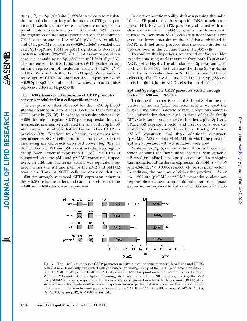
study (17), an Sp1/Sp3 site (�629A) was shown to regulatethe transcriptional activity of the human CETP gene pro-moter. It was thus of interest to analyze the influence of apossible interaction between the �690 and �629 sites onthe regulation of the transcriptional activity of the humanCETP gene promoter. Use of WT, pM2 (�629A allele)and pM1, pM1M2 constructs (�629C allele) revealed thateach Sp1/Sp3 site (pM1 or pM2) significantly decreasedluciferase activity (22–23%, P � 0.05) as compared with aconstruct containing no Sp1/Sp3 site (pM1M2) (Fig. 3A).The presence of both Sp1/Sp3 sites (WT) resulted in sig-nificant repression of luciferase activity (�42%, P �0.0005). We conclude that the �690 Sp1/Sp3 site inducesrepression of CETP promoter activity comparable to the�629 Sp1/Sp3 site, and that the two sites exert an additiverepressive effect in HepG2 cells.
The �690 site-mediated repression of CETP promoter activity is modulated in a cell-specific manner
The repressive effect observed for the �690 Sp1/Sp3site was obtained in HepG2 cells, a cell line that expressesCETP protein (35, 36). In order to determine whether the�690 site might regulate CETP gene expression in a tis-sue-specific manner, we evaluated the role of this Sp1/Sp3site in murine fibroblasts that are known to lack CETP ex-pression (18). Transient transfection experiments wereperformed in NCTC cells, a murine connective tissue cellline, using the constructs described above (Fig. 3B). Inthis cell line, the WT and pM1 constructs displayed signifi-cantly lower luciferase expression (�45%, P � 0.05) ascompared with the pM2 and pM1M2 constructs, respec-tively. In addition, luciferase activity was equivalent be-tween either the WT and pM1 or the pM2 and pM1M2constructs. Thus, in NCTC cells, we observed that the�690 site strongly repressed CETP expression, whereasthe �629 site had no effect, indicating therefore that the�690 and �629 sites are not equivalent.
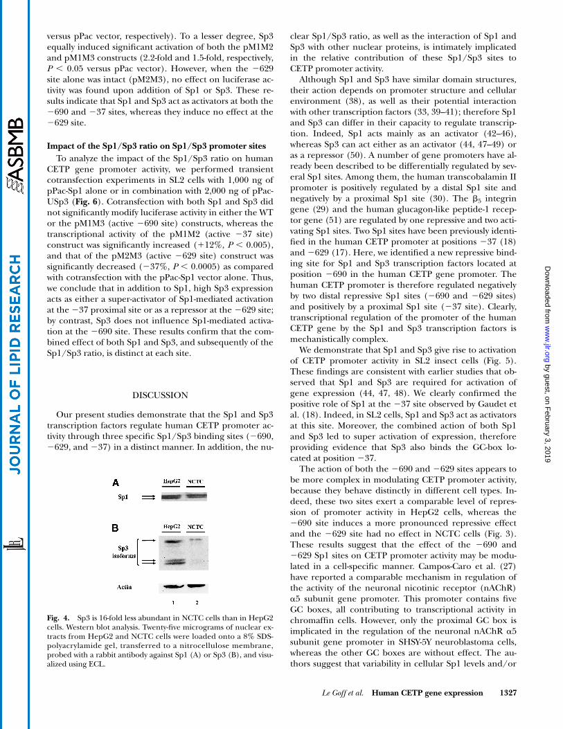
In electrophoretic mobility shift assays using the radio-labeled FP probe, the three specific DNA-protein com-plexes FP1, FP2, and FP3, previously obtained with nu-clear extracts from HepG2 cells, were also formed withnuclear extracts from NCTC cells (data not shown). How-ever, the lower intensity of the FP2 band obtained inNCTC cells led us to propose that the concentration ofSp3 was lower in this cell line than in HepG2 cells.
To confirm this hypothesis, we carried out Western blotexperiments using nuclear extracts from both HepG2 andNCTC cells (Fig. 4). The abundance of Sp1 was similar inboth cell lines (Fig. 4A), whereas the three Sp3 isoformswere 16-fold less abundant in NCTC cells than in HepG2cells (Fig. 4B). These data indicated that the Sp1/Sp3 ra-tio is 16-fold higher in NCTC cells than in HepG2 cells.
Sp1 and Sp3 regulate CETP promoter activity through both the �690 and �37 sites
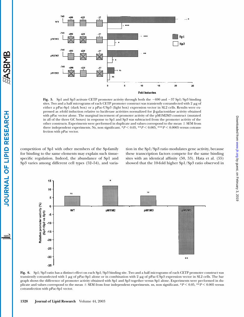
To define the respective role of Sp1 and Sp3 in the reg-ulation of human CETP promoter activity, we used theSL2 cell line, which is devoid of many ubiquitous mamma-lian transcription factors, such as those of the Sp family(37). Cells were cotransfected with either a pPac-Sp1 or apPac-USp3 expression vector and a set of constructs de-scribed in Experimental Procedures. Briefly, WT andpM1M2 constructs, and three additional constructs(pM1M3, pM2M3, and pM1M2M3) in which the proximalSp1 site at position �37 was mutated, were used.
As shown in Fig. 5, cotransfection of the WT construct,which contains the three intact Sp sites, with either apPac-Sp1 or a pPac-Usp3 expression vector led to a signifi-cant induction of luciferase expression (20-fold, P � 0.05and 4.3-fold, P � 0.0005, respectively, versus pPac vector).In addition, the presence of either the proximal �37 orthe �690 site (pM1M2 or pM1M3, respectively) alone wasresponsible for a significant 9-fold induction of luciferaseexpression in response to Sp1 (P � 0.0005 and P � 0.005
Fig. 3. The �690 site represses CETP promoter activity in a cell-specific manner. HepG2 (A) and NCTCcells (B) were transiently transfected with constructs containing 777 bp of the CETP gene promoter with ei-ther the A allele (WT) or the C allele (pM1) at position �629. Two point mutations were introduced in bothWT and pM1 constructs in the Sp1/Sp3 binding site located at position �690, thereby generating the pM2and pM1M2 constructs, respectively. Luciferase activity is expressed in relative luciferase units (RLUs) afterstandardisation for �-galactosidase activity. Experiments were performed in triplicate and values correspondto the mean SD from five independent experiments. *P � 0.05, ***P � 0.0005 versus pM1M2; †P � 0.05,††P � 0.005 versus pM2; ‡P � 0.05 versus pM1.
by guest, on February 3, 2019
ww
w.jlr.org
Dow
nloaded from
Le Goff et al. Human CETP gene expression 1327
versus pPac vector, respectively). To a lesser degree, Sp3equally induced significant activation of both the pM1M2and pM1M3 constructs (2.2-fold and 1.5-fold, respectively,P � 0.05 versus pPac vector). However, when the �629site alone was intact (pM2M3), no effect on luciferase ac-tivity was found upon addition of Sp1 or Sp3. These re-sults indicate that Sp1 and Sp3 act as activators at both the�690 and �37 sites, whereas they induce no effect at the�629 site.
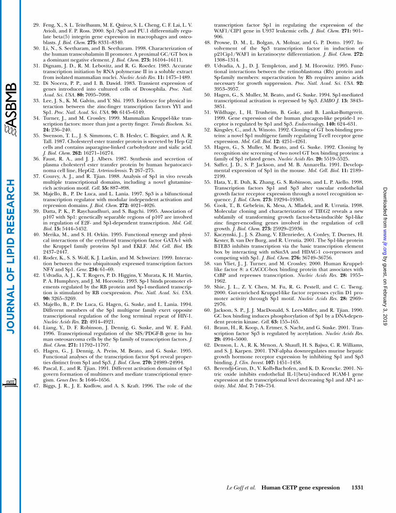
Impact of the Sp1/Sp3 ratio on Sp1/Sp3 promoter sitesTo analyze the impact of the Sp1/Sp3 ratio on human
CETP gene promoter activity, we performed transientcotransfection experiments in SL2 cells with 1,000 ng ofpPac-Sp1 alone or in combination with 2,000 ng of pPac-USp3 (Fig. 6). Cotransfection with both Sp1 and Sp3 didnot significantly modify luciferase activity in either the WTor the pM1M3 (active �690 site) constructs, whereas thetranscriptional activity of the pM1M2 (active �37 site)construct was significantly increased (�12%, P � 0.005),and that of the pM2M3 (active �629 site) construct wassignificantly decreased (�37%, P � 0.0005) as comparedwith cotransfection with the pPac-Sp1 vector alone. Thus,we conclude that in addition to Sp1, high Sp3 expressionacts as either a super-activator of Sp1-mediated activationat the �37 proximal site or as a repressor at the �629 site;by contrast, Sp3 does not influence Sp1-mediated activa-tion at the �690 site. These results confirm that the com-bined effect of both Sp1 and Sp3, and subsequently of theSp1/Sp3 ratio, is distinct at each site.
DISCUSSION
Our present studies demonstrate that the Sp1 and Sp3transcription factors regulate human CETP promoter ac-tivity through three specific Sp1/Sp3 binding sites (�690,�629, and �37) in a distinct manner. In addition, the nu-
clear Sp1/Sp3 ratio, as well as the interaction of Sp1 andSp3 with other nuclear proteins, is intimately implicatedin the relative contribution of these Sp1/Sp3 sites toCETP promoter activity.
Although Sp1 and Sp3 have similar domain structures,their action depends on promoter structure and cellularenvironment (38), as well as their potential interactionwith other transcription factors (33, 39–41); therefore Sp1and Sp3 can differ in their capacity to regulate transcrip-tion. Indeed, Sp1 acts mainly as an activator (42–46),whereas Sp3 can act either as an activator (44, 47–49) oras a repressor (50). A number of gene promoters have al-ready been described to be differentially regulated by sev-eral Sp1 sites. Among them, the human transcobalamin IIpromoter is positively regulated by a distal Sp1 site andnegatively by a proximal Sp1 site (30). The �5 integringene (29) and the human glucagon-like peptide-1 recep-tor gene (51) are regulated by one repressive and two acti-vating Sp1 sites. Two Sp1 sites have been previously identi-fied in the human CETP promoter at positions �37 (18)and �629 (17). Here, we identified a new repressive bind-ing site for Sp1 and Sp3 transcription factors located atposition �690 in the human CETP gene promoter. Thehuman CETP promoter is therefore regulated negativelyby two distal repressive Sp1 sites (�690 and �629 sites)and positively by a proximal Sp1 site (�37 site). Clearly,transcriptional regulation of the promoter of the humanCETP gene by the Sp1 and Sp3 transcription factors ismechanistically complex.
We demonstrate that Sp1 and Sp3 give rise to activationof CETP promoter activity in SL2 insect cells (Fig. 5).These findings are consistent with earlier studies that ob-served that Sp1 and Sp3 are required for activation ofgene expression (44, 47, 48). We clearly confirmed thepositive role of Sp1 at the �37 site observed by Gaudet etal. (18). Indeed, in SL2 cells, Sp1 and Sp3 act as activatorsat this site. Moreover, the combined action of both Sp1and Sp3 led to super activation of expression, thereforeproviding evidence that Sp3 also binds the GC-box lo-cated at position �37.
The action of both the �690 and �629 sites appears tobe more complex in modulating CETP promoter activity,because they behave distinctly in different cell types. In-deed, these two sites exert a comparable level of repres-sion of promoter activity in HepG2 cells, whereas the�690 site induces a more pronounced repressive effectand the �629 site had no effect in NCTC cells (Fig. 3).These results suggest that the effect of the �690 and�629 Sp1 sites on CETP promoter activity may be modu-lated in a cell-specific manner. Campos-Caro et al. (27)have reported a comparable mechanism in regulation ofthe activity of the neuronal nicotinic receptor (nAChR)�5 subunit gene promoter. This promoter contains fiveGC boxes, all contributing to transcriptional activity inchromaffin cells. However, only the proximal GC box isimplicated in the regulation of the neuronal nAChR �5subunit gene promoter in SHSY-5Y neuroblastoma cells,whereas the other GC boxes are without effect. The au-thors suggest that variability in cellular Sp1 levels and/or
Fig. 4. Sp3 is 16-fold less abundant in NCTC cells than in HepG2cells. Western blot analysis. Twenty-five micrograms of nuclear ex-tracts from HepG2 and NCTC cells were loaded onto a 8% SDS-polyacrylamide gel, transferred to a nitrocellulose membrane,probed with a rabbit antibody against Sp1 (A) or Sp3 (B), and visu-alized using ECL.
by guest, on February 3, 2019
ww
w.jlr.org
Dow
nloaded from
1328 Journal of Lipid Research Volume 44, 2003
competition of Sp1 with other members of the Sp-familyfor binding to the same elements may explain such tissue-specific regulation. Indeed, the abundance of Sp1 andSp3 varies among different cell types (52–54), and varia-
tion in the Sp1/Sp3 ratio modulates gene activity, becausethese transcription factors compete for the same bindingsites with an identical affinity (50, 53). Hata et al. (55)showed that the 10-fold higher Sp1/Sp3 ratio observed in
Fig. 5. Sp1 and Sp3 activate CETP promoter activity through both the �690 and �37 Sp1/Sp3 bindingsites. Two and a half micrograms of each CETP promoter construct was transiently cotransfected with 2 �g ofeither a pPac-Sp1 (dark box) or a pPac-USp3 (light box) expression vector in SL2 cells. Results were ex-pressed as x-fold induction relative to luciferase activities normalized for �-galactosidase activity obtainedwith pPac vector alone. The marginal increment of promoter activity of the pM1M2M3 construct (mutatedin all of the three GC boxes) in response to Sp1 and Sp3 was subtracted from the promoter activity of theother constructs. Experiments were performed in duplicate and values correspond to the mean SEM fromthree independent experiments. Ns, non significant. *P � 0.05, **P � 0.005, ***P � 0.0005 versus cotrans-fection with pPac vector.
Fig. 6. Sp1/Sp3 ratio has a distinct effect on each Sp1/Sp3 binding site. Two and a half micrograms of each CETP promoter construct wastransiently cotransfected with 1 �g of pPac-Sp1 alone or in combination with 2 �g of pPac-USp3 expression vector in SL2 cells. The bargraph shows the difference of promoter activity obtained with Sp1 and Sp3 together versus Sp1 alone. Experiments were per formed in du-plicate and values correspond to the mean SEM from four independent experiments. ns, non significant. *P � 0.05, **P � 0.005 versuscotransfection with pPac-Sp1 vector.
by guest, on February 3, 2019
ww
w.jlr.org
Dow
nloaded from

Le Goff et al. Human CETP gene expression 1329
endothelial cells as compared with nonendothelial cellswas responsible for the elevated expression of the kinasedomain receptor promoter in the former, as Sp3 attenu-ated the Sp1-mediated activation of promoter activity.These studies lead us to suggest that the lesser abundanceof Sp3 that we observed in NCTC cells as compared withHepG2 cells may be responsible for the distinct effect atboth the �690 and �629 sites in those two cell lines.Based upon our present results, we propose a mechanismby which cellular Sp3 level may modulate the action of thetwo distal Sp1 sites on CETP promoter activity (Fig. 7).The combined effect of both Sp3 and Sp1 induced signifi-cant repression at the �629 site (pM2M3 construct, Fig.6), whereas Sp1 or Sp3 alone did not exert a significant ef-fect at this site (Fig. 5). Such repression involving Sp3 inSL2 cells is surprising since Majello et al. (38) reportedthat Sp3 cannot act as a repressor on this cellular back-ground. It is for this reason that the repression of the Sp1-mediated activation by Sp3, resulting in competition withSp1 for their common binding site, is frequently observedin SL2 cells (50); however, overexpression of Sp3 inNCTC cells confirmed that Sp3 exerts a repressive effectat the �629 site (data not shown). Thus, in HepG2 cellsthat display high Sp3 levels, Sp3 acts as a repressor at the�629 site. On the other hand, the abundance of Sp3 maynot be sufficient in NCTC cells to repress CETP promoteractivity at the �629 site, and may account for the absenceof effect at this site.
The mechanism involved in the functionality of the�690 site seems to be distinct from that at the �629 site.The fact that on the one hand, Sp1 and Sp3 act as activa-tors at the �690 site (pM1M3 construct, Fig. 5), and that
on the other, Sp3 did not affect Sp1-mediated activationat this site (Fig. 6), suggests that another factor is respon-sible for the repressive effect observed at the �690 site.We speculate that the nonidentified nuclear factor(s) Xinvolved in the formation of the FP3 complex in EMSAexperiments performed with nuclear extracts fromHepG2 and NCTC cells, but not from SL2 cells (data notshown), may explain this repression. The cellular abun-dance of Sp3 might influence the repressive effect medi-ated by this nuclear protein, probably by affecting thebinding or the action of factor X at the �690 site. Thus,in HepG2 cells, the abundance of Sp3 permits only aweak repression (�22%) at the �690 site by factor X,whereas the lower level of Sp3 observed in NCTC cells al-lows a greater degree of repression (�45%) at this site.The hypothesis that an additional protein distinct fromSp1 and Sp3 might be responsible for the repression ob-served at the �690 site is consistent with previous studiesthat report that several other Sp1-like proteins, such asTIEG2 (56), BTEB3 (57), or members of the Krüppel-likefactor family (58, 59), repress promoter activity throughSp1 motifs.
Our data, therefore, strongly suggest that the effect ofboth the �690 and �629 Sp1/Sp3 sites on CETP pro-moter activity might depend on the competitive bindingof Sp1 and Sp3 as well as on the synergistic interactions ofthese two transcription factors, or on their interactionswith other factors at each of these sites. However, as illus-trated in our model, we cannot exclude the possibility thatother mechanisms may modulate the contribution ofthese two Sp1/Sp3 binding sites to regulation of CETPpromoter activity.
Fig. 7. Sp3 levels may modulate the repression of human CETP promoter activity observed through boththe �690 and �629 sites. Both Sp1 and Sp3 are bound at the two �629 (17) and �690 sites located on thehuman CETP promoter, whereas factor X is only present at the �690 site. In HepG2 cells, the high abun-dance of Sp3 i) is sufficient to repress CETP promoter activity at the �629 site (�20%) because Sp3 acts as arepressor at this site when Sp1 is present, and ii) permits only a weak repression (�22%) by factor X at the�690 site. By contrast, the low level of Sp3 in NCTC cells is not sufficient to repress CETP promoter activityat the �629 site, whereas it allows a marked repression (�45%) at the �690 site. This model does not, how-ever, account for total transcriptional promoter activity, because it does not include the contribution of theproximal �37 site.
by guest, on February 3, 2019
ww
w.jlr.org
Dow
nloaded from

1330 Journal of Lipid Research Volume 44, 2003
In conclusion, we demonstrate that both the Sp1 andSp3 transcription factors are required for basal expressionof the human CETP gene and that they exert a dichoto-mous effect on promoter activity. Indeed, the binding ofSp1 and Sp3 leads to transcriptional repression at the twodistal �690 and �629 sites, whereas activation occurs atthe proximal �37 site, thereby illustrating the complexrole of Sp1 and Sp3 in the transcription mechanism. Fi-nally, it is of considerable interest that modulation of thetranscriptional activity of the Sp1 or Sp3 factors by phos-phorylation (60), acetylation (61), or inflammatory cyto-kines such as TNF� (62), or by the action of nitric oxideon such redox-sensitive proteins (63), may potentially re-sult in regulation of CETP gene expression; it can be en-visaged that such modulation of gene expression will bereflected in plasma CETP mass and/or activity, andthence, in HDL phenotype.
INSERM provided generous support for these studies. W.L. wasthe recipient of a Research Fellowship from the French Minis-try of Research and Technology. The authors are indebted toProf. Dr. Guntram Suske (Klinikum der Philipps-UniversitätMarburg, Marburg, Germany) for providing pPac, pPacSp1,and pPacUSp3 vectors; and to Dr. Christine Vesque (InsermU368, Ecole Normale Supérieure, Paris, France) for providingthe pAdh-LacZ expression vector. The authors thank Dr. Thi-erry Huby for stimulating discussion.
REFERENCES
1. Yamashita, S., K. Hirano, N. Sakai, and Y. Matsuzawa. 2000. Molecu-lar biology and pathophysiological aspects of plasma cholesterylester transfer protein. Biochim. Biophys. Acta. 1529(1–3): 257–275.
2. Marotti, K. R., C. K. Castle, T. P. Boyle, A. H. Lin, R. W. Murray, andG. W. Melchior. 1993. Severe atherosclerosis in transgenic mice ex-pressing simian cholesteryl ester transfer protein. Nature. 364: 73–75.
3. Zhong, S., D. S. Sharp, J. S. Grove, C. Bruce, K. Yano, J. D. Curb, andA. R. Tall. 1996. Increased coronary heart disease in Japanese-Amer-ican men with mutation in the cholesteryl ester transfer proteingene despite increased HDL levels. J. Clin. Invest. 97: 2917–2923.
4. Tato, F., G. L. Vega, A. R. Tall, and S. M. Grundy. 1995. Relation be-tween cholesterol ester transfer protein activities and lipoproteincholesterol in patients with hypercholesterolemia and combinedhyperlipidemia. Arterioscler. Thromb. Vasc. Biol. 15: 112–120.
5. McPherson, R., C. J. Mann, A. R. Tall, M. Hogue, L. Martin, R. W.Milne, and Y. L. Marcel. 1991. Plasma concentrations of choles-teryl ester transfer protein in hyperlipoproteinemia. Relation tocholesteryl ester transfer protein activity and other lipoproteinvariables. Arterioscler. Thromb. 11: 797–804.
6. Guerin, M., P. J. Dolphin, and M. J. Chapman. 1994. Preferentialcholesteryl ester acceptors among the LDL subspecies of subjectswith familial hypercholesterolemia. Arterioscler. Thromb. 14: 679–685.
7. de Grooth, G. J., J. A. Kuivenhoven, A. F. Stalenhoef, J. de Graaf,A. H. Zwinderman, J. L. Posma, A. van Tol, and J. J. Kastelein. 2002.Efficacy and safety of a novel cholesteryl ester transfer protein in-hibitor, JTT-705, in humans: a randomized phase II dose-responsestudy. Circulation. 105: 2159–2165.
8. Okamoto, H., F. Yonemori, K. Wakitani, T. Minowa, K. Maeda, andH. Shinkai. 2000. A cholesteryl ester transfer protein inhibitor at-tenuates atherosclerosis in rabbits. Nature. 406: 203–207.
9. McPherson, R., S. M. Grundy, R. Guerra, and J. C. Cohen. 1996.Allelic variation in the gene encoding the cholesteryl ester transferprotein is associated with variation in the plasma concentrations ofcholesteryl ester transfer protein. J. Lipid Res. 37: 1743–1748.
10. Radeau, T., M. Robb, P. Lau, J. Borthwick, and R. McPherson.1998. Relationship of adipose tissue cholesteryl ester transfer pro-tein (CETP) mRNA to plasma concentrations of CETP in man.Atherosclerosis. 139: 369–376.
11. Jiang, X. C., L. B. Agellon, A. Walsh, J. L. Breslow, and A. Tall.1992. Dietary cholesterol increases transcription of the humancholesteryl ester transfer protein gene in transgenic mice. Depen-dence on natural flanking sequences. J. Clin. Invest. 90: 1290–1295.
12. Hirano, R., O. Igarashi, K. Kondo, H. Itakura, and A. Matsumoto.2001. Regulation by long-chain fatty acids of the expression of cho-lesteryl ester transfer protein in HepG2 cells. Lipids. 36: 401–406.
13. Chang, C. K., and J. T. Snook. 2001. The cholesterolaemic effectsof dietary fats in cholesteryl ester transfer protein transgenic mice.Br. J. Nutr. 85: 643–648.
14. Masucci-Magoulas, L., P. Moulin, X. C. Jiang, H. Richardson, A.Walsh, J. L. Breslow, and A. Tall. 1995. Decreased cholesteryl estertransfer protein (CETP) mRNA and protein and increased highdensity lipoprotein following lipopolysaccharide administration inhuman CETP transgenic mice. J. Clin. Invest. 95: 1587–1594.
15. Agellon, L. B., P. Zhang, X. C. Jiang, L. Mendelsohn, and A. R.Tall. 1992. The CCAAT/enhancer-binding protein trans-activatesthe human cholesteryl ester transfer protein gene promoter. J.Biol. Chem. 267: 22336–22339.
16. Chouinard, R. A., Jr., Y. Luo, T. F. Osborne, A. Walsh, and A. R.Tall. 1998. Sterol regulatory element binding protein-1 activatesthe cholesteryl ester transfer protein gene in vivo but is not re-quired for sterol up-regulation of gene expression. J. Biol. Chem.273: 22409–22414.
17. Dachet, C., O. Poirier, F. Cambien, J. Chapman, and M. Rouis. 2000.New functional promoter polymorphism, CETP/-629, in cholesterylester transfer protein (CETP) gene related to CETP mass and highdensity lipoprotein cholesterol levels: role of Sp1/Sp3 in transcrip-tional regulation. Arterioscler. Thromb. Vasc. Biol. 20: 507–515.
18. Gaudet, F., and G. S. Ginsburg. 1995. Transcriptional regulation ofthe cholesteryl ester transfer protein gene by the orphan nuclearhormone receptor apolipoprotein AI regulatory protein-1. J. Biol.Chem. 270: 29916–29922.
19. Gauthier, B., M. Robb, F. Gaudet, G. S. Ginsburg, and R. McPher-son. 1999. Characterization of a cholesterol response element(CRE) in the promoter of the cholesteryl ester transfer proteingene: functional role of the transcription factors SREBP-1a, -2, andYY1. J. Lipid Res. 40: 1284–1293.
20. Jeoung, N. H., W. G. Jang, J. I. Nam, Y. K. Pak, and Y. B. Park. 1999.Identification of retinoic acid receptor element in human choles-teryl ester transfer protein gene. Biochem. Biophys. Res. Commun.258: 411–415.
21. Luo, Y., and A. R. Tall. 2000. Sterol upregulation of human CETPexpression in vitro and in transgenic mice by an LXR element. J.Clin. Invest. 105: 513–520.
22. Luo, Y., C. P. Liang, and A. R. Tall. 2001. The orphan nuclear re-ceptor LRH-1 potentiates the sterol-mediated induction of the hu-man CETP gene by LXR. J. Biol. Chem. 276: 24767–24773.
23. Quinet, E. M., L. B. Agellon, P. A. Kroon, Y. L. Marcel, Y. C. Lee, M. E.Whitlock, and A. R. Tall. 1990. Atherogenic diet increases choles-teryl ester transfer protein messenger RNA levels in rabbit liver. J.Clin. Invest. 85: 357–363.
24. Oliveira, H. C. F., R. A. Chouinard, L. B. Agellon, C. Bruce, L. Ma, A.Walsh, J. L. Breslow, and A. R. Tall. 1996. Human cholesteryl estertransfer protein gene proximal promoter contains dietary choles-terol positive responsive elements and mediates expression in smallintestine and periphery while predominant liver and spleen expres-sion is controlled by 5-distal sequences. Cis-acting sequencesmapped in transgenic mice. J. Biol. Chem. 271: 31831–31838.
25. Suske, G. 1999. The Sp-family of transcription factors. Gene. 238:291–300.
26. Rajakumar, R. A., S. Thamotharan, R. K. Menon, and S. U.Devaskar. 1998. Sp1 and Sp3 regulate transcriptional activity of thefacilitative glucose transporter isoform-3 gene in mammalian neu-roblasts and trophoblasts. J. Biol. Chem. 273: 27474–27483.
27. Campos-Caro, A., C. Carrasco-Serrano, L. M. Valor, S. Viniegra, J. J.Ballesta, and M. Criado. 1999. Multiple functional Sp1 domains inthe minimal promoter region of the neuronal nicotinic receptoralpha5 subunit gene. J. Biol. Chem. 274: 4693–4701.
28. Baker, D. L., V. Dave, T. Reed, and M. Periasamy. 1996. MultipleSp1 binding sites in the cardiac/slow twitch muscle sarcoplasmicreticulum Ca2�-ATPase gene promoter are required for expres-sion in Sol8 muscle cells. J. Biol. Chem. 271: 5921–5928.
by guest, on February 3, 2019
ww
w.jlr.org
Dow
nloaded from
Le Goff et al. Human CETP gene expression 1331
29. Feng, X., S. L. Teitelbaum, M. E. Quiroz, S. L. Cheng, C. F. Lai, L. V.Avioli, and F. P. Ross. 2000. Sp1/Sp3 and PU.1 differentially regu-late beta(5) integrin gene expression in macrophages and osteo-blasts. J. Biol. Chem. 275: 8331–8340.
30. Li, N., S. Seetharam, and B. Seetharam. 1998. Characterization ofthe human transcobalamin II promoter. A proximal GC/GT box isa dominant negative element. J. Biol. Chem. 273: 16104–16111.
31. Dignam, J. D., R. M. Lebovitz, and R. G. Roeder. 1983. Accuratetranscription initiation by RNA polymerase II in a soluble extractfrom isolated mammalian nuclei. Nucleic Acids Res. 11: 1475–1489.
32. Di Nocera, P. P., and I. B. Dawid. 1983. Transient expression ofgenes introduced into cultured cells of Drosophila. Proc. Natl.Acad. Sci. USA. 80: 7095–7098.
33. Lee, J. S., K. M. Galvin, and Y. Shi. 1993. Evidence for physical in-teraction between the zinc-finger transcription factors YY1 andSp1. Proc. Natl. Acad. Sci. USA. 90: 6145–6149.
34. Turner, J., and M. Crossley. 1999. Mammalian Kruppel-like tran-scription factors: more than just a pretty finger. Trends Biochem. Sci.24: 236–240.
35. Swenson, T. L., J. S. Simmons, C. B. Hesler, C. Bisgaier, and A. R.Tall. 1987. Cholesteryl ester transfer protein is secreted by Hep G2cells and contains asparagine-linked carbohydrate and sialic acid.J. Biol. Chem. 262: 16271–16274.
36. Faust, R. A., and J. J. Albers. 1987. Synthesis and secretion ofplasma cholesteryl ester transfer protein by human hepatocarci-noma cell line, HepG2. Arteriosclerosis. 7: 267–275.
37. Courey, A. J., and R. Tjian. 1988. Analysis of Sp1 in vivo revealsmultiple transcriptional domains, including a novel glutamine-rich activation motif. Cell. 55: 887–898.
38. Majello, B., P. De Luca, and L. Lania. 1997. Sp3 is a bifunctionaltranscription regulator with modular independent activation andrepression domains. J. Biol. Chem. 272: 4021–4026.
39. Datta, P. K., P. Raychaudhuri, and S. Bagchi. 1995. Association ofp107 with Sp1: genetically separable regions of p107 are involvedin regulation of E2F- and Sp1-dependent transcription. Mol. Cell.Biol. 15: 5444–5452.
40. Merika, M., and S. H. Orkin. 1995. Functional synergy and physi-cal interactions of the erythroid transcription factor GATA-1 withthe Kruppel family proteins Sp1 and EKLF. Mol. Cell. Biol. 15:2437–2447.
41. Roder, K., S. S. Wolf, K. J. Larkin, and M. Schweizer. 1999. Interac-tion between the two ubiquitously expressed transcription factorsNF-Y and Sp1. Gene. 234: 61–69.
42. Udvadia, A. J., K. T. Rogers, P. D. Higgins, Y. Murata, K. H. Martin,P. A. Humphrey, and J. M. Horowitz. 1993. Sp-1 binds promoter el-ements regulated by the RB protein and Sp-1-mediated transcrip-tion is stimulated by RB coexpression. Proc. Natl. Acad. Sci. USA.90: 3265–3269.
43. Majello, B., P. De Luca, G. Hagen, G. Suske, and L. Lania. 1994.Different members of the Sp1 multigene family exert oppositetranscriptional regulation of the long terminal repeat of HIV-1.Nucleic Acids Res. 22: 4914–4921.
44. Liang, Y., D. F. Robinson, J. Dennig, G. Suske, and W. E. Fahl.1996. Transcriptional regulation of the SIS/PDGF-B gene in hu-man osteosarcoma cells by the Sp family of transcription factors. J.Biol. Chem. 271: 11792–11797.
45. Hagen, G., J. Dennig, A. Preiss, M. Beato, and G. Suske. 1995.Functional analyses of the transcription factor Sp4 reveal proper-ties distinct from Sp1 and Sp3. J. Biol. Chem. 270: 24989–24994.
46. Pascal, E., and R. Tjian. 1991. Different activation domains of Sp1govern formation of multimers and mediate transcriptional syner-gism. Genes Dev. 5: 1646–1656.
47. Biggs, J. R., J. E. Kudlow, and A. S. Kraft. 1996. The role of the
transcription factor Sp1 in regulating the expression of theWAF1/CIP1 gene in U937 leukemic cells. J. Biol. Chem. 271: 901–906.
48. Prowse, D. M., L. Bolgan, A. Molnar, and G. P. Dotto. 1997. In-volvement of the Sp3 transcription factor in induction ofp21Cip1/WAF1 in keratinocyte differentiation. J. Biol. Chem. 272:1308–1314.
49. Udvadia, A. J., D. J. Templeton, and J. M. Horowitz. 1995. Func-tional interactions between the retinoblastoma (Rb) protein andSp-family members: superactivation by Rb requires amino acidsnecessary for growth suppression. Proc. Natl. Acad. Sci. USA. 92:3953–3957.
50. Hagen, G., S. Muller, M. Beato, and G. Suske. 1994. Sp1-mediatedtranscriptional activation is repressed by Sp3. EMBO J. 13: 3843–3851.
51. Wildhage, I., H. Trusheim, B. Goke, and B. Lankat-Buttgereit.1999. Gene expression of the human glucagon-like peptide-1 re-ceptor is regulated by Sp1 and Sp3. Endocrinology. 140: 624–631.
52. Kingsley, C., and A. Winoto. 1992. Cloning of GT box-binding pro-teins: a novel Sp1 multigene family regulating T-cell receptor geneexpression. Mol. Cell. Biol. 12: 4251–4261.
53. Hagen, G., S. Muller, M. Beato, and G. Suske. 1992. Cloning byrecognition site screening of two novel GT box binding proteins: afamily of Sp1 related genes. Nucleic Acids Res. 20: 5519–5525.
54. Saffer, J. D., S. P. Jackson, and M. B. Annarella. 1991. Develop-mental expression of Sp1 in the mouse. Mol. Cell. Biol. 11: 2189–2199.
55. Hata, Y., E. Duh, K. Zhang, G. S. Robinson, and L. P. Aiello. 1998.Transcription factors Sp1 and Sp3 alter vascular endothelialgrowth factor receptor expression through a novel recognition se-quence. J. Biol. Chem. 273: 19294–19303.
56. Cook, T., B. Gebelein, K. Mesa, A. Mladek, and R. Urrutia. 1998.Molecular cloning and characterization of TIEG2 reveals a newsubfamily of transforming growth factor-beta-inducible Sp1-likezinc finger-encoding genes involved in the regulation of cellgrowth. J. Biol. Chem. 273: 25929–25936.
57. Kaczynski, J., J. S. Zhang, V. Ellenrieder, A. Conley, T. Duenes, H.Kester, B. van Der Burg, and R. Urrutia. 2001. The Sp1-like proteinBTEB3 inhibits transcription via the basic transcription elementbox by interacting with mSin3A and HDAC-1 co-repressors andcompeting with Sp1. J. Biol. Chem. 276: 36749–36756.
58. van Vliet, J., J. Turner, and M. Crossley. 2000. Human Kruppel-like factor 8: a CACCC-box binding protein that associates withCtBP and represses transcription. Nucleic Acids Res. 28: 1955–1962.
59. Shie, J. L., Z. Y. Chen, M. Fu, R. G. Pestell, and C. C. Tseng.2000. Gut-enriched Kruppel-like factor represses cyclin D1 pro-moter activity through Sp1 motif. Nucleic Acids Res. 28: 2969–2976.
60. Jackson, S. P., J. J. MacDonald, S. Lees-Miller, and R. Tjian. 1990.GC box binding induces phosphorylation of Sp1 by a DNA-depen-dent protein kinase. Cell. 63: 155–165.
61. Braun, H., R. Koop, A. Ertmer, S. Nacht, and G. Suske. 2001. Tran-scription factor Sp3 is regulated by acetylation. Nucleic Acids Res.29: 4994–5000.
62. Denson, L. A., R. K. Menon, A. Shaufl, H. S. Bajwa, C. R. Williams,and S. J. Karpen. 2001. TNF-alpha downregulates murine hepaticgrowth hormone receptor expression by inhibiting Sp1 and Sp3binding. J. Clin. Invest. 107: 1451–1458.
63. Berendji-Grun, D., V. Kolb-Bachofen, and K. D. Kroncke. 2001. Ni-tric oxide inhibits endothelial IL-1[beta]-induced ICAM-1 geneexpression at the transcriptional level decreasing Sp1 and AP-1 ac-tivity. Mol. Med. 7: 748–754.
by guest, on February 3, 2019
ww
w.jlr.org
Dow
nloaded from